Publisher’s version / Version de l'éditeur:
Canadian Journal of Microbiology, 57, 6, pp. 493-503, 2011-06-03
READ THESE TERMS AND CONDITIONS CAREFULLY BEFORE USING THIS WEBSITE.
https://nrc-publications.canada.ca/eng/copyright
Vous avez des questions? Nous pouvons vous aider. Pour communiquer directement avec un auteur, consultez la
première page de la revue dans laquelle son article a été publié afin de trouver ses coordonnées. Si vous n’arrivez pas à les repérer, communiquez avec nous à PublicationsArchive-ArchivesPublications@nrc-cnrc.gc.ca.
Questions? Contact the NRC Publications Archive team at
PublicationsArchive-ArchivesPublications@nrc-cnrc.gc.ca. If you wish to email the authors directly, please see the first page of the publication for their contact information.
NRC Publications Archive
Archives des publications du CNRC
This publication could be one of several versions: author’s original, accepted manuscript or the publisher’s version. / La version de cette publication peut être l’une des suivantes : la version prépublication de l’auteur, la version acceptée du manuscrit ou la version de l’éditeur.
For the publisher’s version, please access the DOI link below./ Pour consulter la version de l’éditeur, utilisez le lien DOI ci-dessous.
https://doi.org/10.1139/w11-032
Access and use of this website and the material on it are subject to the Terms and Conditions set forth at
Characterization of the bacterial community structure of Sydney Tar
Ponds sediment
Yeung, C. William; Woo, Monica; Lee, Kenneth; Greer, Charles W.
https://publications-cnrc.canada.ca/fra/droits
L’accès à ce site Web et l’utilisation de son contenu sont assujettis aux conditions présentées dans le site LISEZ CES CONDITIONS ATTENTIVEMENT AVANT D’UTILISER CE SITE WEB.
NRC Publications Record / Notice d'Archives des publications de CNRC:
https://nrc-publications.canada.ca/eng/view/object/?id=42421154-e935-47d8-a240-2cf17ffe8153 https://publications-cnrc.canada.ca/fra/voir/objet/?id=42421154-e935-47d8-a240-2cf17ffe8153
Characterization of the bacterial community
structure of Sydney Tar Ponds sediment
C. William Yeung, Monica Woo, Kenneth Lee, and Charles W. Greer
Abstract: The Sydney Tar Ponds is one of the largest toxic waste sites in Canada. The bacterial diversity and abundance in the Sydney Tar Ponds sediment was examined using a 16S rRNA gene clone library and denaturing gradient gel electropho-resis (DGGE) with four different primer sets. The clone library was grouped into 19 phylotypes that could be divided into five phyla: Proteobacteria (56.9%), Actinobacteria (35%), Acidobacteria (4.9%), Firmicutes (2.4%), and Verrucomicrobia (0.8%). Members of the phyla Actinobacteria (represented mainly by Mycobacterium spp.) and Alphaproteobacteria (repre-sented by Acidocella spp.) comprised the majority of the clone library. This study also revealed that the phylogenetic results obtained from clone library analysis and from DGGE analysis, with all the primer sets, showed some variability. However, similar Mycobacterium spp. and Acidocella spp. were found in all the different DGGE analyses, again suggesting that these two genera are dominant in the Sydney Tar Ponds sediment. In addition, DGGE analysis indicated that primer sets targeting the V3 region produced results that were the most similar to those obtained with the clone library.
Key words:Sydney Tar Ponds, bacterial community structure, clone library, DGGE, 16S rRNA gene.
Résumé : Les mares de goudron de Sydney forment un lieu contaminé par les déchets toxiques parmi les plus vastes au Canada. La diversité et l’abondance bactériennes dans les sédiments des mares de goudron de Sydney ont été examinées à l’aide d’une banque génique de clones d’ARNr 16S et par électrophorèse sur gel en gradient dénaturant (DGGE) utilisant quatre paires d’amorces différentes. La banque de clones regroupait 19 phylotypes qui pouvaient se diviser en cinq phylums : Proteobacteria (56,9 %), Actinobacteria (35 %), Acidobacteria (4,9 %), Firmicutes (2,4 %) et Verrucomicrobia (0,8 %). Les membres des phylums des Actinobacteria (principalement représentés par Mycobacterium spp.) et des Alphaproteobacteria (représentés par Acidocella spp.) constituaient la majorité des clones de la banque. Cette étude a aussi révélé que les résultats phylogénétiques obtenus de l’ana-lyse de la banque de clones et de la DGGE, avec les toutes les paires d’amorces, montraient une certaine variabilité. Cependant, les mêmes Mycobacterium spp. et Acidocella spp. ont été trouvées selon toutes les analyses différentes par DGGE, suggérant encore que ces deux genres dominaient les sédiments des mares de goudron de Sydney. De plus, l’analyse par DGGE a indiqué que la paire d’amorces qui ciblait la région V3 générait les résultats les plus similaires à ceux obtenus avec la banque de clones. Mots‐clés :mares de goudron de Sydney, structure de la communauté bactérienne, banque de clones, DGGE, ARNr 16S. [Traduit par la Rédaction]
Introduction
The Sydney Tar Ponds may be the largest toxic waste site in Canada and in recent years has received a lot of govern-ment and public attention. Wastes from over 90 years of steelmaking and coke production have contaminated the water and sediments. It was estimated that more than 700 000 tonnes of coal tar waste with elevated concentrations of polycyclic aromatic hydrocarbons (PAHs as high as 27 800 mg·kg–1), polychlorinated biphenyls (PCBs up to 2600 mg·kg–1), and heavy metals was produced over the life-time of operation (Joint Review Panel 2006). Despite the clo-sure or elimination of the pollution sources in the past 20 years, the Sydney Tar Ponds remain heavily contaminated
and pose a serious risk to human health and the surrounding environment. At present, a remediation program involving solidification and stabilization of the sediments with cement and waste pilings is being implemented (Lee and Jones-Lee 2006).
Although the site is being solidified, biodegradation might still be occurring at the site and could potentially be impor-tant in peripheral areas (e.g., small streams around the former coke oven) that may be subject to leaching. Microorganisms are known to play a major role in aerobic and anaerobic bio-degradation in many contaminated environments (Timmis et al. 2010). To determine whether there is any potential for bioremediation at this site, a fundamental task is to identify the microbial diversity in this environment. The common understanding, however, is that less than 1% of microbes can
Received 17 December 2010. Revision received 28 February 2011. Accepted 2 March 2011. Published at www.nrcresearchpress.com/cjm on 3 June 2011.
C.W. Yeung. National Research Council Canada, Biotechnology Research Institute, 6100 Royalmount Avenue, Montréal,
QC H4P 2R2, Canada; Department of Natural Resource Sciences, McGill University, Ste-Anne-de-Bellevue, QC H9X 3V9, Canada. M. Woo and C.W. Greer. National Research Council Canada, Biotechnology Research Institute, 6100 Royalmount Avenue, Montréal, QC H4P 2R2, Canada.
K. Lee. Fisheries and Oceans Canada, P.O. Box 1006, Dartmouth, NS B2Y 4A2, Canada. Corresponding author: Charles W. Greer (e-mail: Charles.Greer@cnrc-nrc.gc.ca).
Can. J. Microbiol. Downloaded from www.nrcresearchpress.com by National Research Council of Canada on 06/06/11
For personal use only.
be cultured (Fuhrman et al. 1992; Rappé and Giovannoni 2003). The advancement of cultivation-independent molecu-lar techniques, like clone libraries and denaturing gradient gel electrophoresis (DGGE), which are used to characterize the structure of bacterial communities using their rRNA genes, have greatly expanded our knowledge of the phyloge-netic diversity of microorganisms and many of the new mi-crobial groups that have been discovered via these methods are involved in important processes (Karl 2002). One of the most widely used approaches is the construction of 16S rRNA gene clone libraries, which provide sufficiently high resolution to examine the extant microbial diversity (Head et al. 1998). However, the cloning method can be laborious and time-consuming. DGGE, a method used to separate PCR-amplified DNA fragments based on nucleotide sequence dif-ferences (Muyzer et al. 1993), is another extensively used rRNA gene screening method for fast and, at least, semiquan-titative assessment of the diversity and dynamics of dominant microbial species when coupled with the sequencing of sepa-rated 16S rDNA fragments. Röling et al. (2001) previously revealed that the sequence results from bands in DGGE pro-files were comparable to the results obtained from cloning.
The objective of this study was to determine the overall bacterial community composition in this highly contaminated environment using both clone library and DGGE with vari-ous primer sets. Although DGGE and clone library results are known to be comparable to some extent, it was important to compare the results from both methods to ensure that DGGE is sufficiently sensitive to elucidate the dominant mi-crobial community structure in this environment. These re-sults will help to standardize the primer sets needed to produce the most comprehensive information on the bacterial community diversity in the Sydney Tar Ponds.
Materials and methods Sample collection
A water saturated sediment sample (1 part water to 2 parts sediment) was collected at the shoreline of the south tar pond (46.146383N, –60.187283W), a known PCB hotspot (Joint Review Panel 2006), and was stored at 4 °C and shipped over-night to Montréal, Quebec. The sediment was immediately sampled upon arrival (within 48 h of collection) and subsamples were stored at –20 °C for DNA extraction and chemical analysis. Physicochemical and chemical analysis of sediments
The salinity was measured using a Hach sensION 5 con-ductivity meter (Loveland, Colorado, USA). The sediment and water pH were measured using a Fisher Scientific Accu-met pH Accu-meter (model AB15, Fisher Scientific, Nepean, On-tario, Canada). For soil pH, 5 mL of water was added into 5 g of sediment. The mixture was vortexed for 1 min and the pH was measured in the supernatant of the mixture. Sedi-ment dry mass and moisture content were determined from the weight loss after heat treatment (96 h at 105 °C). Pooled sediments were extracted for 18 h with a Soxhlet extractor (model RJ, Fisher Scientific) following a modified version of EPA method 3540 C (Environmental Protection Agency 2003) with 300 mL of glass-distilled dichloromethane (Cale-don, Georgetown, Ontario, Canada). After extraction, the ex-tracts were concentrated to 1 mL on a Turbo Vap
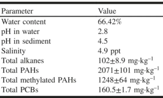
Concentrator (Zymark, Hopkinton, Massachusetts, USA). The extracts were purified using a Solid-Phase extraction (VWR-Canlab, Mont-Royal, Quebec, Canada) column packed with silica gel (activated at 200 °C for 17 h and deactivated with 5% m/v with HPLC grade water; Whatman Laboratory Division, Clifton, New Jersey, USA, 60A 70–230 mesh ASTM for HPLC). The column was washed with glass-distilled hexane (Caledon), the sample was applied as a 1 mL extract, and alkanes and PAHs were eluted with 10 mL of hexane:dichloromethane (4:1 v/v). Extracts of sedi-ments were analyzed using high resolution gas chromatogra-phy (Agilent 6890) coupled to a mass selective detector (Agilent 5973N) (Wilmington, Delaware, USA) in the selec-tive ion monitoring mode using the following gas chromatog-raphy (MDN-5S column 30 m × 0.25 mm, i.d. 0.25 µm film thickness, Supelco, Canada) conditions: cool on-column injection with oven track mode (track 3 °C higher that the oven temperature program) 80 °C hold 2 min ramp at 4 °C·min–1 to 280 °C hold 10 min (King and Lee 2004). PAH standards were obtained from Ultra Scientific (Kings-town, Rhode Island, USA) and Absolute Standards (Hem-den, Connecticut, USA). The total alkane concentration was measured using the method from King and Lee (2004), and the concentration was the sum of 16 alkanes that were de-tected in the samples, including n-decane, undecane, dodec-ane, tridecane, tetradecane, pentadecane, hexadecane, pristane, octadecane, phytane, nonadecane, eicosane, henei-cosane, dohenei-cosane, trihenei-cosane, and tetracosane. The total PAH concentration was the sum of 16 PAHs, including benzo[a]py-rene, anthracene, and pyrene (King and Chou 2003; King and Lee 2004). Total methylated PAH concentrations were the sum of 18 methylated PAHs (King and Lee 2004). Total PCBs were the sum of 125 congeners from IUPAC No. 1 to 209 (King et al. 1996). All analyses were carried out on two replicates, and the mean values obtained were reported. The physicochemical char-acteristics and contaminant concentrations in the sediment are presented in Table 1.
Total community DNA extraction
Total community DNA was extracted from 2 g of sediment sample with an UltraClean soil DNA isolation kit (Mo Bio Laboratories, Carlsbad, California) following the manufac-turer’s protocol. Four separate sediment samples were ex-tracted and the total community DNA was combined. DNA concentration was estimated by agarose gel electrophoresis using 5 µL of purified material against the Lambda HindIII DNA ladder (Amersham Biosciences, Piscataway, New Jer-sey, USA) standard on a 0.7% agarose gel.
Table 1. Physicochemical parameters of the Syd-ney Tar Ponds sediments.
Parameter Value Water content 66.42% pH in water 2.8 pH in sediment 4.5 Salinity 4.9 ppt Total alkanes 102±8.9 mg·kg–1 Total PAHs 2071±101 mg·kg–1
Total methylated PAHs 1248±64 mg·kg–1
Total PCBs 160.5±1.7 mg·kg–1
Can. J. Microbiol. Downloaded from www.nrcresearchpress.com by National Research Council of Canada on 06/06/11
Clone libraries of 16S rRNA genes
For PCR amplification of the full-length 16S rRNA gene, the bacteria-specific forward primer F1 GAGTTT-GATCCTGGCTCAG-3′) and the reverse primer R13 (5′-AGAAAGGAGGTGATCCAGCC-3′) were used (Liesack et al. 1991). Each 50 µL of PCR mixture contained 1 µL of template DNA (∼1 ng), 25 pmol of each oligonucleotide pri-mer, 200 µmol·L–1 (each) dNTP, and 2.5 units of Easy-A cloning enzyme in 10× Easy-A cloning enzyme buffer (Stra-tagene, La Jolla, California). PCR conditions were as follows: an initial denaturation for 5 min at 96 °C, followed by 25 cycles at 94 °C for 1 min, 60 °C for 1 min, and 72 °C for 2 min, with a final extension at 72 °C for 10 min. PCR prod-ucts were loaded onto a 1% agarose gel with SYBR Safe (Molecular Probes, Eugene, Oregon, USA), using a 100 bp ladder (MBI Fermentas, Amherst, New York, USA) to deter-mine the presence, size, and quantity of the PCR products. Three PCRs were combined to minimize bias. The PCR products were purified and combined using Illustra GFX PCR DNA and a Gel Band Purification kit (GE Healthcare, Baie d’Urfé, Quebec) and then purified again with Montage PCR cleanup products (Millipore Corporate, Billerica, Mas-sachusetts, USA). The purified PCR products were cloned using a QIAGEN PCR cloning kit (QIAGEN Inc., Missis-sauga, Ontario, Canada) at an insert:vector ratio of 3:1. The ligations were transformed by electroporation in Escherichia coli XL1-Blue (Stratagene). Transformants were selected on Luria–Bertani medium supplemented with ampicillin (100 mg·L–1), 5-bromo-4-chloro-3-indolyl-b-D -galactopyrano-side (X-Gal; 80 mg·L–1), and isopropyl-b-D -thiogalactopyra-noside (IPTG; 50 µmol·L–1). A total of 123 white colonies were chosen at random, plated on ampicillin-supplemented Luria–Bertani plates, and then incubated overnight. The 16S rRNA gene fragments from each clone were reamplified using vector primers (pDrive-F2: 5′-GTATCGGATCCA-GAATTCGTGA-3′; pDrive-R2: 5′-GAAGCTTGTCGACGA-ATTCAGA-3′), designed based on the available sequences of the pDrive vector (QIAGEN PCR cloning handbook).
The PCR products were used for clone screening by se-quencing at the Université Laval, Laval, Quebec, using the forward vector primer. Forward sequences from each clone were edited with BioEdit version 7.0.5.3 (http://www.mbio. ncsu.edu/BioEdit/bioedit.html; Hall 1999) and were aligned by the MacVector 9.0 software package (Accelrys, Cary, North Carolina, USA). This allowed us to identify groups of
clones containing the same inserts. All sequences hav-ing ≥98% similarity and matchhav-ing the same GenBank se-quence were assigned to the same phylotype. One to four representatives of each phylotype were then chosen for se-quencing of both DNA strands. The sequences were man-ually aligned by comparing forward and reverse sequences with BioEdit. The occurrence of chimeric sequences was de-termined manually with the CHECK_CHIMERA function from the Ribosomal Database Project (RDP) (http://wdcm. nig.ac.jp/RDP/cgis/chimera.cgi?su=SSU, Cole et al. 2003) and Bellerophon (http://comp-bio.anu.edu.au/bellerophon/ bellerophon.pl, Huber et al. 2004). Close relatives of the different phylotypes were tentatively identified by NCBI BLASTN search (http://blast.ncbi.nlm.nih.gov/Blast.cgi). Se-quences were aligned using the MacVector version 9.0 soft-ware package (Accelrys, Cary, North Carolina) with both closely related representatives from NCBI BLASTN and as well as novel complete and partial sequences obtained from NCBI BLASTN. Additional manual alignment was done if necessary. Phylogenetic relationships were constructed with evolutionary distances (Jukes–Cantor distances) and the neighbor-joining method using the MacVector software pack-age. The bootstrap analyses for the phylogenetic trees were cal-culated by running 1000 replicates for the neighbor-joining data. For rarefaction analysis, the rarefaction curves were con-structed using Analytic Rarefaction version 1.3 (http://www. uga.edu/strata/software/index.html). The coverage of the libra-ries was calculated as defined by Good (1953), with the follow-ing formula: C = (1 – n1 / N) × 100, where n1 is the number of phylotypes appearing only once in a library and N is the library size.
PCR–DGGE fingerprinting
Four different sets of primers with a combination of the bacteria-specific forward and reverse primers were used to obtain fragments of the 16S rRNA gene suitable for DGGE analysis. Their sequences and the annealing temperature con-ditions used in this study are detailed in Table 2. All bacte-ria forward primers used for DGGE possessed a GC clamp (5′-GGCGGGGCGGGGGCACGGGGGGCGCGGCGG-GCGGGGCGGGGG-3′) at the 5′ end. This GC clamp stabilizes the melting behavior of the amplified fragments (Sheffield et al. 1989). Each 50 µL of PCR mixture contained 1 µL of template DNA (∼2 ng), 25 pmol of each oligonucleotide primer, 200 µmol·L–1 (each) dNTP, 1 mmol·L–1 MgCl
2, and
Table 2. Primers and PCR conditions used in this study. Primer
set Primer Sequence (5′—3′)
Target
region† Annealing conditions
Amplicon
length (bp) Reference
A F1 GAGTTTGATCCTGGCTCAG V1–V3 65–55 °C, –1 °C per cycle 510 Liesack et al. 1991 519R GTATTACCGCGGCTGCTGG Lane et al. 1985 B U341F CCTACGGGAGGCAGCAG V3–V4 65–55 °C, –1 °C per cycle 435 Muyzer et al. 1993
U803R* CTACCAGGGTATCTAATCC Baker et al. 2003 C U786F* GGATTAGATACCCTGGTAG V5–V8 65–55 °C, –1 °C per cycle 659 Baker et al. 2003 U1401R CGGTGTGTACAAGACCC Felske et al. 1996 D U968F AACGCGAAGAACCTTAC V6–V8 63–53 °C, –1 °C per cycle 449 Felske et al. 1996 U1401R CGGTGTGTACAAGACCC Felske et al. 1996
*Primers U803R (previously U758R) and U786F (previously U758F) are based on the variability map of Van de Peer et al. 1996, as presented in Baker et al. 2003.
†
The V indicates variable region.
Can. J. Microbiol. Downloaded from www.nrcresearchpress.com by National Research Council of Canada on 06/06/11
2.5 units of Taq polymerase (Amersham) in 10× Taq polymerase buffer (100 mmol·L–1 Tris–HCl, pH 9.0; 500 mmol·L–1 KCl; 15 mmol·L–1 MgCl
2). Briefly, after an initial denaturation at 96 °C for 5 min, 10 cycles of touch-down PCR were performed (denaturation at 94 °C for 1 min, annealing for 1 min with an 1 °C per cycle decrease in temperature identified in Table 2, and extension at 72 °C for 1 min). Fifteen additional cycles were performed with the lower annealing temperature. After PCR, the concentra-tion of product was estimated on a 1% agarose gel with SYBR Safe stain (Molecular Probes) and a 100 bp ladder (MBI Fermentas).
DGGE and banding pattern analysis
DGGE was performed as previously described (Muyzer et al. 1993) with some modifications. Briefly, eight PCR reac-tions were combined for each sample from the different pri-mer sets and concentrated by ethanol precipitation. Approximately 550 ng of 16S rRNA PCR product was ap-plied to a lane and was analyzed on an 8% polyacrylamide gel containing a gradient of 40%–65% denaturant (7 mol·L–1 urea and 40% deionized formamide were 100% denaturant). DGGE was performed with a DCode Universal Mutation De-tection System (Bio-Rad, Mississauga, Ontario, Canada). Electrophoresis was performed at a constant voltage of 80 V for 16 h at 60 °C in 1× TAE running buffer. Nucleic acids were visualized by staining with SYBR Gold Nucleic Acid Gel Stain (Molecular Probes) and photographed with the FluoroImager System Model 595 (Molecular Dynamics, Sunnyvale, California). Range-weighted richness (Rr), which is used to reflect the carrying capacity of an ecosystem, was calculated with the following equation: Rr = (N2 × Dg), where N represents the total number of bands in the pattern, and Dg the denaturing gradient concentration between the first and the last band of the pattern (Marzorati et al. 2008). The total number of bands in each lane and the percentage of the gel gradient that it took to cover all the bands were ana-lyzed with GelCompar II version 4.6 (Applied Maths, Sint-Martens-Latem, Belgium). A band was defined as “present” if its peak intensity was at least 3% of the most intense band in the lane. Also, the relative band intensity was estimated using a ChemiImager 4400 (Alpha Innotech, San Leandro, California).
Sequencing and phylogenetic analyses
The top five to six most intensive bands from each lane, identified by relative band intensity analysis, were excised and eluted with 25 µL of dH2O for 48 h at 4 °C before being reamplified with the same set of primers without the GC clamp. One microlitre of DNA was reamplified with the ap-propriate corresponding primer sets as follows: an initial de-naturation for 5 min at 96 °C, followed by 30 cycles at 94 °C for 1 min, 60 °C for 30 s, and 72 °C for 1 min. PCR prod-ucts for sequencing were purified using Illustra GFX PCR DNA and Gel Band Purification kits (GE Healthcare, Baie d’Urfé, Quebec). Sequencing was performed at the Université Laval’s “Plate-forme d’analyses biomoléculaires” using a model ABI Prism 3130XL (Applied Biosystems, Foster City, California) with their respective primers. Raw sequence data were assembled in BioEdit. The sequences were manually aligned by comparing forward and reverse sequences. The
occurrence of chimeric sequences was determined manually with the CHECK_CHIMERA function from the RDP and Bellerophon. Close relatives of different phylotypes were ten-tatively identified by NCBI BLASTN search. Sequences were aligned using MacVector version 9.0 with both closely re-lated representatives from NCBI BLASTN as well as novel complete or partial sequences obtained from NCBI BLASTN. Additional manual alignment was performed if necessary. Phylogenetic relationships were constructed with evolutionary distances (Jukes–Cantor distances) and the neighbor-joining method using the MacVector software pack-age. The bootstrap analyses for the phylogenetic trees were calculated by running 1000 replicates for the neighbor-joining data.
Nucleotide sequence accession numbers
The 16S rRNA gene sequences obtained in this study have been deposited in the GenBank database under accession Nos. HM124388–HM124427.
Results
Physicochemical and chemical parameters of sediments The Sydney Tar Ponds sediments were acidic with a pH of 2.8 in the water fraction and a pH of 4.5 in the sediment (Table 1). The water fraction was relatively fresh with only 4.9 ppt salinity. The hydrocarbon and PCB concentrations were very high (Table 1), similar to the values reported pre-viously (Joint Review Panel 2006).
Clone library
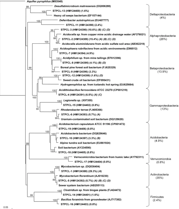
The clone library was composed of 123 clones that grouped into 19 phylotypes (Fig. 1). Five phylotypes ap-peared only once in the library, so the clone library coverage was 96%. The phylotypes could be divided into five phyla as follows: Proteobacteria (56.9% of the total clones), Actino-bacteria (35%), Acidobacteria (4.9%), Firmicutes (2.4%), and Verrucomicrobia (0.8%). The Proteobacteria were highly dominant and comprised 11 different phylotypes with repre-sentatives from four out of five subclasses: Alphaproteobac-teria (26% of the total clones), Betaproteobacteria (13.8%), Gammaproteobacteria (13%), and Deltaproteobacteria (4.1%). Together, Actinobacteria and Alphaproteobacteria phylotypes represented ∼60% of the clone library. The Actinobacteria phylotype was represented mainly by Myco-bacterium spp. The Alphaproteobacteria phylotypes were closely related to Acidocella spp. The Betaproteobacteria, the third major group of clones, were represented by four phylotypes that were mostly of acidophilic origin. Nine out of the 19 phylotypes (49.6% of the clones) had their closest relatives related to acidophilic genera. Only 6.5% of the clones were related to anaerobes: Desulfatimicrobium sp. and Deferribacter sp. from the Deltaproteobacteria, and Clostri-diumsp. and Bacillus sp. from the Firmicutes.
DGGE analysis and comparison of different primer sets The DGGE analysis showed that the different primer sets produced different DGGE band patterns: the patterns from the primer sets F1–519R and U341F–U803R were relatively similar to each other, and the patterns from U786F–U1401R and U968F–U1401R were more similar to each other
Can. J. Microbiol. Downloaded from www.nrcresearchpress.com by National Research Council of Canada on 06/06/11
Fig. 1. Phylogenetic relationship of the 19 bacterial 16S rRNA gene sequences obtained from the Sydney Tar Ponds sediment clone library. The clones are labeled with STPCL (Sydney Tar Ponds Clone Library) followed by a clone number: 1 to 19. The first set of parentheses indicate the GenBank accession number, the second set the percentage of clones in the library for that phylotype, and the last set with letters refer to the DGGE in which they were identified from Fig. 2. The tree was inferred by neighbor-joining analysis of a sequence from each clone. Aquifex pyrophilus was used as the outgroup. Numbers on the nodes are the bootstrap values based on 1000 replicates. The scale bar indicates the estimated number of base changes per nucleotide sequence position.
Can. J. Microbiol. Downloaded from www.nrcresearchpress.com by National Research Council of Canada on 06/06/11
(Fig. 2). However, the major bacterial groups that were iden-tified from all four primer sets were very similar (Figs. 3–6). From each primer set, the top five to six most intense bands were sequenced. From the F1–519R primer set (A) (Fig. 3), the top five most intense bands represented 48.7% of the total intensity in the lane. The most intense band (22.4%) was from a phylotype related to Mycobacterium sp., with a sec-ond band being attributed to another Mycobacterium sp. (5.2%). Other than the Mycobacterium spp., this primer set was able to amplify members of the genera Acidithiobacillus (8.5%), Rhodanobacter (7.1%), and Acidocella (5.5%). With the U341F–U803R primer set (B) (Fig. 4), the top five most intense bands represented 41.1% of the total intensity in the lane. The intensities of all the most intense bands were rela-tively similar, ranging from 11.6% to 4.6%. The two most in-tense bands were from phylotypes related to Acidocella spp., with relative intensities of 11.6% and 9.2%, followed by
those related to Mycobacterium sp. (8.9%), Acidiphilium sp. (6.8%), and Acidobacteria sp. (4.6%). From the U786F– U1401R primer set (C) (Fig. 5), the top five most intense bands represented 45.8% of the total intensity in the lane. The most intense band (21.6%) was from a phylotype related to Mycobacterium sp. Interestingly, there were three different bands attributed to members of Acidocella spp., with relative intensities of 9.2%, 5.6%, and 3.2%, and the last intense band was from a phylotype related to Acidithiobacillus sp. (6.2%). Lastly, from the U968F–U1401R primer set (D) (Fig. 6), the top six most intensive bands represented 77.9% of the total intensity in the lane. The most intense band (54%) was a very broad band identified as closely related to Mycobacte-rium sp. The other bands had lower intensities, with three phylotypes: one related to Acidocella spp. (7.2%, 5.9% and 3%), one to Acidiphilium sp. (7.1%), and one to an unknown Betaproteobacteria (3.8%).
The number of bands and Rr values from each primer set were calculated (Table 3). Primer sets A, B, and C produced similar numbers of bands (19–21) and similarly high Rr val-ues (48.66–76.65), but primer set D produced fewer bands (12) and thus a lower Rrvalue (15.62).
Comparison of clone library and DGGE results
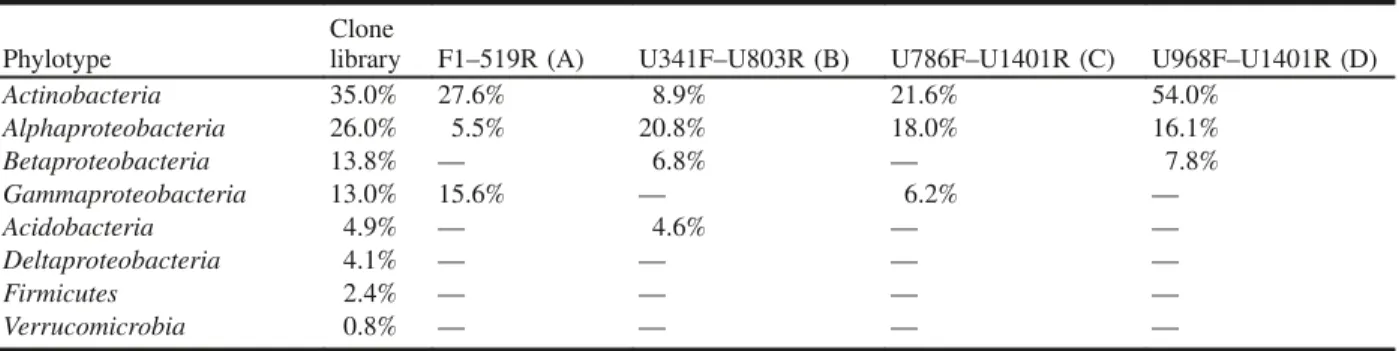
From the clone library analysis, 35% of the clones were re-lated to two Mycobacterium spp., and 26% of the clones were related to two Acidocella spp., making these two genera the most dominant in the clone library, comprising over 50% of the clones (Table 4). Similarly, all four DGGE primer sets were able to amplify members of these two genera (Myco-bacterium and Acidocella) with relatively high intensity bands (Table 4 and Figs. 3–6). Out of the four primer sets, only the F1–519R primer set amplified two phylotypes re-lated to the same Mycobacterium spp. identified in the clone library (Fig. 3), but only one of the two clone-library-identified Acidocella spp. was amplified using this primer set. However, all but the F1–519R primer set amplified phylotypes related to the two Acidocella spp. (Figs. 4–6). In-terestingly, in the clone library, one of the Mycobacterium sp. produced 36 clones, but it was not the one that was seen in the DGGE, except when using the F1–519R primer set. Discussion
Bacterial community structure of the Sydney Tar Ponds sediment
With nearly 100 years of high historical PAHs and PCBs contamination and an extremely low pH, the bacterial diver-sity in the sediment was expected to be low (Lauber et al.
Fig. 2. Denaturing gradient gel electrophoresis fingerprints with dif-ferent primer sets (A, B, C, D) of bacterial assemblages from the Sydney Tar Ponds sediment. The top most intense bands are indi-cated by numbers 1–6.
Table 3. Values of range-weighted richness index (Rr), number of
bands, and gel gradient range to cover all bands derived from de-naturing gradient gel electrophoresis patterns corresponding to mi-crobial community structures from different primer sets.
Primer
set Primer pair
No. of bands
Gel gradient to cover all bands Rr
A F1–519R 20 43.02%–59.93% 67.64 B U341F–U803R 21 41.74%–59.12% 76.65 C U786F–U1401R 19 42.44%–55.92% 48.66 D U968F–U1401R 12 47.67%–58.52% 15.62
Can. J. Microbiol. Downloaded from www.nrcresearchpress.com by National Research Council of Canada on 06/06/11
2009). Mendez et al. (2008) found that the bacterial diversity decreased with an increase in acidity. In their study, while the pH decreased from 7.7 to 2.7 (Sydney Tar Ponds pH = 2.8), the bacterial phylotype richness decreased from 42 phylo-types in the control sample to 24 in the moderately acidic samples to 8 in the extremely acidic tailings samples. Simi-larly, Vivas et al. (2008) found a decrease in bacterial diver-sity in a historical PAH-contaminated soil in comparison with unpolluted soil. With a similarly low pH environment and high PAH contamination, we identified 19 phylotypes from the clone library, suggesting that the bacterial community structure is fairly diverse. With both methods,
Mycobacte-riumspp. and Acidocella spp. were the most dominant bacte-ria, which suggests that regardless of PCR primer bias, the same dominant groups are obtained.
The results are supported by the physicochemical proper-ties of the sediment. The sediments were very acidic and the results revealed that 9 out of 19 phylotypes identified from the clone library were closely related to acidophilic bacteria. Most noticeable among them were Acidocella, Acidosphaera, Acidiphilium, Acidithiobacillus, Acidobacterium, and Acido-bacteria. Although care has to be taken in assigning physio-logical characteristics to organisms identified using only their 16S rRNA gene phylogenetic affiliation, the acidity of the
Fig. 3. Phylogenetic relationship of the five bacterial 16S rRNA gene sequences obtained from the Sydney Tar Ponds sediment with the F1– 519R primer set (A). The bands were labeled with STPDGGE (Sydney Tar Ponds denaturing gradient gel electrophoresis) and their lane and band number from Fig. 2. The first set of parentheses indicate the GenBank accession number, and the second set the percent intensity of the band relative to the total intensity in the lane. The tree was inferred by neighbor-joining analysis of sequence from each clone. Aquifex py-rophiluswas used as the outgroup. Numbers on the nodes are the bootstrap values based on 1000 replicates. The scale bar indicates the estimated number of base changes per nucleotide sequence position.
Fig. 4. Phylogenetic relationship of the 5 bacterial 16S rRNA gene sequences obtained from the Sydney Tar Ponds sediment with the U341F– U803R primer set (B). The bands were labeled with STPDGGE (Sydney Tar Ponds denaturing gradient gel electrophoresis) and their lane and band number from Fig. 2. The first set of parentheses indicate the GenBank accession number and the second set the percent intensity of the band relative to the total intensity in the lane. The tree was inferred by neighbor-joining analysis of sequence from each clone. Aquifex py-rophiluswas used as the outgroup. Numbers on the nodes are the bootstrap values based on 1000 replicates. The scale bar indicates the estimated number of base changes per nucleotide sequence position.
Can. J. Microbiol. Downloaded from www.nrcresearchpress.com by National Research Council of Canada on 06/06/11
sediment supports the observation that these nine retrieved phylotypes were acidophilic. and Uyttebroek et al. (2007) and López et al. (2008) found that members from Mycobac-teriumspp. thrive in acidic environments and could likely be acidophiles. If the phylotypes related to Mycobacterium spp. were also acidophilic, it would bring the total acidophilic clones in the library to ∼85%. The sediment is also character-ized by a high concentration of PAHs and PCBs (Table 1). Strains of Acidocella spp. and Mycobacterium spp. are known to degrade or to be associated with the degradation of hydrocarbons in low pH environments (Dore et al. 2003; Röling et al. 2006; Uyttebroek et al. 2007; López et al. 2008). The results from this study suggested that the micro-bial community associated with the Sydney Tar Ponds
sedi-ment was dominated by heterotrophic acidophiles and potential acidophilic hydrocarbon degraders.
Comparison of the two culture-independent methods This study revealed that the most dominant bacterial groups in the sample were identified independently using the construction of a 16S rRNA gene clone library and PCR– DGGE with four primer sets, but the results from the two methods still showed some differences. One of these differen-ces was due to the use of different primer sets. It is well rec-ognized that the quality of information produced by PCR– DGGE is dependent on both the number and resolution of the amplicons on the gels. Most DGGE users choose primer sets targeting different variable regions of the 16S rRNA
Fig. 5. Phylogenetic relationship of the five bacterial 16S rRNA gene sequences obtained from the Sydney Tar Ponds sediment with the U786F–U1401R primer set (C). The bands were labeled with STPDGGE (Sydney Tar Ponds denaturing gradient gel electrophoresis) and their lane and band number from Fig. 2. The first set of parentheses indicate the GenBank accession number and the second set the percent intensity of the band relative to the total intensity in the lane. The tree was inferred by neighbor-joining analysis of sequence from each clone. Aquifex pyrophiluswas used as the outgroup. Numbers on the nodes are the bootstrap values based on 1000 replicates. The scale bar indicates the estimated number of base changes per nucleotide sequence position.
Fig. 6. Phylogenetic relationship of the six bacterial 16S rRNA gene sequences obtained from the Sydney Tar Ponds sediment with the U968F–U1401R primer set (D). The bands were labeled with STPDGGE (Sydney Tar Ponds denaturing gradient gel electrophoresis) and their lane and band number from Fig. 2. The first set of parentheses indicates the GenBank accession number and the second set the percent intensity of the band relative to the total intensity in the lane. The tree was inferred by neighbor-joining analysis of sequence from each clone. Aquifex pyrophiluswas used as the outgroup. Numbers on the nodes are the bootstrap values based on 1000 replicates. The scale bar indicates the estimated number of base changes per nucleotide sequence position.
Can. J. Microbiol. Downloaded from www.nrcresearchpress.com by National Research Council of Canada on 06/06/11
gene, but there appears to have been little or no systematic evaluation of how the choice of primers influences data quality. Among the limited studies using multiple primer sets, the study conducted by Yu and Morrison (2004) showed that dif-ferent DGGE patterns were produced, with differences in the number and separation of DGGE bands, when targeting dif-ferent variable (V) regions of the bacterial 16S rRNA gene. In this study, four commonly used primer sets were used: two targeting the V3 and flanking regions, and two targeting the V6 and flanking regions (Table 2). These regions theoret-ically have the highest variability within the whole rRNA gene, thus allowing for optimal discrimination of bacterial communities (Muyzer et al. 1993; Heuer et al. 1997; Smalla et al. 1998). Like the results from Yu and Morrison (2004), our results indicated that different primer sets produced dif-ferent DGGE banding patterns. The F1–519R and U341F– U803R primer sets produced patterns that were similar to each other, while the U786F–U1401R and U968F–U1401R patterns were more similar to each other. This is most likely a result of the high variability in the V3 and V6 regions (Yu and Morrison 2004), but the similarity is partially due to the amplification of common regions (overlapping variable re-gions with both primer sets).
Marzorati et al. (2008) proposed a mathematical method using the Rrvalue to reflect the carrying capacity of the sam-pling environment. The assumption is that the broader the carrying capacity of an environment, the higher the probabil-ity that it can host a high number of bands with a wide GC content variability. Therefore, a Rr <10 is characterized as a low range-weighted richness and can be attributed to environ-ments particularly adverse to colonization. A Rr value be-tween 10 and 30 can be correlated with a medium range-weighted richness, and a Rr >30 is typical of very habitable environments. Our results showed that using different primer sets, the Rr values varied greatly from 15.62 with primer set U968F–U1401R (medium range-weighted richness) to 76.65 with primer set U341F–U803R (high range-weighted rich-ness) (Table 3). This suggests that an incorrect choice of pri-mer sets might yield a discrepancy in results that could overestimate or underestimate the richness of the environ-ment.
Several studies have shown that the dominant phylogenetic groups in 16S rRNA gene clone libraries are the same as the dominant in situ groups determined by FISH in natural mi-crobial communities (Cary et al. 1997; Schramm et al. 1998;
Ficker et al. 1999), so most researchers are more comfortable using clone libraries to estimate the relative abundance of bacteria. Fromin et al. (2002) and Murray et al. (1996) sug-gested that the relative band intensity from DGGE is consid-ered to be related to the relative abundance of the corresponding sequence type within the sample. Results ob-tained by Murray et al. (1996) from DGGE analysis sup-ported the relationship between band intensity and relative abundance of the corresponding phylotypes in the template DNA mixture. Our DGGE results (Table 3) revealed that all primer sets amplified the two most dominant groups (Actino-bacteria and Alphaproteobacteria) identified in the clone li-brary. The Betaproteobacteria and Gammaproteobacteria were present in the clone library at nearly the same levels (13.8% and 13%, respectively) but were amplified quite dif-ferently by the primer sets: F1–519R and U786F–U1401R amplified only the Gammaproteobacteria, while U341F– U803R and U968F–U1401R only amplified the Betaproteo-bacteria.
In different studies, the amplicon length can also vary greatly from 80 to 586 bp (list from Yu and Morrison 2004). However, Huber et al. (2009) revealed that the smallest size amplicons (in this case in a clone library) contained more different types of sequences, and accordingly, more diverse members of the community. In terms of DGGE analysis, it is important to obtain the most genetic information to achieve the highest possible resolution. In our case, the results sug-gested that the primer set U341F–U803R, which targets the V3–V4 regions and amplifies the shortest amplicon, gave the highest Rr value (Table 3), indicating a higher diversity and identifying the more abundant groups, which compared well with the result from the clone library (Table 3). However, if the aim is to monitor the changes in Mycobacterium spp., F1–519R would be a better primer set to use.
In conclusion, two culture-independent methods were used to characterize the diversity and richness of bacteria inhabit-ing the Sydney Tar Ponds. Even though the ponds have been exposed to heavy contamination over many generations, the bacterial diversity remains relatively high compared with other contaminated or acidic sites. Mycobacterium spp. and Acidocellaspp. were identified as the most dominant bacteria in the site, using both methods. This study also identified the primer sets F1–519R and U341F–U803R targeting the V3 re-gion of the 16S rRNA gene as the most suitable primer sets for future routine monitoring using PCR–DGGE analyses in this environment.
Table 4. Comparison of the percentage of clones in the clone library and the percentage of the intensive bands in each dena-turing gradient gel electrophoresis lane.
Phylotype
Clone
library F1–519R (A) U341F–U803R (B) U786F–U1401R (C) U968F–U1401R (D) Actinobacteria 35.0% 27.6% 8.9% 21.6% 54.0% Alphaproteobacteria 26.0% 5.5% 20.8% 18.0% 16.1% Betaproteobacteria 13.8% — 6.8% — 7.8% Gammaproteobacteria 13.0% 15.6% — 6.2% — Acidobacteria 4.9% — 4.6% — — Deltaproteobacteria 4.1% — — — — Firmicutes 2.4% — — — — Verrucomicrobia 0.8% — — — —
Can. J. Microbiol. Downloaded from www.nrcresearchpress.com by National Research Council of Canada on 06/06/11
Acknowledgements
The authors thank Peter Weaver for providing samples from the Sydney Tar Ponds, and Jennifer Mason and Tom King, from the Centre for Offshore Oil, Gas and Energy Re-search (COOGER), for their analytical support. We thank Danielle Beaumier for designing of the pDrive primers used in this study.
References
Baker, G.C., Smith, J.J., and Cowan, D.A. 2003. Review and re-analysis of domain-specific 16S primers. J. Microbiol. Methods, 55(3): 541–555. doi:10.1016/j.mimet.2003.08.009. PMID: 14607398.
Cary, S.C., Cottrell, M.T., Stein, J.L., Camacho, F., and Desbruyeres, D. 1997. Molecular identification and localization of filamentous symbiotic bacteria associated with the hydrothermal vent annelid Alvinella pompejana. Appl. Environ. Microbiol. 63(3): 1124– 1130. PMID:16535543.
Cole, J.R., Chai, B., Marsh, T.L., Farris, R.J., Wang, Q., Kulam, S.A., et al.Ribosomal Database Project. 2003. The Ribosomal Database Project (RDP-II): previewing a new autoaligner that allows regular updates and the new prokaryotic taxonomy. Nucleic Acids Res. 31(1): 442–443. doi:10.1093/nar/gkg039. PMID:12520046.
Dore, S.Y., Clancy, Q.E., Rylee, S.M., and Kulpa, C.F., Jr. 2003. Naphthalene-utilizing and mercury-resistant bacteria isolated from an acidic environment. Appl. Microbiol. Biotechnol. 63(2): 194– 199. doi:10.1007/s00253-003-1378-4. PMID:12827325.
Environmental Protection Agency. 2003. US Environmental Protec-tion Agency Test Methods 2003. EPA Methods 3510C, 3540C, and 8100.
Felske, A., Engelen, B., Nübel, U., and Backhaus, H. 1996. Direct ribosome isolation from soil to extract bacterial rRNA for community analysis. Appl. Environ. Microbiol. 62(11): 4162– 4167. PMID:8900007.
Ficker, M., Krastel, K., Orlicky, S., and Edwards, E. 1999. Molecular characterization of a toluene-degrading methanogenic consortium. Appl. Environ. Microbiol. 65(12): 5576–5585. PMID:10584020. Fromin, N., Hamelin, J., Tarnawski, S., Roesti, D., Jourdain-Miserez,
K., Forestier, N., et al. 2002. Statistical analysis of denaturing gel electrophoresis (DGE) fingerprinting patterns. Environ. Microbiol. 4(11): 634–643. doi:10.1046/j.1462-2920.2002.00358.x. PMID: 12460271.
Fuhrman, J.A., Mccallum, K., and Davis, A.A. 1992. Novel major archaebacterial group from marine plankton. Nature, 356(6365): 148–149. doi:10.1038/356148a0. PMID:1545865.
Good, I. 1953. The population frequencies of species and the estimation of population of parameters. Biometrika, 40: 237–264. Hall, T.A. 1999. BioEdit: a user-friendly biological sequence alignment editor and analysis program for Windows 95/98/NT. Nucleic Acids Symp. Ser. 41: 95–98.
Head, I.M., Saunders, J.R., and Pickup, R.W. 1998. Microbial evolution, diversity, and ecology: a decade of ribosomal rRNA analysis of uncultivated microorganisms. Microb. Ecol. 35(1): 1– 21. doi:10.1007/s002489900056. PMID:9459655.
Heuer, H., Krsek, M., Baker, P., Smalla, K., and Wellington, E.M.H. 1997. Analysis of actinomycete communities by specific ampli-fication of genes encoding 16S rRNA and gel-electrophoretic separation in denaturing gradients. Appl. Environ. Microbiol. 63(8): 3233–3241. PMID:9251210.
Huber, J.A., Morrison, H.G., Huse, S.M., Neal, P.R., Sogin, M.L., and Welch, D.B.M. 2009. Effect of PCR amplicon size on assessments of clone library microbial diversity and community
structure. Environ. Microbiol. 11(5): 1292–1302. PMID: 19220394.
Huber, T., Faulkner, G., and Hugenholtz, P. 2004. Bellerophon: a program to detect chimeric sequences in multiple sequence alignments. Bioinformatics, 20(14): 2317–2319. doi:10.1093/ bioinformatics/bth226. PMID:15073015.
Joint Review Panel for the Sydney Tar Ponds and Coke Ovens Sites Remediation Project. 2006. Joint review panel environmental assessment report. Available from www.gov.ns.ca/nse/ea/tarponds/ TarPonds_EnvironmentalAssessmentReport.pdf. ISBN: 0-662-43508-7 (accessed July 2009).
Karl, D.M. 2002. Microbiological oceanography: hidden in a sea of microbes. Nature, 415(6872): 590–591. doi:10.1038/415590b. PMID:11832923.
King, T.L., and Chou, C.L. 2003. Anthropogenic organic contami-nants in American lobster (Homarus americanus) procured from harbours, bays, and inlets of Eastern Canada. Can. Tech. Rep. Fish. Aquat. Sci. 2456. Department of Fisheries and Oceans, Bedford, N.S., Canada.
King, T.L., and Lee, K. 2004. Assessment of sediment quality based on toxic equivalent benzo[a]pyrene concentrations. In Proceedings of the 27th Arctic and Marine Oilspill Program (AMOP), Edmonton, Alta., Canada, 8–10 June 2004. Environmental Science and Technology Division, Environment Canada, Ottawa, Ont., Canada. pp. 793–806.
King, T.L., Haines, B.K., and Uthe, J.F. 1996. Non-, mono-, and di-o-chlorobiphenyls concentrations and their toxic equivalents to 2,3,7,8-tetrachlorodibenzo[p]dioxin in Aroclors and digestive glands from American Lobster (Homarus americanus) captured in Atlantic Canada. Bull. Environ. Contam. Toxicol. 57(3): 465– 472. doi:10.1007/s001289900213. PMID:8672074.
Lane, D.J., Pace, B., Olsen, G.J., Stahl, D., Sogin, M.L., and Pace, N. R. 1985. Rapid determination of 16S ribosomal RNA sequences for phylogenetic analysis. Proc. Natl. Acad. Sci. U.S.A. 82(20): 6955–6959. doi:10.1073/pnas.82.20.6955. PMID:2413450. Lauber, C.L., Hamady, M., Knight, R., and Fierer, N. 2009.
Pyrosequencing-based assessment of soil pH as a predictor of soil bacterial community structure at the continental scale. Appl. Environ. Microbiol. 75(15): 5111–5120. doi:10.1128/AEM. 00335-09. PMID:19502440.
Lee, G.F., and Jones-Lee, A. 2006. Progress toward remediation of the Sydney Tar Ponds: a major Canadian PCB/PAH “superfund” site. Rem. J. 17(1): 111–119.
Liesack, W., Weyland, H., and Stackebrandt, E. 1991. Potential risks of gene amplification by PCR as determined by 16S rDNA analysis of a mixed culture of strict barophilic bacteria. Microb. Ecol. 21(1): 191–198. doi:10.1007/BF02539153.
López, Z., Vila, J., Ortega-Calvo, J.J., and Grifoll, M. 2008. Simultaneous biodegradation of creosote-polycyclic aromatic hydrocarbons by a pyrene-degrading Mycobacterium. Appl. Microbiol. Biotechnol. 78(1): 165–172. doi:10.1007/s00253-007-1284-2. PMID:18074131.
Marzorati, M., Wittebolle, L., Boon, N., Daffonchio, D., and Verstraete, W. 2008. How to get more out of molecular fingerprints: practical tools for microbial ecology. Environ. Microbiol. 10(6): 1571–1581. doi:10.1111/j.1462-2920.2008. 01572.x. PMID:18331337.
Mendez, M.O., Neilson, J.W., and Maier, R.M. 2008. Characteriza-tion of a bacterial community in an abandoned semiarid lead–zinc mine tailing site. Appl. Environ. Microbiol. 74(12): 3899–3907. doi:10.1128/AEM.02883-07. PMID:18424534.
Murray, A.E., Hollibaugh, J.T., and Orrego, C. 1996. Phylogenetic compositions of bacterioplankton from two California estuaries compared by denaturing gradient gel electrophoresis of 16S rDNA
Can. J. Microbiol. Downloaded from www.nrcresearchpress.com by National Research Council of Canada on 06/06/11
fragments. Appl. Environ. Microbiol. 62(7): 2676–2680. PMID: 8779608.
Muyzer, G., De Waal, E.C., and Uitterlinden, A.G. 1993. Profiling of complex microbial populations by denaturing gradient gel electrophoresis analysis of polymerase chain reaction-amplified gene coding for 16S rRNA. Appl. Environ. Microbiol. 59(3): 695– 700. PMID:7683183.
Rappé, M.S., and Giovannoni, S.J. 2003. The uncultured microbial majority. Annu. Rev. Microbiol. 57: 369–394. PMID:14527284. Röling, W.F., van Breukelen, B.M., Braster, M., Lin, B., and van
Verseveld, H.W. 2001. Relationships between microbial commu-nity structure and hydrochemistry in a landfill leachate-polluted aquifer. Appl. Environ. Microbiol. 67(10): 4619–4629. doi:10. 1128/AEM.67.10.4619-4629.2001. PMID:11571165.
Röling, W.F., Ortega-Lucach, M.S., Larter, S.R., and Head, I.M. 2006. Acidophilic microbial communities associated with a natural, biodegraded hydrocarbon seepage. J. Appl. Microbiol. 101(2): 290–299. doi:10.1111/j.1365-2672.2006.02926.x. PMID:16882136. Schramm, A., de Beer, D., Wagner, M., and Amann, R. 1998. Identification and activities in situ of Nitrosospira and Nitrospira spp. as dominant populations in a nitrifying fluidized bed reactor. Appl. Environ. Microbiol. 64(9): 3480–3485. PMID:9726900. Sheffield, V.C., Cox, D.R., Lerman, L.S., and Myers, R.M. 1989.
Attachment of a 40-base-pair G + C-rich sequence (GC-clamp) to genomic DNA fragments by the polymerase chain reaction results in improved detection of single-base changes. Proc. Natl. Acad. Sci. U.S.A. 86(1): 232–236. doi:10.1073/pnas.86.1.232. PMID: 2643100.
Smalla, K., Wachtendorf, U., Heuer, H., Liu, W.T., and Forney, L. 1998. Analysis of BIOLOG GN substrate utilization patterns by microbial communities. Appl. Environ. Microbiol. 64(4): 1220– 1225. PMID:16349535.
Timmis, K.N., McGenity, T., Van Der Meer, J.R., and de Lorenzo, V. 2010. Handbook of hydrocarbon and lipid microbiology. Springer-Verlag, Berlin, Heidelberg, Germany.
Uyttebroek, M., Vermeir, S., Wattiau, P., Ryngaert, A., and Springael, D. 2007. Characterization of cultures enriched from acidic polycyclic aromatic hydrocarbon contaminated soil for growth on pyrene at low pH. Appl. Environ. Microbiol. 73(10): 3159– 3164. doi:10.1128/AEM.02837-06. PMID:17369339.
Van de Peer, Y., Chapelle, S., and De Wachter, R. 1996. A quantitative map of nucleotide substitution rates in bacterial rRNA. Nucleic Acids Res. 24(17): 3381–3391. doi:10.1093/nar/24.17. 3381. PMID:8811093.
Vivas, A., Moreno, B., Del Val, C., Macci, C., Masciandaro, G., and Benitez, E. 2008. Metabolic and bacterial diversity in soils historically contaminated by heavy metals and hydrocarbons. J. Environ. Monit. 10(11): 1287–1296. doi:10.1039/b808567f. PMID:18974897.
Yu, Z., and Morrison, M. 2004. Comparisons of different hypervari-able regions of rrs genes for use in fingerprinting of microbial communities by PCR-denaturing gradient gel electrophoresis. Appl. Environ. Microbiol. 70(8): 4800–4806. doi:10.1128/AEM. 70.8.4800-4806.2004. PMID:15294817.
Can. J. Microbiol. Downloaded from www.nrcresearchpress.com by National Research Council of Canada on 06/06/11