HAL Id: hal-02005297
https://hal.archives-ouvertes.fr/hal-02005297
Submitted on 3 Feb 2019
HAL is a multi-disciplinary open access
archive for the deposit and dissemination of
sci-entific research documents, whether they are
pub-lished or not. The documents may come from
teaching and research institutions in France or
abroad, or from public or private research centers.
L’archive ouverte pluridisciplinaire HAL, est
destinée au dépôt et à la diffusion de documents
scientifiques de niveau recherche, publiés ou non,
émanant des établissements d’enseignement et de
recherche français ou étrangers, des laboratoires
publics ou privés.
Crossmodal Adaptation in Right Posterior Superior
Temporal Sulcus during Face-Voice Emotional
Integration
Rebecca Watson, Marianne Latinus, Takao Noguchi, Oliver Garrod, Frances
Crabbe, Pascal Belin
To cite this version:
Rebecca Watson, Marianne Latinus, Takao Noguchi, Oliver Garrod, Frances Crabbe, et al..
Crossmodal Adaptation in Right Posterior Superior Temporal Sulcus during Face-Voice Emotional
Integration.
Journal of Neuroscience, Society for Neuroscience, 2014, 34 (20), pp.6813-6821.
�10.1523/jneurosci.4478-13.2014�. �hal-02005297�
Behavioral/Cognitive
Crossmodal Adaptation in Right Posterior Superior
Temporal Sulcus during Face–Voice Emotional Integration
Rebecca Watson,
1,2Marianne Latinus,
2,3Takao Noguchi,
4Oliver Garrod,
2Frances Crabbe,
2and Pascal Belin
2,3,51Department of Cognitive Neuroscience, Faculty of Psychology and Neuroscience, Maastricht University, Maastricht 6229 EV, The Netherlands,2Centre for Cognitive Neuroimaging, Institute of Neuroscience and Psychology, University of Glasgow, Glasgow, G12 8QB, United Kingdom,3Neuroscience Institute of Timone, Coeducational Research Unit 7289, National Center of Scientific Research–Aix-Marseille University, F-13284 Marseille, France,4Department of Psychology, University of Warwick, Coventry, CV4 7A, United Kingdom, and5International Laboratories for Brain, Music, and Sound, University of Montreal and McGill University, Montreal, Quebec, Canada, H2V 4P3
The integration of emotional information from the face and voice of other persons is known to be mediated by a number of
“multisen-sory” cerebral regions, such as the right posterior superior temporal sulcus (pSTS). However, whether multimodal integration in these
regions is attributable to interleaved populations of unisensory neurons responding to face or voice or rather by multimodal neurons
receiving input from the two modalities is not fully clear. Here, we examine this question using functional magnetic resonance adaptation
and dynamic audiovisual stimuli in which emotional information was manipulated parametrically and independently in the face and
voice via morphing between angry and happy expressions. Healthy human adult subjects were scanned while performing a happy/angry
emotion categorization task on a series of such stimuli included in a fast event-related, continuous carryover design. Subjects integrated
both face and voice information when categorizing emotion—although there was a greater weighting of face information—and showed
behavioral adaptation effects both within and across modality. Adaptation also occurred at the neural level: in addition to
modality-specific adaptation in visual and auditory cortices, we observed for the first time a crossmodal adaptation effect. Specifically, fMRI signal
in the right pSTS was reduced in response to a stimulus in which facial emotion was similar to the vocal emotion of the preceding stimulus.
These results suggest that the integration of emotional information from face and voice in the pSTS involves a detectable proportion of
bimodal neurons that combine inputs from visual and auditory cortices.
Key words: emotion perception; functional magnetic resonance adaptation; multisensory integration
Introduction
Stimulation in natural settings usually recruits a number of
dif-ferent sensory channels in parallel. Particularly important with
regards to social interaction is the recognition of emotional cues
from both the face and voice of others. A number of regions of the
human brain have been implicated in the integration of these
affective cues, including “convergence zones,” such as the
poste-rior supeposte-rior temporal sulcus (pSTS;
Pourtois et al., 2005
;
Kreif-elts et al., 2007
;
Hagan et al., 2009
,
2013
;
Robins et al., 2009
),
putative “unisensory” regions (i.e., the primary visual and
audi-tory cortices;
de Gelder et al., 1999
;
Pourtois et al., 2000
,
2002
),
and limbic structures, such as the amygdala (
Dolan et al., 2001
;
Klasen et al., 2011
). These regions tend to show greater activity as
measured with neuroimaging techniques such as fMRI in
re-sponse to bimodal face–voice emotional stimulation than to
ei-ther modality alone, leading to them being classified as
“multisensory.”
However, it remains unclear whether the enhanced responses
of these presumed affective multisensory regions reflect
popula-tions of truly bimodal neurons (that receive affective input from
both the visual and auditory modalities) or is simply evoked by
groups of interdigitized, unimodal visual and auditory neurons.
Because of its limited spatial resolution, in which activity from
hundreds and thousands of (potentially, heterogeneous) neurons
within a voxel is averaged out, the traditional fMRI method
can-not distinguish between these two possibilities. Various fMRI
studies defined multisensory regions using specific statistical
cri-teria, but a number of these allow in theory for a purely additive
audio and visual response, which could be evoked simply by
over-lapping face- and voice-sensitive neurons.
Researchers attempted to circumvent the problem of limited
spatial resolution in fMRI by using functional magnetic
reso-nance adaptation (fMR-A;
Grill-Spector et al., 1999
,
2006
). The
logic is as follows: if repetition of a given feature in the
stimula-tion results in a reducstimula-tion of fMRI signal in a given voxel, then
that voxel is assumed to contain neurons that are specifically
involved in processing, or representing, the repeated feature. It
was suggested that fMR-A might also be helpful in distinguishing
between voxels in the multisensory cortex consisting of only
uni-Received Oct. 18, 2013; revised Feb. 21, 2014; accepted March 12, 2014.
Author contributions: R.W., M.L., and P.B. designed research; R.W., T.N., O.G., and F.C. performed research; R.W., M.L., and P.B., analyzed data; R.W. and P.B. wrote the paper.
This work was funded by Biotechnology and Biological Sciences Research Council Grant BBJ003654/1. The authors declare no competing financial interests.
Correspondence should be addressed to Rebecca Watson, Department of Cognitive Neuroscience, Maastricht University, Oxfordlaan 55, Maastricht 6229 EV, The Netherlands. E-mail: rebecca.watson@maastrichtuniversity.nl.
DOI:10.1523/JNEUROSCI.4478-13.2014
sensory neuronal subpopulations and voxels composed of a
mix-ture of unisensory and multisensory populations (
Goebel and
van Atteveldt, 2009
;
Tal and Amedi, 2009
). Varying adaptation
and recovery responses could shed light on the subvoxel
organi-zation of proposed affective multisensory regions: multisensory
neurons should adapt to crossmodal repetitions of emotion
in-formation (e.g., an angry voice followed by an angry face),
whereas unisensory neurons should not.
Here we used fMR-A to investigate crossmodal adaptation to
affective information in faces and voices. We used dynamic
audio-visual stimuli in which affective information was independently and
parametrically manipulated in each modality and used these stimuli
in an efficiency-optimized, “continuous carryover” design (
Aguirre,
2007
) as a means to test whether crossmodal adaptation effect could
be observed, i.e., a significant influence of emotional information in
one modality on neural response to emotional information in the
other modality. Specifically, we hypothesized that, if a cerebral
re-gion contained a sufficiently large proportion of multisensory
audi-tory–visual neurons involved in processing emotional information,
as opposed to interspersed populations of unisensory neurons, that
region should show detectable evidence of crossmodal adaptation.
Materials and Methods
Participants
Eighteen participants (10 males, eight females; mean⫾ SD age, 25 ⫾ 3.7 years) were scanned in the fMRI experiment. All had self-reported nor-mal or corrected-to-nornor-mal vision and hearing. The study was approved by the ethical committee of the University of Glasgow and conformed to the Declaration of Helsinki. All volunteers provided informed written consent before and received payment at the rate of £6/h for participation.
Stimuli
Stimuli consisted of 25 novel face–voice stimuli used previously by our laboratory. Stimulus construction and pretesting has been described pre-viously in full byWatson et al. (2013)and therefore will be described in brief here.
Raw audiovisual content, recorded using a Did3 capture system (Di-mensional Imaging Ltd.), consisted of two actors (one male, one female) expressing anger and happiness in both the face and voice. The sound “ah” was chosen because it contains no linguistic information. Actors were initially instructed to express each emotion with low, medium, and high intensity, with standardized timing when possible. Two final clips per actor (one anger, one happiness) were selected (on the basis of high-intensity production, similar duration) for use.
Audiovisual clips were then split into their audio and visual compo-nents, for within-modality morphing. A landmarked face mesh was ap-plied to each frame of the sequence, which was then used as a basis for morphing in MATLAB (Mathworks). Face morphing consisted of mor-phing between angry dynamic facial expressions (one male and one fe-male) and happy dynamic facial expressions (one male and one fefe-male) within gender. The resulting output was two different face continua (one per gender), each consisting of five within-modality stimuli, morphed between 90% anger and 90% happiness in 20% steps.
Audio output from the audiovisual recordings was processed in Adobe Audition 2.0 (Adobe Systems) and then morphed using the MATLAB-based morphing algorithm STRAIGHT (Kawahara, 2006). Vocal mor-phing ran in parallel with the facial mormor-phing, in that the equivalent individual voices were also morphed between 90% anger and 90% hap-piness in 20% steps, resulting in two vocal continua, one per actor, each consisting of five vocal stimuli.
Within actor, the five dynamic face and five voice stimuli were all equal length. To ensure that all stimuli were of equal length, we edited video and audio clips between actors. Editing was conducted in Adobe Pre-miere 2.0 (Adobe Systems) and consisted of inserting or deleting video frames to match predefined time points (e.g., mouth opening, mouth closing) across clip. We made efforts to ensure that editing occurred
between frames with as little difference in movement as possible to retain the naturalness of the video clip. The editing produced 10 adjusted video clips, each 780 ms long. The audio samples were then also adjusted in accordance with the temporal landmarks identified in the video clips to create 10 vocalizations (five for each actor) of equal length. Within actor, the five visual and five auditory clips were then paired together in all possible combinations. This resulted in a total of 25 audiovisual stimuli for each actor, parametrically varying in congruence between face and voice affective information. Stimuli are illustrated inFigure 1.
Design and procedure
Continuous carryover experiment
In the main experiment, stimuli were presented using the Psychtoolbox in MATLAB via electrostatic headphones (NordicNeuroLab) at a sound pressure level of 80 dB as measured using a Lutron Sl-4010 sound level meter. Before they were scanned, subjects were presented with sound samples to verify that the sound pressure level was comfortable and loud enough considering the scanner noise. Audiovisual movies were pre-sented in two scanning runs (over 2 different days) while the blood oxygenation level-dependent (BOLD) signal was measured in the fMRI scanner. We used a continuous carryover experimental design (Aguirre, 2007). This design allows for measurement of the direct effects (i.e., that of face and voice emotion morph) and the repetition suppression effect, which can be observed in pairs of voices or faces (like in the typical fMRI adaptation experiments).
The stimulus order followed two interleaved N⫽ 25 type 1–index 1 se-quences (one for each of the speaker continua; interstimulus interval, 2 s), which shuffles stimuli within the continuum so that each stimulus is pre-ceded by itself and every other within-continuum stimulus in a balanced manner. The sequence was interrupted seven times with 20 s silent periods, which acted as a baseline, and at the end of a silent period, the last five stimuli of the sequence preceding the silence were repeated before the sequence continued. These stimuli were removed in our later analysis. Participants were instructed to perform a two-alternative forced-choice emotion classi-fication task (responses, angry or happy) using two buttons of an MR-compatible response pad (NordicNeuroLab). They were also instructed to pay attention to both the face and voice but were told they could use the information presented in whatever way they wanted to make their decision on emotion. Reaction times (relative to stimulus onset) were collected using MATLAB with a response window limited to 2 s.
Localization of multisensory regions
In addition to the main experiment, we also used an independent mul-tisensory localizer to identify regions involved in mulmul-tisensory process-ing. We further performed an isolated region of interest (ROI) analysis within these areas to assess whether there were significant crossmodal adaptation effects. Therefore, we were consequently able to infer the neuronal properties (i.e., multisensory vs interdigitized unisensory neu-rons) of these independently established multisensory regions. During an 11 min scanning run, participants were presented with a variety of dynamic audiovisual and unimodal stimuli in 18 different 16 s blocks (for additional details of stimuli, refer toWatson et al., 2014). Thus, each block contained eight different stimuli. These blocks were broadly cate-gorized as follows: (1) faces paired with their corresponding vocal sounds (AV-P); (2) objects (visual and audio) (AV-O); (3) voices alone (A-P); (4) objects (audio only) (A-O); (5) faces alone (V-P); and (6) objects (visual only) (V-O).
Thus, categories 1 and 2 were audiovisual, 3 and 4 were audio only, and 5 and 6 were visual only. There were three different stimulus blocks within each type, each containing different visual/auditory/audiovisual stimuli. A 16 s null event block comprising silence and a gray screen was also created. Each of the 18 blocks was repeated twice, and the blocks were presented pseudorandomly: each block was always preceded and followed by a block from a different category (e.g., a block from the “faces-alone” category could never be preceded/followed by any other block from the faces-alone category). The null event block was repeated six times and interspersed randomly within the presentations of the stim-ulus blocks.
Imaging parameters
Functional images covering the whole brain (32 slices; field of view, 210⫻ 210 mm; voxel size, 3 ⫻ 3 ⫻ 3 mm) were acquired on a 3 T Tim Trio Scanner (Siemens) with a 12 channel head coil, using an echo planar imaging (EPI) sequence (interleaved; TR, 2 s; TE, 30 ms; flip angle, 80°) in both the carryover and localizer experiments. In total, we acquired 1560 EPI volumes for the carryover experiment, split into two scanning ses-sions consisting of 780 EPI volumes, and 336 EPI volumes were acquired for the multimodal localizer. For both the carryover experiment and experimental localizer, the first 4 s of the functional run consisted of “dummy” gradient and radio frequency pulses to allow for steady-state magnetization during which no stimuli were presented and no fMRI data were collected. MRI was performed at the Centre for Cognitive Neuro-imaging (Glasgow, UK).
At the end of each fMRI session, high-resolution T1-weighted struc-tural images were collected in 192 axial slices and isotropic voxels (1⫻ 1⫻ 1 mm; field of view, 256 ⫻ 256 mm2; TR, 1900 ms; TE, 2.92 ms; time to inversion, 900 ms; flip angle, 9°).
Imaging analysis
SPM8 software (Wellcome Department of Imaging Neuroscience, Lon-don, UK) was used to preprocess and analyze the imaging data. First, the anatomical scan was anterior commissure–posterior commissure cen-tered, and this correction applied to all EPI volumes.
Functional data were motion corrected using a spatial transformation that realigned all functional volumes to the first volume of the run and subsequently realigned the volumes to the mean volume. The anatomical scan was coregistered to the mean volume and segmented. The anatom-ical and functional images were then normalized to the Montreal Neu-rological Institute (MNI) template using the parameters issued from the segmentation keeping the voxel resolution of the original scans (1⫻ 1 ⫻ 1 and 3 ⫻ 3 ⫻ 3 mm, respectively). Functional images were then smoothed with a Gaussian function (8 mm FWHM).
EPI time series were analyzed using the general linear model as imple-mented in SPM8. For each subject (first-level analysis), localizer and experimental data were modeled separately.
Localizer data
EPI time series were analyzed using the general linear model as imple-mented in SPM8. Functional data were analyzed in one two-level random-effects design. The first-level, fixed-effects individual partici-pant analysis involved a design matrix containing a separate regressor for each block category (n⫽ 6). These regressors contained boxcar functions representing the onset and offset of stimulation blocks convolved with a canonical hemodynamic response function. To account for residual mo-tion artifacts, the realignment parameters were also added as nuisance covariates to the design matrix. Using the modified general linear model, parameter estimates for each condition at each voxel were calculated and then used to create contrast images for each category relative to baseline: AV-P⬎ baseline, AV-O ⬎ baseline, A-P ⬎ baseline, A-O ⬎ baseline, V-P⬎ baseline, and V-O ⬎ baseline. These six contrast images, from each participant, were taken forward into the second-level two factors (modality and category) ANOVA. The order of conditions was as fol-lows: audiovisual (face⫹ voice); audiovisual (object ⫹ sound); audio only (vocal); audio only (nonvocal); visual only (object); and visual only (face).
We then tested for general audiovisual regions with the conjunction analysis AV⬎ V 32 艚 AV ⬎ A (conjunction null hypothesis;Nichols et al., 2005), including both people and object information (i.e., AV-P⫹ AV-O⬎ V-P ⫹ V-O 艚 AV-P ⫹ AV-O ⬎ A-P ⫹ A-O). This localized regions that showed a higher response to audiovisual stimuli compared with both visual and audio-only stimuli.
Localizer results are reported at a threshold of p⬍ 0.05 (cluster size corrected).
Main functional run: adaptation (continuous carryover)
Functional data were analyzed using four two-level random-effects de-signs: two that examined unimodal carryover effects, and two that exam-ined crossmodal carryover effects.
Unimodal adaptation. For both face and voice unimodal carryover effects, brain activity time-locked to stimulus onset and duration was modeled in separate design matrices against one parametric modulator, which accounted for the absolute percentage difference between the (1)
Figure 1. Experimental stimuli: two sets (1 per actor) of dynamic and time-synchronized audiovisual stimuli were presented within a continuous carryover design (Aguirre, 2007) in interleaved type 1–index 1 sequences over two experimental sessions (sequential presentation indicated by dotted lines in the left panels). Every face morph (extending from 90% angry to 90% happy) was paired with every voice morph (extending from 90% angry to 90% happy) within actor, so to create two sets of 25 face–voice stimuli, parametrically varying in congruence (examples in colored rectangles). In a carryover design, every stimulus precedes and follows every other stimulus, such that each stimulus serves as an adaptor for the following stimulus. The right panels show parts of an example type 1–index 1 sequence. In each experimental run, sequences were blocked by actor. Participants performed a two-alternative forced-choice task (angry or happy) on emotion. The right panels indicate examples of within-block sequences of stimuli. ISI, Interstimulus interval.
face (i.e., unimodal face adaptation) or (b) voice morph levels (i.e., uni-modal voice adaptation) of consecutive biuni-modal stimuli.
Crossmodal adaptation. In two separate design matrices (one for each possible direction of the crossmodal effect, i.e., face-to-voice or voice-to-face), brain activity was modeled against three parametric modulators: (1) the first accounted for the absolute difference between the face morph levels of consecutive stimuli (unimodal face adaptation); (2) the second accounted for the absolute difference between the voice morph levels of consecutive stimuli (unimodal voice adaptation); and (3) the third ac-counted for the crossmodal carryover effect that was either the absolute percentage difference between the (a) voice morph of a stimulus and the face morph of the following stimulus (i.e., voice-to-face crossmodal ad-aptation) or (b) the absolute percentage difference between the face morph of a stimulus and the voice morph of the following stimulus (i.e., face-to-voice crossmodal adaptation). In these design matrices, the latter crossmodal regressor was orthogonalized with respect to the first two unimodal regressors in the SPM routine. In this way, we were able to regress out the variance associated with unimodal effects before examin-ing crossmodal effects. This was to ensure that we did not misinterpret effects apparently related to crossmodal adaptation but in fact attribut-able to unimodal adaptation.
In all four design matrices (unimodal face adaptation, unimodal voice adaptation, face-to-voice adaptation, and voice-to-face adaptation), a linear expansion allowed us to investigate regions in which the signal varied in account with the percentage difference in morph between stim-uli, with a hypothesized linear modulation of signal as the degree of morph level difference increased parametrically. Contrasts for the effects at the first level for each design matrix were entered into four separate second-level, group random-effects analysis, in which we conducted a one-sample t test over first-level contrast images from all participants.
Whole-brain analyses are reported within an audiovisual versus base-line mask (mask threshold, p⬍ 0.001, voxel uncorrected) at a threshold of p⬍ 0.05 (FWE voxel corrected).
ROI analysis
In parallel with the whole-brain analysis, we performed an ROI-based analysis to specifically examine regions highlighted as involved in audio-visual integration in the separate multimodal localizer. Tests of unimodal and crossmodal effects within this ROI were conducted within MarsBar (ROI toolbox for SPM).
Results
Behavioral results
Direct effects
Each participant’s mean categorization values for each audiovisual
emotion morph stimulus (collapsed across actor) was submitted to a
two-factor (face morph and voice morph) repeated-measures ANOVA,
with five levels per factor (percentage of “anger” information in the
morph). This was to assess the overall contributions of face and voice
emotion morph on categorical response. The repeated-measures
ANOVA highlighted a main effect of voice morph (F
(1.14,19.4)⫽ 15.3,
p
⬍ 0.002,
2p
⫽ 0.473) and face morph (F
(2.02,34.3)
⫽ 348, p ⬍ 0.0001,
2p
⫽0.953)andalsoasignificantvoice⫻faceinteraction(F
(5.78,98.1)
⫽
6.78, p
⬍0.0001,
2p
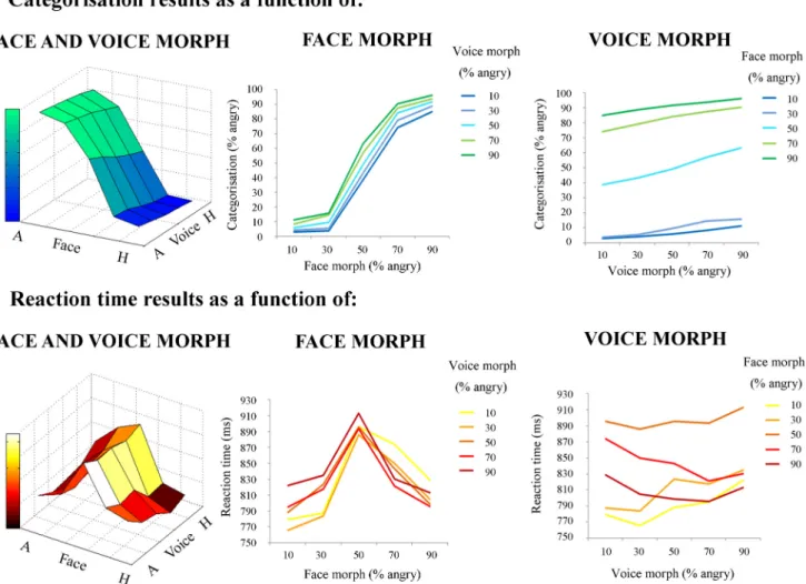
⫽0.285).Thus, it appears that face morph had
a larger driving effect overall on categorization ratings, but its
influence differed depending on what particular voice with which
a face was paired.
In a series of planned comparisons, we further examined at
which points there were significant differences in categorization
ratings between stimuli. We proposed that maximum
incongru-ence between face and voice (i.e., 80% differincongru-ence) would cause
significant shifts in categorization compared with “endpoint”
congruent stimuli (i.e., 10% angry face–10% angry voice; 90%
angry face–90% angry voice). To test these hypotheses, we
per-formed the following paired-sample t tests: (1) 10% angry face–
10% angry voice versus 10% angry face–90% angry voice; (2)
10% angry face–90% angry voice versus 90% angry face–90%
angry voice; (3) 90% angry face–90% angry voice versus 90%
angry face–10% angry voice; and (4) 90% angry face–10% angry
voice versus 10% angry face–10% angry voice. After a
Bonferro-ni’s correction for multiple comparisons (level of significance,
p
⬍ 0.0125), all comparisons were significant (comparison 1:
t
(17)⫽ ⫺24.0, p ⬍ 0.0001; comparison 2: t
(17)⫽ ⫺3.42, p ⬍
0.004; comparison 3: t
(17)⫽ 27.6, p ⬍ 0.0001; and comparison 4:
t
(17)⫽ 2.87, p ⬍ 0.0125, respectively).
Second, each participant’s mean reaction time values for each
stimulus (collapsed across actors) were submitted to another
two-factor (face morph and voice morph) repeated-measures
ANOVA, with five levels per factor (percentage of anger
informa-tion in the morph). As with categorical data, this was to assess the
overall contribution of face and voice emotion morph— or
the “direct effects” of face and voice morph— on reaction times.
The ANOVA of reaction time data highlighted a main effect of
voice morph (F
(2.91,49.6)⫽ 11.8, p ⬍ 0.0001,
2p
⫽ 0.409) and
face morph (F
(2.34,39.7)⫽ 70.6, p ⬍ 0.0001,
2p
⫽ 0.806) and also
a significant interaction between the two modalities (F
(2.90,39.4)⫽
7.40, p
⬍ 0.0001,
2p
⫽ 0.303). Similar to the previous analysis,
face morph drove the speed of categorization more than voice
morph, albeit with different modulating effects at particular
points in the 3D categorization space.
As in our categorization analysis, we proposed that maximum
incongruence between face and voice (i.e., 80% difference) would
take significantly longer to categorize compared with endpoint
congruent stimuli. However, we also expected that some stimuli
that were congruent, but with a lower clarity value (i.e., 50%
angry face–50% angry voice), would take longer to categorize
than endpoint congruent stimuli. To test these hypotheses, we
performed the following paired-sample t tests: (1) 10% angry
face–10% angry voice versus 10% angry face–90% angry voice;
(2) 10% angry face–90% angry voice versus 90% angry face–90%
angry voice; (3) 90% angry face–90% angry voice versus 90%
angry face–10% angry voice; (4) 90% angry face–10% angry voice
versus 10% angry face–10% angry voice; (5) 50% angry face–50%
angry voice versus 10% angry face–10% angry voice; and (6) 50%
angry face–50% angry voice versus 90% angry face–90% angry
voice. After a Bonferroni’s correction for multiple comparisons
(level of significance, p
⬍ 0.008), comparisons 1, 4, 5, and 6 were
significant (comparison 1: t
(17)⫽ ⫺4.72, p ⬍ 0.0001; comparison
4: t
(17)⫽ 3.25, p ⬍ 0.006; comparison 5: t
(17)⫽ 10.67, p ⬍ 0.0001;
and comparison 6: t
(17)⫽ 6.29, p ⬍ 0.0001, respectively), but
comparisons 2 and 3 were not (comparison 2: t
(17)⫽ 1.30, p ⫽
0.210; and comparison 3: t
(17)⫽ ⫺5.80, p ⫽ 0.569, respectively).
All direct behavioral results are illustrated in
Figure 2
.
Adaptation effects
Here the interest was to investigate whether and how difference in
emotion morph affected speed of emotion categorization. We
conducted a hierarchical regression analysis for each subject, in
which there were four regressors (face-to-face emotion morph
difference, voice-to-voice emotion morph difference,
face-to-voice emotion morph difference, and face-to-voice-to-face emotion
morph difference), two of which were covariates in our model
(face-to-face emotion morph difference and voice-to-voice
emo-tion morph difference, i.e., the unimodal effects), and the
depen-dent variable was reaction time. The first five stimulus values
from each experimental block (apart from block one) were
re-moved. This analysis provided two models: one that included
only unimodal adaptation regressors, and a second that included
all four adaptation regressors. In this way, and in parallel with the
fMRI analysis, we ensured that any variance associated with the
two crossmodal adaptation regressors was independent of any
unimodal effects. To analyze the significance at the group level,
we entered individual
 values for each regressor into separate
one-sample t tests, in which they were compared with a
hypothet-ical mean of zero. We first observed that, in the first model, there
were significant unimodal adaptation effects (face: t
(17)⫽
⫺5.019, p ⬍ 0.0001; voice: t
(17)⫽ 8.510, p ⬍ 0.0001). Second, we
found that there was a significant crossmodal adaptation effect
but only in one direction: voice-to-face emotion morph
differ-ence (t
(17)⫽ 13.283, p ⬍ 0.0001) significantly modulated
reac-tion time, but face-to-voice emoreac-tion morph difference did not
(t
(17)⫽ 1.353, p ⫽ 0.194).
fMRI results
Multimodal localizer
A conjunction analysis of the auditory and visual conditions using
the “max” criterion (AV
⬎ A 艚 AV ⬎ V; highlighting multimodal
regions in which response to bimodal stimuli is greater than to each
modality alone) identified a single cerebral region located in the
posterior superior temporal gyrus (pSTG)/STS of the right
hemi-sphere (p
⬍ 0.05, FWE cluster size corrected;
Figs. 3
,
4
c;
Table 1
a).
This cluster defined an ROI for tests of crossmodal adaptation in the
main functional run (see below, ROI analysis).
Affective adaptation (continuous carryover)
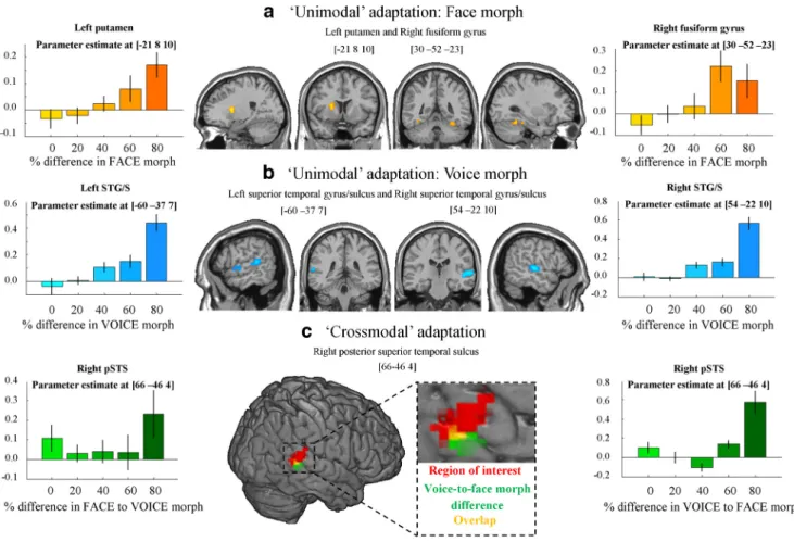
Unimodal adaptation. We observed significant ( p
⬍ 0.05, FWE
voxel corrected) unimodal face adaptation in the left putamen
and right fusiform gyrus (FG;
Table 1
bi) and significant voice
adaptation effects in the bilateral STG/STS and right inferior
frontal gyrus (
Fig. 4
a,b;
Table 1
bii). Generally, the response
heightened as the degree of difference in happiness–anger morph
between consecutive stimuli became larger, either in the auditory
or visual modality.
Figure 2. Behavioral results: direct effects of face and voice emotion morph. a, Categorization results. Categorization (percentage angry responses) as a function of face morph (middle), voice morph (right), and both (left). b, Reaction time results. Reaction time (milliseconds) as a function of face morph (middle), voice morph (right), and both (left). Both face and voice morph were morphed between 10% happy and 90% happy in 20% steps. Both categorization and reaction time results are averaged across actor. Note the greater influence of facial versus vocal emotional cues on behavioral responses. A, Angry; H, Happy.
Figure 3. Imaging results: multimodal localizer. Left, A cluster in the right STG/STS respond-ing more to audiovisual, compared with either visual or auditory, information alone localized using a conjunction analysis (AV⬎ A 艚 AV ⬎ V; conjunction null hypothesis;Nichols et al., 2005). Results were thresholded at p⬍ 0.05 (cluster corrected). Right, Condition effects at the peak voxel of the cluster.
Crossmodal adaptation. No crossmodal effects were significant
at p
⬍ 0.05 (FWE voxel corrected) after having partialed out
unimodal effects. However, at a more liberal threshold of p
⬍
0.001 (voxel uncorrected), a crossmodal carryover effect was
ob-served in the posterior part of the right pSTS (
Fig. 4
c;
Table 1
ci).
Interestingly, this effect was asymmetrical as for the behavioral
effect: activity was observed for voice-to-face emotion morph
difference but not for face-to-voice emotion morph difference.
That is, BOLD signal in response to an AV stimulus was greater in
this region when there was a large difference between the facial
emotion of the current stimulus and the vocal emotion of the
previous one but not when the vocal emotion of the current
stimulus differed from that of the previous face.
ROI analysis
A separate ROI analysis was conducted to test for crossmodal
adaptation effects specifically at locations independently
identi-fied using the “multimodal” localizer (see above). Within this
ROI in the pSTS/STG, there was a significant effect of unimodal
voice adaptation ( p
⬍ 0.005, t ⫽ 2.92), and unimodal face
adap-tation was just above the level of significance ( p
⫽ 0.055, t ⫽
1.68). Furthermore, we observed a significant crossmodal
adap-tation effect in addition to unimodal effects. Again, this effect was
asymmetrical: it was observed for voice-to-face emotion morph
difference ( p
⬍ 0.025, t ⫽ 2.12) but not face-to-voice emotion
morph difference ( p
⫽ 0.24, t ⫽ 0.72).
Discussion
The aim of this study was to examine the cerebral correlates of the
integration of affective information from the face and voice.
Dy-namic face-to-voice stimuli, parametrically varying in emotion,
were used in conjunction with a continuous carryover design to
enable measurements of adaptation effects (
Aguirre, 2007
).
Overall, we demonstrate crossmodal adaptation in the right
pSTS, suggesting the presence of multisensory, integrative
neu-rons in this area.
Behavioral results indicated that emotion categorization and
speed of categorization were modulated in line with parametric
shifts in affective content of the face and voice. Significantly, both
modalities affected emotion perception—an integration effect—
but face morph exerted a far larger influence on behavioral
re-sponses, both categorical and reaction times. This is in line with
other studies, in which emotion categorization has been found
consistently to be more accurate and quicker for faces (
Hess et al.,
1988
;
Collignon et al., 2008
;
Ba¨nziger et al., 2009
).
Figure 4. Imaging results. a, Unimodal face adaptation. Activation in left putamen and right FG in response to varying percentage difference in face morph between consecutive stimuli. Left and right, Parameter estimate at the peak activated voxel of left putamen and right FG, respectively, as a result of varying percentage difference in face morph. Results were thresholded at p⬍0.05(FWEvoxelcorrected) and a minimum cluster size of more than five contiguous voxels. b, Unimodal voice adaptation. Activation in bilateral STG/STS in response to varying percentage difference in voice morph between consecutive stimuli.Leftandright,ParameterestimateatthepeakactivatedvoxelinleftandrightSTG/STS,respectively,asaresultofvaryingpercentagedifferenceinvoicemorph.Resultswerethresholdedatp⬍0.05(FWE voxel corrected) and a minimum cluster size of more than five contiguous voxels. c, Crossmodal adaptation. Red, Results from the independent functional multimodal localizer. An ROI analysis showed that voice-to-face emotion morph difference evoked a significant response in this region ( p⬍0.025,t⫽2.12).Green,ActivationinrightpSTSasaresultofvaryingpercentagedifferencebetweenvoiceandthe following face morph of consecutive stimuli. Results were thresholded at p⬍0.001(voxeluncorrected)withaminimumclustersizeofmorethanfivecontiguousvoxels.Yellow,Overlapbetweenthelocalizer and voice-to-face morph difference activation. Left and right, Parameter estimate at the peak activated voxel of right pSTS as a result of varying percentage difference in face-to-voice morph, and voice-to-face morph, respectively. It should be noted that face-to-voice morph difference did not evoke a significant response in this region.
We also observed adaptation effects at the behavioral level.
Significantly, the crossmodal adaptation effect occurred only in
one direction: the emotion morph difference between a voice and
the following face significantly modulated reaction times,
whereas that of a face and the following voice did not. This
prim-ing effect of vocal information on facial information is consistent
with previous research highlighting crossmodal adaptive effects
in the domain of identity processing (
Ellis et al., 1997
;
Hills et al.,
2010
). Additionally, it should be noted that a recent study in fact
demonstrated adaptation effects between affective face adaptors
and test voices (
Skuk and Schweinberger, 2013
).
Cerebrally, we first observed that both face-to-face and
voice-to-voice emotion morph difference modulated cerebral activity,
namely in the putamen and FG, and bilateral STG/STS and
infe-rior frontal gyrus, respectively. These findings are consistent with
previous research on face and voice emotion perception. For
ex-ample, a recent meta-analysis (
Fusar-Poli et al., 2009
) linked
pro-cessing of emotional faces to increased activation in the putamen,
in particular, that of happy faces. Furthermore, the FG has been
associated consistently with the perception of human faces (
Puce
et al., 1995
;
Kanwisher et al., 1999
;
Haxby et al., 2000
) and has
been shown to be more active during expressive (e.g., fearful) face
processing than neutral faces (
Morris et al., 1998
;
Vuilleumier et
al., 2004
;
Sabatinelli et al., 2011
). Regarding affective voice
pro-cessing, studies showed that the middle temporal gyrus and STS
activate more when people listen to angry as opposed to neutral
speech (
Grandjean et al., 2005
;
Sander et al., 2005
) or when
peo-ple attend to affective prosody compared with the semantic
con-tent of the spoken words (
Mitchell et al., 2003
). Furthermore,
Ethofer et al. (2009)
demonstrated recently successful decoding
of vocal emotions from fMRI responses in bilateral
voice-sensitive areas.
Multisensory neurons in the right pSTS
Central to our main hypothesis, we observed crossmodal
adapta-tion effects during face-to-voice emoadapta-tion integraadapta-tion. Within a
wide-reaching search of any regions responding to audiovisual
information (compared with baseline), we observed a
cross-modal adaptation effect in the right pSTS, a region that has been
well documented as a multimodal region, in both humans (
Beau-champ et al., 2004
;
Ethofer et al., 2006
;
Kreifelts et al., 2007
,
2010
;
Watson et al., 2013
) and nonhuman primates (
Benevento et al.,
1977
;
Bruce et al., 1981
). This effect was small, only significant at
a relatively lenient threshold, but importantly was independent
of any variance elicited by either of the unimodal carryover
ef-fects: our design allowed us to regress out both unimodal face and
voice adaptation effects, ensuring that the variance associated
with crossmodal adaptation was modeled separately from
vari-ance explained by unimodal adaptation effects.
Additionally, this finding was confirmed in a complementary
ROI analysis. Using an independent functional localizer, we
iso-lated a cluster in the right pSTG/STS that responded more to
audiovisual information than to either the visual or auditory
mo-dality alone, using a conjunction analysis (
Goebel and van
Atte-veldt, 2009
;
Kreifelts et al., 2010
;
Love et al., 2011
). We then tested
for crossmodal adaptation within this cluster only, which yielded
a significant effect.
Thus, our results suggest the existence of a sufficiently large
proportion of multisensory neurons in the right pSTS to be
de-tected using fMRI. This finding converges with that of a previous
study that observed a “patchy organization” in this region
con-sisting of interspersed unisensory and multisensory neurons
(
Beauchamp et al., 2004
). We build on that observation by
show-ing that some such multisensory neurons may integrate
informa-tion in the context of affective processing. Furthermore, more
recently,
Kreifelts et al. (2009)
observed that audiovisual
integra-tion of affective signals peaked in the anterior pSTS, at an overlap
of face- and voice-sensitive regions. They proposed that this
im-plies a possible interaction of the underlying voice- and
face-sensitive neuronal populations during the formation of the
audiovisual percept. We argue that such an audiovisual percept
could partly reflect the contribution of populations of
multisen-sory neurons. However, note we do not suggest that right pSTS, a
complex, heterogeneous zone, is exclusively composed of
bi-modal neurons, nor do we suggest that all of face–voice
integra-tion effects in right pSTS are mediated by these bimodal neurons.
An asymmetrical crossmodal adaptation effect
Interestingly, the observed crossmodal adaptation effect was
asymmetrical: activity in both whole-brain and ROI analyses was
driven by the difference between a voice and the following face
but not the difference between a face and the following voice.
Therefore, it appears that voice exerted a stronger adaptive effect
on face than face did on voice. This is in line with our behavioral
data, in which only the difference between a voice and the
follow-ing face significantly modulated reaction times.
With regards to the asymmetry of the observed effect, one
might presume that, if a neuron was multisensory and therefore
“coding” for both stimulus dimensions, both voice-to-face
emo-tion morph difference and face-to-voice emoemo-tion morph
differ-ence would exert similar effects on its response. Why this was not
the case could be attributable to various possibilities. It should be
noted that, as mentioned previously, faces had a stronger effect
on emotion judgment than voices. Therefore, the smaller effect of
voices may have meant that modulations by preceding faces were
even less pronounced and therefore did not reach significance at
the behavioral and neural levels. However, in this experiment, we
explicitly chose not to manipulate our stimuli so to “equate” the
level of difficulty of emotion categorization; rather, the stimuli
were left to reflect a natural situation in which affective
informa-tion conveyed by the face and voice is rarely of equal
informative-Table 1. Imaging results
Coordinates (mm)
Brain regions x y z k t statistic
a, Multimodal localizer
STG/STS 48 ⫺40 13 153 5.23
b, Unimodal adaptation i, Adaptation to face emotion
Putamen ⫺21 8 10 14 7.46
FG 30 ⫺52 ⫺23 8 6.40
ii, Adaptation to voice emotion
STG/STS 54 ⫺22 10 51 7.98
STG/STS ⫺60 ⫺37 7 24 7.37
Inferior frontal gyrus 48 23 22 11 6.32 c, Crossmodal adaptation
i, Adaptation to voice-to-face emotion
STS 66 ⫺46 4 9 4.20
ii, Adaptation to face-to-voice emotion No significant voxels
a, Results from multisensory functional localizer experiment. Contrasts were cluster thresholded at p⬍ 0.05 (FWE cor-rected). MNI coordinates and t scores are from the peak voxel of a cluster. b, Unimodal adaptation results. bi, Adaptation to face emotion. bii, Adaptation to voice emotion. Contrasts were thresholded to display voxels reaching a significance level of p⬍0.05(FWEcorrected)andanadditionalminimumclustersizeofmorethanfivecontiguousvoxels.Contrastswerealso masked by an AV versus baseline contrast thresholded at p⬍0.001(voxeluncorrected).MNIcoordinatesandtscoresare fromthepeakvoxelofacluster.c,Crossmodaladaptationresults.ci,Voice-to-faceadaptation.cii,Face-to-voiceadaptation. Contrasts were thresholded to display voxels reaching a significance level of p⬍ 0.001 (uncorrected) and an additional minimum cluster size of more than five contiguous voxels. Contrasts were masked by an AV versus baseline contrast thresholded at p⬍0.001(voxeluncorrected).MNIcoordinatesandtscoresarefromthevoxelofacluster.
ness. Additional investigation regarding this could involve
including manipulations, such as adding noise to stimuli to
equate categorization difficulty, to investigate whether this
pro-vokes parallel adaptation effects between face and voice, and
voice and face.
Furthermore, alongside unequal direct effects, there may also
have been underlying asymmetries in the unimodal adaptive
ef-fect of each modality, in turn afef-fecting the strength of crossmodal
adaptation in either direction. A recent study (
Ethofer et al.,
2013
) investigated adaptation to faces, voices, and combined
face–voice affective stimuli: these authors found that, although
modality-specific cortices, such as the face-sensitive and
voice-sensitive cortex in the STS, showed a stronger response
habitua-tion for their respective preferred stimulus class, the multisensory
pSTS and orbitofrontal cortex (OFC) showed an adaptive
re-sponse that was equal for both faces and voices. In the pSTS
response habituation was stronger for audiovisual stimuli than
for face-only and voice-only stimuli, whereas in the OFC it was
equal across all three modalities. It would be of interest to see
whether, in these same regions in which adaptation to faces and
voices was approximately equal, there would additionally be
bi-directional crossmodal adaptation effects. However, at this point
at least, our results seem to converge with that of this study in that
the pSTS seems to be a main locus of audiovisual integration
effects.
However, equally, we at the same time argue that there is
perhaps no reason to assume that the effect should be perfectly
symmetrical: indeed, rather than an “all-or-nothing”
phenome-non, such multimodal neurons may receive different proportions
of synapses from visual and auditory neurons, subsequently
in-fluencing the strength of the crossmodal adaptation effect in
ei-ther direction. Furei-thermore, those visual and auditory inputs
could be characterized by differential modulating effects or
weighting on the neural response.
Finally, regarding the pattern of this asymmetrical crossmodal
effect, we noted that, at the peak voxel at least, the effect appeared
to be driven particularly by most extreme morph level difference
(i.e., 80%), perhaps acting as a “tipping point” for the marked
release from adaptation. In other words, in the case of crossmodal
adaptation, it could be possible that there is a precise percentage
difference in emotion between the modalities at which release
from adaptation is triggered rather than a graded linear
paramet-ric effect, as appeared more clearly in the unimodal face and voice
adaptation analyses. That said, it should also be noted that, with
inclusion of the 80% difference condition, the plot of effects had
a strong linear component, and therefore we would still suggest
that the physiology of the effect would be reflected by a linear
expansion of the parametric modulator. However, an interesting
future direction might be to investigate how inclusion of specific
percentage differences in affect morph level would affect the
grading of the adaptive response. This would be particularly
rel-evant to crossmodal adaptation, in which inclusion or exclusion
of particular audiovisual stimuli (and therefore morph
differ-ences) may evoke or extinguish the adaptive effect in either
direc-tion or change the pattern of the effect (e.g., linear to quadratic
response). In this way, we may be able to tap into the more
fine-grained mechanisms of affective face–voice integration.
References
Aguirre GK (2007) Continuous carry-over designs for fMRI. Neuroimage 35:1480 –1494.CrossRef Medline
Ba¨nziger T, Grandjean D, Scherer KR (2009) Emotion recognition from expressions in face, voice, and body: the multimodal emotion recognition test (MERT). Emotion 9:691–704.CrossRef Medline
Beauchamp MS, Argall BD, Bodurka J, Duyn JH, Martin A (2004) Unravel-ing multisensory integration: patchy organization within human STS multisensory cortex. Nat Neurosci 7:1190 –1192.CrossRef Medline
Benevento LA, Fallon J, Davis BJ, Rezak M (1977) Auditory–visual interac-tion in single cells in the cortex of the superior temporal sulcus and the orbital frontal cortex of the macaque monkey. Exp Neurol 57:849 – 872.
CrossRef Medline
Bruce C, Desimone R, Gross CG (1981) Visual properties of neurons in a polysensory area in superior temporal sulcus of the macaque. J Neuro-physiol 46:369 –384.Medline
Collignon O, Girard S, Gosselin F, Roy S, Saint-Amour D, Lassonde M, Lepore F (2008) Audio-visual integration of emotion expression. Brain Res 1242:126 –135.CrossRef Medline
de Gelder B, Bo¨cker KB, Tuomainen J, Hensen M, Vroomen J (1999) The combined perception of emotion from voice and face: early interaction revealed by human electric brain responses. Neurosci Lett 260:133–136.
CrossRef Medline
Dolan RJ, Morris JS, de Gelder B (2001) Crossmodal binding of fear in voice and face. Proc Natl Acad Sci USA 98:10006 –10010.CrossRef Medline
Ellis HD, Jones DM, Mosdell N (1997) Intra- and inter-modal repetition priming of familiar faces and voices. Br J Psychol 88:143–156.CrossRef Medline
Ethofer T, Pourtois G, Wildgruber D (2006) Investigating audiovisual inte-gration of emotional signals in the human brain. Prog Brain Res 156:345– 361.CrossRef Medline
Ethofer T, Van De Ville D, Scherer K, Vuilleumier P (2009) Decoding of emotional information in voice-sensitive cortices. Curr Biol 19:1028 – 1033.CrossRef Medline
Ethofer T, Bretscher J, Wiethoff S, Bisch J, Schlipf S, Wildgruber D, Kreifelts B (2013) Functional responses and structural connections of cortical areas for processing faces and voices in the superior temporal sulcus. Neuroimage 76:45–56.CrossRef Medline
Fusar-Poli P, Placentino A, Carletti F, Landi P, Allen P, Surguladze S, Bene-detti F, Abbamonte M, Gasparotti R, Barale F, Perez J, McGuire P, Politi P (2009) Functional atlas of emotional faces processing: a voxel-based meta-analysis of 105 functional magnetic resonance imaging studies. J Psychiatry Neurosci 34:418 – 432.Medline
Goebel R, van Atteveldt N (2009) Multisensory functional magnetic reso-nance imaging: a future perspective. Exp Brain Res 198:153–164.
CrossRef Medline
Grandjean D, Sander D, Pourtois G, Schwartz S, Seghier ML, Scherer KR, Vuilleumier P (2005) The voices of wrath: brain responses to angry prosody in meaningless speech. Nat Neurosci 8:145–146. CrossRef Medline
Grill-Spector K, Kushnir T, Edelman S, Avidan G, Itzchak Y, Malach R (1999) Differential processing of objects under various viewing condi-tions in the human lateral occipital complex. Neuron 24:187–203.
CrossRef Medline
Grill-Spector K, Henson R, Martin A (2006) Repetition and the brain: neu-ral models of stimulus-specific effects. Trends Cogn Sci 10:14 –23.
CrossRef Medline
Hagan CC, Woods W, Johnson S, Calder AJ, Green GG, Young AW (2009) MEG demonstrates a supra-additive response to facial and vocal emotion in the right superior temporal sulcus. Proc Natl Acad Sci USA 106:20010 – 20015.CrossRef Medline
Hagan CC, Woods W, Johnson S, Green GG, Young AW (2013) Involve-ment of right STS in audio-visual integration for affective speech demon-strated using MEG. PLoS One 8:e70648.CrossRef Medline
Haxby JV, Hoffman EA, Gobbini MI (2000) The distributed human neural system for face perception. Trends Cogn Sci 4:223–233.CrossRef Medline
Hess U, Kappas A, Scherer K (1988) Multichannel communication of emo-tion: synthetic signal production. In: Facets of emoemo-tion: recent research (Scherer K, ed), pp 161–182. Hillsdale, NJ: Erlbaum.
Hills PJ, Elward RL, Lewis MB (2010) Cross-modal identity aftereffects and their relation to priming. J Exp Psychol Hum Percept Perform 36:876 – 891.CrossRef Medline
Kanwisher N, Stanley D, Harris A (1999) The fusiform face area is selective for faces not animals. NeuroReport 10:183–187.CrossRef Medline
Kawahara H (2006) STRAIGHT, exploitation of the other aspect of VOCODER: perceptually isomorphic decomposition of speech sounds. Acoust Sci Tech 27:349 –353.CrossRef
Klasen M, Kenworthy CA, Mathiak KA, Kircher TT, Mathiak K (2011)
pramodal representation of emotions. J Neurosci 31:13635–13643.
CrossRef Medline
Kreifelts B, Ethofer T, Grodd W, Erb M, Wildgruber D (2007) Audiovisual integration of emotional signals in voice and face: an event-related fMRI study. Neuroimage 37:1445–1456.CrossRef Medline
Kreifelts B, Ethofer T, Shiozawa T, Grodd W, Wildgruber D (2009) Cerebral representation of non-verbal emotional perception: fMRI reveals audio-visual integration area between voice- and face-sensitive regions in the superior temporal sulcus. Neuropsychologia 47:3059 –3066.CrossRef Medline
Kreifelts B, Ethofer T, Huberle E, Grodd W, Wildgruber D (2010) Associa-tion of trait emoAssocia-tional intelligence and individual fMRI activaAssocia-tion pat-terns during the perception of social signals from voice and face. Hum Brain Mapp 31:979 –991.CrossRef Medline
Love SA, Pollick FE, Latinus M (2011) Cerebral correlates and statistical criteria of cross-modal face and voice integration. Seeing Perceiving 24: 351–367.CrossRef Medline
Mitchell RL, Elliott R, Barry M, Cruttenden A, Woodruff PW (2003) The neural response to emotional prosody, as revealed by functional magnetic resonance imaging. Neuropsychologia 41:1410 –1421.CrossRef Medline
Morris JS, Friston KJ, Bu¨chel C, Frith CD, Young AW, Calder AJ, Dolan RJ (1998) A neuromodulatory role for the human amygdala in processing emotional facial expressions. Brain 121:47–57.CrossRef Medline
Nichols T, Brett M, Andersson J, Wager T, Poline JB (2005) Valid conjunc-tion inference with the minimum statistic. Neuroimage 25:653– 660.
CrossRef Medline
Pourtois G, de Gelder B, Vroomen J, Rossion B, Crommelinck M (2000) The time-course of intermodal binding between seeing and hearing affec-tive information. Neuroreport 11:1329 –1333.CrossRef Medline
Pourtois G, Debatisse D, Despland PA, de Gelder B (2002) Facial expres-sions modulate the time course of long latency auditory brain potentials. Brain Res Cogn Brain Res 14:99 –105.CrossRef Medline
Pourtois G, de Gelder B, Bol A, Crommelinck M (2005) Perception of facial expressions and voices and of their combination in the human brain. Cortex 41:49 –59.CrossRef Medline
Puce A, Allison T, Gore JC, McCarthy G (1995) Face-sensitive regions in human extrastriate cortex studied by functional MRI. J Neurophysiol 74:1192–1199.Medline
Robins DL, Hunyadi E, Schultz RT (2009) Superior temporal activation in response to dynamic audio-visual emotional cues. Brain Cogn 69:269 – 278.CrossRef Medline
Sabatinelli D, Fortune EE, Li Q, Siddiqui A, Krafft C, Oliver WT, Beck S, Jeffries J (2011) Emotional perception: meta-analyses of face and natu-ral scene processing. Neuroimage 54:2524 –2533.CrossRef Medline
Sander D, Grandjean D, Pourtois G, Schwartz S, Seghier ML, Scherer KR, Vuilleumier P (2005) Emotion and attention interactions in social cog-nition: brain regions involved in processing anger prosody. Neuroimage 28:848 – 858.CrossRef Medline
Skuk VG, Schweinberger SR (2013) Adaptation aftereffects in vocal emo-tion percepemo-tion elicited by expressive faces and voices. PLoS One 8:e81691.CrossRef Medline
Tal N, Amedi A (2009) Multisensory visual–tactile object related network in humans: insights gained using a novel crossmodal adaptation approach. Exp Brain Res 198:165–182.CrossRef Medline
Vuilleumier P, Richardson MP, Armony JL, Driver J, Dolan RJ (2004) Dis-tant influences of amygdala lesion on visual cortical activation during emotional face processing. Nat Neurosci 7:1271–1278.CrossRef Medline
Watson R, Latinus M, Noguchi T, Garrod O, Crabbe F, Belin P (2013) Dis-sociating task difficulty from incongruence in face-voice emotion integra-tion. Front Hum Neurosci 7:744.CrossRef Medline
Watson R, Latinus M, Charest I, Crabbe F, Belin P (2014) People-selectivity, audiovisual integration and heteromodality in the superior temporal sul-cus. Cortex 50:125–136.CrossRef Medline