HAL Id: inserm-02157660
https://www.hal.inserm.fr/inserm-02157660
Submitted on 17 Jun 2019
HAL is a multi-disciplinary open access archive for the deposit and dissemination of sci-entific research documents, whether they are pub-lished or not. The documents may come from teaching and research institutions in France or abroad, or from public or private research centers.
L’archive ouverte pluridisciplinaire HAL, est destinée au dépôt et à la diffusion de documents scientifiques de niveau recherche, publiés ou non, émanant des établissements d’enseignement et de recherche français ou étrangers, des laboratoires publics ou privés.
The Npa1p complex chaperones the assembly of the
earliest eukaryotic large ribosomal subunit precursor
Clément Joret, Régine Capeyrou, Kamila Belhabich-Baumas, Célia
Plisson-Chastang, Rabea Ghandour, Odile Humbert, Sébastien Fribourg,
Nicolas Leulliot, Simon Lebaron, Anthony Henras, et al.
To cite this version:
Clément Joret, Régine Capeyrou, Kamila Belhabich-Baumas, Célia Plisson-Chastang, Rabea Ghan-dour, et al.. The Npa1p complex chaperones the assembly of the earliest eukaryotic large ribosomal subunit precursor. PLoS Genetics, Public Library of Science, 2018, 14 (8), pp.e1007597. �10.1371/jour-nal.pgen.1007597�. �inserm-02157660�
The Npa1p complex chaperones the assembly
of the earliest eukaryotic large ribosomal
subunit precursor
Cle´ment Joret1¤a, Re´gine Capeyrou1, Kamila Belhabich-Baumas1, Ce´lia Plisson-Chastang1, Rabea Ghandour1¤b, Odile Humbert1, Se´bastien Fribourg2, Nicolas Leulliot3, Simon Lebaron1*, Anthony K. Henras1*, Yves Henry1*
1 Laboratoire de Biologie Mole´culaire Eucaryote, Centre de Biologie Inte´grative (CBI), Universite´ de
Toulouse, CNRS, UPS, Toulouse, France, 2 Universite´ de Bordeaux, INSERM U1212, CNRS UMR5320, Bordeaux, France, 3 Laboratoire de Cristallographie et RMN Biologiques, UMR CNRS 8015, Universite´ Paris Descartes, Sorbonne Paris Cite´, Faculte´ de Pharmacie, Paris, France
¤a Current address: RNA Molecular Biology, Fonds de la Recherche Scientifique (F.R.S.-FNRS), Universite´ Libre de Bruxelles (ULB), BioPark Campus, Gosselies, Belgium
¤b Current address: Max Planck Institute of Molecular Plant Physiology, Potsdam, Germany
*lebaron@ibcg.biotoul.fr(SL);henras@ibcg.biotoul.fr(AKH);henry@ibcg.biotoul.fr(YH)
Abstract
The early steps of the production of the large ribosomal subunit are probably the least under-stood stages of eukaryotic ribosome biogenesis. The first specific precursor to the yeast large ribosomal subunit, the first pre-60S particle, contains 30 assembly factors (AFs), including 8 RNA helicases. These helicases, presumed to drive conformational rearrange-ments, usually lack substrate specificity in vitro. The mechanisms by which they are targeted to their correct substrate within pre-ribosomal particles and their precise molecular roles remain largely unknown. We demonstrate that the Dbp6p helicase, essential for the normal accumulation of the first pre-60S pre-ribosomal particle in S. cerevisiae, associates with a complex of four AFs, namely Npa1p, Npa2p, Nop8p and Rsa3p, prior to their incorporation into the 90S pre-ribosomal particles. By tandem affinity purifications using yeast extracts depleted of one component of the complex, we show that Npa1p forms the backbone of the complex. We provide evidence that Npa1p and Npa2p directly bind Dbp6p and we demon-strate that Npa1p is essential for the insertion of the Dbp6p helicase within 90S pre-ribo-somal particles. In addition, by an in vivo cross-linking analysis (CRAC), we map Npa1p rRNA binding sites on 25S rRNA adjacent to the root helices of the first and last secondary structure domains of 25S rRNA. This finding supports the notion that Npa1p and Dbp6p function in the formation and/or clustering of root helices of large subunit rRNAs which cre-ates the core of the large ribosomal subunit RNA structure. Npa1p also crosslinks to snoR-NAs involved in decoding center and peptidyl transferase center modifications and in the immediate vicinity of the binding sites of these snoRNAs on 25S rRNA. Our data suggest that the Dbp6p helicase and the Npa1p complex play key roles in the compaction of the cen-tral core of 25S rRNA and the control of snoRNA-pre-rRNA interactions.
a1111111111 a1111111111 a1111111111 a1111111111 a1111111111 OPEN ACCESS
Citation: Joret C, Capeyrou R, Belhabich-Baumas
K, Plisson-Chastang C, Ghandour R, Humbert O, et al. (2018) The Npa1p complex chaperones the assembly of the earliest eukaryotic large ribosomal subunit precursor. PLoS Genet 14(8): e1007597.
https://doi.org/10.1371/journal.pgen.1007597 Editor: David Tollervey, Univ. of Edinburgh,
UNITED KINGDOM
Received: January 11, 2018 Accepted: July 30, 2018 Published: August 31, 2018
Copyright:© 2018 Joret et al. This is an open access article distributed under the terms of the
Creative Commons Attribution License, which permits unrestricted use, distribution, and reproduction in any medium, provided the original author and source are credited.
Data Availability Statement: NGS analysis files of
raw and processed data were deposited in Gene Expression Omnibus database under the accession number GEO: GSE104281. All other relevant data are within the paper and its Supporting Information files.
Funding: French Ministry of Research PhD
fellowship and Paul Sabatier University ATER contract [to CJ]; Centre National de la Recherche Scientifique (CNRS); The University of Toulouse; and Agence Nationale de la Recherche (ANR,http://
Author summary
Ribosomes, the molecular machines synthesizing proteins, are composed of a small and large subunit, formed by the binding of numerous ribosomal proteins (RPs) to properly folded ribosomal RNAs (rRNAs). RP incorporation as well as processing and folding of rRNAs occur within a succession of ribosomal particles. Formation of the initial pre-60S particle, the first precursor to the large ribosomal subunit, is the least understood step of ribosome biogenesis in eukaryotes. This pre-ribosomal particle contains several assem-bly factors (AFs), including RNA helicases believed to catalyse key conformational rear-rangements. These helicases usually lack substrate specificity on their own. Here, we show that the Dbp6p helicase, a component of the first pre-60S particle and essential for its nor-mal accumulation, associates with a complex of four AFs, including Npa1p. We demon-strate that Npa1p directly binds Dbp6p, forms the backbone of the complex and is required for the integration of Dbp6p within pre-ribosomal particles. We show that Npa1p binds to sequences forming the core of large subunit rRNAs as well as small nucle-olar RNAs required for chemical modification of large subunit rRNAs. Altogether our results suggest that the Npa1p complex plays a crucial role in the chemical modification and folding of large subunit rRNAs.
Introduction
Synthesis of ribosomes is one of the most energy-demanding cellular tasks. In eukaryotes, it involves the transcription by RNA polymerase I (Pol I) of the rRNA precursor to the mature 18S, 5.8S and 25S/28S rRNAs. During transcription, the nascent Pol I transcript assembles with most ribosomal proteins, many small nucleolar ribonucleoprotein particles (snoRNPs) and hundreds of maturation and assembly factors (AFs) [1–3]. In yeast, pre-rRNA cleavage events may start co-transcriptionally [4,5], leading to the release of the first pre-40S pre-ribo-somal particle while assembly of the first pre-60S pre-ribopre-ribo-somal particle is still ongoing on the downstream part of the nascent pre-rRNA. When pre-rRNA cleavages start after completion of transcription, a huge 90S pre-ribosomal particle containing the 35S pre-rRNA is generated, which is then split into the first pre-40S and pre-60S particles. These particles undergo inde-pendent maturation pathways in the nucleolus, nucleoplasm and finally in the cytoplasm, yielding mature 40S and 60S ribosomal subunits competent for translation [1–3].
Assembly and maturation of the initial pre-60S particle are probably the least studied events of ribosome biogenesis in eukaryotes. Formation of this particle involves the co-transcriptional recruitment of approximately 30 AFs and most large subunit ribosomal proteins [6]. Within the nascent pre-60S particle, methylation and pseudouridylation of specific residues of 25S rRNA take place, carried out by snoRNPs. In addition, the 25S and 5.8S rRNAs and flanking sequences start folding [7]. In the mature 60S subunit, the 25S rRNA is folded in 6 structural domains (I to VI), each beginning with a root helix. In both the mature and intermediate pre-60S particles, these root helices are clustered [8,9]. It has been proposed that formation and clustering of the root helices, as well as folding of domain I that forms part of the solvent exposed surface of the large subunit, are key events that take place very early on during large subunit biogenesis and are prerequisites for all downstream maturation steps [10]. The recently published cryo-EM structures of yeast nucleolar pre-60S particles [11–13] support the idea that assembly of the large ribosomal subunit starts with the folding and stabilization of domain I followed by those of domains II and VI.
The Npa1p complex promotes early pre-60S particle assembly
www.agence-nationale-recherche.fr/) [ANR-2010-BLAN-1224 to YH]. The funders had no role in study design, data collection and analysis, decision to publish, or preparation of the manuscript.
Competing interests: The authors have declared
A third of the AFs contained in the initial pre-60S particle are required for normal steady-state accumulation of the particle. These AFs likely play crucial roles in the previously
described pre-rRNA folding and compaction events. This is most probably the case for four of these AFs, namely Dbp6p, Dbp7p, Dbp9p and Prp43p that belong to the family of RNA heli-cases, believed to remodel RNA/RNA and/or RNA/protein interactions [14–16]. Most of these helicases lack substrate specificityin vitro and the mechanisms by which they are targeted to
their specific substrates remain unclear in the majority of cases. Moreover, the precise molecu-lar targets of Dbp6p, Dbp7p and Dbp9p still remain elusive. Early acting molecu-large subunit ribo-somal proteins are also envisaged to play key roles in the assembly and maturation of the initial pre-60S particle, in particular Rpl3 [10]. Rpl3 is necessary for stable assembly of all other large subunit ribosomal proteins [10], possibly because of its proposed role in maintaining the clustering of root helices due to its binding to root helices of domains I and VI.
Genetic and physical interactions linking one of the aforementioned helicases, Dbp6p, and four additional AFs of the first pre-60S particle, namely Npa1p, Npa2p, Nop8p and Rsa3p, have been uncovered [17]. All of these proteins, with the exception of Rsa3p, are essential for growth and are all required for normal production of the large ribosomal subunit [6,17–21]. Rsa3p (25 kDa predicted molecular weight) and Nop8p (57 kDa predicted molecular weight) both feature a putative coiled-coiled domain that may be involved in protein-protein interac-tions. Nop8p also features a putative RNA recognition motif of the RRM type that may allow direct binding of Nop8p to pre-rRNAs. Npa1p and Npa2p are large proteins (203 and 135 kDa predicted molecular weight, respectively) with no obvious already-described protein motif. It has been shown that depletion or inactivation of Npa1p, Npa2p and Dbp6p leads to the accu-mulation of the 35S pre-rRNA, the RNA component of the initial 90S pre-ribosomal particle, suggesting a defect in its maturation, while the steady-state levels of the 27SA2 pre-rRNA, the RNA component of the first pre-60S particle, drop demonstrating that these proteins are required for the production and/or stability of the first pre-60S particle [6,17,19,20] (seeS1 Fig
for a cartoon of pre-rRNA processing inS. cerevisiae).
In the present study, we show that the Dbp6p helicase associates with a core complex formed by Npa1p, Npa2p, Nop8p and Rsa3p via direct interactions with Npa1p and Npa2p. We show that Npa1p forms the backbone of the core complex and is essential for the stable integration of Dbp6p within the initial 90S pre-ribosomal particle. Our mapping ofin vivo
Npa1p-RNA crosslinking sites suggests Npa1p and Dbp6p could play crucial roles in the for-mation and/or clustering of the root helices of large subunit rRNAs and in the control of snoRNA/pre-rRNA interactions.
Results
The Dbp6p helicase associates with a core complex formed by the Npa1p,
Npa2p, Nop8p and Rsa3p proteins
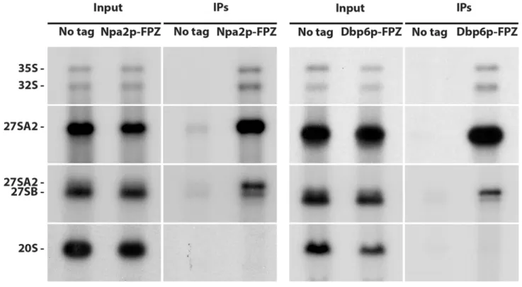
Previous data suggested that Npa1p, Npa2p, Nop8p, Dbp6p and Rsa3p form a complex associ-ated with 90S and early pre-60S pre-ribosomal particles [17]. If this is indeed the case, all these proteins should be specifically associated with the 35S and 32S pre-rRNAs, components of suc-cessive 90S ribosomal particles and with the 27SA2 rRNA, component of the first pre-60S particle. While this has already been demonstrated for Npa1p and Npa2p [6,17], convinc-ing data were lackconvinc-ing for Dbp6, Nop8p and Rsa3p. We therefore performed precipitation experiments using strains expressing FPZ- (Flag-prescission cleavage site-Protein A IgG-bind-ing domain) tagged versions of Nop8p, Dbp6p and Rsa3p and analysed the co-precipitated RNAs by Northern (Fig 1). This analysis confirmed that Nop8p, Dbp6p and Rsa3p, like Npa1p and Npa2p, are mostly associated with the 35S, 33/32S and 27SA2 pre-rRNAs, while displaying
Fig 1. Dbp6p, Nop8p and Rsa3p are components of 90S and 27SA2 pre-rRNA-containing pre-60S particles. Immunoprecipitation experiments were
carried out with IgG sepharose and extracts from cells expressing FPZ-tagged versions of Npa2p, Dbp6p, Nop8p or Rsa3p. Aliquots of input and co-precipitated RNAs (IPs) were submitted to denaturing gel electrophoresis and transferred to nylon membranes. 35S, 32S and 27SA2 pre-rRNAs were detected with probe 23S1, 27SB pre-rRNAs with probe rRNA2.1 and 20S pre-rRNA with probe 20S3.
https://doi.org/10.1371/journal.pgen.1007597.g001
a much weaker association with the 27SB pre-rRNA. No association could be detected with the 20S pre-rRNA, demonstrating that Dbp6p, Nop8p and Rsa3p are not components of pre-40S particles. We conclude that Npa1p, Npa2p, Nop8p, Dbp6p and Rsa3p are recruited within 90S pre-ribosomal particles, remain present within the earliest pre-60S pre-ribosomal particles containing the 27SA2 pre-rRNA and dissociate from 27SB-containing pre-60S particles.
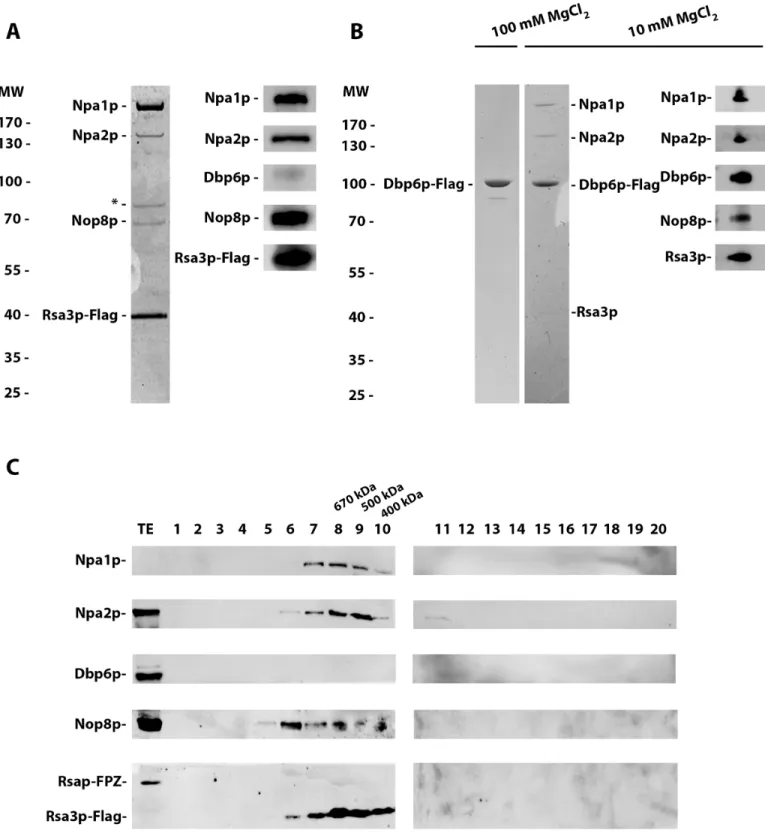
To corroborate that Npa1p, Npa2p, Nop8p, Dbp6p and Rsa3p form a complex, as already indicated by a previous study [17], we performed tandem affinity purifications from extracts of cells expressing FPZ-tagged Rsa3p. Standard protocols lead to the purification of the pre-ribosomal particles with which these proteins are associated. To separate the putative complex from these particles, we employed the same MgCl2concentration (100 mM) that could extract
the Rps3/Enp1p/Ltv1p complex from pre-40S particles [22] and we pelleted the pre-ribosomal particles using two successive ultracentrifugation steps. SDS-PAGE followed by Coomassie blue staining and Western analysis (Fig 2A) of the purified sample demonstrated that under such conditions Npa1p, Npa2p and Nop8p co-purify with Rsa3p. An additional polypeptide co-purifying with Rsa3p of apparent molecular weight close to that of Dbp6p turned out to be a degradation product of Npa1p as shown by mass spectrometry analysis. The lack of Dbp6p in the sample purified using Rsa3p-FPZ as bait prompted us to carry out the tandem affinity purification using Dbp6p-FPZ as bait. When performing such purification at 100 mM MgCl2,
only Dbp6p can be detected after Coomassie blue staining of the purified sample (Fig 2B). However, if the MgCl2concentration is lowered to 10 mM, co-purifying Npa1p and Npa2p are
detected by Coomassie blue staining and Nop8p and Rsa3p by Western blotting (Fig 2B). These data demonstrate that Npa1p, Npa2p, Nop8p and Rsa3p can form one or several com-plexes, with which Dbp6p interacts but such association is magnesium sensitive.
The intensities of the Coomassie-stained polypeptides copurifying with Rsa3p-FPZ indicate that not all proteins are recovered in stoichiometric amounts. This suggests that mixtures of different sub-complexes have been purified reflecting preferential associations between certain partners and/or that multiple copies of the same protein are present in the full complex. To investigate this issue further, the purification sample obtained using Rsa3p-FPZ as bait was subjected to size exclusion chromatography and the eluted fractions were analysed by Western. None of the analysed proteins are present as free polypeptides. Npa1p, Npa2p and Rsa3p, but not Nop8p, are present together in fractions 8 and 9, that contain complexes of approximately 670 and 500 kDa, respectively (Fig 2C). All four proteins are present together in fraction 7, that contains complexes heavier than 700 kDa. Thus at least two types of complexes have been purified using tagged Rsa3p as bait, one containing Npa1p, Npa2p and Rsa3p, the second con-taining these proteins and Nop8p. To confirm that mainly two types of complexes are purified via tagged Rsa3p, the purified sample was analysed by electron microscopy (S2 Fig). Images of negatively stained complexes indeed revealed the presence of two types of particles of 10 or 20 nm in size. The smaller particles could correspond to the Npa1p/Npa2p/Rsa3p complex, the bigger to the Npa1p/Npa2p/Nop8p/Rsa3p complex.
Association between Rsa3p and its partners can take place in absence of de
novo ribosome biogenesis
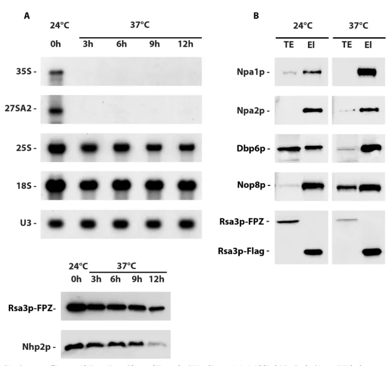
To determine whether Npa1p, Npa2p, Nop8p, Dbp6p and Rsa3p can interact outside pre-ribo-somal particles, we investigated the ability of Npa1p, Npa2p, Dbp6p and Nop8p to co-purify with tagged Rsa3p when RNA pol I transcription was inhibited. To that end, we made use of a temperature sensitiverrn3/RSA3-FPZ strain that expresses Rsa3p-FPZ and a version of the
RNA pol I transcription factor Rrn3p that is inactivated at 37˚C. Northern blot analysis shows that a shift from 24˚C to 37˚C during three hours abolishes 35S pre-rRNA accumulation in the
Fig 2. Npa1p, Npa2p, Nop8p and Rsa3p form complexes. (A) Rsa3p associates with Npa1p, Npa2p and Nop8p. Extracts were prepared from
Rsa3p-FPZ-expressing cells in a buffer containing 100 mM MgCl2and subjected to two consecutive ultracentrifugation steps to pellet pre-ribosomal particles. Tandem affinity
purification of Rsa3p-FPZ was then carried out, proteins co-purified with tagged Rsa3p were TCA precipitated, separated by SDS-PAGE and stained with Coomassie blue (left). The identity of the co-purified proteins was confirmed by Western analysis using specific antibodies (right). The polypeptide marked by an asterisk is a degradation product of Npa1p, identified as such by mass-spectrometry. (B) Dbp6p interacts in a salt-sensitive manner with Npa1p, Npa2p, Nop8p and Rsa3p. Dbp6p-FPZ was subjected to tandem affinity purification using buffers containing 100 mM (left) or 10 mM (right) MgCl2. Tagged Dbp6p and co-purified
proteins were separated by SDS-PAGE and stained with Coomassie blue. Coomassie staining reveals that Npa1p and Npa2p are co-purified with tagged Dbp6p at 10 mM MgCl2, while co-purification of Nop8p and Rsa3p at 10 mM MgCl2is shown by Western analysis with specific antibodies (right). (C) Analysis by size
rrn3/RSA3-FPZ strain (Fig 3A), demonstrating that RNA pol I transcription and hence de novo ribosome biogenesis has been efficiently inhibited. Nevertheless, Rsa3p-FPZ tandem affinity purification followed by Western analysis of the co-purified proteins (Fig 3B) show that Npa1p, Npa2p, Dbp6p and Nop8p can still interact with Rsa3p when RNA pol I transcrip-tion is abolished, suggesting that these proteins can form one or several complexes outside pre-ribosomal particles.
Npa1p and Npa2p interact directly with Dbp6p
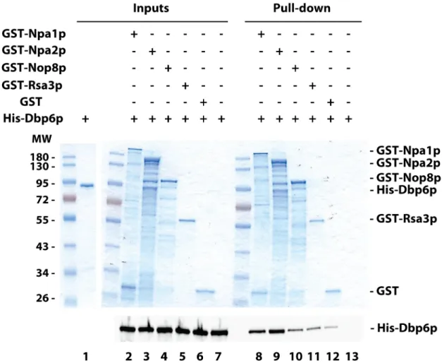
To identify how Dbp6p interacts with the Npa1p-Npa2p-Nop8p-Rsa3p core complex, GST pull-down experiments with recombinant proteins were performed. Purified recombinant His-tagged Dbp6p was incubated with GST-tagged versions of Npa1p, Npa2p, Nop8p or Rsa3p, or GST alone as control, bound to glutathione-coated magnetic beads. Following pre-cipitation of the beads using a magnetic rack, His-tagged protein co-prepre-cipitation was assessed by Western (Fig 4). Higher levels of His-Dbp6p were precipitated with GST-Npa1p or
GST-Npa2p as compared with GST (Fig 4, compare lanes 8 and 9 with lane 12), showing that Dbp6p can directly interact with Npa1p and Npa2pin vitro. These interactions do not rely on
the hypothetical presence of RNAs in the purified protein preparations since extensive RNAse treatment did not impair the interactions (S3 Fig). In contrast, less or no enrichment of His-Dbp6p relative to the GST control was obtained when GST-Nop8p or GST-Rsa3p were used (Fig 4, compare lanes 10 and 11 with lane 12, andS3 Figlower panel, compare lane 4 with lane 5 and lane 8 with lane 9), indicating that under these experimental conditions, no convincing specific interaction between Dbp6p and Nop8p or Rsa3p can be demonstrated. We conclude that Dbp6p interacts with the core complex via direct interactions with Npa1p and Npa2p.
All other binary interactions between purified GST-tagged versions of complex proteins were tested by immunoprecipitation experiments using specific antibodies. Although these antibodies could efficiently precipitate their specific target proteins, we were unable to detect direct protein/protein interactions. This may be due to protein folding problems, the presence of the GST tag and/or inappropriate buffer conditions. We therefore studied interactions between complex members by anin vivo approach (see below).
Npa1p is crucial for core complex formation
We next investigated the role playedin vivo by each member of the complex in the stability of
the other members and in complex formation. To that end, we constructed yeast strains expressing an FPZ-tagged version of one member of the complex and an HA-tagged version of another, the open reading frame of which was placed under the control of theGAL1-10
pro-moter (i.e. strains of the type GAL::HA-protein X/protein Y-FPZ). This allows expression of the HA-tagged protein when the strain is grown in a medium containing galactose and inhibi-tion of its producinhibi-tion when the strain is transferred to a glucose-containing medium. Using these strains, we could show that depletion of Npa1p, Npa2p, Nop8p or Dbp6p has no effect on the steady-state accumulation of the non-depleted complex members (S4,S5,S6andS7
Figs; note that the effects of lack of Rsa3p were not tested because it is the only non-essential member of the complex). We next assessed the effect of depleting one complex member on the ability of the remaining core complex proteins to interact. Tandem affinity purification was performed using a buffer containing 100 mM MgCl2as described above and Rsa3p-FPZ-exclusion chromatography of Rsa3p-containing complexes. The sample obtained following tandem affinity purification performed with extracts from cells expressing Rsa3p-FPZ was subjected to size exclusion chromatography on a superdex 200 10/300 GL column. Proteins from the eluted fractions were analysed by Western using specific antibodies. TE: total extract.
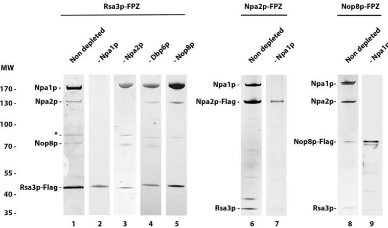
expressing strains depleted of Npa1p, Npa2p, Dbp6p or Nop8p. The purified proteins were analysed by Coomassie blue staining (Fig 5, lanes 1 to 5). This analysis demonstrates that Npa1p depletion prevents Rsa3p interaction with Npa2p and Nop8p (Fig 5, lane 2). In con-trast, depletion of Npa2p, Dbp6p or Nop8p does not prevent Rsa3p association with remaining members of the core complex (Fig 5, lanes 3 to 5). To check whether Npa1p depletion impairs
Fig 3. Rsa3p can still interact with Npa1p, Npa2p, Dbp6p and Nop8p when RNA pol I transcription is inhibited. (A) 35S and 27SA2 pre-rRNA levels are
drastically reduced after shifting the temperature sensitiverrn3/RSA3-FPZ strain to 37˚C. Total RNAs or proteins were extracted from rrn3/RSA3-FPZ cells grown
at 24˚C or shifted to 37˚C for the indicated times and subjected to Northern (top) or Western (bottom) analyses, respectively. The 35S and 27SA2 pre-rRNAs were detected with probe 23S1, 18S and 25S rRNAs with 18S and 25S probes and U3 snoRNA using the anti-U3 probe. The Rsa3p and Nhp2p proteins were detected using specific antibodies. (B) Western analysis of proteins co-purified with tagged Rsa3p. Rsa3p-FPZ was subjected to tandem affinity purification from extracts produced at low MgCl2(10 mM) concentration ofrrn3/RSA3-FPZ cells grown at 24˚C or shifted to 37˚C for 6 hours. Co-purified proteins were analysed by
Western using specific antibodies. TE: total extracts; El: eluted purified samples.
https://doi.org/10.1371/journal.pgen.1007597.g003
interactions between Npa2p and Nop8p, tandem affinity purifications were performed using extracts from Npa2p-FPZ or Nop8p-FPZ expressing strains depleted of Npa1p (Fig 5, lanes 7 and 9). In both cases, no protein co-purifying with the tagged Npa2p or Nop8p baits could be detected. This analysis demonstrates that lack of Npa1p prevents interactions between Rsa3p, Npa2p and Nop8p. These findings suggest that Rsa3p, Nop8p and Npa2p do not directly inter-act with each other, or if they do, those interinter-actions are not possible or stable in the absence of Npa1p.
Our pull-down experiments showed that Npa1p and Npa2p can directly interact with Dbp6p, at leastin vitro. To assess the contribution of Npa1p on Dbp6p interaction with Npa2p in vivo, and vice versa, we performed tandem affinity purifications of Npa1p-FPZ in the
pres-ence or abspres-ence of Npa2p, as well as purifications of Npa2p-FPZ in the prespres-ence and abspres-ence of Npa1p. These purifications were carried out at 10 mM MgCl2to maintain interactions with
Dbp6p, at least in the non-depleted conditions. The co-purification of complex proteins with the Npa1p-Flag or Npa2p-Flag bait was analysed by Western (S8 Fig). This analysis confirms that Npa1p depletion prevents interactions between Npa2p and Nop8p or Rsa3p. In contrast,
Fig 4.In vitro protein-protein interaction assays. Glutathione-coated magnetic beads were mixed with purified recombinant
GST-tagged proteins, washed and incubated with purified recombinant His-Dbp6p. The glutathione-coated magnetic beads were precipitated using a magnetic rack and washed. Pulled-down proteins (Pull-down) and 1/5thof the proteins used in the pull-down experiments (Inputs) were separated by SDS-PAGE. The GST-tagged proteins were visualised by Coomassie blue staining (upper panel) and His-Dbp6p was analysed either by Coomassie blue staining (Inputs) or by Western using anti-His antibodies (lower panel).
the interaction between Npa2p and Dbp6p is maintained in the absence of Npa1p, although it is strongly destabilised. The interaction between Npa1p and Nop8p or Rsa3p persists in the absence of Npa2p, consistent with our previous observation that Npa2p depletion does not prevent interactions between Rsa3p and Npa1p or Nop8p. Strikingly, however, Npa2p deple-tion abolishes the interacdeple-tion between Npa1p and Dbp6p. We conclude that in yeast cells, Dbp6p can interact with Npa2p in absence of other Npa1p complex components, although this interaction is significantly enhanced by the presence of Npa1p. In contrast, Npa2p is essential for the Npa1p-Dbp6p interactionin vivo.
Npa1p is required for stable association of Dbp6p, Npa2p and Rsa3p with
90S pre-ribosomal particles
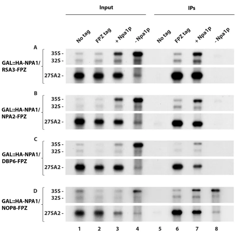
We then analysed the effect of depleting one complex member on the ability of the other mem-bers to associate in a stable fashion with 90S pre-ribosomal particles. We envisaged in particu-lar that due to its RNA recognition motif (RRM), Nop8p might be involved in the tethering of the complex to 35S pre-rRNA, while the putative DEAD box helicase Dbp6p might promote complex integration within pre-ribosomal particles through local remodelling of these parti-cles. To assay for interaction with pre-ribosomal particles, precipitation experiments were per-formed with IgG-sepharose beads and extracts from strains expressing a given FPZ-tagged bait complex protein (that binds to the IgGs) and depleted, or not depleted as control, of another complex protein. Co-precipitated pre-rRNAs were analysed by Northern while the efficiency of bait protein precipitation was assessed by Western. Depletion of Dbp6p has little effect on
Fig 5. Npa1p depletion preventsin vivo interactions between Rsa3p, Npa2p and Nop8p. Tandem affinity purifications were performed using the indicated
strains grown in glucose- (lanes 2, 3, 4, 5, 7 and 9) or galactose-containing medium (lanes 1, 6 and 8). Purified proteins were separated by SDS-PAGE and detected by Coomassie (lanes 1 to 5) or silver staining (lanes 6 to 9).
https://doi.org/10.1371/journal.pgen.1007597.g005
the efficiency of co-precipitation of 35S pre-rRNA with Rsa3p-FPZ (S9 Fig). In contrast, deple-tion of Nop8p strongly reduces the efficiency of co-precipitadeple-tion of 35S pre-rRNA with Rsa3p-FPZ (S10 Fig). This result prompted us to test the effects of Nop8p depletion on the association of another complex protein, Npa2p, with pre-rRNAs. Lack of Nop8p also strongly reduces the efficiency of co-precipitation of 35S pre-rRNA with Npa2p-FPZ (S11 Fig). This reduction is not due to a lower precipitation efficiency of the bait protein under depletion conditions, as assessed by Western (S10andS11Figs, bottom). We conclude that Dbp6p depletion has little effect on Rsa3p integration within 90S pre-ribosomal particles, while Nop8p depletion strongly inhibits, but does not abolish, Rsa3p and Npa2p integration within 90S particles.
We next tested the effects of Npa1p depletion on complex protein interactions with 35S pre-rRNA. Strikingly, lack of Npa1p abolishes 35S pre-rRNA co-precipitation with Rsa3p (Fig 6A, lane 8), Npa2p (Fig 6B, lane 8) and Dbp6p (Fig 6C, lane 8). Such lack of co-precipitation of 35S pre-rRNA is not due to reduced precipitation of the bait proteins (S12 Fig). To our sur-prise, Npa1p depletion does not prevent 35S pre-rRNA co-precipitation with Nop8p (Fig 6D, lane 8). To confirm these results, we assessed the sedimentation on sucrose gradients of com-plex proteins extracted from cells expressing or depleted of Npa1p (S13 Fig). Under non depleted conditions, Rsa3p, Nop8p and Dbp6p are found in fractions containing 60S ribo-somal subunits and in heavier fractions, likely reflecting their association with pre-60S and 90S pre-ribosomal particles, respectively (S13 Fig, left). When Npa1p is depleted, Rsa3p and Dbp6p shift to the top of the gradient, consistent with reduced association with pre-ribosomal particles (S13 Fig, right). In contrast, Nop8p is still present in 60S-containing and heavier frac-tions, consistent with it remaining part of 90S and residual pre-60S particles. The immunopre-cipitation and gradient results therefore indicate that the interactions of Rsa3p and Dbp6p with 90S pre-ribosomal particles are strongly destabilised when Npa1p is absent. In contrast, Nop8p does not require Npa1p for its stable incorporation within pre-ribosomal particles.
Npa1p is not required for Rpl3 integration within 90S pre-ribosomal
particles
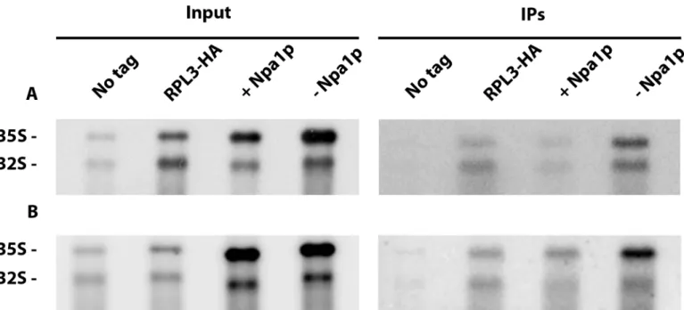
Specific synthetic lethal interactions have been obtained when combining certain mutant alleles of the genes encoding Npa1p, Npa2p, Nop8p, Rsa3p or Dbp6p and mutant alleles of the gene encoding ribosomal protein Rpl3 [17]. Such genetic interactions may reflect functional links between the Npa1p-containing complex and Rpl3, a ribosomal protein crucial for the maturation of 27SA2 pre-rRNA containing pre-60S particles and assembly of the large ribo-somal subunit [10]. It has been proposed that Rpl3 plays a crucial role in the folding of the cen-tral core of 25S rRNA, by binding to both the 3’ end of 25S rRNA and the 5’end of 5.8S rRNA [10]. We first tested the possibility that the Npa1p-containing complex might be required for the recruitment and/or the stable integration of Rpl3 within 90S pre-ribosomal particles. This hypothesis was investigated by an anti-HA immunoprecipitation experiment using strain
GAL::npa1/RPL3-HA, as fusion of the FPZ tag to Rpl3 strongly affected growth. The study of
interactions between Rpl3 and the 35S pre-rRNA by precipitation experiments having never been reported to our knowledge, we first performed such experiments at low salt concentra-tion (50 mM KCl), since interacconcentra-tions between small ribosomal subunit proteins and the 35S pre-rRNA are known to be salt sensitive [23]. Depletion of Npa1p did not affect the efficiency of co-precipitation of 35S pre-rRNA with Rpl3-HA at 50 mM KCl relative to the non depleted controls (Fig 7A). This indicates that Npa1p is not required for the recruitment of Rpl3 within 90S pre-ribosomal particles. Npa1p might nevertheless be required for the stabilisation of Rpl3 within 90S pre-ribosomal particles. To test this idea, we performed anti-HA immunoprecipita-tion experiments at 400 mM KCl. Even at this salt concentraimmunoprecipita-tion, depleimmunoprecipita-tion of Npa1p did not
Fig 6. Effects of Npa1p depletion on the efficiency of pre-rRNA co-precipitation with Rsa3p-FPZ (A), Npa2p-FPZ (B), Dbp6p-FPZ (C) and Nop8p-FPZ (D).
Precipitation experiments were carried out with IgG-sepharose beads and extracts from the indicated strains of the typeGAL::HA-NPA1/PROTEIN Y-FPZ grown in
galactose-containing medium (+ Npa1p) or shifted 8 hours to glucose-containing medium (- Npa1p), as well as from thePROTEIN Y-FPZ (FPZ tag) and the parental
BY4742 (No tag) strains grown in glucose-containing medium as controls. RNAs were extracted from aliquots of the input extracts (Input) and from the IgG-sepharose beads after precipitation and washing (IPs). Pre-rRNAs were analysed by Northern using the 23S1 oligonucleotide probe.
https://doi.org/10.1371/journal.pgen.1007597.g006
decrease the co-precipitation efficiency of 35S pre-rRNA with Rpl3-HA relative to controls (Fig 7B). We conclude that Npa1p is required neither for the recruitment nor for the stabilisa-tion of Rpl3 within 90S pre-ribosomal particles.
Npa1p binds 25S rRNA adjacent to Rpl3 binding sites and the root helices
of the first and last secondary structure domains of 25S rRNA
Since the Npa1p complex is not required for the recruitment of Rpl3, it could instead collabo-rate with Rpl3 during an early critical step of large ribosomal subunit assembly. If this is the case, we might expect Npa1p to bind to (pre)-rRNA sequences located in the vicinity of Rpl3 binding sites. To identify Npa1p pre-rRNA binding sites, we performed UV cross-linking and analysis of cDNAs (CRAC) [24] followed by deep sequencing of the cDNAs obtained. The sequences retrieved with tagged Npa1p that are most highly enriched compared to the untagged BY4742 control are snoRNA, 25S rRNA and ITS2 sequences (S14 Fig). The direct cross-linking sites can be identified by the presence within those sequences of small deletions or point mutations [24]. Five major Npa1p cross-linking sites can be identified within 25S rRNA (Fig 8AandS15 Fig). Strikingly, most of these are positioned in the immediate vicinity (in the primary or conserved secondary structure of 25S/5.8S rRNAs) of rRNA sequences con-tacted by Rpl3 in the mature 60S subunit, including root helices of rRNA domains I and VI (Fig 8A). When positioned on the recently published structure of a nucleolar pre-60S particle in so-called state C [11], all these Npa1p cross-linking sites surround the root helix of domain I (that includes the 5’ end of 5.8S) and the root helix of domain VI (that includes the 3’ end of 25S) (Fig 8B).
Fig 7. Effects of Npa1p depletion on interactions of Rpl3 with pre-rRNAs. Precipitation experiments were carried out at 50 mM KCl (A) or 400 mM KCl (B)
with anti-HA agarose beads and extracts from theGAL::npa1/RPL3-HA strain grown in galactose-containing medium (+ Npa1p) or shifted 16 hours to
glucose-containing medium (- Npa1p), as well as from theRPL3-HA and the parental BY4742 (No tag) strains grown in glucose-containing medium as controls. RNAs
were extracted from aliquots of the input extracts (Input) and from the anti-HA agarose beads after precipitation and washing (IPs). Pre-rRNAs were analysed by Northern using the 23S1 oligonucleotide probe.
Fig 8. Npa1p binding sites on 25S rRNA determined by CRAC. (A) Npa1p binding sites positioned on the 25S rRNA secondary
structure. Npa1p binding site are highlighted in blue and cross-linked nucleotides identified by point mutations are circled in brown. Also indicated are positions of modifications (green) directed by snR5, snR10, snR42, snR61, the complementary sequences to snR190 (snR190 a and b, green), the binding sites of Rpl3 ([8], yellow), Rrp5p (pink) [37] and Prp43p (grey) [36]. (B) Positions of the Npa1p cross-linking sites (CLS 1-5b) on the rRNA 3D structure of the state C nucleolar pre-60S particle ([11]; pdb code: 6EM1). 25S and 5.8S rRNAs are represented in pale blue and yellow, respectively. Rpl3 is shown in magenta. Root helices 1 (R1) and 6 (R6) are represented by a black (25S) and yellow (5.8S) helix (R1), and by a green helix (R6).
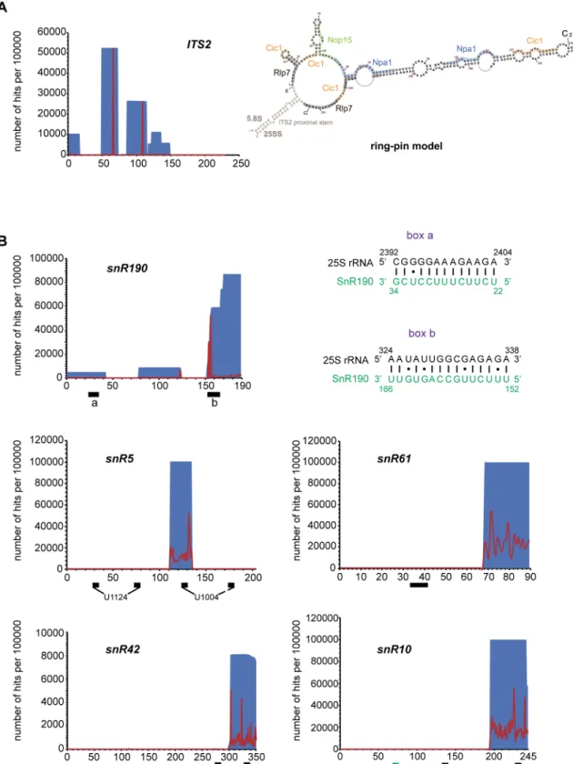
Npa1p was also found cross-linked to two sites within ITS2 (Fig 9A). In the proposed “ring-pin” model of ITS2 secondary structure [7], Npa1p is positioned at the base and in the central region of the hairpin structure (Fig 9A), where no AF binding had so far been reported.
Npa1p binds to snR190 snoRNA and to snoRNAs involved in decoding
center and peptidyl transferase center modifications
A high proportion of reads in the Npa1p-HTP data set (15%) correspond to snoRNAs, while comparatively very few snoRNA sequences were retrieved in the case of the BY4742 control (S14 Figand S3 Table inS3 Text). The reads that map to the box C/D snoRNA snR190 gene are by far the most abundant (S3 Table inS3 Text). This snoRNA is predicted to guide 2’O methylation of 25S rRNA G2395 residue, but this methylation has never been detected even with high throughput detection methods [25–27] and snR190 might instead function as a fold-ing chaperone. In support of this hypothesis, we detected within snR190 a so far unreported region of 15 nucleotide perfect complementarity with a sequence of 25S rRNA located in the vicinity of Npa1p cross-linking site n˚1 (box b, Figs8Aand9B). Other abundant reads map to sequences of snoRNAs that bind within the PTC (snR10, snR42) or in domain II of 25S rRNA (snR5, snR61). Analysis of point mutations indicates that Npa1p cross-links to one of the pseu-douridylation pockets of snR5, snR10 and snR42 (Fig 9B), suggesting that at least in the case of these snoRNAs, interactions with Npa1p may take place while they are bound to pre-rRNAs. Altogether, these data are compatible with the hypothesis that the Npa1p complex promotes pre-rRNA folding events within the earliest pre-60S particle and/or modulates interactions between 25S rRNA and snoRNAs within these particles.
Discussion
The Npa1p complex: Structure and requirements for integration into 90S
pre-ribosomal particles
Formation and maturation of the earliest pre-60S particle are probably the least understood stages of ribosome biogenesis. No overall particle structure is available, although a recent high throughput pre-rRNA probing has provided important insights into the folding of the 35S and 27SA2 pre-rRNAs [7]. Moreover, very recently, structures of nucleolar pre-60S particles have been reported that seem to lack the Npa1p complex and probably correspond to particles closely downstream from the first pre-60S particle in the maturation pathway [11–13]. Of the approximately 30 AFs present in the first pre-60S particle [6], a third are required for its pro-duction and/or stability. Their precise molecular roles remain unknown. The latter AFs include the Rrp5p/Noc1p/Noc2p complex [28], the RNA helicases Prp43p, Dbp6p, Dbp7p and Dbp9p, the RNA-binding proteins Nop4p and Nop8p and the Npa1p, Npa2p and Rsa3p pro-teins. Our work provides strong evidence that Npa1p, Npa2p, Nop8p and Rsa3p form a core complex, with which Dbp6p interacts in a magnesium sensitive fashion. Since Npa1p, Npa2p, Nop8p and Dbp6p co-purify with Rsa3p when RNA pol I transcription is inhibited, it is proba-ble that the complex forms prior to its incorporation in 90S pre-ribosomal particles. Likewise, it was demonstrated that Noc1p, Noc2p and Rrp5p, members of 90S and the first pre-60S par-ticles, can form a complex outside pre-ribosomal particles [28,29]. The order of and require-ment for assembly of early pre-60S particle components with nascent pre-ribosomal particles was recently investigated [30]. This study suggests that at least Npa1p and Npa2p require tran-scription of the full 25S rRNA sequence for stable association with nascent pre-ribosomal par-ticles. All members of the Npa1p complex are associated with the 27SA2 pre-rRNA,
Fig 9. Npa1p binds to ITS2 and to snoRNAs. (A) Npa1p cross-links to ITS2. Left: Number per 100000 reads and positions of reads
from the NPA1-HTP CRAC experiment on the ITS2 sequence. Positions and numbers of reads are indicated in blue, positions of mutations are indicated in red. Right: Positions of Npa1p cross-linking sites (blue, with mutated residues indicated in brown) on the “ring-pin” model of ITS2 structure [7]. Binding sites of Nop15p (green), Cic1p/Nsa3p (light orange) and Rlp7p (grey) [7,39–41] are also indicated. (B) Npa1p cross-links to a subset of snoRNAs. Number per 100000 reads and positions of reads from the NPA1-HTP CRAC experiment on the corresponding snoRNA sequences. Positions and numbers of reads are indicated in blue, positions of mutations are
27SB pre-rRNAs, suggesting that they dissociate together prior to formation of intermediate, 27SB pre-rRNA-containing, pre-60S particles. Npa1p, of 203 kDa predicted molecular weight, is by far the largest protein of the complex and seems to form its backbone. When Npa1p is absent, Npa2p, Nop8p and Rsa3p can no longer interactin vivo. We also show that Dbp6p
binds directly to both Npa1p and Npa2pin vitro. Moreover, our data point to a direct in vivo
interaction between Npa2p and Dbp6p and demonstrate that Npa2p is essential for the Npa1p-Dbp6p interactionin vivo. These data suggest that Npa1p binds directly to Npa2p,
Nop8p, Rsa3p and Dbp6p, that Npa2p, Nop8p and Rsa3p do not interact directly in absence of Npa1p, while Dbp6p also binds to Npa2p (see model shown inS16 Fig). These conclusions are fully consistent with the results of a large scale yeast two-hybrid screen between pre-60S parti-cle AFs [31]. Moreover, another large scale double hybrid analysis conducted with Chaeto-mium thermophilum AFs strongly suggests a direct interaction between Npa1p and Rsa3p [32].
Npa1p is also crucial for the stable integration of most complex members within 90S pre-ribosomal particles. Immunoprecipitation experiments indicate that depletion of Npa1p inhib-its interactions between Rsa3p, Npa2p and Dbp6p with the 35S pre-rRNA, while they show that Nop8p can still interact with this pre-rRNA. Sucrose gradient sedimentation experiments also suggest strongly reduced interactions of Rsa3p and Dbp6p with pre-ribosomal particles when Npa1p is depleted and confirm that Nop8p can still interact with pre-ribosomal particles, maybe because it possesses RNA-binding activity [21]. Although depletion of Nop8p does not abolish integration of Npa2p and Rsa3p within 90S pre-ribosomal particles, it significantly destabilizes this integration. We propose that Nop8p strengthens the interaction of the Npa1p complex with pre-ribosomal particles through direct binding to the pre-rRNA mediated by its RRM domain. In contrast, the absence of Dbp6p does not significantly impair Rsa3p integra-tion within 90S pre-ribosomal particles nor does it prevent interacintegra-tions between Rsa3p, Npa1p, Npa2p and Nop8p. These data suggest that in the absence of Dbp6p, the Npa1p/ Npa2p/Nop8p/Rsa3p core complex can form and efficiently associate with 90S pre-ribosomal particles. However, this core complex is probably not fully functional since it lacks enzymatic activity. We envisage that the Npa1p/Npa2p/Nop8p/Rsa3p core complex is necessary not only for the stable integration of the Dbp6p helicase at the correct site within 90S pre-ribosomal particles but also for the modulation of its enzymatic activities. Further experiments are needed to test the latter hypothesis.
Functional and structural links between the Npa1p complex and Rpl3,
Rrp5p, Prp43p, Nop15p and Cic1p/Nsa3p
Specific synthetic lethal interactions have been uncovered between mutant alleles of genes encoding Npa1p complex components and mutant alleles of the large subunit Rpl3 ribosomal protein gene [17]. These genetic interactions suggested that the Npa1p complex and Rpl3 may functionally interact. Rpl3 is a crucial ribosomal protein which is necessary for the processing of the 27SA2 pre-rRNA at the A3 site and for the stable association with pre-ribosomal parti-cles of most other large subunit ribosomal proteins [10]. In its absence, the first pre-60S parti-cle is formed, but probably because it is misassembled, the 27SA2 pre-rRNA it contains fails to be processed and the particle is rapidly turned over [10,33]. Rpl3 is also the largest ribosomal protein in yeast, which associates co-translationally with a specific chaperone, Rrb1p [34,35]. This association shields Rpl3 from degradation and promotes its nuclear import. Since Rrb1p
indicated in red. Black boxes indicate positions of anti-sense sequences (snR190, snR61) or pseudouridylation pockets (snR5, snR10, snR42). The green box indicates the position of snR10 anti-sense sequence complementary to 18S rRNA. The complementarities between snR190 boxes a and b (novel anti-sense sequence) and 25S rRNA are drawn next to the snR190 graph.
is not a stable component of pre-ribosomal particles, it likely dissociates from Rpl3 prior to or during Rpl3 integration within 90S pre-ribosomal particles. We reasoned that the Npa1p com-plex might facilitate the integration of Rpl3 within 90S particles, possibly by promoting its release from Rrb1p. However, our data clearly show that Npa1p is required neither for Rpl3 recruitment within 90S pre-ribosomal particles nor for the stabilisation of its integration. We cannot formally exclude the possibility that Nop8p, which can be stably integrated within 90S particles in the absence of Npa1p, promotes Rpl3 recruitment on its own.
In the mature yeast 60S ribosomal subunit, Rpl3 interacts with the 3’ end of 25S rRNA and the 5’ end of 5.8S rRNA [8]. The 3’end of 25S rRNA is engaged in a short double helix struc-ture. Likewise, the 5’end of 5.8S rRNA forms a small helix with a sequence of 25S rRNA. These two short helices lie at the base of the secondary structure domains VI and I of 25S rRNA, respectively. They are clustered in the mature 60S subunit with the short helices at the base of the four other secondary structure domains of 25S rRNA (namely domains II, III, IV and V). Gamalinda and colleagues have proposed that clustering of these root helices is a key initial step required for the subsequent correct folding, stabilization and compaction of large subunit rRNAs [10]. According to a recent high throughput pre-rRNA structure probing, formation of root helix I (5’end of 5.8S rRNA base-paired to 25S rRNA) is a very early, probably co-tran-scriptional event [7]. Moreover, in the earliest nucleolar pre-60S particle structure reported so far (state A of [11]), the entire domain I and parts of domains II and VI are already stabilised [11], supporting the notion that large subunit rRNA stabilization and compaction are initiated by those of domains I, II and VI. In the better resolved structure of a nucleolar pre-60S particle in state C [11], root helices of domains I (containing the 5’ end of 5.8S) and VI (containing the 3’ end of 25S) are already clustered. When positioned on this structure, Npa1p cross-linking sites surround on all sides these root helices (Fig 8B). Npa1p may play a role in bringing root helices of domains I and VI together. In the state C structure, Rpl3 is associated with the root helix of domain VI, but contrary to the situation in intermediate pre-60S particles purified via Nog2p [9], it does not yet seem to be able to contact the root helix of domain I (bearing in mind that an internal segment of Rpl3 is missing in the published state C structure). Thus Npa1p may intervene prior to Rpl3 in bringing domains I and VI together. After Npa1p disso-ciation from pre-60S particles, Rpl3 may maintain the compacted state of root helices I and VI.
Interestingly, partially overlapping binding sites for Rrp5p and the helicase Prp43p, two components of the earliest pre-60S particles [6], have been identified by CRAC next to Npa1p cross-linking site n˚ 1 adjacent to the root helix I (Fig 8A) [36,37]. Whether simultaneous binding of Rrp5p and Prp43p in that region can occur remains to be determined given the par-tial overlap of the cross-linking sites. Npa1p could interact simultaneously or sequenpar-tially with both proteins while bound to this 25S rRNA domain. Direct physical interactions between Npa1p and Rrp5p or Prp43p are supported by the findings that Npa1p co-purifies with Rrp5p after cross-linking under RNase treatment and highly denaturing conditions [37], while the
Chaetomium thermophilum orthologues of Npa1p and Prp43p are found to interact in a yeast
two-hybrid assay [32]. Finally, we identified a novel potential interaction site for snR190 snoRNA next to the Rrp5p binding site in domain I (Fig 8A) and both Npa1p (this work) and Rrp5p [37] interact with snR190. All these observations lead us to propose that Rrp5p (together with associated Noc1p and Noc2p), Prp43p, snR190 and the Npa1p complex collaborate in early pre-rRNA structural rearrangements. In particular, snR190 could act as a specific chaper-one to control the formation of the root helix of domain V (PTC) and to draw together this root helix with that of domain I. Dbp6p, which displays ATPase activityin vitro (manuscript
in preparation), could use ATP hydrolysis to drive pre-rRNA conformational rearrangements and/or to promote snR190/pre-rRNA interaction or dissociation (see below). We note in that respect that substitution of a key amino acid in the active site of Dbp6p prevents growth
without affecting the steady-state accumulation of the protein, strongly suggesting that the enzymatic activity of Dbp6p is essential for large ribosomal subunit production [38].
Two prominent Npa1p cross-linking sites were also found in ITS2 (Fig 9A), in between the binding sites of Nop15p and Cic1p/Nsa3p [39–41]. All three AFs are present in the earliest pre-60S particles [6]. They could participate to the folding of ITS2, a very early event [7] and/ or to the protection of ITS2 prior to the recruitment of the dedicated processing enzymes.
Intimate physical interactions between Npa1p and a subset of modification
guide snoRNAs interacting with 25S rRNA
The CRAC analysis indicates that Npa1p is cross-linked to snoRNAs that bind to the PTC or domain II (decoding center) of the 25S rRNA (Figs8Aand9B,S14 Fig, S3 Table inS3 Text). In addition to snR190 (see above), the next most frequent reads map to the sequences of modi-fication guide snoRNAs snR42, snR10 (that bind to the PTC), snR5 and snR61 (that bind to domain II). Remarkably, snR42 and snR10 feature among the snoRNAs we found most effi-ciently precipitated with tagged Npa1p [6]. Npa1p cross-linking sites 2 and 3 on the 25S rRNA (Fig 8A) lie in the immediate vicinity of snR190, snR42 and snR10 binding sites. Moreover, Npa1p is cross-linked to one of the pseudouridylation pockets of snR5, snR10 and snR42 (Fig 9B), which may indicate that Npa1p mostly contacts these snoRNAs while they are bound to pre-rRNAs. This interpretation is supported by the finding that the efficiency of co-precipita-tion of snR5, snR10 and snR42 with tagged Rsa3p-FPZ is more than halved when RNA Pol I transcription is inhibited (S17 Fig). The Npa1p complex could assist the association and/or dissociation of snoRNAs with or from pre-rRNAs by providing two activities. The non-enzy-matic components of the complex, in particular Npa1p and Npa2p, could form a rigid struc-tural framework maintaining the overall cohesion of the pre-rRNA structure, when local secondary structures unfold to allow snoRNA binding and refold upon their dissociation. Such a role has also been proposed for Rrp5p which remarkably, is also cross-linked to snR10 and snR42 [37]. This activity of Npa1p and Rrp5p could in fact result from their function in promoting the stability of the compacted state of root helices and/or the convex solvent side of the particles (see above). The enzymatic component of the Npa1p complex, Dbp6p, might pro-vide the needed ATPase/helicase activities, possibly assisted by Prp43p, directly or indirectly involved in the control of snoRNA/pre-rRNA interactions.
Materials and methods
Yeast strains and media
Strains expressing Npa1p-FPZ, Npa2p-FPZ, Nop8p-FPZ, Dbp6p-FPZ and Rsa3p-FPZ have been obtained as follows. The open reading frames (ORFs) encoding the composite FPZ tag (Flag—PreScission protease cleavage site—Z sequence derived fromS. aureus Protein A) and
theURA3 gene have been amplified by PCR using plasmid pBS1539-FPZ [42] and primers listed in S1 Table inS1 Text. The resulting PCR fragments have been transformed into strain BY4742 (MATα, his3Δ1, leu2Δ0, lys2Δ0, ura3Δ0) and clones having integrated the URA3 gene
have been selected on YNB (Yeast Nitrogen Base) medium lacking uracil.
A strain expressing Rpl3-HA has been produced by transforming strain BY4742 with a PCR product obtained using plasmid pFA6a-3HA-His3MX6 [43] and primers listed in S1 Table inS1 Text. Clones having integrated theHIS3 gene have been selected on YNB medium
lacking histidine.
Strains expressing the HA-tagged ORF of Npa1p, Npa2p, Nop8p or Dbp6p under the con-trol of theGAL1-10 promoter and FPZ-tagged versions of Npa1p complex proteins have been
produced as follows. PCR products generated using primers listed in S1 Table inS1 Textand plasmid pFA6a-kanMX6-PGAL1-3HA [43] have been transformed into strainsNPA1-FPZ, NPA2-FPZ, DBP6-FPZ, NOP8-FPZ and RSA3-FPZ. Selection for clones having integrated the
kanMX6 resistance cassette has been performed on YPGal medium supplemented with G418 (Gibco, 200μg/ml final concentration).
StrainGAL::npa1/RPL3-HA has been obtained by transforming the GAL::npa1 strain with a
PCR cassette produced with plasmid pFA6a-3HA-His3MX6 [43] and primers listed in S1 Table inS1 Text. Clones having integrated theHIS3 gene have been selected on YNB medium
lacking histidine. TheGAL::npa1 strain was generated by transforming BY4742 with a PCR
cassette produced with plasmid pFA6a-kanMX6-PGAL1 [43] and primers listed in S1 Table in
S1 Text. Selection for clones having integrated the kanMX6 resistance cassette has been carried out as described above.
Strainrrn3-8/RSA3-FPZ has been produced by transforming strain rrn3-8 (ade5, his7-2, leu2-112, trp1-289, ura3-52, rrn3-8) with a FPZ-URA3 cassette flanked by RSA3 sequences
pro-duced as described above. Clones having integrated theURA3 gene have been selected on YNB
medium lacking uracil.
Strain expressing Npa1p-HTP (His tag—Tev cleavage site—Z sequence derived fromS. aureus Protein A) has been obtained by transforming BY4742 with a PCR cassette produced
using plasmid pBS1539-HTP [24] and primers listed in S1 Table inS1 Text. Clones having integrated theURA3 gene have been selected on YNB medium lacking uracil.
Strains have been grown in YP (1% yeast extract, 1% peptone) supplemented with 2% galac-tose or 2% glucose or YNB (6.8 g/L) supplemented with ammonium sulphate (20 g/L) and 2% galactose or 2% glucose.
Escherichia coli strains and plasmids
E. coli strains directing expression of His-Dbp6p and His-Rsa3p were obtained by
transform-ingE. coli Rosetta λDE3 with plasmids pET-DBP6 and pRC52, respectively. pET-DBP6 was
obtained by cloning into a modified pET-15b (Novagen) plasmid in the NdeI and BamHI restriction sites [44] aDBP6 ORF PCR cassette by Ligation Independent Cloning (LIC)
method using oligonucleotides listed in S2 Table inS2 Text. pRC52 was obtained by inserting into XhoI-digested pET-15b a PCR cassette obtained by amplifying theRSA3 ORF using
oligo-nucleotides listed in S2 Table inS2 Text.E. coli strains directing expression of GST-Rsa3p,
GST-Nop8p, GST-Dbp6p, GST-Npa2p and GST-Npa1p were obtained by transformingE. coli
RosettaλDE3 with plasmids pRC55, pRC56, pRC57, pRC58 and pRC59, respectively. pRC55, pRC56, pRC57, pRC58 and pRC59 were obtained by inserting into XhoI-digested pGEX-6P1 a PCR cassette obtained by amplifying theRSA3, NOP8, DBP6, NPA2 and NPA1 ORFs,
respec-tively, using oligonucleotides listed in S2 Table inS2 Text. The cloning of all ORFs into XhoI-digested pGEX-6P1 was performed with the kit In-Fusion HD Cloning (Clontech). AnE. coli
strain directing expression of MBP-Nop8p-His was obtained by transformingE. coli Rosetta
λDE3 with plasmid pRC60. pRC60 was obtained by inserting into EcoRI-digested pMAL-c2X a PCR cassette obtained by amplifying theNOP8 ORF from plasmid pRC56 using
oligonucleo-tides listed in S2 Table inS2 Text.
Purification of His-tagged and GST-tagged proteins
E. coli Rosetta λDE3 bacteria transformed with plasmids pET-DBP6, pRC52 or pRC60,
direct-ing expression of His-Dbp6p, His-Rsa3p or MBP-Nop8p-His, respectively, are grown at 37˚C in 2YT medium (16 g/L tryptone, 10 g/L yeast extract, 5 g/L NaCl) supplemented with 100μg/ ml ampicillin and 25μg/ml chloramphenicol to an A600of 0.6. Recombinant protein
expression is then induced with 0.5 mM isopropylβ-D-thiogalactoside (IPTG), and culturing is continued overnight at 18˚C. Bacteria are harvested by centrifugation, resuspended in 50 mM Tris-HCl pH 7.5, 500 mM NaCl, 10% glycerol, 10 mM imidazole, 0.1% Triton-X100, 5 mM dithiothreitol (DTT), 0.5 mM phenylmethylsulfonyl fluoride and frozen at -80˚C. Bacteria are thawed and sonicated on ice in the presence of a complete EDTA-free protease inhibitor cocktail (Roche). The soluble fraction is subjected to affinity chromatography using a HisTrap FF column (GE Healthcare) and His-tagged proteins are eluted with a linear gradient of imid-azole. The eluted MBP-Nop8p-His protein is recovered at this step and stored at -80˚C. In the case of His-Dbp6p and His-Rsa3p proteins, affinity chromatography on HisTrap FF column is followed by size exclusion chromatography on a Superdex 200 column. Eluted fractions with imidazole containing His-tagged proteins are pooled and the proteins of interest are purified by size exclusion chromatography with a Superdex 200 gel filtration column (GE Healthcare) in 20 mM Tris-HCl pH 8, 100 mM potassium chloride, 10% glycerol, 1 mM DTT. The purified His-tagged proteins are stored at -80˚C.
TheE. coli Rosetta λDE3 bacteria transformed with plasmids pRC55 to pRC59 are used to
produce GST-tagged proteins. Cells are grown at 37˚C in 2YT medium supplemented with 100μg/ml ampicillin and 25 μg/ml chloramphenicol to an A600of 0.6. Recombinant protein
expression is then induced with 0.5 mM isopropylβ-D-thiogalactoside (IPTG) and culturing is continued overnight at 18˚C. Bacteria are harvested by centrifugation, resuspended in 50 mM Tris-HCl pH 7.5, 150 mM NaCl, 10% glycerol, 0.1% Triton-X100, 5 mM DTT and stored at -80˚C. Bacteria are first incubated with 300μg/ml of lysozyme for 30 min on ice and soni-cated in the presence of Protease Inhibitor cocktail (Sigma-Aldrich). The clarified lysate is sub-jected to a Generon Proteus “1-step batch” Midi Spin column (CliniSciences) loaded with Glutathione Sepharose 4 FF resin (GE Healthcare). The GST-tagged proteins are eluted with 20 mM reduced glutathione in 50 mM Tris-HCl pH 8.8, 150 mM NaCl, 10% glycerol, 0.1% Triton-X100, 5 mM DTT. The GST-tagged proteins are concentrated with Amicon Ultra Cen-trifugal Filter (Millipore) with a molecular weight cut-off of 30 000 Da and/or desalted with Zeba Spin Desalting column (Thermo Scientific) with a molecular weight cut-off of 7000 Da. GST-tagged proteins are stored at -80˚C in 20 mM Tris-HCl pH 8, 150 mM potassium chlo-ride, 10% glycerol, 1 mM DTT.
Production of antibodies
Anti-Rsa3p, anti-Dbp6p and anti-Nop8p antibodies were produced in rabbit using purified recombinant His-Rsa3p, His-Dbp6p and MBP-Nop8p-His proteins, respectively. Anti-Npa1p or anti-Npa2p antibodies were produced in rabbit using a mix of two synthesized peptides located at the N- and C-termini of each protein and were purified against only one peptide. All polyclonal antibodies were generated by the Agro-Bio company (La Ferte´ Saint-Aubin, France) following a standard immunization protocol.
GST pull-down assays
A total of 2μg of each purified recombinant GST-tagged protein are incubated under gentle shaking for 2 h at 4˚C in 150μl IP buffer (50 mM Tris-HCl pH 8, 150 mM potassium chloride, 5 mM magnesium chloride, 0.2% Nonidet P-40) with 10μl of Glutathione Magnetic Beads (Pierce), previously coated with 3% BSA during 2 h. The beads are washed three times with IP buffer and are incubated with 2μg of purified His-Dbp6p in 200 μl IP buffer with gentle shak-ing for 1 h at 16˚C. The beads are washed three times with IP buffer and proteins retained on the beads using a magnetic rack are eluted with SDS-PAGE loading buffer (100 mM Tris-HCl
pH 6.8, 4% SDS, 20% glycerol, 200 mM DTT, 0.04% bromophenol blue). Co-precipitated pro-teins are analyzed by Western blotting using anti-His (GE Healthcare) antibodies.
Tandem-affinity purification of FPZ-tagged proteins
Yeast cell powder is produced using a PM 100 planetary ball mill (Retsch) from a cell pellet obtained from 12 L of yeast culture grown to an O.D.600of 0.6–0.8. 25 g of cell powder are
dis-solved in 30 ml of buffer A (50 mM Tris-HCl pH 7.4, 150 mM NaCl, 10 or 100 mM MgCl2,
0.1% Igepal) to which EDTA-free protease inhibitors (Roche) are added. The sample is centri-fuged in a Beckman Optima L-100 XP ultracentrifuge 2 h at 39 000 rpm, 4˚C, in a Beckman Ti 50.2 rotor, with no brake. The resulting supernatant is subjected to a second ultracentrifuga-tion step during 45 min at 39 000 rpm, 4˚C. The pH of the clarified extract is adjusted to between 7.4 and 8 using 1 M Tris-HCl, pH 8. 50 ml of clarified extract are loaded on three 20 ml columns (Biorad) containing 600μl IgG sepharose beads (6 fast Flow, GE Healthcare). The beads are incubated with the extract during 30 min, 4˚C, with gentle agitation. Beads are then washed with 60 ml of buffer B (20 mM Hepes pH 7.4, 100 mM KOAc, 150 mM NaCl, 10 or 100 mM MgCl2, 1 mM DTT, 0.02% Tween, 0.1% Triton X-100), then incubated 2 h at 4˚C
with 40 units PreScission protease (GE Healthcare) in 800μl of buffer B. 3 ml of column elu-ates are loaded on a 10 ml Biorad column containing 200μl of anti-Flag beads (Sigma). The column is washed with 40 ml of buffer C (10 mM Tris-HCl pH 7.4, 150 mM NaCl, 10 or 100 mM MgCl2, 1 mM DTT). Purified complexes are eluted by adding 5 times 200μl of buffer C
supplemented with 2 x Flag peptide (100μg/ml, IGBMC Strasbourg).
Gel filtration analysis
Complexes purified by tandem affinity chromatography are concentrated on Vivaspin col-umns (Sartorius) by adding 3% BSA and centrifuging 3 times 2 min in a bench centrifuge (Eppendorf 5415D) at 13 000 rpm, 4˚C. 300μl of concentrated samples are injected in a Super-dex 200 10/300 GL (GE Healthcare) gel filtration column, previously equilibrated with a buffer containing 10 mM Tris-HCl pH 7.4, 150 mM NaCl, 100 mM MgCl2, 1 mM DTT. Eluted
frac-tions of 500μl are collected, from which proteins are precipitated using TCA and analysed by Western.
Immunoprecipitation experiments
Yeast cell pellets corresponding to 1 L of culture grown to O.D.6000.6–0.8 are re-suspended in
2 ml of buffer containing 20 mM Tris-HCl pH 8.0, 5 mM MgAc, 50, 200, or 400 mM KCl, 0.2% Triton-X100, 1 mM DTT, EDTA free protease inhibitors (Roche), 0.1 unit/μl of RNasin (Promega). 400μl of acid-washed glass beads (Sigma) are added to 800 μl of cell suspensions. Yeast cells are broken by three runs of vigorous 2 min 30 s vortexing, with 2 min incubation of samples in ice between each run. Extracts are clarified by two consecutive 5 min centrifugation steps at 13 000 rpm in an Eppendorf bench centrifuge. The absorbance at 260 nm of the clari-fied extracts is measured using a Nanodrop 2000 spectrophotometer. For an absorbance at 260 nm of 100, 500μl of clarified extracts are used. The required volumes of clarified extracts are mixed with 15μl of anti-HA agarose beads (Sigma, EZview Red Anti-HA Affinity Gel) or 15 μl of IgG-sepharose beads (6 fast flow, GE Healthcare) previously equilibrated in a buffer con-taining 20 mM Tris-HCl pH 8.0, 5 mM MgAc, 50, 200 or 400 mM KCl, 0.2% Triton-X100, 1 mM DTT. Sample volumes are adjusted to 1 ml final with the buffer used for extract prepara-tion. Samples are incubated during 1 h 30 min at 4˚C on a shaking table, centrifuged 30 s at 2000 rpm in an Eppendorf bench centrifuge and supernatants are discarded. Beads are washed 5 times by adding 1 ml of a buffer containing 20 mM Tris-HCl pH 8.0, 5 mM MgAc, 50, 200
or 400 mM KCl, 0.2% Triton-X100, 1 mM DTT, followed by a 30 s centrifugation step at 2000 rpm and removal of supernatants. Bead samples from which RNAs will be extracted are washed twice more in an identical manner. Bead samples used for protein analysis are washed twice more using 1 ml of a buffer containing 20 mM Tris-HCl pH 8.0, 5 mM MgAc, 50, 200, or 400 mM NaCl, 0.2% Triton-X100, 1 mM DTT. After removal of the supernatants, 50μl of a buffer containing 100 mM Tris-HCl pH 8.0, 4% SDS, 20% glycerol, 0.04% bromophenol blue, 200 mM DTT are added to the samples used for the analysis of precipitated proteins. Samples destined for RNA analysis are treated as follows. To the bead samples are added 160μl of 4 M guanidine isothiocyanate, 4μl 20 mg/ml glycogen (Roche), 80 μl of a 10 mM Tris-HCl pH 8.0, 1 mM EDTA pH 8.0, 100 mM NaAc solution, 120μl of phenol and 120 μl of chloroform. Sam-ples are vortexed, incubated 5 min at 65˚C, vortexed again, then centrifuged 5 min at 13 000 rpm, 4˚C. 240μl of aqueous phase are collected and mixed with 120 μl phenol and 120 μl chlo-roform. Samples are vortexed, then centrifuged 5 min at 13 000 rpm, 4˚C. Nucleic acids from 240μl of aqueous phase are precipitated using 750 μl ethanol during 12 h at -20˚C. During the incubation of the clarified extracts with the beads, total RNAs are extracted from 1/10thof the extract volumes used for the immunoprecipitations. Extract volumes are adjusted to 100μl final by adding the required volume of buffer used for extract preparation. 4μl of 20 mg/ml glycogen (Roche), 200μl of 4 M guanidine isothiocyanate, 150 μl of a solution containing 10 mM Tris-HCl pH 8.0, 1 mM EDTA pH 8.0, 100 mM NaAc, 225μl phenol and 225 μl chloro-form are added. Samples are vortexed, incubated 5 min at 65˚C, vortexed again, then centri-fuged 5 min at 13 000 rpm, 4˚C. 400μl of aqueous phase are extracted with 200 μl phenol and 200μl chloroform. After 5 min centrifugation at 13 000 rpm, 380 μl of aqueous phase are extracted with 380μl of a 1:1 phenol/chloroform mix. After 5 min centrifugation at 13 000 rpm, nucleic acids from 300μl of aqueous phase are precipitated with 1 ml ethanol during 12 h at -20˚C. After precipitation of total and immunoprecipitated RNAs, a 60 min centrifugation at 13 000 rpm, 4˚C, is carried out. Pellets are washed with 1 ml 70% ethanol and samples are centrifuged again 45 min at 13 000 rpm, 4˚C. After removal of the supernatants, the pellets are dried at room temperature and re-suspended in 20μl (in the case of total RNAs) or 4 μl (in the case of co-precipitated RNAs) of water.
Northern analysis
Electrophoresis of glyoxal-treated RNAs through agarose gels are performed as reported [45]. Northern analyses of U3, snR5, snR10 and snR42 snoRNAs, pre-rRNAs and mature rRNAs are carried out using32P-labelled oligodeoxynucleotide probes. Sequences of antisense oligo-nucleotides used to detect these RNAs are the following: anti-U3 probe (5’ATGGGGCTCAT CAACCAAGTTGG3’), anti-snR5 (5’GTCTACTTCCAGCCATTTGC3’), anti-snR10 (5’CAT GGGTCAAGAACGCCCCGGAGGGG3’), anti-snR42 (5’TCAAACAATAGGCTCCCTAA AGCATCACAA3’), 20S3 (5’TTAAGCGCAGGCCCGGCTGG3’), rRNA2.1 (5’GGCCAGC AATTTCAAGTTA3’), 23S1 (5’GATTGCTCGAATGCCCAAAG3’), 18S (5’CCGTCAGT GTAGCGCGCGTGCGGCCC3’) and 25S (5’CTCACGACGGTCTAAACCC3’). Blots are hybridized with 5’ end-labelled oligonucleotide probes using Rapid Hyb Buffer (GE Healthcare).
Western analysis
Proteins are separated by electrophoresis on 10% SDS-polyacrylamide gels in Laemmli run-ning buffer (Tris-Glycine-SDS) and transferred overnight at 20 V, 4˚C on Hybond-C extra (GE Healthcare) membranes in a transfer buffer containing 1X Laemmli running buffer, 20% ethanol. Membranes are saturated by 1 h incubation in 200 ml of buffer containing 1 X PBS,
0.5% Tween 20 and 5% milk. Specific proteins are detected by incubation in 1 X PBS, 0.5% Tween 20 and 5% milk with the following antibodies: anti-HA-HRP (Clone 3F10, Roche) diluted 1:1000; rabbit PAP (Dako) diluted 1:10 000; anti-Nhp2p (produced in rabbit) diluted 1:5000; anti-Npa1p (produced in rabbit) diluted 1:5000; anti-Npa2p (produced in rabbit) diluted 1:1000; anti-Nop8p (produced in rabbit) diluted 1:2000; anti-Rsa3p (produced in rab-bit) diluted 1:10 000; anti-Dbp6p (produced in rabrab-bit) diluted 1:10 000. After incubation with primary antibodies, membranes are washed 4 times with 200 ml of buffer containing 1 X PBS, 0.5% Tween 20 and 1% milk, or the same buffer lacking milk if no secondary antibodies are used. Anti-rabbit IgG-HRP conjugate (Promega) are used when needed as secondary antibod-ies diluted 1:10 000. Membranes are then washed 4 times with 200 ml of buffer containing 1 X PBS, 0.5% Tween 20. Proteins decorated by the antibodies are detected by chemiluminescence using ECL reagents (Biorad) and ImageQuant LAS 4000 biomolecular imager (GE
Healthcare).
Negative staining and transmission electron microscopy observations
Continuous carbon grids (Cu 300 Mesh, Quantifoil) are glow-discharged for 20 s on a Pelco easyGlow device (Ted Pella). 3.5μl of purified sample are then immediately deposited onto the grids and the excess liquid is blotted with a Whatmann 4 filter paper after 1 min incubation. Two successive drops of 3.5μl of 1% uranyl acetate staining solution are then applied on the grids and the second one rests on the grid 20 s before final blotting. Grids are then air dried and observed on the Jeol JEM 2100 LaB6electron microscope of the Toulouse
cryo-EM facility (METI,https://www-meti.biotoul.fr), with an acceleration voltage of 200 kV. Images are recorded on an US 1000 CCD camera (Gatan), with a calibrated pixel size of 4.5Å.
CRAC analysis
This analysis was performed using the Npa1p-HTP expressing strain and the megatron as cross-linking tool as described in [37].
Deep-sequencing and bio-informatics analyses
To build the cDNA library, 5’ adapters are linked at the 5’ end (BY4742: 5’-invddT-ACACr GrArCrGrCrUrCrUrUrCrCrGrArUrCrUrNrNrNrUrArArGrC-OH-3’; NPA1-HTP: 5’-invddT-ACACrGrArCrGrCrUrCrUrUrCrCrGrArUrCrUrNrNrNrArUrUrArGrC-OH-3’), and a 3’ linker (5’-AppTGGAATTCTCGGGTGCCAAG/ddC/-3’) is added at the 3’ end. Sam-ples are analysed by Illumina sequencing using HiSeq. Barcodes, adapters and low quality reads are eliminated using Flexbar (http://sourceforge.net/projects/flexbar/). Remaining reads are aligned to the yeast genome using Novoalign (http://www.novocraft.com). Down-stream analyses including the pileups are performed using the pyCRAC tool suite (http:// sandergranneman.bio.ed.ac.uk/Granneman_Lab/pyCRAC_software.html). Hits repartition per million of sequences are produced using pyReadCounter.py—m 1000000 option. Different pileups of hits for each gene are obtained using pyPileup.py—L 50—limit = 100000 options.
NGS analysis files of raw and processed data were deposited in Gene Expression Omnibus database under the accession number GEO: GSE104281. In addition, the NPA1-HTP CRAC data displayed in the UCSC genome browser can be accessed via the following URL:https://genome. ucsc.edu/cgi-bin/hgTracks?db=sacCer3&lastVirtModeType=default&lastVirtModeExtraState= &virtModeType=default&virtMode=0&nonVirtPosition=&position=chrXII%3A460923% 2D466869&hgsid=642906575_AycMTIJwrsUKtASq0SLaYLJEWCOF.