Sub-ppb mercury detection in real environmental samples with an improved Rhodamine-based detection system
Texte intégral
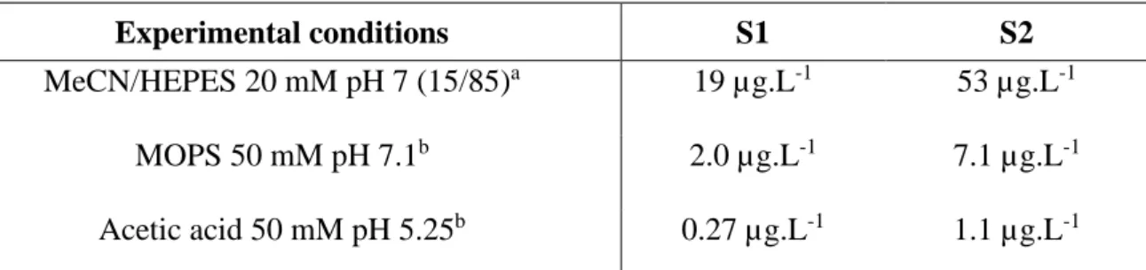
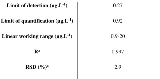
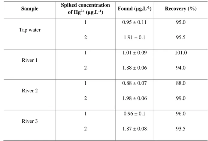
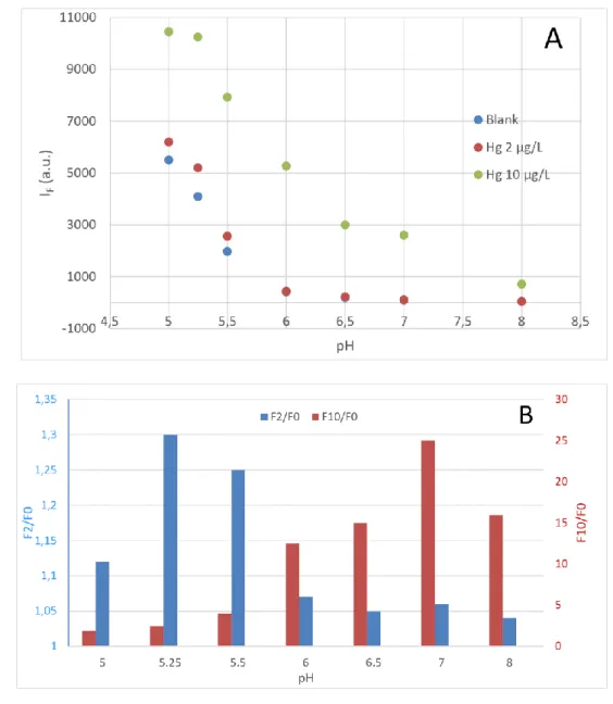
Figure

Documents relatifs
Collaborative systems thinking is an emergent behavior of teams resulting from the interactions of team members and utilizing a variety of thinking styles, design processes,
Comparison of O 3 profiles measured by MIPAS-B and ILAS-II (data versions 1.4 (v1.4) and 2 (v2)) on 20 March 2003 above northern Scandinavia together with differences and com-
At the request of the Municipality of Ciserano, one of the five included in the Zingonia area not far from Milan, Gennaro Castellano has submitted this project and
Calculez combien d’argent est rendu pour chaque transaction.. Coˆ ut des Articles Montant Pay´e
This integrated live hybrid- ization machine (LHM), which will soon be commercially available, allows real-time measurement of the hybrid- ization and melting of
were built to a height of one metre higher than the winter high water level (4.8 m), while the fourth island, designed to accommodate a light pier, came to 3.7 m higher than
This adjustment was done assuming an EB structure and using the mean speed vertical profile in each of the basins, calculated using all the float velocities (separately for
A plutono-volcanic acid suite occurs in a wide, long zone across the Guiana Shield (Venezuela, northern part of Roraima state, central Guyana, western Suriname) whereas a smaller
