Controlled Release of Gentamicin from Polyelectrolyte
Multilayers to Treat Implant-Related Infection
by
A
Joshua Seth Moskowitz
B.S. Materials Science and EngineeringCornell University (2007) M.S. Chemical Engineering Practice Massachusetts Institute of Technology (2009)
SUBMITTED TO THE DEPARTMENT OF CHEMICAL ENGINEERING IN PARTIAL FULFILLMENT OF THE REQUIREMENTS FOR THE DEGREE OF
DOCTOR OF PHILOSOPHY IN CHEMICAL ENGINEERING AT THE
MASSACHUSETTS INSTITUTE OF TECHNOLOGY JUNE 2012
@ 2012 Massachusetts Institute of Technology. All rights reserved.
Signature of Author:
JoshL67S. Moskowitz Department of Chemical Engineering July 30, 2010
Certified by:
Paula T. Hammond Bayer Chair Professor of Chemical Engineering Thesis Supervisor
Accepted by:
Patrick S. Doyle Professor of Chemical Engineering Graduate Officer
Controlled Release of Gentamicin from Polyelectrolyte
Multilayers to Treat Implant-Related Infection
by
Joshua Seth Moskowitz
Submitted to the Department of Chemical Engineering on July 30, 2010 in Partial Fulfillment of the Requirements for the Degree of
Doctor of Philosophy in Chemical Engineering
Abstract
Polyelectrolyte multilayered (PEM) coatings were fabricated to incorporate and release the small, hydrophilic antibiotic gentamicin from implant surfaces for infection control. The use of a cationic hydrolytically cleavable poly(p-amino ester) rendered these films biodegradable, yielding both diffusion-based and surface-erosion based release of this therapeutic. The Layer-by-Layer (LbL) assembly platform was used to create conformal, micron scale reservoirs with highly tunable drug release. Film release profiles were engineered through film architecture design and post-processing crosslinking techniques. Delivery of gentamicin was sustained for weeks, which is a significant improvement from previous gentamicin-releasing LbL systems. To gain better insight on the mechanisms of release and aid in rational film design, a theoretical treatment of the physical system was performed. These results include an analytical mathematical model describing the release of drug per surface area of film as a function of time as well as a computational model that simulates the time-dependent concentration profiles in these LbL systems.
These erodible, antibiotic coatings were demonstrated to be bactericidal against
Staphylococcus aureus, an infectious microorganism that is highly relevant to implant-related
infections. Film degradation products were generally nontoxic toward MC3T3-E1 osteoprogenitor cells. A reproducible in vivo rabbit bone infection model was developed to test the PEM coatings against sterile, uncoated placebos; subsequent in vivo experimentation demonstrated the proof-of-principle that an antibiotic-eluting LbL film can efficaciously treat a pre-existing implant-related infection.
One further application was studied which combined the release-based mechanism of these erodible films with a permanent, contact-killing LbL film. This combination has the treatment benefit of an initial burst release of antibiotic, prevents biofilm formation, and reduces the probability of developing antibiotic resistance due to the prolonged presence of sublethal concentrations of gentamicin.
Thesis Supervisors Paula T. Hammond, Ph.D
Bayer Professor and Executive Officer of Chemical Engineering Massachusetts Institute of Technology
Thesis Committee Myron Spector, Ph.D Professor of Orthopedic Surgery
Harvard Medical School
Darrell J. Irvine, Ph.D Professor of Materials Science Massachusetts Institute of Technology
J. Christopher Love, Ph.D Professor of Chemical Engineering Massachusetts Institute of Technology
Acknowledgements
I have been fortunate to work with a truly amazing and talented set of people throughout my graduate experience. The work presented within this thesis has been strongly inspired by these
interactions.
My premier appreciation is extended to Professor Paula T. Hammond, who has been my greatest mentor throughout the duration of my graduate experience at MIT. She provided me with an opportunity to work on a challenging, fascinating, cutting-edge research project and
offered extensive guidance and intellectual support throughout the duration of my
involvement. Her open-mindedness afforded me, on many occasions, to pursue avenues of my greatest interest. Her most unique and defining attribute that had the greatest impact on me was her energetically cheerful, positive attitude that would only enhance my daily motivation. I would like to thank my thesis committee, Professors Myron Spector, Darrell J. Irvine, and J. Christopher Love, for offering their sincere and constructive advice on how to overcome my greatest research challenges. Their collaboration has significantly impacted the efficiency and quality of my research. It has been academically stimulating and an absolute pleasure for me to work with such a supportive committee.
I thank Professor Robert E. Cohen for his brainchild PhDCEP program and his support of my PhDCEP incoming class of 2007. Were it not for this unique doctoral track, I would have enrolled in the Department of Materials Science and Engineering.
I would like to acknowledge Professors George Malliaras (Ucole Nationale Superieure des Mines de Saint-ttienne), Shu Yang (University of Pennsylvania), and Ali Yazdani (Princeton University), each for inviting me into their labs and allowing me to pursue independent
research. Collectively, they have spurred my decision to continue onto higher education. Dr. Helen Chuang and her colleague Jeff Easley were integral in helping me get comfortable in the lab, and trained on most of the relevant techniques that enabled me to successfully carry out the responsibilities associated with this thesis. Helen's continued collaboration has not only been helpful for sharing ideas and discussing experiments, but has also provided much personal support. I am sincerely grateful for these altruistic interactions.
Within the Hammond group, Dr. Raymond Samuel's expertise as an orthopedic surgeon has helped me better understand how to develop an in vivo infection model. He has
additionally contributed an enormous sum to all aspects of my research (and life) by providing his worldly and astute advice at my request. Ray was nothing short of an 'alpha-colleague', and I feel fortunate to have had the opportunity to work so closely with such a talented and selfless person. Furthermore, I hope that one day I will have the same zest for life that I see in Ray.
I owe my appreciation to the LbL subgroup- Mara Lee Macdonald, Renee Smith, Dan Schmidt, Anita Shukla, and Jessie Wong for their insightful discussions. I have had the added
enjoyment of pursuing a team-based project with Jessie Wong, which has since strengthened our friendship.
I wish to thank Michael Blaisse, a brilliant undergraduate chemistry student who will begin his tenure as a Ph.D student at the University of California at Berkeley next year. Mike has given me the opportunity to take the role of a mentor and has worked very diligently under my leadership throughout the duration of my graduate work. He has contributed significantly to both the experimental and theoretical aspects of this thesis, and I consider him my closest teammate within the Hammond Lab. I hope that Mike's experience was as rewarding for him as it was for me.
For the in vivo work, I am sincerely grateful for the efforts of Dr. Hu-Ping Hsu, who performed all animal surgeries. Most of what I know about surgical technique has been acquired via my apprenticeship with Dr. Hsu. He has helped me think through the surgical aspects of the animal model and was a vital component to the success of this project. Drs. Jean C. Lee, Mitch Harris, and Scott Martin each contributed substantially to development of the in
vivo infection model. Dr. Lee's expertise in microbiology essentially shaped the data collection and analysis procedures for the in vivo experiment. Dr. Harris and Dr. Martin gave me the opportunity to witness a live orthopedic surgery and generously spent their time meeting with me to discuss the details of the infection model. I thank the entire E25 staff in the MIT-DCM facility, and specifically regard the efforts of Dr. Alison Hayward and Catrina Wong, who were invaluable assets and teachers of animal care and anesthesia. I wish to thank Ellen Buckley and Nikki Lew for microbiological analysis, and Dr. Wayne Schwark (Cornell University) for
gentamicin quantification assays.
I appreciate the administrative staff that have allowed for the smooth functioning of my graduate experience-Linda Mousseau, Suzanne Easterly, Katie Lewis, and Christine Preston have been extraordinarily supportive. Christine, in particular, has handled countless issues and requests from me.
I acknowledge Steve Chapin, Byron Masi, Justin Quon, and Adel Ghaderi for their periodic sanity checks. These great friends have contributed to the richness of my MIT experience, and have certainly provided for some excellent memories, which I hope we continue to produce.
Ultimately, my personal, emotional, social, athletic, and academic wellbeing have all relied on those closest to me: my family and friends. This thesis is dedicated to you, as it is you who have made me who I am. To my mom and dad, who have given me endless support, unrestricted love, and a true opportunity to succeed-you believed in me and I have made it. To my sister and brother, who each have mutually pushed me to pursue my goals and strive to give my best-you both will do great things and I will be here for you as you have been there for me. To my grandparents, Gerry, Arnie, Phyllis, and Reuben (a.k.a. Ron), for their continuous love and care throughout my life-you are so special to me and I'm grateful to have all four of
you to see me through this program. To my aunts, uncles, and cousins-we are fortunate to have such a close family. To my closest friends, who have shaped my life and continue give me something to look forward to each week. To Kelsey Ann Handstad, the one who I will share forever with, I love you.
JOSHUA SETH MosKOWITZ
Massachusetts Institute of Technology, July 31, 2010
J. S. Moskowitz is thankful for financial support by the National Institutes of Health, National Institute of Aging (5R01AG029601-03). This work made use of the Center for Materials Science and Engineering shared facilities, the animal facilities of MIT's Division of Comparative Medicine, the Institute of Soldier Nanotechnologies (ISN), as well as the Robert Langer Laboratory.
Biography
Joshua Seth Moskowitz was born in Nashua, NH on October 21, 1984. He was raised in this city until his departure for college upon graduation from Bishop Guertin High School. He learned advanced mathematics at a very early age through questions posed by his grandfather Ron, which were rewarded with silver dollars or 50-cent pieces. In high school, he continued to find solace in mathematics and spent a summer teaching himself calculus via an online course provided through Stanford University. This independent effort eventually set the stage for achievement of the BGHS Mathematics Trophy upon graduation.
Josh was an avid soccer player through his adolescent years, representing the state of NH at the USYSL regional championships on four occasions and ultimately captaining his high school team during his senior year. His affinity for the mathematics and sciences, and enduring interest in soccer guided him to Carnegie Mellon University for the 2003-2004 academic year, during which he participated on the varsity soccer team. He made many friends and greatly enjoyed his time in Pittsburgh, but began to realize that competitive soccer would not continue to guide his future. Josh's distaste for quitting something that he started made it difficult to justify staying at Carnegie Mellon and leaving the varsity soccer team. Therefore, he decided that to accept a transfer offer at Cornell University and join his good friend and old high school teammate, Timothy Ulm.
Tim integrated Josh into the Cornell social network, and Josh quickly fell in love with Ithaca, NY. He pursued a degree in Materials Science and Engineering, in part due to the fascinating research prospects. After completing a semester long class taught by Professor
George Malliaras, Josh was contacted by George himself about a research position in his lab. Without hesitation, he began his first experience as a researcher under the unparalleled guidance of Jason Slinker (who has recently accepted a professorship at the University of Texas
at Dallas). His work was focused on improving the efficiency and lifetime of organic light emitting diodes. The remainder of Josh's fulfilling tenure at Cornell was peppered with research, classes, intramural soccer, and fun. He participated in two National Science Foundation funded research programs, one in Professor Shu Yang's lab at the University of Pennsylvania (2005) working on magnetically functionalized polymer nanopost arrays and the other in Professor Ali Yazdani's lab at Princeton University (2006) working on graphene. Each experience reinforced his decision to pursue graduate work.
Upon graduation, Josh finally moved back "home" to greater Boston to begin work on an advanced degree. Although offered admission to the Sc.D program through the Department of Materials Science and Engineering at MIT, he elected to switch majors and embark on a unique program offered through the Department of Chemical Engineering. This program, the Ph.D in chemical engineering practice (CEP), offered a joint opportunity with the MIT Sloan
with world-class business education. Such an opportunity, in light of his grandfather's history of founding a successful technology-based company and Josh's rapidly growing interest in management, was not to be matched. MIT has since been a wondrous boon for him, constantly presenting intellectual challenges and testing his will to achieve. He has met a truly diverse and talented collection of individuals who have each uniquely contributed to his defining
experience. After completing an application-driven, original research project, and earning an education in management, Josh is excited come full circle by integrating his problem solving skills as a researcher with his superior teamwork and leadership experience as a business student to address conventional industrial problems. As of this writing, Josh has accepted a Senior Consultant position at Deloitte (U.S. - Boston) to begin in October 2012.
Table of Contents
ACKNOW LEDGEM ENTS...5
BIOGRAPHY ... 9
TABLE OF CONTENTS...11
TABLE OF FIGURES ... 15
TABLE OF TABLES ... 23
CHAPTER 1 INTRODUCTION ... 24
1.1 INCIDENCE, ECONOMICS, AND DRUG-DEVICE COMBINATIONS... 24
1.2 OUTLINE OF THESIS...25
1.3 ADVANTAGES TO LOCAL DELIVERY OF DRUGS... 27
1.4 IMPLANT-RELATED INFECTION AND BIOFILMS...27
1.5 ORTHOPEDIC IMPLANTS AND OSTEOMYELITIS: OPPORTUNITY ... 28
1.6 LAYER-BY-LAYER FILMS FOR DRUG DELIVERY: SOLUTION... 30
1.7 SPECIFIC AIMS OF THESIS ... 32
CHAPTER 2 ENGINEERING PEM COATINGS FOR THE PROLONGED RELEASE OF ANTIBIOTICS...33
2.1 DESIGNING THE ANTIMICROBIAL FILM USING ELECTROSTATIC LAYER-BY-LAYER SELF-ASSEMBLY... 33
2.1.1 Liposom es ... 35
2.1.2 Alternative Film Architectures ... 47
2.2 CHARACTERIZATION OF THE PERFORMANCE OF [POLY1/PAA/GS/PAA]N... 55
2.2.1 Growth, Erosion, and Release of [Poly 1/PAA/GS/PAA]n. ... 56
2.2.2 In Vitro Activity against Staphylococcus Aureus ... 58
2.2.3 Cytotoxicity toward M C3T3-E1 Cells... 59
2.3 POST-PROCESSING TECHNIQUES FOR CONTROLLED RELEASE... 62
2.3.1 Crosslinking ... 62
2.3.2 Sponge Effect ... 67
CHAPTER 3 THEORETICAL TREATMENT OF DRUG RELEASE FROM PEM COATINGS...69
3.1 INTRODUCTIO.N ... 69
3.2 THE M ATHEMATICAL FRAMEWORK...69
3.2.1 Defining Erosion-Based Release...72
3.3 M ODEL PREDICTIONS...79
3.4 VALIDATING THE ANALYTICAL M ODEL FOR EXISTING SYSTEMS...83
3.5 POSSIBLE EXTENSIONS TO THE ANALYTICAL M ODEL...85
3.6 NUMERICAL TREATMENT OF DIFFUSION-BASED RELEASE ... 87
CHAPTER 4 DESIGN OF A REPRODUCIBLE IN VIVO BONE INFECTION MODEL ... 93
4.1 INTRODUCTION ... 93
4.2 EXPERIMENTAL ... 95
4.2.1 M aterials...95
4.2.2 Colonization of Im plants. ... 95
4.2.3 Approval of In Vivo Procedures. ... 96
4.2.4 Experim ental Bone Infection M odel... 96
4.2.5 Analysis of Infection. ... 98
4.2.6 Statistical Analysis ... 99
4.3 RESULTS AND DISCUSSION ... 100
4.3.1 Reproducibility of the Bone Infection M odel...100
4.3.2 Lim itations to the M odel...103
4.4 CONCLUSIONS...104
CHAPTER 5 IN VIVO EVALUATION OF ANTIBIOTIC POLYELECTROLYTE MULTILAYER COATINGS ... 105
5.1 INTRODUCTIO.N ... 105
5.2 EXPERIMENTAL ... 107
5.2.1 M aterials...107
5.2.2 Preparation of Polyelectrolyte Solutions ... 107
5.2.3 Polyelectrolyte Deposition. ... 108
5.2.4 In Vitro Characterization of Drug Release and Film Erosion ... 108
5.2.5 Approval of In Vivo Procedures ... 109
5.2.6 Rem oval of Colonized Rod and Insertion of a Test Im plant. ... 110
5.2.7 Sam pling Schedule and Analysis of Infection. ... 111
5.2.8 Statistical Analysis. ... 112
5.3 RESULTS AND DISCUSSION ... 113
5.3.1 Release and Erosion Characteristics of Treatm ent Sam ples ... 113
5.3.2 Local Infection Quantifiaction ... 116
5.3.3 Local Gentam icin Quantification ... 120
5.3.4 Lim itations to the Study ... 122
CHAPTER 6 DUAL FUNCTIO NAL BACTERICIDAL COATINGS...125
6.1 INTRODUCTIO.N ... 125
6.2 EXPERIMENTAL ... 127
6.2.1 M aterials...127
6.2.2 Synthesis of Polym ers...127
6.2.3 Preparation of Polyelectrolyte Solutions...128
6.2.4 LbL Film Assem bly...128
6.2.5 Characterization of Film Grow th, Erosion, and Release...130
6.2.6 Bactericidal Activity of Film s ... 131
6.2.7 In Vitro Cytotoxicity: Adhesion and Proliferation of Cells ... 132
6.2.8 Sam ple Sizes and Data ... 132
6.3 RESULTS AND DISCUSSION ... 133
6.3.1 Design of Com bination Film s with Dual Functionality ... 133
6.3.2 Characterization of Combination Films: Growth, Erosion, and Release...133
6.3.3 Bactericidal Activity ... 136
6.3.4 Cytotoxicity: Adhesion and Proliferation of Cells on Film s ... 139
6.4 CONCLUSIONS...141
CHAPTER 7 O UTLOOK...142
7.1 SUMMARY ... 142
7.2 FUTUR E W ORK ... 144
7.2.1 Delivery of M ultiple A gents ... 144
7.2.2 Revam ping the In Vivo M odel ... 144
7.2.3 Biodegradable Polyanions ... 145
7.3 CONCLUSIONS...146
CHAPTE R 8 CAPSTONE ... 147
8.1 INTRODUCTION...147
8.2 PAYERS, PROVIDERS, AND PATIENTS- ADOPTION OFA NEW M EDICAL TECHNOLOGY...148
8.3 GROW ING SUPPLY AND DEMAND IMBALANCE IN HEALTHCARE ... 150
8.4 ENABLING INTENSIVISTS TO REACH MORE PATIENTS...152
8.5 THE TELE-ICU PLATFORM ... 153
8.5.1 M arket Penetration of Tele-ICU Technology...155
8.5.2 Vendor Alternatives for Tele-ICU Technology ... 156
8.8 IMPLEMENTATION AND BARRIERS...162
8.8.1 Perceptions of Tele-ICU...162
8.8.2 Operation...163
8.8.3 Barrier 1: Costs...166
8.8.4 Barrier 2: Data Integration ... 167
8.8.5 Barrier 3: Change M anagem ent and Relationships ... 168
8.8.6 Lifestyle Im provem ents Using the Tele-ICU ... 170
8.9 FINANCIAL IMPACT AND PAYBACK...171
8.10 NATIONAL SUPPORT AND OUTLOOK FOR TELE-ICU ... 175
8.11 CONCLUSIONS...176
APPENDIX: MATLAB CODE FOR NUMERICAL TREATMENT OF DRUG RELEASE...177
Table of Figures
Figure 2-1. (Left) Structure of gentamicin. Sites protonated in fully charged state are indicated. Information on R1 groups and their relative content percentages can be obtained from
Mediatech, Inc. (Lot #: 61098046). (Right) In vitro efficacy of gentamicin sulfate against S. aureus (ATCC 49230) in CMHB over a 16 h incubation period at 37 "C. Data represent the mean ± standard deviation of triplicate sam ples. ... 33 Figure 2-2. Structure of Poly 1. ... 34 Figure 2-3. (Left) AFM and (right) SEM images of liposomes on a silicon surface. ... 37 Figure 2-4. (Left) Average diameter of liposome-PLL complexes formed using PLL with molecular weight of 28 kDa as a function of mass ratio. (Right) Zeta-potential measurements of the same samples. Data represent the mean ± standard deviation of multiple measurements performed on a single prepared sam ple for each point. ... 40 Figure 2-5. Diameter and zeta potential of liposomes stored at 4 C without serum (top), 25 and 37 *C without serum (middle), and 4 *C with serum (bottom) as a function of time. Data represent the mean ± standard deviation of multiple measurements performed on a single prepared sam ple for each point. ... 41 Figure 2-6. Gentamicin loss as a function of time for unstabilized liposomes stored at various temperatures in 100 mM NaOAc, pH 5.0. One sample was prepared and re-measured throughout the experim ent for each temperature... 42 Figure 2-7. Determination of the MIC of GS-Lipo (top) and PLL-GS-Lipo (bottom) against S.
aureus. Data represent the mean ± standard deviation of triplicate samples...44 Figure 2-8. Cumulative gentamicin release for [Poly 1/HA/GS/HA]5o plotted on the left ordinate
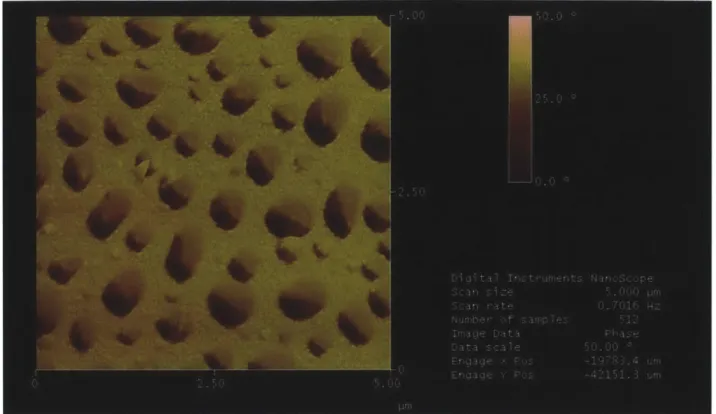
and [Poly 1/HA/PLL-Lipo-GS/HA]so plotted on the right ordinate. Data sets represent values fro m o ne sa m p le each ... 4 5 Figure 2-9. AFM phase image of [Poly 1/HA/PLL-Lipo-GS/HA]2.... ... . . 46
Figure 2-10. (Left) Release profile of (Poly 1/HA/GS/HA)(CHI/PAA/GS/HA)n with n = 25 and n = 50. (Right) Comparison in total drug release between (Poly 1/HA/GS/HA)(CHI/PAA/GS/HA)n and (Poly 1/HA/GS/HA)(CHI/ PAA-DOPA/GS/HA)n at n = 25 and 50 octolayers. Data sets represent values from one sam ple each. ... 51 Figure 2-11. (Left) Comparison in total drug release between (Poly 1/PAA/GS/HA)50,
(CHI/PAA/GS/HA)50, and (CHI/HA/GS/HA)50. (Right) Comparison in total drug release between
(Poly 1/PAA/GS/PAA)50, (Poly 2/PAA/GS/HA)5o, and (Poly 1/HA/GS/PAA)50. Each data set
contains information from one sample. Data sets were generated in batches of three-one of each film architecture-and normalized to the maximum release value for that particular batch. Thus, each graph contains three independent repeat experiments. Data sets represent values fro m o ne sa m p le each ... 5 3 Figure 2-12. Comparison in total drug release between (Poly 1/PAA/GS/PAA)50, (Poly
1/PAA/GS/PAA)(CHI/ PAA/GS/HA)25, (Poly 1/PAA/GS/HA)50, and (Poly
1/PAA/GS/HA)(CHI/PAA/GS/HA) 25. Data sets were produced in a similar manner to those
presented in Figure 2-11 and contain values from one sample each...54
Figure 2-13. (Left) Cumulative amount of gentamicin released from [Poly 1 / Anion / GS / Anion]50 films. (Right) Normalized plot shows differences in release kinetics. Data sets
represent the mean ± standard deviation of n = 4 samples for 1.25 MDa PAA, n = 6 for 100 kDa PA A , a n d n = 8 fo r H A . ... 5 6 Figure 2-14. (Left) Growth curve for [Poly 1/PAA/GS/PAA]n. SEM images are provided for the circled data points. Thicknesses were measured by profilometry at four predetermined points on each substrate and averaged over three replications. The error bars represent the average root mean squared roughness from triplicate samples and quadruplicate measurements per sample. (Right) SEM images of the growing film on a silicon surface at (A) n = 25, (B) n = 50, (C) n = 100, and (D) n = 200 layers. White arrows mark the film edge. Nota bene: the scale changes betw een (B) and (C). ... 57 Figure 2-15. Untreated (left) and EtO treated (right) titanium rods coated with [Poly
(measured perpendicular to the long axis of the rod) against S. aureus after overnight incubation at 37 *C. As a control, the sample is referenced to a commercially available BD Sensi-Disc. The lighter color at the rod surface is a result of the ruptured agar and not the presence of bacteria. The scale bar is in centim eters ... 59 Figure 2-16. (A) MTT metabolic activity of MC3T3 cells after 16-18 hr treatment in elution buffer normalized to negative control (i.e. cells incubated in standard medium). The p-value was computed using a single-tailed student's t-test assuming equal variances, and data represent the mean ± standard deviation for three different samples. Images of MC3T3 cells subjected to 16-18 hr treatment in (B) 100 and (C) 200 tetralayer elution buffer. Live cells are represented by a blue nucleus surrounded by green cytoplasm. Dead cells are represented by a red nucleus. (D) Percentage (and standard deviation) of dead cells calculated from triplicate images. Scale bar hash marks are spaced 100 ptm apart. ... 62
Figure 2-17. Release profiles of [LPEI/SPS]10+[Polyl/PAA/GS/PAA] 5o treated with various
crosslinking conditions in PBS at 37 "C. Data sets represent the mean ± standard deviation of trip licate sa m p les...6 3 Figure 2-18. Microdilution assays determining the minimum inhibitory concentration of drug eluted from A) untreated films, B) films heated at 140 *C for 30 minutes, and C) films heated at 170 *C for 30 minutes. All graphs are normalized to the negative control. All data represent the average values of three separate measurements from a single sample. Similar results were observed for two additional repeat experiments each using a separate sample (data not s h o w n )...6 5 Figure 2-19. FTIR spectra of one sample first untreated, then baked at 140 0C for 30 minutes, and finally baked again overnight at 170 *C. (Left) Spectra between wavenumbers 900 and 1900, and (right) spectra between wavenumbers 2000 and 3800. Data were selected as characteristic examples amongst six repeat experiments. All data sets were reported used a m in im u m o f 3 2 sca n s...6 6 Figure 2-20. Effect of increasing the concentration of the gentamicin dip bath on release kinetics in (left) ultra pure water and (right) modified simulated body fluid. The differences in
total release quantities were a result of the pH of the release medium as discussed in more detail in Chapter 3. Data sets represent the mean ± standard deviation of triplicate samples. .68 Figure 2-21. Effect of ending on a drug deposition step without a final rinse. Each of the three samples shown here are compared against their '+' counterpart which contains an additional
(Poly 1/PAA/GS)1 without a final rinse cycle. Data sets represent individual samples. ... 68
Figure 3-1. Growth curve of [Poly 1/PAA/GS/PAA]n used for model validation. Measurements were made on triplicate samples and error bars indicate the rms roughness values (note: use of standard deviation of thickness would have yielded much tighter error bars). For the thickness measurements, all coefficients of variation were less than 0.5... 70 Figure 3-2. Effect of ionic strength (left) and pH (right) on erosion of [Poly 1/PAA]1oo and [Poly
1/PAA/GS/PAA]50. Measurements represent the mean ± standard deviation of triplicate
sa m p le s ... 7 1 Figure 3-3. Effect of pH on the relative thickness/roughness (i.e. smoothness index) of [Poly 1/PAA]1oo (left) and [Poly 1/PAA/GS/PAA]50 (right) after erosion in 100 mM NaOAc for 19 hr at
37 *C. Measurements represent the mean ± standard deviation of triplicate samples...71 Figure 3-4. (Clockwise from top left) Initial state with the LbL film immersed in aqueous environment; water diffuses into the film as predicted by scaling arguments; hydrolysis takes place at rates that are dictated by local pH and temperature; simultaneously, ionic bonds continue to break and re-form at rates that are controlled, in part, by the concentration of salts (collision theory); after sufficient hydrolysis, it becomes kinetically possible for all remaining ionic bonds to break such that material can dissociate from the film surface. ... 74 Figure 3-5. Top: (A) Kinetic erosion profiles of [Poly 1/PAA/GS/PAA]50 in 100 mM NaOAc pH 5.0 at different temperatures, and (B) corresponding release profiles. All measurements represent the mean ± standard deviation of triplicate samples. Bottom: Arrhenius relationships for the erosion rate constant (Reff) using zero-order (linear) or first-order (exponential) interpolated representations of the data sets in (A). (C) The Arrhenius relationship for the first day of erosion or (D) the full duration studied. ... 75
Figure 3-6. Physical diffusion problem describing drug release from film into infinite medium. 76
Figure 3-7. The average concentration of drug remaining in the film depletes with time as drug mass is released. The partition coefficient accounts for the preference of drug to remain in the film versus the infinite m edium at equilibrium . ... 77 Figure 3-8. (Top, left) Model (3-25 fit to the release data from the (Poly 1/PAA/GS/PAA)200 +
(Poly 1/PAA/GS)1 architecture described in Chapter 5 via a least squares regression. (A) Effect
of increasing parameter A, (B) effect of increasing parameter B, and (C) effect of increasing p a ra m e te r C . ... 8 0 Figure 3-9. Model prediction of release profile after increasing the concentration of diffusive drug by a factor of 2 while keeping all other values constant. Model 10 is the non-phenom enological m odel described in this chapter ... 82 Figure 3-10. Model prediction of release profile after increasing (left) and decreasing (right) the diffusivity of the drug by a factor of 10 while keeping all other values constant...83
Figure 3-11. Model prediction of release profile after increasing (left) and decreasing (right) the thickness of the LbL film by a factor of 2 while keeping all other values constant. ... 83 Figure 3-12. Clockwise from top left: Release of dextran sulfate (DS) from erodible films made with (AB1/DS)20, (A2/DS)20, (A3/DS)20, and (A4/DS)20... ... . . 84
Figure 3-13. (Left) (Poly 1/PAA/GS/PAA)200 + (Poly 1/PAA/GS)1 with R2 = 0.997 and (right) (Poly
1/Chondroitin/ Vancomycin/Chondroitin)60 with R2 = 0.994. ... 85
Figure 3-14. (Left) Effect of increasing initial drug concentration in the film (Co) while holding all other parameters constant. (Right) Effect of decreasing the aqueous drug diffusivity (D) while holding all other param eters constant... 89 Figure 3-15. (Left) Effect of increasing the total film thickness (h) while holding all other parameters constant. (Right) Effect of increasing total film thickness (h) while allowing the total mass of drug in the system to scale with film thickess. ... 89 Figure 3-16. Space-time concentration profile of drug. Initially all drug is loaded into the first
Figure 3-17. Example analytical fit of the simulated first 1.8 minutes of release from a 20 micron film eluting drug with an aqueous diffusivity of 4.0 x 10⁻⁷ cm2/s and an initial film
concentration of 50,000 ptg/cm3 . . . .91 Figure 4-1. (A and B) MicroCT images of the rabbit femur. The drill site is marked with the red 'X'. (C) Drilled defect in the medial femoral condyle of the left hind limb. (D) Defect site with implant set in place. (E and F) Closing of the surgical site. (G) Closed wound...98
Figure 4-2. Blood agar plates of explanted PMMA rods that were rolled down the center of the plate and streaked for qualitative analysis of device surface colonization. Each plate corresponds to one rabbit. Each white dot corresponds to a single CFU. ... 101 Figure 5-1. (A) Opened defect site with infected PMMA implant exposed. (B) The PMMA implant has been extracted leaving a void. (C) Without washing the defect site, a titanium sample implant is press-fit into the void. (D) Anterior-posterior radiograph showing the placement of the titanium peg in the medial femoral condyle of the left hind limb. ... 111 Figure 5-2. Schematic of the hypothesized magnitude of infection in the defect site as a function of time. The initial surgery described in Chapter 4 is conducted at day -4. The direct exchange described in this chapter is conducted at day 0. All n-values correspond to the total num ber of animals euthanized in that particular test group. ... 112 Figure 5-3. (A) Normalized film erosion from [Poly 1 / PAA / GS / PAA]200 films and normalized
GS release from [Poly 1 / PAA / GS / PAA]200 + [Poly 1 / PAA / GS ]i films. Erosion values are normalized to initial film thickness with the error bars representing the normalized rms roughness averaged over triplicate samples. Release values are normalized to the final release quantity (582 lpg/cm2
) with error bars representing standard deviation of triplicate samples. Release data have truncated for convenience of comparison. (B) Cumulative antibiotic release from [Poly 1/PAA/GS/PAA]200 + [Poly 1/PAA/GS]1 films in m-SBF at 37 *C before (replotted from
(A)) and after an effective dose of ethylene oxide gas. Inset contains data from the first three days. All values are reported as mean ± standard deviation of triplicate samples. ... 114 Figure 5-4. SEM images of the PEM film on a titanium implant (i.e. substrate) before implantation (A and C), and after 4 days incubation in the defect site (B and D). ... 117
Figure 5-5. (A) Blood agar plates of explanted titanium rods that were rolled down the center of the plate and streaked for qualitative analysis of device surface colonization. Each plate corresponds to one rabbit. Each white dot corresponds to a single CFU. Explants from the four day treatment group (top row) are compared to explants from the four day placebo group (bottom row). None of the sterile plates are depicted. (B) Explants from the seven day treatment group (top row) are compared to explants from the seven day placebo group (bottom row). (C) Final counts (Mean ± SD) of log-transformed Staphylococcus aureus CFU data in femoral condyles at day zero and after direct exchange. Raw data are available in Table 5-1.
... 1 1 8 Figure 5-7. Antibiotic susceptibility of bacteria recovered from four of the six infected rabbits in the seven day treatment group. Each number (17, 25, 40, 41) corresponds to one animal. Data represent mean ± standard deviation of triplicate measurements per sample. ... 120 Figure 6-1. Structure of DM LPEI (compliments of Jessie W ong)...128 Figure 6-2. Schematic of the combination film with the permanent bactericidal base film deposited first, the erodible adhesion film deposited second, and the erodible GS-eluting film deposited on top (courtesy of Ksenia Tim achova). ... 130 Figure 6-3. Growth curve and roughness of (Poly 1/PAA/GS/PAA)n on top of (DMLPEI/PAA)1o(Poly 1/PAA)5. Dat a r e p r e se n t t h e m e a n ± st a n d a r d d ev i a t i o n o f t r i p l ica t e
sa m p le s ... 1 3 4 Figure 6-4. Degradation profile (left) and GS-release profile (right) of combination films. Data represent the mean ± standard deviation of triplicate sam ples...135 Figure 6-5. Kirby Bauer assays of GS-releasing films eroded for increasing amounts of time in PBS at 37"C. Row 1 shows (LPEI/SPS)io(Poly 1/PAA/GS/PAA)20 and row 2 shows
(DMLPEI/PAA)1 0(Poly 1/PAA)s(Poly 1/PAA/GS/ PAA)20. All samples were tested with GS-susceptible S. aureus except for the 4-day samples. This figure was reproducible over three replications (data not shown) and was produced in collaboration with Jessie Wong. ... 136
Figure 6-6. Mediaborne assay with increasing time of incubation in S. aureus broth; top row shows bare silicon substrates; bottom row shows (DMLPEI/PAA)10 films with degradable top
films completely eroded (courtesy of Jessie Wong). This figure was reproducible over three replications (data not show n)...138 Figure 6-7. (Top) Samples incubated with fluorescently tagged albumin for 1 hour at 370C. The
intensity of green color correlates with the concentration of albumin. (Bottom) Samples incubated with albumin solution and then subjected to the mediaborne assay. This figure was reproducible over three replications (data not shown) and was prepared by Jessie Wong...139 Figure 6-8. Microscope images of MC3T3-E1 cells seeded on (A) blank glass substrates or (B) (DMLPEI/PAA)10. Images are representative images selected from triplicate samples and at
least triplicate images per sample. Scale bar hash marks represent 100 pm. ... 140 Figure 6-9. MTT data showing metabolic activity of cells seeded on (DMLPEI/PAA)1o films
compared to an uncoated control. There is no statistical difference between any data pair. Data represent mean ± standard deviation of triplicate samples...141 Figure 8-1. [This figure was extracted from reference [208]]. The lines indicate the base model (i.e. expected) scenario. The darker shaded areas represent the upper and lower bounds of the sensitivity analysis. For demand, ICU use was varied by ± 10%. For supply, the number of hours worked by intensivists was varied by ± 10%. The lighter shaded area corresponds to an increase in coverage from one third of all ICU patients to two thirds of all ICU patients. FTE = full time e q u iv a le nt...1 5 1 Figure 8-2. Most hospitals aim to staff their own local intensivist teams to provide critical care. According to the study by Dara et al., these hospitals should staff more than one intensivist per 15 ICU beds. Clipart was used with permission from Arcadia Solutions (Burlington, MA)...152 Figure 8-3. The tele-ICU model allows intensivists in a centralized command center to access all patient information from connected ICU beds to help assess patients and make decisions on critical care. The tele-ICU does not replace local bedside care teams. It enhances them with additional coverage and support of an off-site intensivist. This is particularly important for
healthcare systems that do not currently employ local intensivists. Clipart was used with perm ission from Arcadia Solutions (Burlington, M A). ... 154 Figure 8-4. Market penetration and location of the 41 active command centers in 2010. Data w ere extracted fro m [207]...155 Figure 8-5. Timeline of events leading up to and through the UMMMC study by Lilly etal. ....157 Figure 8-6. VISICU tele-ICU station. Image was extracted from Google Images...164
Table of Tables
Table 3-1. Comparison between Simulation Inputs and Analytical Fit Outputs. ... 91 Table 4-1. Bone Infection M odel: Significant Blood Changes ... 102 Table 5-1. Raw CFU Data from Bone H omogenates.*...119 Table 8-1. Increase in adherence to best clinical practices at UMMMC and each community hospital (this page) [215], and Sutter Health (next page). ... 159 Table 8-2. Sensitivity analysis for payback. (Top) Fixed costs. (Bottom) Variable Costs. ... 174 Table 8-3. Estimated savings based on reduction in average LOS, total patient throughput, duration of study, and average cost of ICU per day...175
Chapter 1
Introduction
1.1
Incidence, Economics, and Drug-Device Combinations
Most implanted devices are associated with orthopedics as over 800,000 joint replacements are performed in North America each year-reflecting a 200% increase between 1999 and 2002 alone [1, 2]. Primary joint replacements (also known as arthroplasties) are typically performed on patients with arthritis or major injury to alleviate pain and restore motion to the joint. These operations add up to an annual expenditure of $1.4 billion in the United States for total hip arthroplasty and $2.59 billion for total knees with 2/3 of these costs being offset by Medicare [3]. As expected, the number of revision surgeries is also on the rise at 17.5% for hips and 8.2% for knees [1]. Since revision surgeries require extended use of hospital resources and surgeon time, subtracting 1% from each of these values would have saved $112.6 million and $100 million for hips and knees respectively [1, 4].
In order to promote implant success, there has been rising interest in the design and implementation of combination devices. According to the United States Food and Drug Administration, a combination device is an apparatus that includes two or more regulated components (i.e. drugs, devices, or biologics) that are combined and produced as a single entity
[5]. Drug-device combination products have garnered increasing attention from both
pharmaceutical and medical device companies as a general strategy to address persisting complications in clinical practice. Coordinated design of such devices has the potential to greatly improve both device performance as well as the associated quality of life for the patient. Since efficacy of a drug-device combination is generally not a linear combination of adding existing technologies together, these products can offer synergistic advantages over administering both the drug and device separately in their conventional forms.
This thesis focuses on the incorporation of the small, hydrophilic antibiotic gentamicin into polyelectrolyted multilayered (PEM) surface coatings for tunable, local, and sustained delivery from implants based on Layer-by-Layer (LbL) assembly. These films are specifically
designed to treat an existing implant-related infection. Among the attractive features of LbL (which are discussed at length in section 1.6), the critical advantage that places LbL on the cutting edge of existing technologies is its potential to release multiple therapeutics, simultaneously or sequentially, and thus allow for smart design of local therapeutic delivery from drug-device combinations for optimized treatment. For the stated case of orthopedic implants, the pertinent set of complications that could be addressed include pain, inflammation, infection, and long term loosening of the implant. The work presented herein was conducted under a financial support parcel whose overarching aim was to develop a thin film solution to address this precise set of complications. Nevertheless, the end goal of a multi-therapeutic product necessitates design and optimization of individual multi-therapeutic systems. The work in this thesis specifically addresses the antibiotic delivery component for the treatment of infection.
1.2
Outline of Thesis
Chapter 1 highlights the problem of device-associated infections, introduces the benefits of local drug release, and explains how polyelectrolyte multilayered (PEM) coatings stand out from competing technologies as an approach to addressing these issues. Using orthopedic implants as the model substrate for these combinations, the goal of this research was to develop an antibiotic coating that would be capable of eradicating an existing device-associated infection in a direct exchange arthroplasty. In orthopedics, the conventional method to treat device-associated infection is a two-stage process involving removal of the infected device, radical and extensive debridement of dead or infected tissue, local insertion of a high-concentration antibiotic-eluting spacer, six weeks of intravenous antibiotic therapy, and a follow-up surgery to re-implant a new sterile device. An antibiotic coating that is sufficiently effective against an existing infection would promote a one-stage re-implantation procedure that calls for direct exchange of the infected implant with a new, drug-coated implant without the need for antibiotic therapy or a second surgery. To this end, design of the coating requires burst-release of drug to immediately eradicate existing infection followed by weeks of sustained local delivery as phrophylaxis. Chapter 1 outlines the work that has been done toward this goal, and
Chapter 2 articulates the strategies and details of achieving the desired release profile from PEM films using an engineering-driven, application-focused approach. Among the techniques for controlling the loading and release of gentamicin are: liposome encapsulation, complex film architectures, thermal crosslinking, and the 'sponge' effect. The films are shown to load substantial amounts of cargo, retain drug activity upon release, and have some level of biocompatibility with osteoprogenitor cells.
Chapter 3 offers a theoretical view of the release behavior of gentamicin from the PEM systems. A simple mathematical framework is developed and applied to describe the drug release as a function of time. Using the model, predictions are made regarding the tenability of release profiles by changing drug concentration, film thickness, and drug diffusivity.
Chapter 4 presents a reproducible in vivo bone infection model using New Zealand White rabbits. The model was designed to simulate a contaminated one stage arthroplasty where the animals were intentionally given implant-related infection using a Staphylococcus aureus surface-colonized peg, which was press-fitted into a drilled defect in the rabbit femur. This model set the foundation for an in vivo clinical comparison between film-coated implants and sterile, uncoated placebos; results are discussed in detail in Chapter 5.
Chapter 6 builds upon the work in the first five chapters and demonstrates the versatility of LbL systems. This chapter focuses on a side project which combines two independent film architectures that each has unique bactericidal functionality. The combination films are first release-killing, and then become contact-killing upon exhaustion of the therapeutic cargo. Although this dual mechanism concept is demonstrated for releasable gentamicin, it can be extended to other therapeutics offering a powerful, multi-functional design for drug-device combinations.
After thesis summarization in Chapter 7, Chapter 8 is appended as a capstone that integrates the technology described in this thesis with a broader commentary regarding the state U.S. healthcare, and describes a new enabling technology that may help recent medical developments penetrate the market.
1.3
Advantages to Local Delivery of Drugs
Local drug delivery offers significant advantages over systemic drug delivery. Among the most important are smaller overall doses, less susceptibility to the development of resistance, and avoidance of systemic drug exposure, which can be toxic [5]. The reduced dosages correlate with reduced costs. Furthermore, therapeutic entities such as genes and proteins cannot be
delivered through standard alimentary or parenteral tracts due to harsh conditions in the former and rapid clearance in the latter. With respect to antibiotics in drug-device combinations, there can be the added benefit of direct mitigation of device-associated infections. Local antibiotic delivery systems should be designed to initially provide effective doses of drug straight to the target site to combat elevated post-operative infection risk, and then maintain therapeutic levels of release over prolonged periods of time to undermine any latent infection [6]. The release kinetics are to be uniquely designed for a particular drug, device, implant site, and disease, while ensuring that device performance is not impaired.
1.4
Implant-related Infection and Biofilms
Over two million nosocomial infections occur in the United States annually with an average hospital cost around $15,000 [5]. More than half of these are associated with implanted devices [3]. Implant-associated infection can occur due to any implanted medical device such as minimally invasive contact lenses, to temporary urinary catheters and endotracheal tubes, to permanent cardiac valves and orthopedic implants [7]. Specifically for orthopedics, the economic and health related penalties to treat an implant-related infection add up to a total cost of about five to seven times that of the initial surgery, longer hospital residency with limited mobility, and the potential for skeletal defects, limb shortening, renewed disability, and death [8, 9]. Today, with the increased use of medical devices and onset of a major orthopedic implant market due to the aging baby-boomer population, the need to address the common clinical problem of hospital-related infection is paramount.
Pathogens can be introduced to the implant surface by exogenous organisms on the skin, non-sterile surgical tools, the local environment, or even systemically circulating bacteria, which can become spontaneously pathogenic upon attachment to the implant surface. The
latter is particularly concerning since such an event can take place at any time (even years) after implantation. Upon attachment, the bacteria rapidly proliferate and create a protective mucopolysaccharide matrix known as a biofilm [10]. Within the biofilm environment therapeutics have a lower diffusion rate and bacteria have decreased metabolism, each which lends to enhanced antibiotic resistance [11]. The biofilm allows sessile colonies to communicate more effectively, exchange genetic material (thus allowing antibiotic-resistance transfer), and elude host humoral response. Biofilms can propagate infection by giving rise to planktonic satellites that can travel to other, non-colonized surfaces. Due to the fact that mature biofilms generally do not respond to administered therapeutics or host immune responses, their existence generally requires removal of the implant before local and systemic treatment can become effective [12]. The two major drug-device combination strategies that are currently employed to control infection include the use of drugless anti-adhesive materials that prevent bacteria attachment and direct incorporation of drugs into or onto a medical device [13].
1.5
Orthopedic Implants and Osteomyelitis: Opportunity
Among the millions of orthopedic implants inserted annually, bone-implant integration is a common clinical problem that leads to bone resorption, loosening, and opportunistic infection. One study demonstrated the presence of bacteria in over 72% of implants extracted for aseptic loosening [14]. Despite the standard pre-operative procedures that include antibiotic prophylaxis, antimicrobial shower, shaving, and application of disinfectants, infection rates on the order of 1% still persist. Therefore recent efforts have been directed at integrating drug delivery with these devices to accelerate bone formation and healing while also treating infection [15].
Osteomyelitis is the inflammatory response and accompanying bone deconstruction caused by an infecting microorganism. It is commonly associated with the existence of a biofilm. Staphylococcus aureus is the single largest contributor to osteomyelitis and accounts for two-thirds of operative specimen isolates [16]. Conventional therapies to treat osteomyelitis are often inadequate due to limited blood supply to the skeletal tissue and poor
drug penetration into the infected bone [17]. Simply increasing the systemic dosage can cause toxic side effects and is therefore not a resolution. Thus, local drug delivery from the surface of implants is attractive since it poses a viable alternative to systemic techniques.
The gold standard technique to control orthopedic implant infection is the use of antibiotic loaded bone cement (ALBC). ALBC uses poly(methyl methacrylate) (PMMA) (also known as acrylic) that has been loaded with clinically relevant antibiotics such as gentamicin, vancomycin, or tobramycin [18-21]. One study that surveyed the Scandinavian arthroplasty registers followed up on more than 240,000 total hip replacements, and reported a 50% reduction in the infection rate using these cements [22]. The use of pre-blended bone cement has been widely accepted in Europe for decades, and became approved for use in the United States in 2003. Bone cements typically release their therapeutic cargo in bi-phasic manner. First, there is a burst release of drug that takes place on the order of one day followed by a much longer tail of incomplete release that lasts for weeks. The release behavior of these systems is controlled by the loading ratio of drug to polymer, bulk porosity of the cement, surface area, and roughness [23-25].
There is substantial in vivo evidence that these cements perform superior to unloaded cements with respect to infection prevention [5]; however, despite the positive advantages to using ALBC, there are some drawbacks that have been elucidated. Pharmacokinetic studies suggest that elution of gentamicin from PMMA cements is imperfect; less than 50% of the total cargo is released within 4 weeks, after which continuous release was ceased [26-28]. Diffusive elution of drug from cements often has unstable and unpredictable release kinetics [29, 30]. This can lead to large fluctuations in the local drug concentration causing local tissue toxicity, or worse, emergence of drug-resistant pathogens [31]. In the event of infection or failure, revision
becomes very difficult, requiring the removal of the cemented nonbiodegradable PMMA before osteogenesis can help fill the defect site. In such a procedure, bone substance is lost, and residual PMMA debris can cause fibrous encapsulation and foreign body response [32]. Also, the polymerization reaction is highly exothermic [33], which limits the types of therapeutics that can be delivered from bone cement (e.g. growth factors would be denatured). This heat,
For ALBC specifically, drug release leads to the formation of pores within the cement matrix, which provides a highly favorable environment to harbor any remaining bacteria [31]. Finally, ALBC is well-mixed, thus negating the opportunity for smart design of complex or sequential release profiles of individual therapeutics. As of 2007, only five ALBC composites were approved for clinical use by the United States' Food and Drug Administration, and their use is restricted to prophylaxis rather than treatment of existing infection [16]. If the traditional 11% usage rate of ALBC in primary arthroplasties were increased to 50%, the economic effect of replacing plain cement with ALBC-requiring an average increase of $300 per packet-was estimated to increase the overall health care costs in the United States by $117 million [35]. This would be offset by the savings salvaged from a supposed lower overall infection rate. Given the current prevalence of total joint replacements, the infection rate would need to be reduced from 1.5% to 0.3% (i.e. 80% reduction in incidence rate).
These drawbacks leave open a window of opportunity for the development of a next-generation therapeutic coating. Currently, there is no approved coating for the treatment of existing infection. Recent alternative materials for use as drug releasing surface coatings include hydroxyapatites [36], biodegradable poly(L-lactic acid) [37], poly(lactic-co-glycolic acid) [38], sol-gels [39, 40], biodegradable polyhydroxyalkanoates [41], and others. Release rates from materials that depend on rate of surface degradation can be accurately tuned by changing the coating composition. Overall, there is need for an implant coating with tunable, release-based infection control that does not present host toxicity issues or inhibit incorporation of other delicate therapeutics such as anti-inflammatory agents and growth factors while imparting a permanent biofilm-resistant functionality. To date, no such combination film meets all of these specifications.
1.6
Layer-by-Layer Films for Drug Delivery: Solution
The true birth of LbL assembly took place in 1966 with a publication authored by Ralph Iler who studied the buildup of charged inorganic colloidal multilayers at DuPont [42]. After a couple relatively latent decades, the 'modern age' of this technology began as it was thrust into the limelight during the '90s [43], in part by Gero Decher's publication in Science [44]. Decher
pioneered the general use of polyelectrolytes for multilayered systems [45, 46] and helped publish the first textbook on this subject in 2003 [47]. LbL assembly of alternately charged polyelectrolytes has now become a well-established method for 'bottom up' engineering of surface coatings [48]. PEM films have the potential to make an important impact in the medical world as a drug delivery vehicle. They produce conformal coatings on most surfaces [49-51] with nanometer thickness precision [52] and highly tunable drug loading [53]. Functional components are incorporated into the film at the exact layer of interest [44-47, 54] so that these films can be engineered to deliver multiple different drugs with complex release profiles [48, 55]. The LbL fabrication process is simple, economical, and gentle. One of the major advantages to this technique is the all-aqueous fabrication condition, which allows for the integration of biologically active materials such as proteins [56], DNA [57], peptides [58, 59], and enzymes [60] without denaturing or loss of function. Additionally, as with any local drug delivery system, lower doses are required, less systemic drug exposure is endured, and for
antibiotics, resistance is mitigated [5].
As a result of the rising interest in drug-device combinations, coatings for orthopedic implants are an excellent application for localized drug delivery and prevention of infection [40, 61]. This provides an opportunity for PEMs to be incorporated onto the surfaces of these prostheses, and make an unparalleled impact since PEMs can be built with painkillers [62, 63], antibiotics [53], and growth factors [64-66] to sequentially deliver the exact treatment necessary for optimal recovery. PEMs will release their therapeutic components upon degradation, and can be designed to do this in specific settings such as in the presence of enzymes [67-69], small molecules [70-72], reducing agents [73], electrochemical potential [74, 75], and most importantly for the proposed application, aqueous physiological environment [76].
To address the latter mechanism of degradation, Professor David Lynn and colleagues have designed a class of hydrolytically degradable synthetic polycations, which can serve as both a structural component in a PEM film and a transient element that is cleaved by water, hence facilitating disassembly [48, 77, 78]. By adjusting the hydrophobicity of these polycations, the degradation rates and corresponding release rates can be tuned [79]. As a
result, the use of these polycations for drug delivery applications has been the topic of several recent publications [63, 80-83]. Degradable PEM films are currently the only thin film drug coating technology that enables large payload, tunabilty, sustainable release, and the potential for delivery of multiple therapeutic agents with co- or sequential release profiles.
1.7
Specific Aims of Thesis
The work presented in this thesis describes the engineering of a thin film that erodes in top-down fashion when subjected to physiological condition via the use of a hydrolytically degradable poly(P-amino ester). This allows the antibiotics payload to be released locally with precise dosage and timing. Films are designed to be highly conformal, functionalizable, biocompatible, easy to process, and economical. The specific drug of interest for this work is the small hydrophilic molecule gentamicin, which previously faced the problem of premature diffusion out of PEM films [53]. The application of interest is the treatment and prevention of orthopedic implant-related osteomyelitis, which generally develops as a result of S. Aureus. This research involves the design, construction, and characterization of films that can be applied to orthopedic implants such that they effectively kill S. Aureus when placed in physiological condition. To advance this technology, the proposed research is broken down into three specific aims:
1. Demonstrate control over dosage and release rates of gentamicin from polyelectrolyte multilayers with the particular objective of attaining release duration on the order of weeks
2. Establish reproducible bone infection model in vivo
Chapter 2
Engineering PEM Coatings for the Prolonged
Release of Antibiotics
2.1
Designing the Antimicrobial Film Using Electrostatic Layer-by-Layer
Self-Assembly
With respect to film components, the design of layer-by-layer systems for the release of antibiotics in physiological environment has three basic functional requirements that must be addressed: (1) incorporation of a therapeutic, (2) a mechanism of release, and (3) any other materials necessary to permit stable layer-by-layer film growth without compromising biocompatibility. Gentamicin sulfate is a small, water-soluble aminoglycoside antibiotic with five amine groups that are protonated at low pH to a maximum charge of +5.0 (Figure 2-1, left), and thus easily incorporated into thin film systems that rely on electrostatic interactions [53]. Gentamicin has a minimum inhibitory concentration below 0.156 pg/mL against S. aureus UAMS-1 (Figure 2-1, right) which is consistent with literature values for other strains of S. aureus [84]. Because gentamicin is nephrotoxic and ototoxic at elevated systemic levels, the low concentration necessary for local delivery via LbL systems is advantageous.
R, 1.400 1.200 SE 1.000 c 0.800 O H2N - 0.600 -~ 0.400 0.20 2 N0 @l 0.200 H2N 0 m'm-) aiIINH2 HN- n nn oi
1k
Hd 0 00Gentamicin Sulfate Concentration
(pg/ml)
HO
Figure 2-1. (Left) Structure of gentamicin. Sites protonated in fully charged state are indicated. Information on

![Figure 2-8. Cumulative gentamicin release for [Poly 1/HA/GS/HA]so plotted on the left ordinate and [Poly 1/HA/PLL-Lipo-GS/HA] 5 o plotted on the right ordinate](https://thumb-eu.123doks.com/thumbv2/123doknet/14482598.524417/45.918.211.704.520.852/figure-cumulative-gentamicin-release-plotted-ordinate-plotted-ordinate.webp)


![Figure 2-13. (Left) Cumulative amount of gentamicin released from [Poly 1 / Anion / GS / Anion] 50 films](https://thumb-eu.123doks.com/thumbv2/123doknet/14482598.524417/56.918.112.797.115.370/figure-left-cumulative-gentamicin-released-poly-anion-anion.webp)
![Figure 2-14. (Left) Growth curve for [Poly 1/PAA/GS/PAA]. SEM images are provided for the circled data points](https://thumb-eu.123doks.com/thumbv2/123doknet/14482598.524417/57.918.108.799.112.410/figure-left-growth-curve-images-provided-circled-points.webp)

![Figure 3-2. Effect of ionic strength (left) and pH (right) on erosion of [Poly 1/PAA]1OO and [Poly 1/PAA/GS/PAA] 50](https://thumb-eu.123doks.com/thumbv2/123doknet/14482598.524417/71.918.104.804.352.600/figure-effect-ionic-strength-right-erosion-poly-poly.webp)
