HAL Id: hal-02150792
https://hal.archives-ouvertes.fr/hal-02150792
Submitted on 9 Oct 2020
HAL is a multi-disciplinary open access
archive for the deposit and dissemination of
sci-entific research documents, whether they are
pub-lished or not. The documents may come from
teaching and research institutions in France or
abroad, or from public or private research centers.
L’archive ouverte pluridisciplinaire HAL, est
destinée au dépôt et à la diffusion de documents
scientifiques de niveau recherche, publiés ou non,
émanant des établissements d’enseignement et de
recherche français ou étrangers, des laboratoires
publics ou privés.
algae and land plant lineages
V. Gorelova, O. Bastien, O. de Clerck, S. Lespinats, F. Rébeillé, D. van der
Straeten
To cite this version:
V. Gorelova, O. Bastien, O. de Clerck, S. Lespinats, F. Rébeillé, et al.. Evolution of folate biosynthesis
and metabolism across algae and land plant lineages. Scientific Reports, Nature Publishing Group,
2019, 9 (1), pp.5731. �10.1038/s41598-019-42146-5�. �hal-02150792�
www.nature.com/scientificreports
evolution of folate biosynthesis and
metabolism across algae and land
plant lineages
V. Gorelova
1,4, O. Bastien
3, O. De Clerck
2, S. Lespinats
3, F. Rébeillé
3& D. Van Der straeten
1Tetrahydrofolate and its derivatives, commonly known as folates, are essential for almost all living organisms. Besides acting as one-carbon donors and acceptors in reactions producing various important biomolecules such as nucleic and amino acids, as well as pantothenate, they also supply one-carbon units for methylation reactions. Plants along with bacteria, yeast and fungi synthesize folates de novo and therefore constitute a very important dietary source of folates for animals. All the major steps of folate biosynthesis and metabolism have been identified but only few have been genetically characterized in a handful of model plant species. The possible differences in the folate pathway between various plant and algal species have never been explored. In this study we present a comprehensive comparative study of folate biosynthesis and metabolism of all major land plant lineages as well as green and red algae. The study identifies new features of plant folate metabolism that might open new directions to folate research in plants.
Tetrahydrofolate (THF) and its derivatives, known as folates or B9 vitamins, are essential elements in the metab-olism of all living organisms, except methanogenic and sulfate-reducing Archaea that use tetrahydromethano-pterin (H4MPT) or its derivative tetrahydrosarcinatetrahydromethano-pterin (H4SPT) in their C1 metabolism1. The THF molecule is composed of a pterin moiety, para-aminobenzoic acid (pABA) and a glutamate tail. To the N5 and N10 posi-tions of THF, one-carbon (C1) units of various oxidation states are attached (Fig. 1A). Essentially, folates act as donors and acceptors of C1 units in C1 transfer reactions that are involved in many major metabolic pro-cesses. Folates are used in synthesis of nucleic acids (with the exception of cytosine), amino acids, pantothenate, formyl-methionyl-tRNA, and S-adenosyl-methionine (SAM). While animals are mainly depend on their dietary sources for folate supply, bacteria, fungi and plants synthesize folates de novo.
In plants, folate biosynthesis localizes to three subcellular compartments. Pterin and pABA are synthesized in cytosol and plastids, respectively. Subsequently, the THF molecule is assembled in mitochondria, and the gluta-mate tail is added (Fig. 1B).
Like other folate-producing organisms, plants synthesize p-ABA in two steps. The first step is catalysed by aminodeoxychorismate synthase (ADCS) (Fig. 1B). Similar to their fungal2,3 and protozoan4 counterparts, Arabidopsis and tomato ADCSs exist as a bifunctional protein with two functional domains: the glutamine ami-dotransferase domain (GAT) homologous to E. coli PabA and the chorismate binding domain homologous to E.
coli PabB5,6.
The second step in the biosynthesis of pABA is mediated by aminodeoxychorismate lyase (ADCL) that catal-yses the beta-elimination of pyruvate and aromatization of the ADC ring to produce pABA7,8.
Synthesis of the pterin moiety starts with the conversion of GTP into dihydroneopterin triphosphate and formate, catalysed by GTP cyclohydrolase I (GTPCHI) (Fig. 1B). Studies on spinach, tomato and Arabidopsis demonstrated that plant GTPCHI has two functional domains that are essential for the enzymatic activity, as nei-ther domain has a complete set of the residues required for substrate binding and catalysis9–11. Dihydroneopterin undergoes dephosphorylation that proceeds in two steps. The first step is the removal of pyrophosphate by
1Department of Biology, Laboratory of Functional Plant Biology, Ghent University, K.L Ledeganckstraat 35, 9000,
Ghent, Belgium. 2Department of Biology, Phycology Research Group, Ghent University, Krijgslaan 281, 9000, Gent,
Belgium. 3Laboratoire de Physiologie Cellulaire Vegetale, UMR168 CNRS-CEA-INRA-Universite Joseph Fourier
Grenoble I, Bioscience and Biotechnologies Institute of Grenoble, CEA-Grenoble, 17 rue des Martyrs, 38054, Grenoble, Cedex 9, France. 4Present address: Department of Botany and Plant Biology, Laboratory of Plant Biochemistry and
Physiology, University of Geneva, Quai E. Ansermet 30, 1211, Geneva, Switzerland. Correspondence and requests for materials should be addressed to D.V.D.S. (email: Dominique.VanDerStraeten@UGent.be)
Received: 28 August 2018 Accepted: 25 March 2019 Published: xx xx xxxx
cytosolic Nudix hydrolase12. The second step is catalysed by a non-specific phosphatase13. The final step of the synthesis of the pterin moiety is mediated by dihydroneopterin aldolase (DHNA), which cleaves the lateral side chain of dihydroneopterin, releasing glycolaldehyde and 6-hydroxymethyldihydropterin (HMDHP)14.
Synthesis of THF in mitochondria starts with pyrophosphorylation of HMDHP and its subsequent coupling with pABA that results in the formation of dihydropteroate (DHP). These two reactions are mediated by HMDHP pyrophosphokinase (HPPK) and dihydropteroate synthase (DHPS) enzymatic activities. These two enzymes were found to be coupled on a single polypeptide in several plant species, such as pea15,16, Arabidopsis17, rice and wheat18. DHP is further converted to dihydrofolate (DHF) in the reaction mediated by dihydrofolate synthetase (DHFS), which attaches the first glutamate residue to the carboxyl moiety of pABA in DHP (Fig. 1B). The penul-timate step of folate biosynthesis is catalysed by dihydrofolate reductase (DHFR) that reduces DHF into THF. DHFR activity can be performed by a monofunctional enzyme as in mammals, or by a bifunctional protein, coupled with thymidylate synthase (TS), as in protozoa. Among plant species, bifunctional DHFR-TS genes were described in carrot19, pea20, maize21 and Arabidopsis22.
In the pathway leading to THF-Glun synthesis, two reactions mediate the attachment of glutamate. The first
reaction is catalysed by DHFS. In the second reaction catalysed by folylpolyglutamate synthetase (FPGS), the polyglutamate tail of THF-Glun is formed by the sequential addition of glutamate residues to THF-Glu1 (Fig. 1B).
The polyglutamate tail can be shortened or removed by the activity of vacuolar gamma-glutamyl hydrolases (GGH) that play an important role in regulation of folate homeostasis23–25.
Enzymes involved in the interconversion of various folate species are important components of folate metabo-lism not only for folate-producing organisms but also for those that rely on their diet for the folate supply26. These enzymes orchestrate folate metabolism and therefore regulate the flux of C1 units in response to environmental or developmental cues. Therefore, their activities have to be tightly regulated.
There are several sources of one-carbon units for folate metabolism in plants. Serine is an important entry point to 5,10-methylene-tetrahydrofolate (5,10-CH2-THF) in all eukaryotes. Loading of THF occurs upon
con-version of Ser into Gly, mediated by SHMT, which hooks a methylene group onto THF, in a reversible reaction
Figure 1. Structure of the THF molecule and folate biosynthesis. (A) Structure of THF molecule. (B)
Folate biosynthesis in higher plants. Precursors: GTP, guanosine triphosphate; DHN-P3, dihydroneopterin
triphosphate; DHN-P, dihydroneopterin monophosphate; DHN, dihydroneopterin; DHM, dihydromonapterin; HMDHP, 6-hydroxymethyldihydropterin; HMDHP-P2, 6-hydroxymethyldihydropterin pyrophosphate; DHP,
dihydropteroate; DHF, dihydrofolate; THF, tetrahydrofolate; THF-Glu(n), tetrahydrofolate polyglutamate; ADC,
aminodeoxychorismate; pABA, para-aminobenzoic acid. Enzymes: GTPCHI, GTP cyclohydrolase I; DHN-P3
-diphosphatase, dihydroneopterin triphosphate pyrophosphatase; DHNA, dihydroneopterine aldolase; HPPK, HMDHP pyrophosphokinase; DHPS, dihydropteroate synthase; DHFS, dihydrofolate synthetase; DHFR, dihydrofolate reductase; FPGS, folylpolyglutamate synthetase; ADCS, aminodeoxychorismate synthase; ADCL, aminodeoxychorismate lyase; GGH, gamma-glutamyl hydrolase.
www.nature.com/scientificreports
www.nature.com/scientificreports/
(Fig. 2)27,28. SHMT is assumed to operate in the cytosol, plastids, and mitochondria29. In the latter compart-ment, the production of 5,10-CH2-THF is mainly mediated by Gly cleavage through the action of the T-protein
of glycine decarboxylase (GDC), a reaction of paramount importance in photorespiration, typical for C3 plants. (Fig. 2). Another source of one-carbon units for folate metabolism is formate. Formate is converted to 10-formyl-tetrahydrofolate (10-CHO-THF) in the reaction catalysed by 10-CHO-THF synthetase (FTHFS)30. Subsequently, 10-CHO-THF can be metabolized in purine biosynthesis or converted to 5,10-methenyl-THF (5,10-CH+-THF) via 5,10-CH
2-tetrahydrofolate dehydrogenase/5,10-CH+-tetrahydrofolate cyclohydrolase
(MTHFD-MTHFC) (Fig. 2)31. MTHFC and MTHFD activities were found to be coupled on a bifunctional poly-peptide in pea31 and Arabidopsis32.
The reverse reaction resulting in hydrolysis of 10-formyl-THF into THF and formate is carried out by 10-CHO-THF deformylase (10-FDF) (Fig. 2). Two 10-FDF genes were characterized in Arabidopsis and found to be crucial for photorespiration32.
There are two alternative ways to generate 5,10-CH+-THF. The first one involves 5-formyltetrahydrofolate
cycloligase (5-FCL) that catalyses the conversion of 5-formyltetrahydrofolate (5-CHO-THF) into 5,10-CH+-THF
(Fig. 2). Unlike other folate species, 5-CHO-THF is not used as a one-carbon (C1) donor but acts as a potent inhibitor of SHMT and several other enzymes of C1 metabolism33. Therefore, 5-CHO-THF is considered to be a regulator of C1 metabolism. The second way to generate 5,10-CH+-THF involves glutamate formiminotransferase
(GFT), a folate-dependent enzyme that takes part in histidine degradation in mammals and some bacteria. GFT mediates the transfer of a formimino group from formiminoglutamate to THF, producing 5-formimino-THF which is then converted to 5,10-CH+-THF by the action of formiminotetrahydrofolate cyclodeaminase (FTHFC)
(Fig. 2)34. Biochemical and genetic characterization of plant GFT is still poor35.
The flux of one-carbon units from folate metabolism demands the activity of methylenetetrahydrofolate reductase (MTHFR) that catalyses the reduction of 5,10-CH2-THF to 5-methyltetrahydrofolate (5-CH3-THF)
which enters the methyl cycle (Fig. 2).
While biochemistry of plant folate biosynthesis and metabolism is considered well characterized, their com-partmentalisation and regulation await further studies. The task could become utmost challenging considering that the regulation of folate metabolism might differ between species. Examples showing that biofortification strategies are not equally efficient for all crop species corroborate this notion36. Unfortunately, genes of folate biosynthesis and metabolism were characterised only in few model plant species. Increasing availability of genome-scale data provides an excellent opportunity to trace the origin of the pathway components in algae, to identify possible differences in regulation and compartmentalisation of folate metabolism in various plant species and to plan future biofortification approaches that will take into account individual features of folate metabolism of a given species. To identify the possible differences and to trace their emergence during evolution, we ventured into a comparative genomic and phylogenetic study including all major land plant lineages as well as red and green algae. Our study provides a comprehensive view on the folate biosynthesis and metabolism throughout the plant kingdom and points out novel aspects of folate metabolism. We demonstrate that bifunctionality of enzymes in folate biosynthesis and enzymes involved in interconversion of folate species is a common feature for algae and higher plant species. Our comparative study shows that the number of genes, localization and the structure of the isoforms they encode is highly conserved across algae and land plants. Moreover, our findings underscore
Figure 2. Interconversion of folate species. THF, tetrahydrofolate; 5-CH3-THF, 5-methyltetrahydrofolate;
5,10-CH2-THF, 5,10-methylenetetrahydrofolate; 5,10-CH+-THF, 5,10-methenyltetrahydrofolate;
5-CHO-THF, 5-formyltetrahydrofolate; 10-CHO-5-CHO-THF, 10-formyltetrahydrofolate. SHMT, serine hydroxymethyl transferase; GDC, glycine decarboxylase complex, FTHFC, formiminotetrahydrofolate cyclodeaminase; 5-FCL, 5-formyltetrahydrofolate cycloligase; GFT, glutamate formiminotransferase; FTHFS, 10-CHO-THF synthetase; MTHFD-MTHFC, 5,10- CH2-THF dehydrogenase/5,10-CH+-THF cyclohydrolase; MTHFR,
the notion that folate metabolism of different subcellular compartments is characterized by specific sources of one-carbon units.
Results
Taxon sampling for the analysis and identification of putative orthologs in folate
metabo-lism.
Our comparative study is largely focused on land plant species. A moss Physcomitrella patens, a lycophyteSelaginella moellendorfii and an ancient angiosperm species Amborella trichopoda were included in the study.
Four monocotyledonous genomes, namely, Musa acuminata, Oryza sativa, Sorghum bicolor, Triticum aestivum and Zea mays and sixteen dicotyledonous genomes, namely, Arabidopsis thaliana, Brassica rapa, Capsella rubella,
Carica papaya, Citrus sinensis, Cucumis sativus, Fragaria vesca, Glycine max, Gossipium raimondii, Linum usitatis-simum, Medicago truncatula, Phaseolus vulgaris, Populus trichocarpa, Solanum lycopersicum, Solanum tuberosum
and Vitis vinifera were analysed.
Several green algae members of Charophyta, namely, Ostreococcus tauri, Ostreococcum lucimarinus and
Micromonas pusilla (Prasinophyceae), Chlamydomonas reinhardtii and Volvox carteri (Chlorophyceae), Chlorella vulgaris and Chlorella variabilis (Trebouxiophyceae), Ulva mutabilis (Ulvophyceae), and Klebsormidium flaccidum
(Klebsormidiophyceae) were used in the study. Two red algae, namely, Chondrus crispus (Florideophyceae) and
Cyanidioschyzon merolae (Cyanidiophyceae) were also covered.
To identify putative orthologs of folate pathway genes in the species listed above, full-length protein sequences from Arabidopsis thaliana were used as a query to run a BLAST protein sequence alignment37. To infer putative orthology the selected protein sequences were scrutinized for the presence of functional protein domains using the Pfam database38. The selected protein sequences were also surveyed for the presence of subcellular targeting signals and for the pattern of conserved protein motifs.
ADCs.
Our comparative analysis shows that ADCS is encoded by a single copy gene in genomes of all analysed algal species as well as in most analysed higher plant species (Fig. 3). Being in line with reported targeting of Arabidopsis ADCS to chloroplasts7 our prediction of the subcellular localization suggested that ADCS homologs in algae and higher plants possess chloroplast targeting signals (Fig. 3).Examination of selected protein sequences for the presence of functional domains revealed that besides glu-tamine amidotransferase domain (GAT) and chorismate binding domains, algal and higher plant ADCS bear homology to the N terminal region domain of anthranilate synthase component I (alpha subunit) (Fig. 3). ADCS proteins from P. patens, P. trichocarpa, F. vesca, A. thaliana and C. rubella contain one or two additional domains (Fig. 3), possibly manifesting a process of neofunctionalization of ADCS within these species. The motif pat-tern (sequence within which motifs appear) is conserved throughout algal and land plant species (Supplemental Fig. 1).
ADCL.
While algae possess a single copy of ADCL, the majority of higher plant species contains two or threeADCL genes (Fig. 4). Like those of algae, the genomes of A. thaliana, C. rubella and B. rapa also encode a single ADCL gene, indicating loss of the second ADCL gene during evolution of Brassicaceae. Our phylogenetic study revealed that ADCL genes of higher plants fall into two clades (Fig. 4). Interestingly, each clade contains an ADCL homolog from every analysed higher plant species. The presence of an ADCL gene from the A. trichopoda genome in both clades suggests that the divergence of ADCL genes occurred before diversification of flower-ing plants. The prediction of the subcellular localization revealed that all proteins from the clade containflower-ing A.
trichopoda_XP_006833333.1 have a chloroplast localization signal, whereas proteins from the clade containing
the A. trichopoda_XP_011623669.2 isoform are exclusively cytosolic (Fig. 4). Moreover, the P. patens genome also encodes two cytosolic and one plastidial ADCL isoform, further confirming that duplication and diversification of ADCL genes occurred during early evolution of land plants (Fig. 4). The plastidial localization of GFP-tagged ADCL from Arabidopsis has been previously demonstrated7, while a confirmation for cytosolic ADCL remains unreported.
All analysed ADCL polypeptides contain a single Aminotransferase class I and II (PF00155.20) domain (Fig. 4). The motif pattern is well conserved among the analysed algal and higher plant species (Supplemental Fig. 2).
GTPCHI.
In silico analysis demonstrates that while algae have a single GTPCHI isoform, the majority ofana-lysed higher plant species possesses two or more GTPCHI isoforms (Supplemental Fig. 3). Prediction of sub-cellular targeting suggested cytosolic localization of all analysed proteins, supporting the experimental data for Arabidopsis and tomato GTPCHI proteins11.
Our findings reveal the presence of two GTPCHI domains throughout algae and higher plant lineages, with the exception of L. usitatissimum GTPCHI that possesses three domains, the first being duplicated (Supplemental Fig. 3). Remarkably, the two GTPCHI domains show very different conserved motif composition, that further suggests their strong divergence (Supplemental Fig. 4).
HppK-DHps.
Genomes of the majority of analysed algae and higher plant species contain a single HPPK-DHPS gene (Supplemental Fig. 5). Prediction of their subcellular localization suggested mitochondrial localization of the majority of studied HPPK-DHPS isoforms (Supplemental Fig. 5). This finding is in line with experimentally determined mitochondrial targeting of HPPK-DHPS proteins in pea15 and Arabidopsis39.Our comparative study shows that the bifunctionality is conserved across algae and land plant species, as HPPK and two DHPS domains (DHPS1 and DHPS2) were identified in most analysed proteins (Supplemental Fig. 5). While all higher plant species possess two DHPS domains, some algae and lower land plant species (S.
www.nature.com/scientificreports
www.nature.com/scientificreports/
additional domains (Supplemental Fig. 5). Our in silico protein analysis indicates that the motif pattern is well conserved (Supplemental Fig. 6).
DHFS.
Phylogenetic analysis revealed that DHFS is encoded by a single copy gene in genomes of most ana-lysed species, with the exception of G. max and M. truncatula genomes which are characterized by high poly-ploidy (Supplemental Fig. 7). Being in line with experimental evidence for Arabidopsis DHFS39, our analysis suggested that DHFS in higher plant species localizes to mitochondria, while in algae it can also localize to cytosol or plastids (Supplemental Fig. 7).In silico analysis revealed that DHFS proteins contain the same set of functional domains as FPGS
polypep-tides (see below): FPGS1, FPGS2 and muramyl ligase (Supplemental Fig. 7). Although DHFS and FPGS share similar biochemical function, sequences of these two proteins diverged immensely. The divergence is further
Figure 3. Phylogenetic analysis, subcellular localization and domain composition of ADCS proteins. Species
names are followed by protein identifiers. The bar indicates the mean distance of 2.0 changes per amino acid residue. The numbers at the branching points indicate the percentage of times that each branch topology was found during bootstrap analysis (n = 1000). The box contains predicted functional domains. Schemes on the right represent domain organisation of analysed proteins (colored boxes represent functional domains, lengths of black lines correspond to lengths of proteins. The scale bar below shows protein containing 500 amino acids). Cyt, cytosolic localization; chl, plastidial localization. Indication of double localization (e.g. cyt chl) for a single protein implies its probable localization to both compartments.
illustrated by the protein sequence analysis which demonstrates that DHFS and FPGS proteins have very dif-ferent patterns of conserved motifs (compare Supplemental Figs 8 and 11). It has been previously reported that Arabidopsis DHFS is more similar to the DHFS-FPGS gene from E. coli than to Arabidopsis FPGS genes39.
DHFR.
Our phylogenetic analysis demonstrates that single-copy DHFR-TS genes are present in algal genomes, while DHFR-TS in higher plant species is encoded by two or more genes (Supplemental Fig. 9). Prediction of subcellular localization suggested that DHFR-TS isoforms can be targeted to multiple compartments (Supplemental Fig. 9), corroborating our previous finding of multiple subcellular targeting of DHFR-TS isoforms in Arabidopsis40. The data in Supplemental Fig. 9 demonstrate that genomes of all analysed higher plant and algal species encode bifunctional DHFR-TS enzymes.A previous study of Arabidopsis DHFR-TS demonstrated that one of the isoforms lacks enzymatic activity and acts as an inhibitor of its family members. Phylogenetic analysis suggested that such inhibitory isoforms may be also present in Arabidopsis’ closest relatives, B. rapa and C. rubella40. The present study shows that the inhibitory isoforms lack the last protein motif, while all other analysed DHFR-TS proteins retain it (Supplemental Fig. 10). It is plausible that this motif is important for the DHFR and TS activities, possibly being essential for a proper protein conformation.
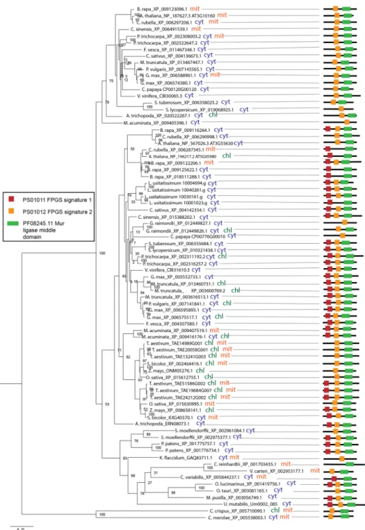
FPGS.
The present study shows that, unlike algae, land plant species possess two or more FPGS isoforms. Being in line with experimental data39,41 our prediction of subcellular localization suggests that the isoformsFigure 4. Phylogenetic analysis, subcellular localization and domain composition of ADCL proteins. Species
names are followed by protein identifiers. The bar indicates the mean distance of 0.6 changes per amino acid residue. The numbers at the branching points indicate the percentage of times that each branch topology was found during bootstrap analysis (n = 1000). Schemes on the right represent domain organisation of analysed proteins (color boxes represent functional domains, lengths of black lines correspond to lengths of proteins. The scale bar below shows protein containing 500 amino acids). The box contains predicted functional domains. Cyt, cytosolic localization; chl, plastidial localization.
www.nature.com/scientificreports
www.nature.com/scientificreports/
localize to multiple subcellular compartments, namely, mitochondria, cytosol and plastids (Fig. 5). Analysis of protein sequences revealed the presence of three functional domains in FPGS polypeptides: a muramyl ligase domain and two FPGS domains (FPGS1 and FPGS2). Our phylogenetic analysis demonstrated that FPGS of higher plants branch into two clades, each containing a gene from A. trichopoda (Fig. 5). The two clades differ by their domain composition. The clade containing the A. trichopoda_ERN08073.1 isoform retains both FPGS func-tional domains, while the clade comprising the A. trichopoda_XP_020522267 isoform lacks the FPGS1 domain.
Figure 5. Phylogenetic analysis, subcellular localization and domain composition of FPGS proteins. Species
names are followed by protein identifiers. The bar indicates the mean distance of 1.0 change per amino acid residue. The numbers at the branching points indicate the percentage of times that each branch topology was found during bootstrap analysis (n = 1000). Schemes on the right represent domain organisation of analysed proteins (color boxes represent functional domains, lengths of black lines correspond to lengths of proteins. The scale bar below shows protein containing 500 amino acids). The box contains predicted functional domains. Cyt, cytosolic localization; chl, plastidial localization; mit, mitochondrial localization. Indication of double localization (e.g. cyt chl) for a single protein implies its probable localization to both compartments.
In previous work it was shown that mit AtFPGS, lacking the FPGS1 domain, could complement a yeast FPGS mutant39, indicating that the absence of this domain did not impair the activity. Prediction of targeting signals suggests that subcellular localization of FPGS proteins varies within each clade (Fig. 5). This indicates that local-ization signals of FPGS were gained in each lineage individually.
GGH.
Our analysis shows that GGH is encoded by multiple genes in several algal and land plant genomes (Supplemental Fig. 12). Interestingly, GGH is the only enzyme of the folate pathway that is encoded by several genes in genomes of algae species. This observation corroborates the notion that the enzyme plays an important role in regulation of folate pool24 and highlights the demand of a more profound investigation of its function. Being in line with previous studies of GGH enzymes from Arabidopsis and tomato23,25 our subcellular localization analysis predicted existence of secretory signals in all analysed GGH proteins (Supplemental Fig. 12). Protein analysis revealed that all analysed GGH proteins possess a single peptidase C26 (PF07722.13) functional domain (Supplemental Fig. 12) and show a highly conserved motif pattern (Supplemental Fig. 13).GDC and sHMt.
The in silico analysis revealed that the GDC T-protein is encoded by a single gene in algal genomes, while land plant species possess single or multiple GDC T-protein genes (Supplemental Fig. 14). The predicted mitochondrial localization of the analysed GDC isoforms is in line with its role in photorespiration42. Protein analysis suggested the presence of two functional domains: an aminomethyltransferase folate-binding domain (PF01571.20) and a glycine cleavage T-protein C-terminal barrel domain (PF08669.10). Phylogenetic analysis suggested that genomes of both algae and land plants encode multiple SHMT isoforms that fall into three big clades. According to our prediction of subcellular localization, each clade comprises isoforms with either mitochondrial, cytosolic or plastidial localization, confirming previous findings in pea and potato20,28 (Supplemental Fig. 15).FTHFS.
Our comparative study demonstrates that FTHFS is encoded by a single gene in all examined species, with the exception of G. max, L. usitatissimum and P. trichocarpa that have two FTHFS genes (Supplemental Fig. 16). In agreement with the finding that FTHFS activity is associated with the cytosolic fraction of pea cot-yledons, our prediction of subcellular localization suggested that FTHFS resides exclusively in the cytosol of all analysed algae and land plant species (Supplemental Fig. 16). Protein analysis revealed that all analysed FTHFS proteins possess a single formate-tetrahydrofolate ligase (PF01268.18) functional domain (Supplemental Fig. 16) and show a highly conserved motif pattern (Supplemental Fig. 17).10-FDF.
Phylogenetic analysis reveals the presence of single genes encoding 10-FDF in algal genomes, whereas land plant species might possess several 10-FDF genes (Supplemental Fig. 18). Being in agreement with reported mitochondrial targeting of Arabidopsis 10-FDF isoforms and their role in photorespiration32, our subcellular localization prediction suggests mitochondrial localization of the analysed 10-FDF proteins (Supplemental Fig. 18). Our protein analysis demonstrates that all analysed 10-FDF bear a single formate-tetrahydrofolate ligase domain (PF01268.18) and have a highly similar motif pattern (Supplemental Figs 18 and 19).MTHFD-MTHFC.
In silico analysis indicates that higher plant species contain multiple MTHFD-MTHFCisoforms that fall into three clades, according to their predicted subcellular localization in cytosol, mitochon-dria and plastids, each comprising a gene from A. trichopoda (Supplemental Figs 20–22). Each clade includes a MTHFD-MTHFC homolog from A. trichopoda, suggesting their emergence early during speciation of flowering plants. Analysis of protein sequences showed that all analysed proteins possess the NAD(P)-binding (PF02882.18) and MTHFD-MTHFC catalytic (PF00763.22) domains, with the exception of two L. usitatissimum plastidial MTHFD-MTHFC isoforms that bear an additional domain identified as ACT (Supplemental Figs 20–22). Our study shows that the motif pattern is well conserved (Supplemental Figs 23–25).
5-FCL.
Supplemental Fig. 26 illustrates a comparative analysis for 5-FCL. It is encoded by a single gene in algal genomes, in contrast to some higher plant species, which contain two 5-FCL coding genes. As P. patens genome contains two 5-FCL genes, its duplication presumably occurred during speciation of land plants or earlier. Prediction of subcellular localization has shown that the analysed 5-FCL proteins are mainly targeted to mito-chondria but can also reside in plastids and in the cytosol (Supplemental Fig. 26). The prediction is in line with the experimentally determined mitochondrial localization of 5-FCL activity in pea43. All analysed proteins have a similar motif pattern (Supplemental Fig. 27) and bear a single 5-formyltetrahydrofolate cycloligase (PF01812.19) functional domain (Supplemental Fig. 26); no other domains could be identified.GFT.
GFT appears to be encoded by a single gene in algae, whereas genomes of land plants can contain single or multiple GFT genes (Supplemental Fig. 28). Subcellular localization prediction suggested that GFT isoforms predominantly reside in the cytosol in higher plants, but can also be targeted to both mitochondria and plastids, as for instance, in A. trichopoda, C. sinensis, L. usitatissimum, O. sativa, P. trichocarpa and Z. mays (Supplemental Fig. 28). Protein analysis demonstrated that GFT proteins possess a single formiminotransferase domain (PF07837.11) and show a well conserved motif pattern throughout algal and land plant lineages (Supplemental Fig. 29). G. raimondii, L. usitatissimum, C. sinensis and C. sativus bear an additional domain.MTHFR.
Our comparative analysis illustrates that unlike those of algal species, genomes of most land plant species encode several MTHFR isoforms (Supplemental Fig. 30). Prediction of subcellular localization suggested that MTHFR of algae and land plant species reside in the cytosol (Supplemental Fig. 30). The cytosolic locali-zation of MTHFR is in line with the assumption that the cytosol is the predominant location of the one-carbon flux from folate metabolism toward methionine production44. Analysis of protein sequences demonstrated thatwww.nature.com/scientificreports
www.nature.com/scientificreports/
MTHFR of algae and land plants contain a single MTHFR functional domain (Supplemental Fig. 28) and show a well conserved motif pattern (Supplemental Fig. 31).
Evolution of DHFR-TS and HPPK-DHPS in plants versus other organisms.
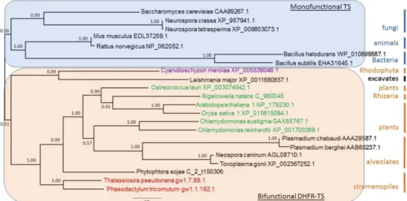
In the green lineage, the assembly of the folate molecule occurs within the mitochondria (Fig. 1) and involves two bifunctional enzymes, DHFR-TS and HPPK-DHPS. This is not always the case in other organisms where these activities can be driven by monofunctional enzymes. It is well recognized that the fusion between DHFR and TS is a milestone in evolu-tion, illustrating the separation between unikonts and bikonts45. To test whether the other bifunctional enzyme of the folate biosynthesis pathway follows the same evolutionary history as DHFR-TS, we compared the phyloge-netic evolution of the TS and DHPS domains in wide range of organisms, including bacteria, protists (Alveolata, fungi, red algae (Rhodophyta), Rhizaria, stramenopiles), plants and animals.ts.
The phylogeny based on the maximum likelihood (ML) for the TS domain from 22 species representative of the various kingdoms clearly illustrates the evolutionary separation between organisms having monofunctional TS (unikonts) versus those having bifunctional DHFR-TS (bikonts) (Fig. 6).Thus, the phylogeny of the TS domain clearly revealed a monophyletic origin of the bikont clade. To obtain a better idea of the relative distance between these various species, we represented the ML phylogeny in a three-dimensional space (Fig. 7).
This figure represents a ‘sequence space’. It is the projection in 3D of the highly multidimensional space con-taining all the possible sequences of the TS domain46,47. To assign a position to each sequence in this three dimen-sional space, non-linear mapping methods are used to offer a configuration of points preserving as much as possible the distances observed between the various sequences in the original space (see Material and Methods). With such methods, axes become arbitrary and every rotation or symmetry is admissible48. The meaning of this representation is therefore essentially carried by distance: the distance between two points on the figure tends to display the distance between species. Links between points show the ML phylogenic tree, and thus the figure combines distance-based and ML-based phylogenies.
Such representation can provide additional information compared to the previous one. For example, Toxoplasma gondii, an apicomplexan having a bifunctional DHFR-TS, was positioned far from other apicom-plexa but close to organisms such as Leishmania major, an excavate, a situation different from that seen in Fig. 6. This illustrates that two points relatively close in the sequence space are not necessarily close from an evolutionary point of view. For example, Bigelowiella natans and Mus musculus, or Toxoplasma gondii and Saccharomyces cerevisiae may appear close in the sequence space whereas they are not related in the ML tree. In other words, the evolutionary distances (as shown in the ML tree) can be large, despite relative similarities between the sequences. It is also clear that monofunctional (in blue) and bifunctional (in red) branches occupy a different space in this three-dimensional representation, indicating a totally independent evolution of the TS domain after its fusion with DHFR. In addition, the location of the Toxoplasma/Neospora group, far from that of the Plasmodium group in the sequence space could explain the relatively low support value of 0.57 (Fig. 6) calculated for the branch sep-aration leading to Chlamydomonas on one hand and alveolates on the other hand.
Figure 6. Phylogenetic trees of the protein sequences of the TS domain constructed using maximum likelihood
method Phyml (see text). The selected model using Bayesian information criterion was LG + G + I with gamma shape parameter estimate = 1.117 and the proportion of invariable site estimate = 0.207. Nodes values represents the Bayesian posterior probabilities branch support. The species colour code corresponds to the type of plastid pigments, as follows: purple, chlorophyll a; green, chlorophyll a and b; red, chlorophyll a and c.
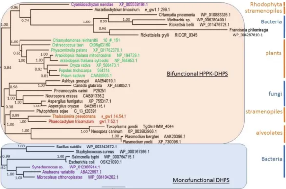
DHps.
The maximum likelihood phylogenetic tree obtained for the DHPS domain appears very different from that of TS. In this case also, we identified organisms having a monofunctional DHPS and others having a bifunctional HPPK-DHPS (Fig. 8).Animals, which depend on their diet for folate supply, do not possess DHPS activity. Most of the mono-functional DHPS are found in bacteria (unikonts), but some bacteria display also a bimono-functional enzyme. From this point of view, it is interesting to note that within gamma proteobacteria there are species displaying mono-functional DHPS (Escherichia coli, Salmonella typhi) whereas others (Francisella philomiragia, Ricketsellia grylli) display a bifunctional HPPK-DHPS. This is also true for the alpha proteobacteria (containing Rickettsia bellii,
Wolbachia), while beta proteobacteria (not represented here) and firmicutes (Bacillus subtilis, Staphylococcus aureus) contain mostly monofunctional DHPS. These data raise the question of the origin of the fusion of the two
domains. Likewise, fungi (unikonts) also display a bifunctional HPPK-DHPS, as found in plants (bikonts). Thus, the separation unikonts/bikonts does not apply for this enzyme, suggesting that the evolutionary events leading to the two bifunctional enzymes HPPK-DHPS and DHFR-TS were completely different. The 3D representation (Fig. 9) also shows a different situation compared with the one obtained with the TS domain. In this case, mono-functional and bimono-functional DHPS do not occupy a different space. Monomono-functional enzymes (blue branches) occupy a small space compared to the bifunctional HPPK-DHPS (red branches) which is widely spread within the entire sequence space. This might suggest more constraint on the monofunctional enzyme, which evolved less than the bifunctional one. It is interesting to note that in Fig. 7 as well as in Fig. 9, the green lineage is always compacted in a rather small space, whereas the apicomplexa are quite widely distributed. This is indicative of a much higher mutational rate in the latter compared with the former.
Discussion
Folate biosynthesis and metabolism in higher plants have been studied for almost two decades. Despite the wealth of genetic and biochemical evidence, many questions remain to be addressed. Particularly, the regulation and compartmentalisation of folate biosynthesis and metabolism in various plant lineages require scrutiny. Our com-parative study encompasses all steps of folate biosynthesis and interconversion of folate species in land plants, as well as green and red algae and reveals novel aspects of folate metabolism that can support further experimental studies in the field and help to design effective biofortification strategies. An earlier comparative genomic analysis of bacterial and plant folate metabolism identified new bacterial GTPCHI and FPGS gene families and predicted a bacterial folate transporter49.
Our comparative study revealed that while algae possess single isoforms of the studied genes, plant species tend to have multiple isoforms regulating the same steps in folate metabolism. The multiple isoforms could derive from whole genome duplication or duplication of certain genomic regions, contributing to the development of organelle-specific features of folate metabolism of land plants. Interestingly, three steps of folate biosynthesis, namely ADCS, HPPK-DHPS and DHFS, are catalysed by single isoforms in most analysed land plant species. The restriction of the number of these genes to a single copy in land plant genomes might reflect the necessity for a tight regulation of the abundance of folate intermediates. Being in line with the known inhibition by folate
Figure 7. Phylogenetic representation in the protein sequence space for TS domains. Non-linear mapping
methods are designed to offer a configuration of points in this multidimensional space that is representative of the observed distances. With this method, axes become arbitrary and every rotation or symmetry is admissible. In this figure, the distance between two data points on the figure tends to display the distance between species. Links between points show the ML phylogenic tree presented in the Fig. 6. Blue branches: monofunctional TS, red branches: bifunctional TS. Brown letters, bikont cluster; brown letters in bold, the green lineage; black letters, unikont cluster. Abbreviations for genus names: Arab_tha1, Arabidopsis thaliana 1; Ory_sat1, Oryza
sativa 1; Chla_rei, Chlamydomonas reinhardtii; Chla_eug, Chlamydomonas eustigma; Ostr_tau, Ostreococcus tauri; Plas_ber, Plasmodium berghei; Plas_cha, Plasmodium chabaudi; Toxo_gon, Toxoplasma gondii; Neos_can, Neospora caninum; Cyan_mer, Cyanidioschyzon merolae; Leis_maj, Leishmania major strain Friedlin; Phae_tri, Phaeodactylum tricornutum; Thal_pse, Thalassiosira pseudonana; Phyt_soj, Phytophthora sojae; Mus_musc, Mus musculus; Rat_nor, Rattus norvegicus; Neur_cra, Neurospora crassa; Neur_tet, Neurospora tetrasperma; Sach_
cer, Saccharomyces cerevisiae; Metha_ja, Methanocaldococcus jannaschii; Baci_sub, Bacillus subtilis; Baci_hal,
www.nature.com/scientificreports
www.nature.com/scientificreports/
Figure 8. Phylogenetic trees of the protein sequences of the DHPS domain constructed using maximum
likelihood method Phyml (see text). The selected model using Bayesian information criterion was LG + G + I with gamma shape parameter estimate = 1.058 and the proportion of invariable site estimate = 0.047. Nodes values represents the Bayesian posterior probabilities branch support. The species colour code corresponds to the type of plastid pigments, as follows: purple, chlorophyll a; green, chlorophyll a and b; and red, chlorophyll a and c.
Figure 9. Phylogenetic representation in the protein sequence space for DHPS domains. Non-linear mapping
methods are designed to offer a configuration of points in this multidimensional space that is representative of the observed distances. With this method, axes become arbitrary and every rotation or symmetry is admissible. In this figure, the distance between two data points on the figure tends to display the distances between species. Links between points show the ML phylogenic tree presented in the Fig. 8. Blue branches: monofunctional enzyme, red branches: bifunctional enzyme. Brown letters, bikont cluster; brown letters in bold, the green lineage; black letters, unikont cluster. Abbreviations for genus names: Arab_thM, Arabidopsis thaliana Mitochondrial; Arab_thC, Arabidopsis thaliana Cytosolic; Popu_tri, Populus trichocarpa; Pisu_sat, Pisum
sativum; Oryz_sat, Oryza sativa; Phys_pat, Physcomitrella patens; Chla_rei, Chlamydomonas reinhardtii; Ostr_
tau, Ostreococcus tauri; Plas_yoe, Plasmodium yoelii; Plas_ber, Plasmodium berghei; Toxo_gon, Toxoplasma
gondii; Neos_can, Neospora caninum; Cyan_mer, Cyanidioschyzon merolae; Phae_tri, Phaeodactylum tricornutum; Thal_pse, Thalassiosira pseudonana; Aura_lim, Aurantiochytrium limacinum; Phyt_soj, Phytophthora sojae; Aspe_fum, Aspergillus fumigatus; Aspe_ory, Aspergillus oryzae; Neur_cra, Neurospora crassa; Pneu_car, Pneumocystis carinii; Ashb_gos, Ashbya gossypii; Cand_gla, Candida glabrata; Bige_nat, Bigelowiella natans; Rick_gry, Rickettsiella grylli; Chla_pne, Chlamydia pneumonia; Rick_bel, Rickettsia bellii;
Wolb_dro, Wolbachia Drosophila simulans; Fran_phi, Francisella philomiragia; Esch_col, Escherichia coli; Salm_ typ, Salmonella typhi; Baci_sub, Bacillus subtilis; Stap_aur, Staphylococcus aureus; Syne_cho, Synechococcus sp.; Anab_var, Anabaena variabilis; Micr_cht, Microcoleus.
intermediates both at the transcript and the protein levels50, this notion suggests that biofortification strategies aiming to enhance folate content should involve elevation of the expression of the downstream genes of folate biosynthesis pathway, namely, ADCS, HPPK-DHPS and DHFS. Such an approach has been already successfully employed to elevate folate content in potato51.
It remains to be addressed why certain enzymes of folate biosynthesis are encoded by single isoforms in duplication-prone land plant genomes. Exceptions with two ADCS (in B. rapa, G. max and V. vinifera), HPPK-DHPS (G. max and L. usitatissimum) and DHFS (G. max and M. truncatula) are also intriguing. On the one hand, one can ascribe the existence of duplicated isoforms to genetic redundancy inherent to plant genomes. On the other hand, it is possible that the duplicated isoforms might implement a different function or reflect a difference in the regulation of folate biosynthesis. In the future, it will also be important to address whether the duplicated genes encode functional enzymes. Our analysis of conserved amino acids for several folate pathway enzymes, namely, GTPCHI, ADCS, HPPK-DHPS, DHFS and MTHFR, suggests that the isoforms included in our study bear enzymatic activity (Supplemental Figs 32–36). Interestingly, two out the five plant species that bear two copies of ADCS and HPPK-DHPS are naturally rich in folates, namely, B. rapa and G. max. Apart from ADCS and HPPK-DHPS, B. rapa genome bears several copies of GTPCHI. The B. rapa case echoes the bioforti-fication strategy applied for potato, where ADCS, GTPCHI and HPPK-DHPS genes have been overexpressed51. It is tempting to speculate that such an enhancement approach might prove efficient for species that bear a single copy of the abovementioned genes. However versatile, Arabidopsis is not a flawless model to be used to study regulation of plant metabolism. As highlighted by the study of bifunctional DHFR-TS40 and by the present study, Arabidopsis and other species from Brassicaceae, namely, C. rubella and B. rapa have unique features that are not shared with species from other land plant lineages. Thus, species from Brassicaceae bear enzymatically inactive DHFR-TS homologs that inhibit activity of active homologs, thereby regulating availability of THF40. This regu-lation seems not to be operating in other plant lineages. Brassicaceae members also exceptionally lack cytosolic ADCL isoform (Fig. 4). Moreover, Arabidopsis bears a cytosolic HPPK-DHPS isoform that might be involved in stress response17. Interestingly, this feature is unique to Arabidopsis and not shared even with other Brassicaceae species (Supplemental Fig. 5). It is tempting to assume that Arabidopsis might differ from species of other plant lineages in its regulation of folate biosynthesis. Taking this into consideration, drawing parallels from studies using Arabidopsis should be done with caution.
Prediction of subcellular localization allowed to identify novel aspects of folate biosynthesis and metabo-lism. Although it must be kept in mind that these in silico predictions have to be experimentally confirmed, our study suggests that ADCL can localize to the cytosol. The presence of the cytosolic isoform in every analysed land plant species, except species from Brassicaceae, might indicate an important function different from that in folate biosynthesis. To date, the cytosolic role of the enzyme remains unknown. It is tempting to speculate that cytosolic ADCL contributes to pABA production that so far has been only reported to occur in plastids in the plant cell. This assumption is in line with studies showing cytosolic localization of certain enzymes operating in the shikimate pathway that provides chorismate for pABA synthesis. If this scenario is operative, there should be a cytosolic conversion of chorismate to aminodeoxychorismate (ADC) similar to that performed by ADCS in plastids; alternatively, ADC can be exported from plastids. As a folate independent, stress-related function for the cytosolic isoform of HPPK-DHPS, an enzyme catalysing the successive step in the folate biosynthesis, has been suggested previously52, it is possible that the cytosolic ADCL might also fulfil a role in stress response. Generally speaking, the intracellular compartmentalization of folate biosynthesis established for Arabidopsis (depicted in Fig. 1) seems to be a general feature for land plants. However, the situation might be more complex in algae since the predicted localization of the enzymes appeared more fluctuating from one species to another. For example, there is no putative DHFR activity located in the mitochondria of most algae, and in K. flaccidum the entire folate biosynthesis pathway (except for the ADCS activity) could take place in the cytosol.
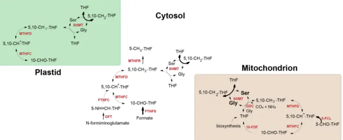
Analysis of subcellular compartmentalization of genes involved in interconversion of folate species sug-gests that the sources of one-carbon units in plant cells are specific for different subcellular compartments and this specificity is highly conserved across higher plant lineages (Fig. 10). Thus, conversion of 5-CHO-THF, a putative folate storage form, by 5-FCL and the flux of one-carbon units from 10-CHO-THF to 5,10-CH2-THF
via the sequential action of 10-FDF and GDC are assumed to mainly occur in mitochondria. In the cytosol, one-carbon units appear to be mainly derived from the conversion of Ser to Gly catalysed by SHMT. However, the conversion offormate to 10-CHO-THF mediated by FTHFS and the conversion of N-formiminoglutamate into 5-formimino-THF catalysed by GFT can also serve as a source of one-carbon units. As plastids seem to lack the abovementioned enzymes, they are assumed to mainly obtain their C1-THF derivatives from the activity of SHMT and the transport of metabolically active folate forms from mitochondria and cytosol. Because of the pres-ent lack of reliable in silico tools for prediction of subcellular localization of algal proteins, localization of folate pathway enzymes in the algae species analysed cannot be unequivocally concluded. Presently, only experimental approaches are able to reveal the localization of enzymatic steps of folate biosynthesis and metabolism.
Our comparative analysis shows that HPPK and DHPS as well as DHFR and TS activities are coupled on a single bifunctional polypeptide across algae and land plant lineages. This is not the case in all organisms, and, as a matter of fact, the fusion between the DHFR and TS genes coincides with the appearance of biflagellate organ-isms. Indeed, unikonts, with one cilium, show monofunctional enzymes, while bikonts, with two cilia, possess bifunctional enzymes. Our results confirm this well-recognised milestone in the phylogenetic tree of evolution45. However, this is not the case for the fusion of the HPPK and DHPS genes which have a completely different evolutionary history compared with DHFR and TS. Indeed, the clade for the bifunctional HPPK-DHPS contains bacteria and fungi (unikonts) as well as plants and alveolates (bikonts). It is interesting to note that all eukary-otes analysed here were grouped in the clade containing the bifunctional enzyme (Fig. 8). Since HPPK-DHPS is a mitochondrial enzyme, it is possible that the bacteria at the origin of the endosymbiotic event that resulted in the advent of mitochondria already contained a bifunctional HPPK-DHPS, and that the gene coding for this
www.nature.com/scientificreports
www.nature.com/scientificreports/
enzyme was thereafter transferred to the nucleus. It was proposed that the ancient bacterium likely at the origin of mitochondria belonged to the α-proteobacteria class53. Since α-proteobacteria contain species displaying either monofunctional DHPS or bifunctional HPPK-DHPS such as those presented here (Rickettsia, Wolbachia), it would be interesting to know whích α-proteobacterial lineage provided the mitochondrial ancestor. Several stud-ies hint towards the group of Rickettsiales, but this conclusion is today highly challenged54,55.
Furthermore, our study reveals that all analysed ADCS homologs, in addition to GAT and chorismate binding domains, possess an anthranilate synthase (AS) domain. Anthranilate synthase (AS) catalyses the first step in the biosynthesis of tryptophan from chorismate56. It is thus tempting to speculate that ADCS of algae and higher plant species are capable of anthranilate synthesis. If the AS domain of ADCS is functional, the protein might play a role in tryptophan synthesis that is known to occur in plastids of higher plants57 and subsequently contribute to auxin biosynthesis, known to use Trp as a major precursor58. Alternatively, it may play a regulatory role therein. A possible contribution of ADCS to auxin synthesis might link the folate and auxin pathways. Interestingly, folate has previously been reported to influence auxin distribution during seedling development, together with sucrose59. Furthermore, it was recently shown that root gravitropism is regulated by a crosstalk between PABA (product of the ADCS + ADCL reaction), ethylene and auxin in Arabidopsis60.
In several higher plant species some folate pathway enzymes, namely, ADCS, HPPK-DHPS, plastidial MTHFD-MTHFC and GFT, bear additional functional domains. The ACT domain residing on the polypeptide chain of bifunctional MTHFD-MTHFC of L. usitatissimum is often found in proteins regulated by amino acids. It is tempting to speculate that this domain might be involved in regulation of the protein by Ser and Gly abun-dance. However, the domain is absent in all other analysed species, even in the closest relatives of L.
usitatissi-mum. Apparently, this domain appeared in MTHFD-MTHFC as a result of a random insertion during genome
rearrangement of L. usitatissimum. Presence of additional domains in ADCS, HPPK-DHPS and GFT cannot be immediately linked to a specific function. Moreover, the rare occurrence of the additional domains suggests that they occurred in result of random insertions.
In conclusion, the present comparative study revealed novel features of folate metabolism and emphasized the need for further understanding of its regulation in different plant species. Our study suggests that the subcellular localization of folate biosynthesis might be different between algae and land plants, indicating that the localiza-tion established in Arabidopsis might not be an absolute physiological requirement. Our phylogenetic analysis implies that duplication of genes controlling certain steps in the folate biosynthesis pathway might have served as a way to increase folate production in certain species. Consequently, the information on the gene numbers could be informative for planning biofortification strategies for related species. The analyses of DHFR/TS and HPPK/DHPS (Figs 6–9) suggest that different steps in the biosynthetic pathway might have a unique evolutionary history.
Once experimentally verified, our findings will be useful for development of species-specific biofortification strategies and better understanding of roles of folate metabolism in plant physiology.
Figure 10. Sources of one-carbon units in the plant cell. Arrows in bold indicate sources of one-carbon units.
THF, tetrahydrofolate; 5-CH3-THF, 5-methyltetrahydrofolate; 5,10-CH2-THF, 5,10-methylenetetrahydrofolate;
5,10-CH+-THF, 5,10-methenyltetrahydrofolate; 5-CHO-THF, 5-formyltetrahydrofolate; 10-CHO-THF,
10-formyltetrahydrofolate; Ser, serine; Gly, glycine. SHMT, serine hydroxymethyltransferase; GDC, glycine decarboxylase complex, FTHFC, formiminotetrahydrofolate cyclodeaminase; 5-FCL, 5-formyltetrahydrofolate cycloligase; GFT, glutamate formiminotransferase; FTHFS, 10-CHO-THF synthetase; MTHFD-MTHFC, 5,10- CH2-THF dehydrogenase/5,10-CH+-THF cyclohydrolase; MTHFR, methylenetetrahydrofolate reductase;
Materials and Methods
Identification of putative orthologs and protein analysis.
To identify putative orthologs of folate pathway genes, full-length protein sequences from Arabidopsis thaliana were used as a query to run a blastp with E-value ≤ 1e-10 cutoff 37. To infer putative orthology the selected protein sequences were scrutinized for the presence of functional protein domains using the Pfam database38 and ScanProsite61. The conserved motifs were identified using the MEME suite62. Analysis of conserved amino acids was conducted using BLAST Conserved Domain Database tool63.phylogenetic analysis.
Full-length amino acid sequences were aligned and manually corrected and edited in MEGA 764 using MUSCLE software65. The phylogenetic relationship was inferred using the maximum likeli-hood method implemented in RaxML66 using WAG + G + I model that was selected by ProtTest67. The maximum likelihood tree was evaluated with 1000 bootstrap replicates. Phylogenetic trees were visualized using FigTree. The method described above was used to build phylogenetic trees for Figs 3–5 and Supplemental Figs 3, 5, 7, 9, 12, 14–16, 18, 20–22, 26, 28, 30.For the comparison of TS and DHPS domains from plants and other organisms, amino acid sequences were retrieved from NCBI resources, JGI genome portal or specific databases such as, plasmoDB and TAIR. Phylogenetic studies of the bifunctional enzymes DHFR-TS and HPPK-DHPS were performed with the largest domain in each case, i.e. TS and DHPS which have been identified using Conserved Domain Search Service v3.1563 Parameters: Expect Value threshold = 0.01, Composition based statistics adjustment. Amino acid domains sequences were aligned in an iterative process using ClustalX software68. During the process, alignments were curated using both ClustalX 2.0 and Jalview 269. The phylogenetic relationship was inferred using the maximum likelihood method PhyML70 with the Smart Model Selection71 under the Bayesian information criterion (BIC)72. The maximum likelihood tree was evaluated with aBayes73 and phylogenetic trees were visualized using FigTree.
The phylogenetic trees obtained from the maximum likelihood method PhyML were represented in the pro-tein sequence space, where each residue in the propro-tein is represented by a dimension with 20 possible positions along that axis, corresponding to the possible amino acids47,74,75. This space is high-dimensional since its dimen-sion is 20 to the power of the number of residues in the sequence. Nevertheless, it can be represented in a 3D space by using Multidimensional scaling methods which consider the distances between data in the original high dimensional space and associate a configuration of points in a lower dimensional space (the so called rep-resentation space, which is here a 3D Euclidean space)76,77. Obviously, distances cannot be always preserved and distortions appear78. Most often, the preservation of short distances is favored so as to preserve local properties. Consequently, axes have no specific meaning and maps must be considered as invariant by translation, rotation and symmetry. The information in maps is carried by distances between points. Here we used the Sammon’s mapping79. It should be emphasized that multidimensional scaling offers an intuitive representation of the orig-inal data structure (the similarity between sequences). Distances between sequences were computed using the PAM250 matrix and the Phylip prodist program80. The method described above was used to build phylogenetic trees for Figs 6 and 8.
Protein subcellular localization.
Subcellular localization of analysed proteins was predicted using TargetP81, SherLoc282, MultiLoc83, PredAlgo84. Double localizations originated from predictions by different tools.References
1. Chistoserdova, L., Vorholt, J. A., Thauer, R. K. & Lidstrom, M. E. C1 transfer enzymes and coenzymes linking methylotrophic bacteria and methanogenic Archaea. Science 281, 99–102 (1998).
2. Edman, J. C., Goldstein, A. L. & Erbe, J. G. Para‐aminobenzoate synthase gene of Saccharomyces cerevisiae encodes a bifunctional enzyme. Yeast 9, 669–675 (1993).
3. James, T. Y. et al. The pab1 gene of Coprinus cinereus encodes a bifunctional protein for para aminobenzoic acid (PABA) synthesis: implications for the evolution of fused PABA synthases. Journal of Basic Microbiology 42, 91–103 (2002).
4. Triglia, T. & Cowman, A. F. Plasmodium falciparum: a homologue of p-aminobenzoic acid synthetase. Experimental Parasitology
92, 154–158 (1999).
5. Basset, G. J. et al. Folate synthesis in plants: the p-aminobenzoate branch is initiated by a bifunctional PabAPabB protein that is targeted to plastids. Proceedings of the National Academy of Sciences of the United States of America 101, 1496–1501 (2004). 6. Camara, D., Richefeu-Contesto, C., Gambonnet, B., Dumas, R. & Rébeillé, F. The synthesis of pABA: Coupling between the
glutamine amidotransferase and aminodeoxychorismate synthase domains of the bifunctional aminodeoxychorismate synthase from Arabidopsis thaliana. Archives of Biochemistry and Biophysics 505, 83–90 (2011).
7. Basset, G. J. et al. Folate synthesis in plants: the last step of the p‐aminobenzoate branch is catalyzed by a plastidial aminodeoxychorismate lyase. The Plant Journal 40, 453–461 (2004).
8. Green, J. M., Merkel, W. K. & Nichols, B. P. Characterization and sequence of Escherichia coli pabC, the gene encoding aminodeoxychorismate lyase, a pyridoxal phosphate-containing enzyme. Journal of Bacteriology 174, 5317–5323 (1992). 9. Auerbach, G. et al. Zinc plays a key role in human and bacterial GTP cyclohydrolase I. Proceedings of the National Academy of
Sciences 97, 13567–13572 (2000).
10. Nar, H. et al. Active site topology and reaction mechanism of GTP cyclohydrolase I. Proceedings of the National Academy of Sciences
92, 12120–12125 (1995).
11. Basset, G. et al. Folate synthesis in plants: the first step of the pterin branch is mediated by a unique bimodular GTP cyclohydrolase I. Proceedings of the National Academy of Sciences 99, 12489–12494 (2002).
12. Klaus, S. M. et al. A nudix enzyme removes pyrophosphate from dihydroneopterin triphosphate in the folate synthesis pathway of bacteria and plants. Journal of Biological Chemistry 280, 5274–5280 (2005).
13. Suzuki, Y. & Brown, G. M. The biosynthesis of folic acid XII. Purification and properties of dihydroneopterin triphosphate pyrophosphohydrolase. Journal of Biological Chemistry 249, 2405–2410 (1974).
14. Goyer, A. et al. Folate biosynthesis in higher plants. cDNA cloning, heterologous expression, and characterization of dihydroneopterin aldolases. Plant Physiology 135, 103–111 (2004).
15. Rébeillé, F., Macherel, D., Mouillon, J. M., Garin, J. & Douce, R. Folate biosynthesis in higher plants: purification and molecular cloning of a bifunctional 6‐hydroxymethyl‐7,8‐dihydropterin pyrophosphokinase/7,8‐dihydropteroate synthase localized in mitochondria. The EMBO Journal 16, 947–957 (1997).
www.nature.com/scientificreports
www.nature.com/scientificreports/
16. Mouillon, J.-M., Ravanel, S., Douce, R. & Rébeillé, F. Folate synthesis in higher-plant mitochondria: coupling between the dihydropterin pyrophosphokinase and the dihydropteroate synthase activities. Biochemical Journal 363, 313–319 (2002). 17. Storozhenko, S. et al. Cytosolic Hydroxymethyldihydropterin Pyrophosphokinase/Dihydropteroate Synthase from Arabidopsis
thaliana A Specific Role in Early Development and Stress Response. Journal of Biological Chemistry 282, 10749–10761 (2007). 18. Gillies, S. A., McIntosh, S. R. & Henry, R. J. A cereal crop with enhanced folate: Rice transgenic for wheat HPPK/DHPS (2008). 19. Luo, M., Orsi, R., Patrucco, E., Pancaldi, S. & Cella, R. Multiple transcription start sites of the carrot dihydrofolate
reductase-thymidylate synthase gene, and sub-cellular localization of the bifunctional protein. Plant Molecular Biology 33, 709–722 (1997). 20. Neuburger, M., Rébeillé, F., Jourdain, A., Nakamura, S. & Douce, R. Mitochondria are a major site for folate and thymidylate
synthesis in plants. Journal of Biological Chemistry 271, 9466–9472 (1996).
21. Cox, K., Robertson, D. & Fites, R. Mapping and expression of a bifunctional thymidylate synthase, dihydrofolate reductase gene from maize. Plant Molecular Biology 41, 733–739 (1999).
22. Lazar, G., Zhang, H. & Goodman, H. M. The origin of the bifunctional dihydrofolate reductasethymidylate synthase isogenes of Arabidopsis thaliana. The Plant Journal 3, 657–668 (1993).
23. Akhtar, T. A. et al. Tomato γ-glutamylhydrolases: expression, characterization, and evidence for heterodimer formation. Plant
Physiology 148, 775–785 (2008).
24. Akhtar, T. A. et al. A central role for gamma‐glutamyl hydrolases in plant folate homeostasis. The Plant Journal 64, 256–266 (2010). 25. Orsomando, G. et al. Plant γ-Glutamyl Hydrolases and Folate Polyglutamates Characterization, Compartmentation, and
Co-Occurrence in Vacuoles. Journal of Biological Chemistry 280, 28877–28884 (2005).
26. Ravanel, S., Douce, R. & Rebeille, F. In Advances in Botanical Research Vol. 59, 67–106 (Elsevier, 2011).
27. Hanson, A. D. & Roje, S. One-carbon metabolism in higher plants. Annual Review of Plant Biology 52, 119–137 (2001).
28. Mouillon, J. M. et al. Glycine and serine catabolism in non‐photosynthetic higher plant cells: their role in C1 metabolism. The Plant
Journal 20, 197–205 (1999).
29. Besson, V., Neuburger, M., Rébeillé, F. & Douce, R. Evidence for three serine hydroxymethyltransferases in green. Plant Physiol.
Biochem 33, 665–673 (1995).
30. Kirk, C. D., Imeson, H. C., Zheng, L.-L. & Cossins, E. A. The affinity of pea cotyledon 10-formyltetrahydrofolate synthetase for polyglutamate substrates. Phytochemistry 35, 291–296 (1994).
31. Kirk, C. D., Chen, L., Imeson, H. C. & Cossins, E. A. A 5, 10-methylenetetrahydrofolate dehydrogenase: 5, 10-methenyltetrahydrofolate cyclohydrolase protein from Pisum sativum. Phytochemistry 39, 1309–1317 (1995).
32. Collakova, E. et al. Arabidopsis 10-formyl tetrahydrofolate deformylases are essential for photorespiration. The Plant Cell 20, 1818–1832 (2008).
33. Stover, P. & Schirch, V. The metabolic role of leucovorin. Trends in Biochemical Sciences 18, 102–106 (1993).
34. Jeanguenin, L. et al. Moonlighting glutamate formiminotransferases can functionally replace 5-formyltetrahydrofolate cycloligase.
Journal of Biological Chemistry 285, 41557–41566 (2010).
35. Guo, W. et al. In ICPM conference Dalian, China (2017).
36. Strobbe, S. & Van Der Straeten, D. Folate biofortification in food crops. Current Opinion in Biotechnology 44, 202–211 (2017). 37. Altschul, S. F., Gish, W., Miller, W., Myers, E. W. & Lipman, D. J. Basic local alignment search tool. Journal of Molecular Biology 215,
403–410 (1990).
38. Bateman, A. et al. The Pfam protein families database. Nucleic Acids Research 30, 276–280 (2002).
39. Ravanel, S. et al. Tetrahydrofolate biosynthesis in plants: molecular and functional characterization of dihydrofolate synthetase and three isoforms of folylpolyglutamate synthetase in Arabidopsis thaliana. Proceedings of the National Academy of Sciences 98, 15360–15365 (2001).
40. Gorelova, V. et al. Dihydrofolate reductase/thymidylate synthase fine-tunes the folate status and controls redox homeostasis in plants. The Plant Cell 29, 2831–2853 (2017).
41. Mehrshahi, P. et al. Functional analysis of folate polyglutamylation and its essential role in plant metabolism and development. The
Plant Journal 64, 267–279 (2010).
42. Oliver, D. J. The glycine decarboxylase complex from plant mitochondria. Annual Review of Plant Biology 45, 323–337 (1994). 43. Roje, S., Janave, M. T., Ziemak, M. J. & Hanson, A. D. Cloning and characterization of mitochondrial 5-formyltetrahydrofolate
cycloligase from higher plants. Journal of Biological Chemistry 277, 42748–42754 (2002).
44. Isegawa, Y., Watanabe, F., Kitaoka, S. & Nakano, Y. Subcellular distribution of cobalamin-dependent methionine synthase in Euglena gracilis Z. Phytochemistry 35, 59–61 (1993).
45. Stechmann, A. & Cavalier-Smith, T. Rooting the eukaryote tree by using a derived gene fusion. Science 297, 89–91 (2002). 46. Bastien, O. et al. Analysis of the compositional biases in Plasmodium falciparum genome and proteome using Arabidopsis thaliana
as a reference. Gene 336, 163–173 (2004).
47. Bastien, O., Ortet, P., Roy, S. & Maréchal, E. A configuration space of homologous proteins conserving mutual information and allowing a phylogeny inference based on pair-wise Z-score probabilities. BMC Bioinformatics 6, 49 (2005).
48. Degret, F. & Lespinats, S. In MATEC Web of Conferences. 10002 (EDP Sciences) (2018).
49. de Crécy-Lagard, V., El Yacoubi, B., de la Garza, R. D., Noiriel, A. & Hanson, A. D. Comparative genomics of bacterial and plant folate synthesis and salvage: predictions and validations. BMC Genomics 8, 245 (2007).
50. Gorelova, V., Ambach, L., Rébeillé, F., Stove, C. & Van Der Straeten, D. Folates in Plants: Research Advances and Progress in Crop Biofortification. Frontiers in Chemistry 5 (2017).
51. De Lepeleire, J. et al. Folate biofortification of potato by tuber-specific expression of four folate biosynthesis genes. Molecular Plant
11, 175–188 (2018).
52. Navarrete, O. et al. A folate independent role for cytosolic HPPK/DHPS upon stress in Arabidopsis thaliana. Phytochemistry 73, 23–33 (2012).
53. Yang, D., Oyaizu, Y., Oyaizu, H., Olsen, G. J. & Woese, C. R. Mitochondrial origins. Proceedings of the National Academy of Sciences
82, 4443–4447 (1985).
54. Fitzpatrick, D. A., Creevey, C. J. & McInerney, J. O. Genome phylogenies indicate a meaningful α-proteobacterial phylogeny and support a grouping of the mitochondria with the Rickettsiales. Molecular Biology and Evolution 23, 74–85 (2005).
55. Martijn, J., Vosseberg, J., Guy, L., Offre, P. & Ettema, T. J. Deep mitochondrial origin outside the sampled alphaproteobacteria.
Nature 557, 101 (2018).
56. Kawamura, M., Keim, P. S., Goto, Y., Zalkin, H. & Heinrikson, R. L. Anthranilate synthetase component II from Pseudomonas putida. Covalent structure and identification of the cysteine residue involved in catalysis. Journal of Biological Chemistry 253, 4659–4668 (1978).
57. Radwanski, E. R. & Last, R. L. Tryptophan biosynthesis and metabolism: biochemical and molecular genetics. The Plant Cell 7, 921 (1995).
58. Mano, Y. & Nemoto, K. The pathway of auxin biosynthesis in plants. Journal of Experimental Botany 63, 2853–2872 (2012). 59. Stokes, M. E., Chattopadhyay, A., Wilkins, O., Nambara, E. & Campbell, M. M. Interplay between sucrose and folate modulates
auxin signaling in Arabidopsis. Plant Physiology 162, 1552–1565 (2013).
60. Nziengui, H. et al. Root gravitropism is regulated by a crosstalk between para-aminobenzoic acid, ethylene, and auxin. Plant