HAL Id: hal-02103512
https://hal.archives-ouvertes.fr/hal-02103512
Preprint submitted on 18 Apr 2019
HAL is a multi-disciplinary open access
archive for the deposit and dissemination of sci-entific research documents, whether they are pub-lished or not. The documents may come from teaching and research institutions in France or abroad, or from public or private research centers.
L’archive ouverte pluridisciplinaire HAL, est destinée au dépôt et à la diffusion de documents scientifiques de niveau recherche, publiés ou non, émanant des établissements d’enseignement et de recherche français ou étrangers, des laboratoires publics ou privés.
Robust non-adiabatic T 2 -preparation using universal
parallel- transmit k T -point pulses for 3D FLAIR
imaging at 7 Tesla
Vincent Gras, Eberhard Pracht, Franck Mauconduit, Denis Le Bihan, Tony
Stöcker, Nicolas Boulant
To cite this version:
Vincent Gras, Eberhard Pracht, Franck Mauconduit, Denis Le Bihan, Tony Stöcker, et al.. Robust non-adiabatic T 2 -preparation using universal parallel- transmit k T -point pulses for 3D FLAIR imaging at 7 Tesla. 2019. �hal-02103512�
Robust non-adiabatic T
2-preparation using universal
parallel-transmit k
T-point pulses for 3D FLAIR imaging at 7 Tesla
Vincent Gras
1, Eberhard D. Pracht
2, Franck Mauconduit
3, Denis Le Bihan
1, Tony
Stöcker
2,4, Nicolas Boulant
11
NeuroSpin, Commissariat à l’Energie Atomique (CEA), Université Paris-Saclay,
Gif sur Yvette, France
2
German Center for Neurodegenerative Diseases (DZNE), Bonn, Germany
3Siemens Healthineers, Saint-Denis, France
4
Department of Physics and Astronomy, University of Bonn, Bonn, Germany
Corresponding author: Dr. Nicolas Boulant, CEA Saclay, 91191 Gif sur Yvette
Cedex, France. Email: [email protected]
Running head: T2-preparation using universal kT-point pulses in 3D FLAIR
imaging
Tables: 0
Figures: 5
Word count: 2706
Key words: ultra-high field, parallel transmission, RF pulse design, universal
pulse, kT-points, FLAIR, T2-preparation
Abstract
Purpose: The Fluid Attenuated Inversion Recovery (FLAIR) sequence is a pillar technique to detect brain lesions in MRI. At ultra-high field, the lengthening of T1 often advocates a T2-weighting
preparation module to regain signal and contrast between tissues, which can be affected by transmit radiofrequency (RF) field inhomogeneity. In this note, we report an extension of a previous FLAIR study that now incorporates the T2-preparation with parallel transmission calibration-free universal
pulses to mitigate the problem.
Methods: The preparation consisted of a 90°--180°--90° module to implement an effective inversion
in the cerebral spinal fluid and a saturation in the brain tissues. Care was taken for the pulses to have the desired phase relationship in every voxel by appropriate pulse design. The RF pulse design made use of the kT-point parametrization and was based on a database of 20 B1+ and B0 maps previously acquired
on different subjects at 7T. Simulations and experiments on 5 volunteers, not contained in the database, were performed for validation.
Results: Simulations reported very good inversion efficiency for the preparation module with 8% variation, with respectively four and six times less power and SAR than for the adiabatic version. Experiments revealed FLAIR images free of B1+ artefacts.
Conclusion: This work contributes further to the panel of 3D sequences validated and now available with universal pulses at 7T. The drop in power and SAR demand compared to adiabatic pulses in the T2-preparation leads to more freedom for the design of the readout train.
Introduction
The Fluid Attenuated Inversion Recovery (FLAIR) sequence is of major importance for the detection of cortical lesions (1), e.g. in patients with multiple sclerosis. At ultra-high field (UHF), it has been reported that the increased image resolution enabled by the inherent boost in Signal-to-Noise Ratio (SNR) leads to superior diagnostic capability (2-4). For whole-brain T2-weighted imaging compatible with clinical
routine, the protocol often incorporates variable flip angle readouts to comply with Specific Absorption Rate (SAR) guidelines and power limit issues. But as shown previously (5-7), T2-weighted sequences
at UHF are particularly sensitive to the radiofrequency (RF) field inhomogeneity, making their direct implementation in the Circularly Polarized (CP) mode at 7T highly problematic due to severe signal dropouts. Parallel transmission (pTx) provides to the user a valuable set of degrees of freedom to mitigate the problem but its inherent workflow has been a major obstacle to transpose all the developments made by the community to clinical routine. The approach typically consists of first measuring subject-specific B1+ and B0 maps, post-processing the data and performing on the fly RF
pulse design, which altogether can cumulate 10 minutes or more over an entire exam. It requires expertise and it is subject to human and technical errors. Moreover, it is based on field maps that are typically acquired at the beginning of the exam and thus are prone to change if subsequent patient motion occurs. To bypass the calibration procedure while being robust against intersubject/position variability, a calibration-free “universal pulse” (UP) approach, which consists of designing RF pulses offline on a database of good-quality B1+ and B0 maps pre-acquired on different subjects, was first proposed in (8).
The concept was successfully tested with home-made and commercial coils at 7T with the MPRAGE (8,9), the 2D GRE (9) and the variable Flip Angle Turbo Spin Echo (vFA-TSE) (7) sequences, yielding good mitigation of the RF inhomogeneity problem at a mild cost in performance compared to a subject-based tailored approach. An inversion pulse prior to the vFA-TSE sequence was also inserted to suppress the Cerebral Spinal Fluid (CSF) signal and implement a 3D SPACE (Sampling Perfection with Application of optimized Contrasts using different flip angle Evolution)-FLAIR sequence (7). However, although the flip angle throughout the brain was uniform, the final images lacked contrast because of the lengthening of the T1 with field strength. For this reason, it is advocated to insert a T2-preparation
module (10,11) before the readout to effectively boost the signal and the contrast between white and gray matter while still suppressing the CSF signal (see Fig. 1 for a Bloch simulation illustration (12)). This preparation can consist of an adiabatic half passage, a delay, an adiabatic full passage, another delay and a last adiabatic half passage (11) but can also use up to 4 adiabatic refocusing pulses to be more robust against B1+ and B0 inhomogeneity (10). However, because they are SAR and power
intensive, adiabatic approaches can lead to sub-optimal duty cycles, sub-optimal readout trains (RF pulse design, flip angle evolution train) and detrimental magnetization transfer effects (10). Finally, when surrounded by crusher gradients their long durations can lead to flow-induced artefacts.
In this work, we report a whole-brain 3D FLAIR implementation at 7T in which the T2
-preparation module is built from calibration-free universal pulses to mitigate the RF inhomogeneity problem. The design of the pulses for the vFA-TSE readout is similar to a previous work (7) and thus will not be detailed here. Instead, here we report the design of RF pulses tailored to the T2-preparation,
as well as simulations and in vivo experiments on 5 healthy volunteers to validate its design concept.
Theory
The T2-preparation module is shown in Fig. 2. It consists of a 90° excitation, a delay TE/2, a 180°
refocusing pulse (dephased by 90° with respect to the excitation), same delay TE/2 and same 90° excitation. The overall targeted effect is to invert the CSF magnetization and saturate brain tissue uniformly in space, despite heterogeneous B1+ and B0 fields. In terms of RF pulse design, one of the
challenges associated with this task is to synthetize the correct rotation matrix at each location in the region of interest (the brain), not just flip angles. For the effects of the 90° and 180° pulses to be coherent over the whole T2 preparation module, the 90° pulses ideally should yield 90° rotation angles with
transverse rotation axis. The latter requirement is strong given the amount of B1+ and B0 inhomogeneity
(6) and the peak/average power limits imposed by the hardware and the safety guidelines. This problem was circumvented in (7) for the vFA-TSE readout module by observing that the rotation matrix of a pulse could be well approximated under some circumstances by a free precession, followed by a rotation with purely transverse rotation axis and the same free precession. That way, the free precessions could be “absorbed” in the delays naturally occurring in the MRI sequence while the pulses behaved
effectively as if they were purely transverse rotations. In principle, the leading 90° excitation of the vFA-TSE readout could be used for the T2-preparation module while the 180° could be synthetized by
cascading two such 90° pulses. Let us denote by R the rotation matrix characterizing the 90° pulse. According to the theory (7), this rotation may be rewritten as R = R (∆B )R (θ)R (∆B ) where R and R (θ)denote rotations about the z and a purely transverse axis respectively. Hence, doubling the waveform would yield the rotation R = R (∆B )R (θ)R (∆B )R (∆B )R (θ)R (∆B ), in general different from the desired rotation R (∆B )R (2θ)R (∆B ), except if ∆B =0. As a result, simply doubling the 90° pulse of the vFA-TSE readout would yield good results for up to moderate B0 values
only. For the refocusing pulse of the T2 preparation module, we thus propose to design a specific B0
-robust universal kT-point inversion pulse. We observe that if a 180° flip angle is reached, then the axis
of rotation necessarily is transverse (13), and the pulse is refocusing. Denoting by α and β the Cayley-Klein parameters of the rotation matrix, propagating an initial magnetization along z and looking at its projection along –z yields: [0 1] 𝛼 −𝛽∗
𝛽 𝛼∗ 10 = 𝛽. Thus, by unitarity of the matrix (|𝛼| + |𝛽| = 1),
if |β| = 1 then α = 0, i.e. the axis of rotation is transverse. One convenient way to construct a phase-coherent pair of 90° and 180° pulses hence is to design a unique refocusing 180° pulse whose internal structure is the cascade of two identical waveforms. By doing this, the objective function to minimize in this case is simply a flip angle Normalized Root Mean Square Error (NRMSE) with 180° target. Taking the first half of the gradient and RF waveforms then leads to the desired result: a rotation angle halved (90°) and the same, transverse, rotation axis. The proposed approach thus remains in essence as simple as a conventional flip angle NRMSE minimization problem while it inherently promotes more phase coherent solutions compared to the approach consisting of designing a 90° pulse and doubling it.
Methods
The 180° rotation of the T2-preparation module was designed using the kT-point parametrization (14),
under explicit power and SAR constraints (15) and with simultaneous optimization of the k-space trajectory (16). According to the theory above, it consisted of 5 kT-point pulses applied twice back to
back, leading to a total of 10 kT-points and a duration of 2 ms (160 µs per sub-pulse and 40 µs gradient
blips). The first half of the waveform was extracted to yield the phase coherent 90° pulse. The UPs were designed on a database of 20 subjects whose field maps were acquired in a previous study (age 40 ± 15 years old, 50 % women) as an attempt to cover significant intersubject variability yet with a reasonable number of scans (9). The flip angle NRMSE averaged over the database subjects was the cost function to minimize. The vFA-TSE readout flip angle train was synthetized using Mugler’s approach (17) and was built from a single, scalable, refocusing UP (7). The designed pulses were integrated in the T2
-preparation module of the sequence and were implemented on a Magnetom 7T scanner (Siemens Healthcare, Erlangen, Germany) equipped with the Nova 8TX-32RX (Nova Medical, Wilmington, MA, USA) head coil and a SC72 whole body gradient insert (40 mT/m nominal amplitude and 200 T/m/s maximum slew rate). Neither tailored pTx calibration nor online pulse design were performed, just as in single channel mode. The scans were conducted on 5 adult volunteers in the “protected mode” supervision (Siemens step 2.3) with average power limits of 1.5 W per channel and 8 W total at the coil plug. The study was approved by the local ethics committee and the 5 volunteers gave written informed consent. The sequence parameters were the following: TR = 7 s, TI = 2100 ms, TE = 100 ms, 1×1×1 mm3 resolution, matrix size = 256×208×144, GRAPPA (R = 22), total scan time = 9 min 48 s. To
demonstrate the gain in contrast brought by the preparation, an additional scan was performed on 2 of the volunteers with the T2 preparation module replaced by a single UP inversion (3.7 ms duration). Brain
images were segmented by using the FSL FMRIB Automated Segmentation Tool (FAST) (18) to confirm contrast quality. After measuring their respective B1 and B0 maps, Bloch simulations of the
action of the T2 preparation module were performed for the scanned subjects to verify good inversion
and saturation properties for CSF (T1 = 4.4 s, T2 = 2 s) and brain tissue (T1 = 2 s, T2 = 0.06 s) respectively.
For comparison, a similar simulation was performed for the preparation consisting of one hard tip-down 90° pulse, 4-hyperbolic secant adiabatic refocusing pulses (duration = 9 ms, maximum frequency modulation = 706 Hz), one hard tip-up 90° pulse and a final inversion adiabatic pulse (duration = 17.1 ms, maximum frequency modulation = 700 Hz), as proposed in (10) and implemented in (19) with pTx. Analysis of the longitudinal magnetization homogeneity was performed just after the T2-preparation and
of magnetization transfer effects for the UP and the adiabatic-based T2 preparation modules, the same
simulation was performed for the macromolecular protons of the brain tissues with T1=1.6 s and T2=10
µs (20). The saturation of the macromolecular pool (21), i.e. the ratio of the longitudinal magnetization at the end of the preparation to the one prior to it, hereby was obtained for the two T2 preparation
techniques. For simplicity, the exchange between the free and macromolecular protons was ignored during the preparation due to its relatively low rate (2.4 s-1 and 4.3 s-1 for gray and white matter
respectively (22)) at the time scale of interest (the duration of the T2 preparation module , i.e. 100 ms).
Given that TI ≫ T2,it was also neglected during the recovery of the brain tissues to follow the
longitudinal magnetization of the same macromolecular protons considered during the T2 preparation,
in the limit that the two proton pools have similar T1 relaxation times.
Results
The returned average power per channel with the kT-point UPs was 0.13 W for the whole T2
-preparation, thus requiring 4 times less power than the adiabatic version1 (19). Electromagnetic field
simulations provided by the coil manufacturer on a head model and validated in the laboratory also returned 6 times less peak 10-g SAR than for the adiabatic approach. The saturation coefficient of the macromolecular pool averaged over the whole-brain amounted to 0.16 and 0.68 for the adiabatic and UP approaches respectively, just at the end of the preparation module, and 0.77 and 0.91 just before the vFA-TSE readout. Intersubject variability was found to be not more than 10%. The flip angle NRMSEs of the 90° and 180° UPs of the preparation module were 7.5 ± 1.5 % and 10.1 ± 1.2 % when evaluated over the 20 database subjects, so better than the 13 % NRMSE obtained at 3T in single channel CP mode (23). The simulated longitudinal magnetization is reported in Fig. 3 for the 5 scanned subjects, for the adiabatic and UP implementations. The preparation effectively results into a saturation for brain tissues while maintaining an inversion for the CSF. Just after the preparation, standard deviations in units of M0 are 0.10 and 0.23 for the brain tissue and CSF respectively with the adiabatic approach, versus 0.02
and 0.07 with UPs. As shown in (19), and confirmed by our simulations, remaining inhomogeneities at the end of the T2 preparation largely disappear with the regrowth of the longitudinal magnetization
(M (TI) = M (0)e + (1 − e )), making the readout in fact far more critical for signal and contrast homogeneity. After recovery, the numbers above become 0.03 and 0.14 for the adiabatic case, versus 0.01 and 0.04 for the UPs, yielding good homogeneity in both cases.
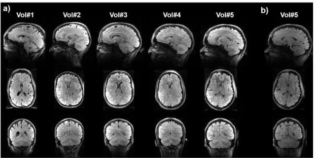
Fig. 4.a reports the in vivo results on the 5 volunteers. Good contrast was obtained consistently throughout the tests with no apparent B1+ artefact. As a reference, for comparison, the result obtained
with a single UP inversion instead of the T2 preparation is provided in Fig. 4.b for volunteer #5. It
demonstrates a lack of contrast if a T2 preparation module is not incorporated. The gain in contrast
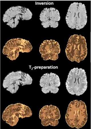
brought by the preparation translates directly into better segmentation results (see Fig. 5).
Discussion
We have reported the design and implementation of a T2-preparation module with parallel
transmit kT-point UPs. The phase coherence between the 180° and the 90° excitation pulses was enforced
by parametrizing the former with two successive identical waveforms, the first (or second) half constituting the excitation. Smaller, phase coherent, rotations incidentally could be synthetized this way by extrapolating the method. We showed in (9) that universal inversion pulses could reach flip angle NRMSEs less than 5 %. Here the NRMSE obtained across the database subjects for the refocusing pulse was 10.1 ± 1.2 %, thus indicating a small price to pay to achieve phase coherence with this pulse construction. Alternatively, the doubling of the waveforms to achieve 180° could be abandoned, but then at the expense of a more involved and complicated pulse design for the excitation where the phase in each voxel would need to be tracked (6).
A whole-brain FLAIR study with a T2-preparation module and with pTx was also reported in
(19). The pulse design was based on a direct signal control model where each RF pulse in the vFA-TSE readout train made use of a different RF shim setting to target a prescribed signal evolution for a given T1 and T2 pair. A UP version, still making use of RF shim pulses, was also successfully implemented,
albeit with some moderate loss of performance compared to the subject-based tailored approach. To be more robust against the RF field inhomogeneity inherent to 90° hard pulses, the T2 preparation used 4
results by showing that the vFA-TSE readout indeed is far more critical for the signal and contrast homogeneity than the T2-preparation, as imperfections in the preparation largely disappear with
magnetization recovery. The main benefit of the proposed kT-point universal pulse approach hence is
the reduced power (×4) and SAR (×6) demands, leading most importantly to more freedom to optimize the readout train. Incidentally, we could also show in simulation significant reduction of the saturation of the macromolecular protons caused by the preparation. Those differences yet again fade after magnetization recovery and do not likely lead to big effects in this particular application. Finally, the shorter duration of the refocusing universal pulses (2 ms versus 9 ms) can be beneficial to limit flow artefacts in the presence of crusher gradients occurring in the T2-preparation.
Conclusion
We have reported the design and implementation of a T2-preparation module with pTx universal pulses
in the 3D FLAIR sequence at 7T. Simulations and measurements validated the approach, which yields comparable signal homogeneity to the adiabatic version but with significantly less power, SAR and pulse duration. This complements the toolbox of universal pulses already available in 3D sequences and widens the applicability of the technique in general for use in clinical routine. The solutions have also been shown to be robust with respect to inter-site variability (24). They are available upon reasonable request with the appropriate sequences.
Acknowledgements
The authors thank Drs. Alexandre Vignaud and Catherine Oppenheim for valuable discussions. The research leading to these results has received funding from the European Research Council under the European Union’s Seventh Framework Program (FP7/2013-2018), ERC Grant Agreement n. 309674, and from the ERPT equipment program of the Leducq Foundation.
References
1. Hajnal JV,De Coene B, Lewis PD, et al. High signal regions in normal white matter shown by heavily T2-weighted CSF nulled IR sequences. J Comput Assist Tomogr 1992;16:506-513.
2. Kilsdonk ID, Wattjes MP, Lopez-SorianoA, Kuijer JP, de Jong MC, de Graaf WI, Conijn MM, Polman CH, Luijten PR, Geurts PR, Geerlings MI, Barkhof F. Improved differentiation between MS and vascular brain lesions using FLAIR at 7T. Eur. Radiol. 2014;24:841-849.
3. Trattnig S, Springer E, Bogner W, Hangel G, Strasser B, Dymerska B, Lima Cardoso P, Robinson SD. Key clinical benefits of neuroimaging at 7T. NeuroImage 2018;168:477-489.
4. de Graaf WL, Zwanenburg JJM, Visser F, Wattjes MP, Pouwels PJW, Geurts JJG, Polman CH, Barkhof F, Luijten PR, Castelijns JA. Lesion detection at seven Tesla in multiple sclerosis using magnetization prepared 3D-FLAIR and 3D-DIR. Eur. Radiol 2012;22:221-231.
5. Eggenschwiler F, O’Brien KR, Gruetter R, Marques JP. Improving T2-weighted imaging at high field through the use of kT-points. Magn. Reson. Med. 2014;71:1478–1488.
6. Massire A, Vignaud A, Robert B, Le Bihan D, Boulant N, Amadon A. Parallel-transmission-enabled three-dimensional T2-weighted imaging of the human brain at 7 Tesla. Magn. Reson. Med. 2015;73:2195–2203.
7. Gras V, Mauconduit F, Vignaud A, Amadon A, Stöcker T, Boulant N. Design of universal parallel-transmit refocusing kT-point pulses and application to 3D T2-weighted imaging at 7T. Magn. Reson.
Med.2018;80:53-65.
8. Gras V, Vignaud A, Amadon A, Le Bihan D, Boulant N. Universal pulses: A new concept for calibration-free parallel transmission. Magn. Reson. Med. 2017;77:635–643.
9. Gras V, Boland M, Vignaud A, Ferrand G, Amadon A, Mauconduit F, Bihan DL, Stöcker T, Boulant N. Homogeneous non-selective and slice-selective parallel-transmit excitations at 7 Tesla with universal pulses: A validation study on two commercial RF coils. PLOS ONE 2017;12:e0183562. doi: 10.1371/journal.pone.0183562.
10. Visser F, Zwanenburg JJ, Hoogduin JM, Luijten PR. High-resolution magnetization-prepared 3D-FLAIR imaging at 7.0 Tesla. Magn. Reson. Med. 2010;64:194–202.
11. Nezafat R, Ouwerkerk R, Derbyshire AJ, Stuber M, McVeigh ER. Spectrally selective B1-insensitive
T2 magnetization preparation sequence. Magn. Reson. Med. 2009;61:1326-1335.
12. Pracht ED, Gras V, Boulant N, Stöcker T. Whole-brain FLAIR imaging at 7T employing universal pulses. In proceedings of the 26th annual ISMRM meeting, 2018, p 0585, Paris, France.
13. Massire A, Cloos MA, Vignaud A, Le Bihan D, Amadon A, Boulant N. Design of non-selective refocusing pulses with phase-free rotation axis by gradient ascent pulse engineering algorithm in parallel transmission at 7T. J. Magn Reson. 2013;230:73-83.
14. Cloos MA, Boulant N, Luong M, Ferrand G, Giacomini E, Le Bihan D, Amadon A. kT-points: short
three-dimensional tailored RF pulses for flip angle homogenization over an extended volume. Magn. Reson. Med. 2012;67:72-80.
15. Hoyos-Idrobo A, Weiss P, Massire A, Amadon A, Boulant N. On Variant Strategies to Solve the Magnitude Least Squares Optimization Problem in Parallel Transmission Pulse Design and Under Strict SAR and Power Constraints. IEEE Trans. Med. Imaging 2014;33:739–748.
16. Gras V, Luong M, Amadon A, Boulant N. Joint design of kT-points trajectories and RF pulses under explicit SAR and power constraints in the large flip angle regime. J. Magn. Reson. 2015;261:181 – 189. 17. Mugler JP III, Kiefer B, Brookeman JR. Three-dimensional T2-weighted imaging of the brain using very long spin-echo trains. In: Proceedings of the 8th Annual Meeting of ISMRM, 2000, p 687, Denver, CO, USA.
18. Zhang Y, Brady M, Smith S. Segmentation of brain MR images through a hidden Markov random field model and the expectation-maximization algorithm. IEEE Trans Med Imaging 2001;20:45-57. 19. Beqiri A, Hoogduin H, Sbrizzi A, Hajnal JV, Malik SJ. Whole-brain 3D FLAIR at 7T using direct signal control. Magn. Reson. Med. 2018. doi: 10.1002/mrm.27149.
20. Stanisz GJ, Odrobina EE, Pun J, Escaravage M, Graham SJ, Bronskill MJ, Henkelman RM. T1 and
21. Matson G, Liu H. Computer simulations of 3D MPRAGE in human brain with inclusion of inadvertent magnetization transfer effects. In proceedings of the 19th annual ISMRM meeting, 2010, p
3008, Stockholm, Sweden.
22. Sled JG, Pike BG. Quantitative Imaging of magnetization transfer exchange and relaxation properties in vivo using MRI. Magn. Reson. In Med. 2001;46:923-931.
23. Boulant N, Le Bihan D, Amadon A. Strongly modulating pulses for counteracting RF inhomogeneity at High Fields. Magn. Reson. Med. 2008;60:701-708.
24. Wu X, Gras V, Vignaud A, Mauconduit F, Boland M, Stöcker T, Ugurbil K, Boulant N. The travelling pulses: multicenter evaluation of universal pulses at 7T. In proceedings of the 26th annual
Footnote
1The 0.34 W indicated in ref (19) is for TR=8 s, and thus becomes 0.39 W for TR=7 s. A private
communication with one of the authors revealed that this number did not include the final inversion pulse, necessary to invert the CSF longitudinal magnetization. Given the information provided about that pulse, the overall total power was estimated as 0.58 W.
List of captions
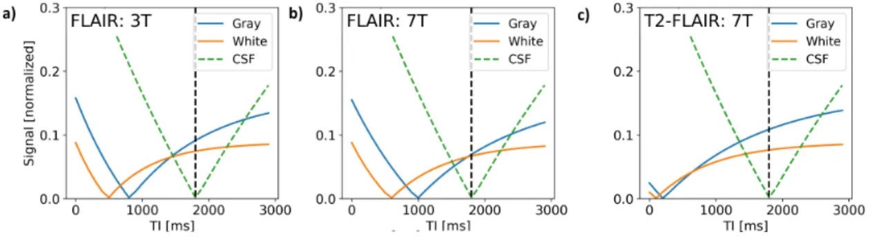
Figure 1. Bloch equation simulation results for the FLAIR. The simulations took literature values for T1 and T2 at 3T and at 7T. The simulation results are provided for 3T (a) and 7T (b) with just inversion
recovery, c) for 7T with a T2 preparation module. The lengthening of T1 from 3T to 7T attenuates the
signal and contrast between brain tissues. Inserting the T2 preparation boosts the contrast between gray
and white matter while suppressing the CSF signal.
Figure 2. Sequence diagram of the implemented FLAIR sequence with magnetization preparation. The turbo spin echo (TSE) readout train uses variable flip angle refocusing universal pulses (UPs). The T2
-preparation module inverts the CSF signal while saturating brain tissue to regain signal and contrast between grey and white matter. For more optimality, in the proposed work the design of the pulses in the T2-preparation is independent from the design of the pulses in the readout train.
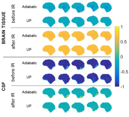
Figure 3. Bloch simulation results of the T2-preparation module. Assuming perfect spoiling, the result
is represented as the longitudinal magnetization (units of M0) for the brain tissue (top) and CSF (bottom).
The distributions are plotted on sagittal slices for the 5 scanned subjects. Comparisons are provided between the UP and the adiabatic pulse implementations, just after the preparation module and just prior to the vFA-TSE readout (i.e. after Inversion Recovery (IR)). Both methods return good homogeneity at the end of the recovery with perhaps little imperfections at the bottom of the brain for the CSF with the adiabatic approach (see also Figure 4 in (19)).
Figure 4. In-vivo FLAIR imaging results at 7T with no bias field correction. a) Sagittal, axial and coronal views obtained with pTx universal pulses on 5 volunteers. The CSF is effectively suppressed throughout the brain while good contrast between white matter and grey matter is obtained. b) Same views for volunteer #5 obtained with a single UP inversion replacing the preparation. Comparison between the two illustrates the contrast gain achieved by means of the T2 preparation module.
Figure 5. Segmentation results with FSL on volunteer #5. Top: results with the UP inversion only. Bottom: results with the UP T2-preparation approach. Three tissue-segmentation (three colors) was