HAL Id: hal-02643815
https://hal.inrae.fr/hal-02643815
Submitted on 28 May 2020
HAL is a multi-disciplinary open access
archive for the deposit and dissemination of
sci-entific research documents, whether they are
pub-lished or not. The documents may come from
teaching and research institutions in France or
abroad, or from public or private research centers.
L’archive ouverte pluridisciplinaire HAL, est
destinée au dépôt et à la diffusion de documents
scientifiques de niveau recherche, publiés ou non,
émanant des établissements d’enseignement et de
recherche français ou étrangers, des laboratoires
publics ou privés.
Contribution of uniformly 13C-enriched
sterigmatocystin to thestudy of its pulmonary
metabolism
Odile Cabaret, Olivier Puel, Françoise Botterel, Michel Péan, Stéphane
Bretagne, Marcel Delaforge
To cite this version:
Odile Cabaret, Olivier Puel, Françoise Botterel, Michel Péan, Stéphane Bretagne, et al..
Contribu-tion of uniformly 13C-enriched sterigmatocystin to thestudy of its pulmonary metabolism. Rapid
Communications in Mass Spectrometry, Wiley, 2011, 25 (19), pp.2704-2710. �10.1002/rcm.5068�.
�hal-02643815�
Contribution of uniformly
13
C
‐enriched sterigmatocystin to the
study of its pulmonary metabolism
†
Odile Cabaret
1,2, Olivier Puel
3, Françoise Botterel
1,2, Michel Pean
4,5,6, Stéphane Bretagne
1,2and Marcel Delaforge
7*
1
UMR BIPAR, U‐PEC, AFSSA, ENVA, Faculté de Médecine, Créteil Cedex F‐94010, France
2AP‐HP, Groupe hospitalier Henri Mondor‐Albert Chenevier, Laboratoire de Parasitologie‐Mycologie, Créteil Cedex F‐94010,
France
3ToxAlim INRA, Equipe d′Immunologie‐Toxicologie, Toulouse F‐31027, France 4
CEA, DSV, IBEB, Groupe de Recherches Appliquées en Phytotechnologie, Saint‐Paul‐lez‐Durance, F‐13108, France
5CNRS, UMR Biologie Végétale & Microbiologie Environnementale, Saint‐Paul‐lez‐Durance, F‐13108, France 6
Aix‐Marseille Université, Saint‐Paul‐lez‐Durance, F‐13108, France
7CEA Saclay, iBiTec‐S, SB2SM and URA CNRS 2096, Gif sur Yvette Cedex F‐91191, France
Mycotoxins are secondary metabolites offilamentous fungi which can cause a wide range of systemic effects. Human health effects of inhaled mycotoxins remain poorly documented, despite the large amounts present, associated with air‐ borne particles. Among these mycotoxins, sterigmatocystin is one of the most prevalent. Because its chemical structure is close to that of the aflatoxins, we studied its metabolism and its cellular consequences when in contact with the airway epithelium, using the mass spectral signature from the 10%13C uniformly enriched sterigmatocystin. The metabolism
was studied in vitro, using recombinant cytochrome P450s enzymes, and in porcine tracheal epithelial cell (PTEC) primary cultures at an air‐liquid interface. The metabolites were analyzed by high‐performance liquid chromatography coupled with tandem mass spectrometry detection. Expressed enzymes and PTECs were exposed to uniformly 13C‐ enriched sterigmatocystin to confirm the relationship between sterigmatocystin and its metabolites because this isotopic cluster shape is conserved for all metabolites and their product ions. Incubation of sterigmatocystin with recombinant cytochrome P450 1A1 led to the formation of three metabolites identified as monohydroxysterigmatocystin, dihydroxysterigmatocystin and one glutathione adduct, the latter after the formation of a transient intermediate. In the PTEC cultures, sterigmatocystin metabolism resulted in a glucuro‐conjugate. Two other products were detected, a sulfo‐conjugate and a glucuro‐conjugate of hydroxysterigmatocystin upon cytochrome P450 1A1 induction. This is the first study to report sterigmatocystin metabolism in airway epithelium, and it suggests that, contrary to the aflatoxins, sterigmatocystin is mainly detoxified into its conjugates and is unable to produce significant amounts of reactive metabolites in respiratory cells, at least in pigs. Copyright © 2011 John Wiley & Sons, Ltd.
Mycotoxins are secondary metabolites of filamentous fungi which can cause a wide range of acute and chronic systemic effects. As xenobiotics, these compounds can be metabolized by some of the host enzymes. These enzymes, primarily hepatic cytochromes P450 (CYP), ensure the oxidation of many hydrophobic exogenous compounds, increasing their polarity and thus assuring their biliary or urinary elimina-tion. This transformation can involve a reduction of the parental compound toxic effects (reduction of ergotism by oxidation of the ergot alkaloids, e.g.[1]) or, in other cases, can involve a toxic activation, as in the case of aflatoxins.[2]
These modified compounds are then conjugated to polar com-pounds in conjugation reactions. Glucuronic acid moieties
are transferred to substrate molecules that contain oxygen, nitrogen, sulfur or carboxyl groups. Sulfonic acid moieties are transferred from a donor molecule to an acceptor alcohol or amine and the conjugation of reduced glutathione to electrophilic centers, on a wide variety of substrates.
Most publications on mycotoxins deal with hazards due to toxic effects after their gastrointestinal uptake.[3] There are fewer data available concerning toxicity and metabolism after inhalation, despite the detection of mycotoxins in bioaerosols and dust at workplaces and in homes[4,5] and their association with inhaled spores.[6,7]
Among mycotoxins, sterigmatocystin (STG) is one of the most prevalent in the environment. STG has been detected in carpet dust from damp dwellings[8]and in dwellings after waterflooding.[9,10]Aspergillus versicolor is the most efficient producer of STG, which is a biochemical precursor in the biosynthesis of aflatoxin.[11]
After ingestion, STG is carcino-genic in animal models[12–14] and it is classified as possibly carcinogenic in humans.[15]STG is activated by CYP to a liver carcinogen.[16] To date, STG metabolites have never been studied in airway epithelium.
* Correspondence to: M. Delaforge, CEA Saclay, iBiTec‐S, SB2SM and URA CNRS 2096, Gif sur Yvette Cedex F‐91191, France.
E‐mail: marcel.delaforge@cea.fr
† Presented at the 6th Congress of the French Society of
Stable Isotopes (Société Française des Isotopes Stables, SFIS) held 26–29 October 2010 in Toulouse, France.
Received: 24 January 2011 Revised: 11 April 2011 Accepted: 19 April 2011 Published online in Wiley Online Library
Rapid Commun. Mass Spectrom. 2011, 25, 2704–2710 (wileyonlinelibrary.com) DOI: 10.1002/rcm.5068
The aim of this study was to elucidate the metabolism of STG after contact with airway epithelium and to assess any possible cellular consequences. For this purpose, we used uniformly 13C‐enriched STG to confirm the metabolic
relationship to STG. Preliminary studies consisted of identi-fying STG metabolites in recombinant CYP enzymes, and thus the metabolism upon exposure of porcine tracheal epithelial cell (PTEC) primary cultures to STG.
EXPERIMENTAL
Chemicals
Ham’s F‐12 nutrient medium, Dulbecco’s modified Eagle’s medium, penicillin/streptomycin, amphotericin B, fetal calf serum, 0.05% trypsin‐EDTA (1X) and phosphate‐buffered saline were purchased from Invitrogen Life‐Technologies (Cergy‐Pontoise, France). Gentamicin, collagen IV, commer-cial12C STG (molecular weight (MW) = 324.3) with purity of
98%, acetonitrile, reduced nicotinamide adenine dinucleotide phosphate (NAPDH), glutathione (GSH),β‐naphthoflavone, and ammonium acetate were obtained from Sigma (Saint Quentin Fallavier, France). Ultroser G was purchased from BioSepra SA (Cergy‐Saint‐Christophe, France); dimethyl sulf-oxide (DMSO) from Prolabo (VWR International, Fontenay sous Bois, France).
Production of uniformly13C‐enriched STG
Production of uniformly 13C‐enriched STG was performed
using autoclaved 10%‐13
C wheat grains (Triticum aestivum cv. Ardente) as the exclusive nutrient source for the fungal culture, as previously described.[17] The grains were moist-ened to a water activity value of 0.98 before sterilization. Around 30 g of this plant material was inoculated with a suspension of 2.105 Emericella nidulans conidia. STG was
extracted from 14‐day‐old cultures with chloroform. The raw extract was fractioned by automatedflash chromatography (Teledyne Isco, Lincoln, NE, USA). The fractionation was performed on a normal‐phase RediSep 12 g column (Tele-dyne Isco) at aflow rate of 25 mL/min with linear gradient. The solvent system was 10% acetic acid in toluene as solvent A and 10% acetic acid in ethyl acetate as solvent B. Initially solvent B was present at 0% and increased to 45.5% within 10 min. The fraction volume was set to 5 mL. The presence of STG in fractions was checked by high‐performance liquid chromatography (HPLC) with diode‐array detection (Kontron Instruments, Neufahru, Germany) with a 250 × 4.6 mm Modul‐cart Strategy 5 µm C18‐2 column (Interchim, Montluçon, France). Gradient chromatography (run of 46 min) was per-formed with 0.2% acetic acid in water (eluent A)/acetonitrile (eluent B) as the mobile phase at aflow rate of 1.5 mL/min. The compounds were eluted by starting from 31% solvent B for 30 minfollowedbyalineargradientupto90%within5 min.After an isocratic elution for 5 min, the gradient was reduced to its initial value within 5 min, and remained at this value for thefinal 5 min. The13C STG was purifiedfrompositive fractionsbysemi‐ quantitative HPLC. The HPLC unit was fitted with a 250 × 7.8 mm Modul‐cart Strategy 5 µm C18‐2 column (Inter-chim) and a diode‐array detector. The gradient program was the same as above, except that theflow rate was increased to 4.5 mL/min. After production of uniformly13C‐enriched STG
the presence of an isotopic cluster close to that expected from theoretical evaluation[17] was checked by mass spectrometry
(MS).
Metabolite studies by human cytochrome P450 enzymes Bactosome®‐expressed human cytochromes P450 (CYP) were obtained from Cypex (Tebu‐bio, Le Perray en Yvelines, France). We tested the main human CYP differing by their active site and thus by their substrate specificity.[18]Based on
the structural homology of STG with aflatoxins, we con-sidered the CYP isoforms 1A, 2A and 3A, reported to metabolize aflatoxin B1.[19–22] The metabolism of both
commercial12C STG and13C STG was studied as previously
described.[23]
STG metabolism in porcine tracheal epithelial cells (PTECs) Preparation of porcine primary cultures was performed with trachea obtained from the Centre de Recherche Chirurgicale of the Henri‐Mondor Hospital (Créteil, France) as previously described.[24] After proliferation during the first week, the
PTECs reached the differentiated state during a second week of culture with the presence of an apical side at an air interface and a basal side at a liquid contact. PTECs were exposed when cell differentiation was stable (after 14 days of culture) for 48 h to 1 mL of 1μM U10%13C STG through the basal side of the culture. After this exposure the medium was collected for extraction. To examine the effect of amounts of CYP1A1, the cells were pre‐treated for 24 h with 25 μM β‐ naphthoflavone, a well‐known CYP1A inducer, which led to an increase of CYP1A expression, or with 0.2% DMSO as control. The basal medium was then removed and the STG incubated for an additional 48 h. Controls were obtained after 48 h incubation with 13C STG in coated wells without cells and with 0.2% DMSO incubation in cell culture medium. Before extraction, the proteins were precipitated by addition of 500μL cold methanol. After 30 min at −20°C, the mixture was centrifuged at 10 000 rpm for 5 min. Then 3 mL of water were added to the supernatant and the resulting solution was purified with a SepPack C18 cartridge (Waters Corp., Milford, MA, USA). The samples were eluted with 1 mL acetonitrile. This extract was reduced to dryness by speed‐ vac concentration (Savant SC‐200, Savant Instruments, Farmingdale, NY, USA) and then dissolved in the HPLC solvent (50% 10 mM ammonium acetate in water, 50% acetonitrile).
HPLC/MS/MS analysis
HPLC/MS/MS analyses were performed on an Esquire ion trap mass spectrometer (Bruker Daltonics GmbH, Bremen, Germany) with an electrospray ionization source operating in alternative positive/negative ionization mode, coupled with a 150 × 2.1 mm C18 Kromasil column (Interchim). The flow rate was 0.2 mL/min of a 25 min linear gradient ranging from 10 to 80% B (90 % acetonitrile in water) into A (90% 10 mM ammonium acetate in water, 10% acetonitrile). Electrospray ionization was performed at 4.5 kV and the capillary temperature was set to 310°C using a nitrogen gasflow rate of 40 mL/min and an auxiliary gas flow of 10 mL/min. Collision‐induced dissociation of the selected precursor ion was carried out in the presence of helium with a collision Metabolism of U13C‐sterigmatocystin in vitro and ex vivo
wileyonlinelibrary.com/journal/rcm
Copyright © 2011 John Wiley & Sons, Ltd. Rapid Commun. Mass Spectrom. 2011, 25, 2704–2710
voltage of 0.70 V using a trap isolation width of 3 m/z units in order to isolate the whole isotopic cluster. Optimizations of MS and MSn signals were carried out using infusion of commercial STG. Injections were performed in 50:50 acetoni-trile/water containing 10 mM ammonium acetate. Data acquisition and reprocessing were performed using Bruker Daltonics Data Analysis 3.2 software.
The SMARTCyp 1.5.3 program[25]was used to predict CYP
metabolism. Mass Frontier 7.0 Spectral Interpretation Soft-ware (Thermo Fisher Scientific, San Jose, CA, USA) was used to simulate MS fragmentation in positive ion mode.
RESULTS AND DISCUSSION
Uniform13C enrichment in STG
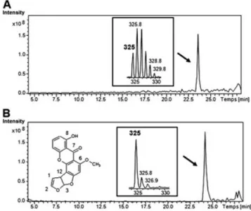
Before its use in metabolic studies, we checked that 13C‐ enriched STG presented an isotopic cluster characteristic of 10%13C enrichment. The MS spectrum in positive ion mode contained the [M + H]+ ion at m/z 325 and also M + 1 (m/z
326), M + 2 (m/z 327), M + 3 (m/z 328), M + 4 (m/z 329) and M + 5 (m/z 330) ions (Fig. 1). These peaks are in a 0.50:1.00:0.93:0.57:0.23:0.02 ratio relative to the most intense
ion at m/z 326, and the isotopic ratios are close to those expected from theoretical evaluation for 10 % 13C enrichment.[17]
The accuracy on the obtained isotope pattern determined for 15 different patterns was inferior to 20% for each peak. Metabolite studies by human P450 enzymes
In order to avoid the use of human microsomes which contain around 20 different CYP isoforms, we used recombinant human CYP which are obtained from insect cells transfected with only a specific CYP cDNA. A 30 min incubation of either commercial
12C or 10%‐13
C STG with 0.2μM recombinant human CYP1A1 enzyme in the presence of NADPH led to the significant formation of two metabolites M1 and M2. These metabolites, having retention time (tR) = 19.5 min (M1) and 15.5 min (M2),
were more polar than STG (tR= 24.8 min). They had a molecular
weight difference of one (+16 Da) (MW = 340) or two oxygen atoms (+32 Da) (MW = 356), respectively, relative to STG (Table 1). The metabolites formed using 10%‐13C STG exhibited
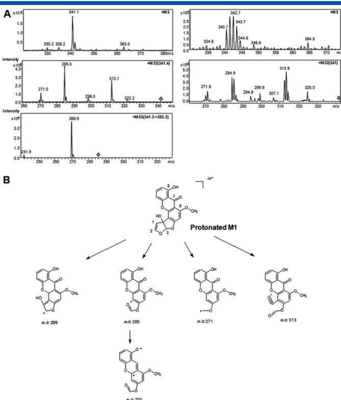
isotopic clusters similar to that of STG. Their [M + 1]+ and [M + 2]+ions were more intense than [M]+. Collision‐induced
fragmentation of protonated M1 led to product ions at m/z 313, 299, 285, and 271 in positive ion mode (Fig. 2). Incubations in the presence of GSH led to the formation of a third metabolite (M3) having tR= 11 min and an ion at m/z 644 in negative ion mode,
which corresponds to an increase of 321 Da relative to the molecular weight of STG (Table 1). Incubations of STG with other human CYP isoforms led to the formation of M1 and M3 for CYP1A2, M3 for CYP3A4, and no metabolism for CYP2A6 and CYP2A13. All the metabolites exhibited an isotopic cluster profile in their mass spectra (Fig. 2), which confirmed their connection to STG. The MS2product ions also conserved an isotopic cluster (Fig. 2).
Based on its mass, its polarity and the monooxygenase activity of CYP, M1 was assigned as monohydroxysterigmatocystin (Table 1). CYP metabolism prediction using the SMARTCyp program indicates that two sites (C3 and C12) in STG bear a labile proton for CYP hydroxylation. The fragmentation pathway of protonated M1 in positive ion mode is in accordance with hydroxylation on C12 (Fig. 2). Interestingly, it had been demonstrated that CYP1A2 oxidizes aflatoxin B1in the same
position, leading to aflatoxin M1.[26]
M3 was assigned as a GSH adduct of an oxidized metabolite (Table 1). This oxidation may occur in the 1‐2‐ position since the STG‐glutathione adduct formed on STG 1,2‐oxide has been observed.[27]
Other possible structures include oxidation of the phenol ring to a reactive quinone which can then form a glutathione adduct.[28]The detection Figure 1. Representative HPLC/MS chromatograms and
isotopic cluster of uniformly 13C‐enriched sterigmatocystin after 10% 13C enrichment (A) and natural 12C sterigmato-cystin with its structure (B).
Table 1. Molecular weights (MW) for the compounds described in this study MW
(Da) Detection(m/z)
Sterigmatocystin 324 Positive and negative ion mode (325+, 323–) M1: monohydroxysterigmatocystin 340 Positive and negative ion mode (341+, 339–) M2: dihydroxysterigmatocystin 356 Negative ion mode (355–)
M3: GSH adduct of an oxidized metabolite 645 Negative ion mode (644–) M4: sterigmatocystin‐glucuro‐conjugate 500 Negative ion mode (499–) M5: sulfo‐conjugate of monohydroxysterigmatocystin 420 Negative ion mode (419–) M6: glucuro‐conjugate of monohydroxysterigmatocystin 516 Negative ion mode (515–)
of a glutathione adduct after in vitro metabolism through CYP1A and CYP3A4 activities strongly suggests the forma-tion of a transient reactive epoxide of STG, as has been described using either human hepatic microsomes or the Ames test.[27,29] Such metabolic pathways have been de-scribed for aflatoxin B1 hepatotoxicity.[30] These mutagenic
properties could be related to the pulmonary carcinomas described in mice after oral administration of STG.[15,31]
M2 was assigned as dihydroxysterigmatocystin (Table 1). M2 may be formed by hydrolysis of M3 or by M1 hydroxylation. CYP metabolism prediction using the SMARTCyp program indicates that C3 is the most probable site for CYP hydroxylation in monohydroxysterigmatocystin (M1). Thus, M2 could also be a catechol or a para‐diphenol on the phenyl ring, and it may be metabolized to a quinone.
Figure 2. Representative HPLC/MS, MS2and MS3chromatograms of the ion at m/z 341 in positive ionization
mode (M + 16, metabolite M1); with natural 12C sterigmatocystin on the left and with 13C‐enriched sterigmatocystin on the right (A). Proposed fragmentation pathways of protonated‐M1 in positive ion mode (B). Metabolism of U13C‐sterigmatocystin in vitro and ex vivo
wileyonlinelibrary.com/journal/rcm
Copyright © 2011 John Wiley & Sons, Ltd. Rapid Commun. Mass Spectrom. 2011, 25, 2704–2710
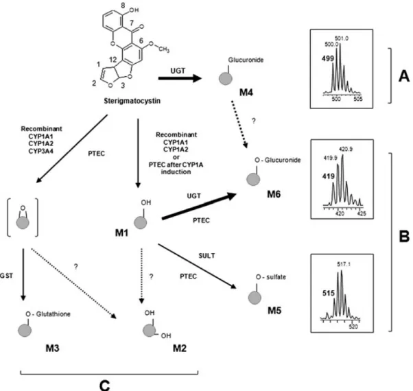
STG metabolism in PTECs
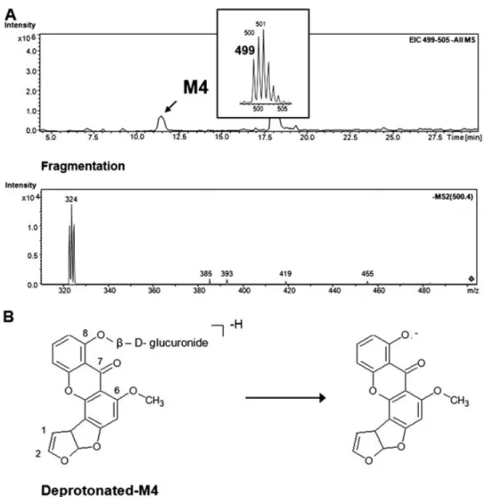
After 48 h incubation with PTECs, only one STG metabolite, M4, was formed and this was identified as a glucuro conjugate. M4 was more polar than STG (at tR= 11.5 min)
and had an isotopic cluster shifted to M + 176 relative to STG (MW = 500) (Fig. 3, Table 1). Fragmentation of deprotonated M4 in negative ion mode led to the formation of the MS cluster of deprotonated STG. The glucuronide formation was confirmed after hydrolysis withβ‐glucuronidase (data not shown). Only glucuronide conjugation with the free hydroxyl function on C8 is compatible with the mass of M4. Glucuronidation has been involved in metabolism of STG, and a STG glucuronide was detected in the urine of rat or mon-key.[32,33] Glucuronidation has been identified as an important pathway in the respiratory detoxification of environmental carcinogens such as polycyclic aromatic hydrocarbons.[34]
After 24 h exposure of PTECs to 25μM β‐naphthoflavone, 48 h STG incubation led to the formation of two new polar metabolites, M5 and M6, detected in negative ion mode. These metabolites had tR values of 12.5
and 5.5 min and an isotopic cluster shifted to M + 96 (MW = 420) and M + 192 (MW = 516), respectively, relative to STG (Fig. 3). M5 was identified as a sulfo‐ conjugate (M + 16 + 80) and M6 as a glucuro‐conjugate (M + 16 + 176) of monohydroxysterigmatocystin (Table 1). The product ions of deprotonated M5 and M6 show an M + 16 isotopic cluster, which could correspond to a monohydroxysterigmatocystin product (M1 or hydroxyl-ation on another lochydroxyl-ation). Therefore, CYP1A1 metabolism may result in M1 formation, with rapid transformation into M5 and M6. Evidence for the epoxide formation and the subsequent formation of dihydroxysterigmatocystin (M2) or the glutathione conjugate (M3) was not found in porcine primary cells.
Figure 3. Representative HPLC/MS/MS chromatograms showing formation of ste-rigmatocystin metabolite in PTEC exposed to 1 μM sterimatocystin through the basal side of cultures (A): m/z 499 in negative ionization mode (M4) and MS2 spectrum of
M4 metabolite. Proposed fragmentation pathways of deprotonated M4 in negative ion mode (B).
CONCLUSIONS
Our study shows that STG is metabolized in porcine epithelial airway cells through phase 1 and 2 metabolism (Fig. 4, Table 1). This is thefirst study to report metabolite formation and de-toxification pathways in the pulmonary stage using primary cells at an air‐liquid interface. This metabolism seems close to that of aflatoxin B1with the formation of oxidized metabolites, a
glu-tathione adduct and glucuronides. However, further studies are necessary to precisely locate the sites of oxidation. STG is main-ly detoxified through glucuronidation (glucuronidation of STG and monohydroxysterigmatocystin). We also suggest that the metabolism of STG in the porcine airway tract in vitro is unable to produce significant amounts of reactive epoxide metabolites. Finally, the uniformly enriched molecules with stables isotopes are very powerful tools for metabolism studies. Since the isotopic cluster signature is preserved for all the enriched metabolites, mass spectrometric analysis is easier than the classical approach.
Acknowledgements
We thank Sandrine Blondel and Anaïs Roussel for technical assistance. These studies were supported in part by AFSSET (Grant Numbers EST‐2007‐63 and EST‐2008‐27). We thank Andrew Lilley for English correction.
REFERENCES
[1] M. A. Peyronneau, M. Delaforge, R. Riviere, J. P. Renaud, D. Mansuy. High affinity of ergopeptides for cytochromes P450 3A. Importance of their peptide moiety for P450 recognition and hydroxylation of bromocriptine. Eur. J. Biochem. 1994, 223, 947.
[2] E. M. Isin , F. P. Guengerich. Complex reactions catalyzed by cytochrome P450 enzymes. Biochim. Biophys. Acta 2007, 1770, 314.
[3] J. W. Bennett, M. Klich. Mycotoxins. Clin. Microbiol. Rev. 2003, 16, 497.
Figure 4. Putative pulmonary sterigmatocystin metabolism. Pathway A (top) indicates formation of a sterigmatocystin‐glucuro conjugate (metabolite M4) in ’normal’ airway epithelial cell cultures. Pathway B indicates formation of a sulfo conjugate and a glucuro conjugate of monohydroxyster-igmatocystin (metabolites M5 and M6, respectively) in’CYP1A1‐induced’ airway epithelium cultures. CYP1A1 amounts were increased by 24 h exposure to 25μM β‐naphthoflavone. For high levels of CYP1A1 and in the presence of glutathione‐S‐transferases and glutathione, transient epoxide formation led to the glutathione conjugate or to dihydrodiol (pathway C, bottom).
Metabolism of U13C‐sterigmatocystin in vitro and ex vivo
wileyonlinelibrary.com/journal/rcm
Copyright © 2011 John Wiley & Sons, Ltd. Rapid Commun. Mass Spectrom. 2011, 25, 2704–2710
[4] E. Bloom, L. F. Grimsley, C. Pehrson, J. Lewis, L. Larsson. Molds and mycotoxins in dust from water‐damaged homes in New Orleans after hurricane Katrina. Indoor Air 2009, 19, 153.
[5] J. Bunger, G. Westphal, A. Monnich, B. Hinnendahl, E. Hallier, M. Muller. Cytotoxicity of occupationally and environmentally relevant mycotoxins. Toxicology 2004, 202, 199.
[6] K. Khoufache, O. Puel, N. Loiseau, M. Delaforge, D. Rivollet, A. Coste, C. Cordonnier, E. Escudier, F. Botterel, S. Bretagne. Verruculogen associated with Aspergillus fumigatus hyphae and conidia modifies the electrophysiological properties of human nasal epithelial cells. BMC Microbiol. 2007, 7, 5. [7] W. G. Sorenson, D. G. Frazer, B. B. Jarvis, J. Simpson, V. A.
Robinson. Trichothecene mycotoxins in aerosolized conidia of Stachybotrys atra. Appl. Environ. Microbiol. 1987, 53, 1370. [8] S. Engelhart, A. Loock, D. Skutlarek, H. Sagunski, A. Lommel, H. Farber, M. Exner. Occurrence of toxigenic Aspergillus versicolor isolates and sterigmatocystin in carpet dust from damp indoor environments. Appl. Environ. Microbiol. 2002, 68, 3886.
[9] T. Tuomi, K. Reijula, T. Johnsson, K. Hemminki, E. L. Hintikka, O. Lindroos, S. Kalso, P. Koukila‐Kahkola, H. Mussalo‐Rauhamaa, T. Haahtela. Mycotoxins in crude building materials from water‐damaged buildings. Appl. Environ. Microbiol. 2000, 66, 1899.
[10] K. F. Nielsen, S. Gravesen, P. A. Nielsen, B. Andersen, U. Thrane, J. C. Frisvad. Production of mycotoxins on artificially and naturally infested building materials. Mycopathologia 1999, 145, 43.
[11] K. Yabe, H. Nakajima. Enzyme reactions and genes in aflatoxin biosynthesis. Appl. Microbiol. Biotechnol. 2004, 64, 745.
[12] R. H. Adamson. Induction of hepatocellular carcinoma in nonhuman primates by chemical carcinogens. Cancer Detect. Prev. 1989, 14, 215.
[13] K. Fujii, H. Kurata, S. Odashima, Y. Hatsuda. Tumor induction by a single subcutaneous injection of sterigmato-cystin in newborn mice. Cancer Res. 1976, 36, 1615. [14] M. Mabuchi. Sequential hepatic changes during
sterigma-tocystin‐induced carcinogenesis in the rat. Jpn J. Exp. Med. 1979, 49, 365.
[15] I.A.R.C. Sterigmatocystin. IARC Monogr. Eval. Carcinog. Risk Chem. Man. 1976, 10, 245.
[16] I. R. McConnell, R. C. Garner. DNA adducts of aflatoxins, sterigmatocystin and other mycotoxins. IARC Sci. Publ. 1994, 49.
[17] F. Bravin, R. C. Duca, N. Loiseau, M. Pean, O. Puel, M. Delaforge. Production and use of mycotoxins uniformly enriched with stable isotopes for their dosage in biological samples. World Mycotoxins J. 2008, 1, 275.
[18] F. P. Guengerich, in Human Cytochrome P450 Enzymes, (Ed: P. R. Ortiz de Montellano), Plenum, New York, 2005. [19] C. L. Crespi, B. W. Penman, et al. Human cytochrome
P450IIA3: cDNA sequence, role of the enzyme in the metabolic activation of promutagens, comparison to
nitrosamine activation by human cytochrome P450IIE1. Carcinogenesis 1990, 11, 1293.
[20] X. Y. He, L. Tang, S. L. Wang, Q. S. Cai, J. S. Wang, J. Y. Hong. Efficient activation of aflatoxin B1 by cytochrome P450 2A13, an enzyme predominantly expressed in human respiratory tract. Int. J. Cancer 2006, 118, 2665.
[21] J. D. Kelly, D. L. Eaton, F. P. Guengerich, R. A. Coulombe Jr. Aflatoxin B1 activation in human lung. Toxicol. Appl. Pharmacol. 1997, 144, 88.
[22] T. R. Van Vleet, P. J. Klein, R. A. Coulombe Jr. Metabolism of aflatoxin B1 by normal human bronchial epithelial cells. J. Toxicol. Environ. Health A 2001, 63, 525.
[23] O. Cabaret, O. Puel, F. Botterel, M. Pean, K. Khoufache, J. M. Costa, M. Delaforge, S. Bretagne. Metabolic detoxication pathways for sterigmatocystin in primary tracheal epithelial cells. Chem. Res. Toxicol. 2010, 23, 1673.
[24] K. Khoufache, O. Cabaret, C. Farrugia, D. Rivollet, A. Alliot, E. Allaire, C. Cordonnier, S. Bretagne, F. Botterel. Primary in vitro culture of porcine tracheal epithelial cells in an air‐liquid interface as a model to study airway epithelium and Aspergillus fumigatus interactions. Med. Mycol. 2010, 48, 1049.
[25] Available: http://www.farma.ku.dk/smartcyp/
[26] F. P. Guengerich, W. W. Johnson, T. Shimada, Y. F. Ueng, H. Yamazaki, S. Langouet. Activation and detoxication of aflatoxin B1. Mutat. Res. 1998, 402, 121.
[27] K. D. Raney, T. Shimada, D. H. Kim, J. D. Groopman, T. M. Harris, F. P. Guengerich. Oxidation of aflatoxins and sterigmatocystin by human liver microsomes: significance of aflatoxin Q1 as a detoxication product of aflatoxin B1. Chem. Res. Toxicol. 1992, 5, 202.
[28] E. S. Krol, J. L. Bolton. Oxidation of 4‐alkylphenols and catechols by tyrosinase: ortho‐substituents alter the mecha-nism of quinoid formation. Chem. Biol. Interact. 1997, 104, 11.
[29] T. Shimada, C. L. Hayes, H. Yamazaki, S. Amin, S. S. Hecht, F. P. Guengerich, T. R. Sutter. Activation of chemically diverse procarcinogens by human cytochrome P‐450 1B1. Cancer Res. 1996, 56, 2979.
[30] J. S. Wang, J. D. Groopman. DNA damage by mycotoxins. Mutat. Res. 1999, 424, 167.
[31] G. M. Zwicker, W. W. Carlton, J. Tuite. Long‐term adminis-tration of sterigmatocystin and Penicillium viridicatum to mice. Food Cosmet. Toxicol. 1974, 12, 491.
[32] J. J. Olson, F. S. Chu. Immunochemical Studies of Urinary Metabolites of Sterigmatocystin in Rats. J. Agric. Food Chem. 1993, 41, 250.
[33] P. G. Thiel, M. Steyn. Urinary excretion of the mycotoxin, sterigmatocystin by vervet monkeys. Biochem. Pharmacol. 1973, 22, 3267.
[34] R. W. Dellinger, J. L. Fang, G. Chen, R. Weinberg, P. Lazarus. Importance of UDP‐glucuronosyltransferase 1A10 (UGT1A10) in the detoxification of polycyclic aromatic hydrocarbons: decreased glucuronidative activ-ity of the UGT1A10139Lys isoform. Drug Metab. Dispos. 2006, 34, 943.