HAL Id: hal-02084968
https://hal.archives-ouvertes.fr/hal-02084968
Submitted on 30 Mar 2019
HAL is a multi-disciplinary open access
archive for the deposit and dissemination of sci-entific research documents, whether they are pub-lished or not. The documents may come from teaching and research institutions in France or abroad, or from public or private research centers.
L’archive ouverte pluridisciplinaire HAL, est destinée au dépôt et à la diffusion de documents scientifiques de niveau recherche, publiés ou non, émanant des établissements d’enseignement et de recherche français ou étrangers, des laboratoires publics ou privés.
A drug based model to predict enhanced transdermal
and blood brain barrier drug deliverability using
ultrasonic methods
Clifford Fong, South Australia
To cite this version:
Clifford Fong, South Australia. A drug based model to predict enhanced transdermal and blood brain barrier drug deliverability using ultrasonic methods. [Research Report] Eigenenergy, Adelaide, Australia. 2019. �hal-02084968�
A drug based model to predict enhanced transdermal and blood brain barrier drug deliverability using ultrasonic methods
Clifford W. Fong
Eigenenergy, Adelaide, South Australia, Australia.
Email: cwfong@internode.on.net
Keywords: Ultrasound enhanced permeability, Transdermal, Blood Brain Barrier, Ultrasound enhanced drug deliverability model, Chemotherapeutic drugs, Drug molecular properties, Quantum mechanics
Abbreviations: Ultrasound US, Focussed Ultrasound FUS, blood brain barrier BBB, stratium corneum SC,
bis-chloroethylnitrosourea BCNU, dipole moment DM, molecular volume MV, Transdermal Permeability TP, Therapeutic US Enhanced Transdermal Permeability TUSTP, Low Frequency US Transdermal Permeability LFUSTP,
Diphytanoylphosphatidylcholine DPhPC (or ester-DPhPC), ΔGdesolvation free energy of water desolvation, ΔGlipophilicity free energy of
lipophilicity or hydrophobicity, ΔGdesolv,CDS free energy of water desolvation of the cavitation dispersion solvent structure (CDS),
ΔGlipo,CDS free energy of lipophilicity or hydrophobicity for the CDS, DM dipole moment DM, R2 multiple correlation
coefficient, F the F test of significance, SEE standards errors for the estimates, SE(ΔGdesolvation) standard errors of ΔGdesolvation,
SE(ΔGlipophilicity), standard errors of ΔGlipophilicity, SE(Dipole Moment) standard errors for dipole moments, SE (Molecular
Volume) standard errors for molecular volumes as calculated from “t” distribution statistics,
Abstract
A previously described equation using the free energy of water desolvation (ΔGdesolv,CDS), the
lipophilicity free energy (ΔGlipo,CDS), dipole moment and molecular volume that describes the
ability of drugs to cross the blood brain barrier and other cell membranes has been shown to apply to transdermal permeation when combined with ultrasound treatment. The dipole moment is particularly important when seeking to predict how drugs will interact with the transient pores in the cell membrane or blood brain barrier under ultrasound treatment.
A quantum mechanical DFT analysis of the molecular interaction of mannitol and BCNU interacting with a surrogate membrane based on DPhPC has shown that the unexpected greater US induced permeability of mannitol compared to BCNU is due to differences in how the dipoles of mannitol and BCNU interact with the electric dipole potential of the lipid bilayers of the blood brain barrier, specifically by interacting with the phosphatidylcholine headgroup not the ester group. This result indicates that the design of therapeutic drugs to cross dermal and BBB membranes should consider whether such drugs can interact with the phosphatidylcholine headgroup of the membranes thereby possibly lowering the dipole potential of the lipid bilayer and hence enhancing drug permeation.
The observed increase in electrical conductivity of the stratium corneum of the skin and blood brain barrier upon ultrasound treatment is an important factor which determines the transient permeabilization enhancement of drugs. It is likely that the dipole moment of permeating drugs may be a critically important factor that interacts with the membrane dipole potential of the stratium corneum of the skin and blood brain barrier upon ultrasound treatment. Transient pores are known to be involved when electrical fields are exerted on biological membranes, with
conductance spikes occurring during the opening and closing of these transient pores. The opening and closing of lipidic pores also involve electrically invisible (silent) pre-pores.
Introduction
The use of therapeutic ultrasound to enhance the transport of drugs through biological tissue is well established. Conventional drug delivery (not associated with a carrier system) by systemic intravenous injection, topical application, and oral means can be enhanced by ultrasonic
techniques. Sustained release formulations of drugs, such as micelles and liposomes, which can sequester drugs in their lipophilic membranes or hydrophilic aqueous cores) (liposomes) or lipophilic cores (micelles) have also shown enhanced transport with ultrasound (US). Focussed US using pulsing techniques can be advantageous because it uses lower power preventing tissue damage or necrosis. It has been shown that US increases the permeability of cell membranes by creating transient holes in the membrane as a result of acoustic cavitation events, thereby facilitating drug transport. [1-3]
Sonoporation is the general term for the use of US with microbubble mediated cavitation to generate transient pores in the walls of blood vessels to enhance drug deliverability.
Microbubbles are gas filled bubbles stabilized by a surfactant such as a protein or polymer at their surface. Drugs can be loaded onto the shell or into the void of the microbubble, and surface modification can allow specific binding to cells at a target site. Application of US triggers the release of drugs at the target site.
Drug deliverability can be triggered by stable cavitation (where microbubbles oscillate without collapsing in an acoustic field) or inertial cavitation of microbubbles (where microbubbles collapse in field causing secondary mechanical effects). Stable and inertial cavitation exert mechanical forces on adjacent tissues. Cell membrane permeabilizations caused by acoustic cavitation are considered reversible and cell membranes usually return to their original
conformation quickly. Several microbubble formulations are already FDA approved, and many others are in clinical trials. Sonoporation relies on ultrasonic parameters such as acoustic
pressure, pulse length, duty cycle, repetition rate, and exposure duration, as well as microbubble properties such as size, gas species, shell material, interfacial tension, and surface rigidity. Lower US frequencies produce better therapeutic outcomes because of reduced attenuation and deeper tissue penetration. [1-4]
The FDA has recently approved the use of focussed US (FUS), guided by magnetic-resonance imaging, to concentrate sound waves on a specific spot in a patient’s head to treat Parkinson disease. The FDA has also recently approved a clinical trial specifically aimed at temporarily disrupting the blood brain barrier (BBB) without causing tissue damage, with the ultimate aim of dramatically enhancing the ability of clinicians to treat brain diseases since the vast majority of drugs cannot permeate the BBB. The trial involves injecting microscopic inert gas-filled bubbles into a patient's bloodstream and then oscillating the microbubbles with highly targeted pulsed US
waves, thus stretching the brain blood vessel walls to create temporary openings, lasting about four to six hours.
Ultrasonic drug delivery has been widely studied and therapeutic agents such as genetic material, proteins and chemotherapeutic agents have shown that the use of microbubbles enormously enhances their delivery. Attaching the genetic material such as DNA or drugs directly to the microbubbles or to gas-containing liposomes enhances gene uptake even further enhances uptake in tissues such as cardiac, vascular, skeletal muscle, tumor and fetal tissue. US enhanced
transdermal delivery of insulin and other proteins and hormones result from the reversible disruption of the structure of the stratus corneum. Drugs can also be delivered utilizing microbubbles, micelles and liposomes carriers, whereby the cavitation can disrupt the carrier vesicle and release the drug, as well as permeabilizing cell membranes and blood capillaries. [1-5] The permeabilization of cell membranes caused by sonoporation using microbubbles has been shown to involve endocytosis as well as other mechanisms driven by acoustic pressure.
The therapeutic delivery of drugs using US can be achieved by using (a) the free drug with US, (b) the free drug co-administered with US and microbubbles, or (c) using drug impregnated microbubbles with US. Both (a) and (b) suffer from lack of direct targeting the desired tissue, whereas (c) can target specific tissue by tailoring the microbubble surface to allow specific binding to cells at a target site. However pursuing clinical trials and obtaining approval for (c) ie therapeutic regimens and FDA approval for specific tailored microbubbles with US protocols will be a far more demanding route to clinical use than those required for (a) or (b) where approved drugs and approved microbubbles can be used with an approved US protocol.
The BBB represents a formidable challenge to therapeutic treatment of brain diseases. Following US BBB disruption it’s possible to deliver therapeutics including small molecules, nanoparticles, liposomes and even stem cells. Sonication has been used to open the BBB in mice, to deliver antibodies to alpha synuclein, a protein that occurs in patients with Parkinson’s disease, resulting in enhanced antibody delivery, reduction of alpha synuclein levels and a change in the animals’ symptoms. The antibody is several hundred times the size of typical Parkinson’s disease drugs. [6]
However, while there has been extensive research and trials of US enhanced drug delivery in various cells and tissues, there is little known about what are the particular characteristics of various drugs that can show enhanced drug delivery. Since cavitating gas bodies such as
oscillating gas bubbles or microbubbles are critical factors in drug delivery efficacy, clearly the interaction of drugs with the gas bubbles or gas containing microbubbles and their release upon sonication becomes a crucial factor in predicting US enhanced drug delivery and efficacy. Changes to the permeability of tissues and capillaries upon US treatment have been well characterized, but the characteristics of the therapeutic drug and interaction with the disrupted cell membranes has not been well addressed, other than molecular weight (size) of the drug being a significant factor.
This study aims to:
(1) Evaluate what are the molecular properties of drugs that determine their permeability of cell membranes, particularly in the skin and BBB.
(2) Evaluate how US permeabilizes cell membranes and whether a previous documented model of the ability of drugs to cross cell membranes and the BBB can be used to predict what drugs can more easily cross cell membranes and the BBB after US treatment.
Results
This section analyzes available systematic data for the deliverability of various drugs and molecules across the skin or BBB from literature sources, in sections (a) to (c) below, and then analyzes that data (in d) using a model we previously developed that can accurately describe how various drug molecular properties determine the permeability of these drugs to cross cell
membranes and the BBB.
(a) Delivery of encapsulated drugs using FUS microbubble techniques
Konofagou at al [9-11] have shown that drugs could potentially be impregnated into the lipid coating of microbubbles and then released at the blood brain barrier near pulsed focussed US induced BBB openings that allow various drugs to be transported across the BBB. FUS is focussed on specific regions of the brain, where the microbubbles oscillate and increase in size (up to a critical 8 micron size) and result in openings in the BBB. The acoustic pressure appears to be the driving force for the microbubble activity.
The delivery of some large molecules has been demonstrated using FUS with microbubbles, including MRI contrast agents such as Omniscan (573 Da), Evans Blue (961Da), Herceptin (148 kDa), doxorubicin (544 Da), and rabbit anti-Ab antibodies. FUS with microbubbles was shown to allow molecules up to 2000 Da to cross the BBB. [8-9] In particular, Dextrans of 3,10 and 70kDa crossed the BBB, with the 3 kDa dextran having greater permeability. The 10 kDa dextran showed similar permeability as the 3 kDa dextran, but with less homogenous concentrated diffusion. However, 70 kDa dextran showed numerous punctuated regions of higher concentration, indicating a more heterogeneous overall distribution than the 3 and 10 kDa dextran, suggesting that drug molecular factors other than molecular size determined the in vivo FUS microbubble induced permeability. [11,12]
The use of various FUS microbubble preclinical techniques to enhance drug delivery to the brain has been reviewed, and includes multifunctional theranostic multibubbles with FUS-induced BBB opening to treat brain tumours. For example the use of BCNU impregnated microbubbles with FUS to treat brain glioma in rats has shown a 12% increase in efficacy compared to BCNU alone, but also lowered BCNU disposition in the liver by 4.8 fold, demonstrating the lower side effect toxicity of the microbubble encapsulation. [13]
(b) Transdermal delivery of free drugs using US techniques
The use of US to enhance the transdermal delivery of drugs (sonophoresis) is a well established method. [1-5] Mitragotri et al [14-17] have systematically investigated how some common drugs and molecules show enhanced transdermal permeability. Using therapeutic US techniques (1 MHz, 1 W/cm2) the permeability of benzene, butanol, caffeine, corticosteroid, estradiol,
0.84 W/cm2), aldosterone, butanol, salicyclic acid, corticosterone, estradiol, sucrose, and water were also examined. The strong dependence on US frequency was attributed to cavitation effects. At a given intensity, the enhancement decreased with increasing ultrasound frequency. An increase in homogeneity of transport was noticed with increased frequency, with the
maximum around 60 kHz. Generally low frequency US enhances transdermal permeation by up to 1000 times higher than that induced by therapeutic US. [14-17]
The stratium corneum (SC) of the skin was identified as the skin layer showing US enhanced permeability. A 30% decrease in skin electrical resistance was a notable effect directly related to the enhanced transdermal permeability. [14-17]
(c) Delivery of free drugs across the BBB using US techniques
Liu et al [18] have examined how water, mannitol, BCNU, inulin (5kDa), dextran (70 KDa) and gd-lipo can be transported across the BBB of porcine brain tissue under US conditions. The degree of permeability enhancement was strongly dependent on US parameters, increasing proportionally with the energy density, duty cycle and inversely with the frequency at a given energy. US at frequencies of 85 kHz, 174 kHz, and 1 MHz enhanced the in vitro permeation of tritium labeled molecules across porcine brain tissue. A maximum enhancement of 24-fold was observed at 85 kHz and 1,200 J/cm2. Drug delivery in vivo exposure in the brain of a non-human primate to 1-MHz ultrasound was also shown not to cause any detectable damage. A rough relationship exists between the enhancement in permeability under US and the molecular weight of the transported drugs. The BBB permeability of dextrans of 3,10 and 70kDa showed
molecular factors other than just molecular size determined the in vivo FUS microbubble induced permeability. [11,12]
A linear relationship between BBB permeability enhancement and electrical membrane conductivity enhancement for mannitol was observed for all of the frequencies and energy densities. [18]
A comparison of the permeability of mannitol and the brain cancer drug BCNU in porcine brain tissue showed that despite the large molecular differences between mannitol and BCNU, and the difference in passive permeability (3.51 x 10⁻⁶ and 1.9 x 10⁻⁶ cm/min respectively) the relative enhancement under US of mannitol was significantly greater than that of BCNU. [18]
(d) A model to predict the permeation of drugs across cell membranes and the blood brain barrier
We have previously developed a four parameter model that has been shown to apply to the transport and anti-cancer and metabolic efficacy of various drugs. The model, equation 1, is based on establishing linear free energy relationships between the four drug properties and various biological processes. Equation 1 has been previously applied to passive and facilitated diffusion of a wide range of drugs crossing the BBB, the active competitive transport of tyrosine kinase inhibitors by the hOCT3, OATP1A2 and OCT1 transporters, and cyclin-dependent kinase inhibitors and HIV-1 protease inhibitors. The model also applies to PARP inhibitors, the
anti-bacterial and anti-malarial properties of fluoroquinolones, and active organic anion transporter drug membrane transport, and some competitive statin-CYP enzyme binding processes. There is strong independent evidence from the literature that ΔGdesolvation, ΔGlipophilicity, the dipole moment
and molecular volume are good inherent indicators of the transport or binding ability of drugs. [19-28]
Equation 1:
Transport or Binding = ΔGdesolvation + ΔGlipophilicity + Dipole Moment + Molecular Volume
Or eq 1(a)
Transport or Binding = ΔGdesolv,CDS + ΔGlipo,CDS + Dipole Moment + Molecular Volume
A modified form of equation 1, eq 1(a), using the free energy of water desolvation (ΔGdesolv,CDS)
and the lipophilicity free energy (ΔGlipo,CDS) where CDS represents the non-electrostatic first
solvation shell solvent properties, may be a better approximation of the cybotactic environment around the drug approaching or within the protein receptor pocket, or the cell membrane surface or the surface of a drug transporter, than the bulk water environment outside the receptor pocket or cell membrane surface. The CDS includes dispersion, cavitation, and covalent components of hydrogen bonding, and hydrophobic effects. Desolvation of water from the drug (ΔGdesolv,CDS)
before binding in the receptor pocket is required, and hydrophobic interactions between the drug and protein (ΔGlipo,CDS) is a positive contribution to binding. ΔGlipo,CDS is calculated from the
solvation energy in n-octane. We have previously shown that ΔGlipo,CDS is highly linearly
correlated with log P, the octanol-water partition coefficient. [22] In some processes, the influence of some of the independent variables is small and can be eliminated to focus on the major determinants of biological activity.
The deliverability of molecules across SC of the skin [14-17] and the BBB [18] as shown in Table 1has been analyzed by the general equation 1(a). The data sets for the molecular properties have been normalized (by dividing the calculated molecular volumes in water by a factor of 20) to allow direct comparison of the coefficients of the independent variables. While the statistical significance of some of the equations is low, the object is to directly compare the effect of US treatment on a small number of the same drugs by comparing the ratios and signs of the four independent variables.
Transdermal Permeability (TP1): benzene, butanol, caffeine, corticosteroid, estradiol, progesterone, testosterone Equation 2(a) TP1 = -118.7ΔGdesolv,CDS - 0.75ΔGlipo,CDS - 50.7Dipole Moment – 32.8Molec Vol - 83.1
Where R2 = 0.701, SEE = 56.1, SE(ΔG
desolvCDS) = 89.3, SE(ΔGlipoCDS) = 21.2, SE(Dipole Moment) = 31.6, SE(MV) = 27.0,
F=1.17, Significance=0.51
Therapeutic US Enhanced Transdermal Permeability (TUSTP): benzene, butanol, caffeine, corticosteroid, estradiol, progesterone, testosterone Equation 2(b)
TUSTP = 6.75ΔGdesolv,CDS - 0.92ΔGlipo,CDS + 1.05Dipole Moment + 2.52Molec Vol + 7.1
Where R2 = 0.554, SEE = 5.04, SE(ΔG
desolvCDS) = 8.06, SE(ΔGlipoCDS) = 1.90, SE(Dipole Moment) = 2.83, SE(MV) = 2.42,
F=0.62, Significance=0.69
Comparison of the DM/ΔGlipo,CDS, MV/ΔGlipo,CDS and ΔGdesolv,CDS/ΔGlipo,CDS ratios in eq 2(a) and
eq 2(b) show that transdermal permeation is dominantly negatively dependent upon DM, MV and ΔGdesolv,CDS whereas when therapeutic US is used to increase permeability, DM, MV and
ΔGdesolv,CDS have about the same positive importance as lipophilicity of the drugs in this data set.
However, the low statistical precision of eq 2(a) and 2(b) means that these comparisons are only indicative. These observations are consistent with a major mechanistic change in the dermal permeability upon US.
Transdermal Permeability (TP2): aldosterone, butanol, salicyclic acid, corticosteroid, estradiol, sucrose, water Equation 3(a)
TP2 = 0.91ΔGdesolv,CDS - 1.07ΔGlipo,CDS - 0.20Dipole Moment - 0.10Molec Vol + 2.14
Where R2 = 0.558, SEE = 0.81, SE(ΔG
desolvCDS) = 0.41, SE(ΔGlipoCDS) = 0.45, SE(Dipole Moment) = 0.17, SE(MV) = 0.28,
F=2.89, Significance=0.27
Low Frequency US Transdermal Permeability (LFUSTP): aldosterone, butanol, salicyclic acid, corticosteroid, estradiol, sucrose, water Equation 3(b) LFUSTP = -950.3ΔGdesolv,CDS + 1174.3ΔGlipo,CDS 500.8Dipole Moment + 558.8Molec Vol
-56.2
Where R2 = 0.862, SEE = 679.6, SE(ΔG
desolvCDS) = 344.3, SE(ΔGlipoCDS) = 376.1, SE(Dipole Moment) = 143.4, SE(MV) = 24.1,
F=10.37, Significance=0.088
Eq 3(a) and (b) use a different set of drugs from those used in eq 2(a) and (b), and contain more hydrophilic drugs (salicylic acid, sucrose, water). Comparison of the DM/ΔGlipo,CDS,
MV/ΔGlipo,CDS and ΔGdesolv,CDS/ΔGlipo,CDS ratios in eq 3(a) and eq 3(b) show that transdermal
permeation is dominantly negatively dependent upon DM and MV, but positively dependent upon ΔGdesolv,CDS. However when low frequency US is used to increase permeability, DM and
ΔGdesolv,CDS have about the same negative importance as lipophilicity of the drugs in this data set,
but MV has a positive importance compared to lipophilicity. These observations possibly indicate that low frequency US dermal permeability could involve different changes to the dermal SC compared to therapeutic US as used in eq 2(b) or alternatively a result of the greater number of hydrophilic drugs in the data set used for eq 3(b).
Unfortunately there is little available systematic drug data from investigations of the US enhanced permeability of the BBB. However, a direct comparison of BBB permeabilities of mannitol and BCNU is available, [18] and the molecular properties have been calculated. (see Table 1). Assuming eq 1(a) describes the BBB permeability of these drugs, then it appears that the greater permeability of mannitol is attributed to its lower lipophilicity (ΔGlipo,CDS -3.34
compared to -4.44 for BCNU) since the ΔGdesolv,CDS, DM and MV are essentially the same for
both. The similarity in drug water desolvation for mannitol and BCNU suggest that transport paths across the BBB are not aqueous in nature, but dominated by hydrophobic mechanisms. It is noted that mannitol has a greater permeability than BCNU under both passive and US enhanced conditions.
There is clear evidence that molecular weight (or MV) strongly influences BBB permeability as can be seen for dextran (70kDa) and inulin (5kDa) in Table 1, particularly since comparison of the notional C12 oligomer units for these biopolymers show similar ΔGdesolv,CDS, ΔGlipo,CDS, DM
and MV values, indicating that overall molecular size of the biopolymers is the dominant determining factor in permeability.
Discussion
An important observation in US enhanced permeability of dermal and BBB membranes is the observation that increased permeability is accompanied by increased electrical conductivity of the membranes. [14-18] Any mechanism that deals with how drug transport can be enhanced by US must account for this seminal observation. Reversible and irreversible membrane US
permeabilization is postulated to be related to the number of transient cavitation events. [29] It is also known that electrical fields promote pore formation in biological membranes, and
conductance spikes were associated with the opening and closing of transient pores. The opening and closing of lipidic pores also involve electrically invisible (silent) pre-pores. [30] It is then conceivable that US induced permeabilization of dermal and BBB membranes involves transient and reversible US induced cavitation-electrical polarization of membranes. The typical
biological cell membrane has a electrical potential between the inside and outside of the cell of about -40 to -80 mV. The cell membrane (a phospolipid protein bilayer) is a dielectric with a dielectric constant of ca 9. Hence US cavitation events can disrupt the cell bilayer and induce changes to the membrane electrical potential.
The total electrical potential across a cellular lipid membrane consists of (a) the transmembrane potential due to differences in the salt concentrations on both sides of the membrane, (b) the surface potential due to the charged head groups of the phospholipid molecules and the attracted ions that accumulate at the surface of the membrane, (c) The dipole potential comprised of a few dipole potentials within and on the surface of the membrane. The dipole potential is the largest dominant component of membrane electric field strength. [32,33]
The dipole potential of a lipid bilayer consists of phospholipid carbonyls in the head group regions of the bilayer, the ester linkage between the head group and the fatty acid chains and the terminal end of the fatty acid chain. Layers of structured water are also attached to the surface of the bilayer, resulting in orientation of the water dipoles. The zwitterionic (P- - N+)
phosphatidylcholine lipid head group carries charges, and are notionally electrical neutral. The influence of molecules interacting with the lipid bilayer and changing the membrane dipole potential depends on how these molecules interact with the lipid bilayer: for example the
orientation and strength of the interaction with the phospholipid carbonyls in the head groups, or with the ester linkage or the terminal ends of the fatty acid chains will have dramatically
different effects on the dipole potential. [19,31,32,33] The dipole moment of the permeating drugs will depend on their particular molecular structures.
Electrical fields have been shown to promote pore formation in biological membranes, and particularly conductance spikes are associated with the opening and closing of transient pores. The opening and closing of lipidic pores also involve electrically invisible (silent) pre-pores. [30] So the dynamic interaction of the dipole of various drugs interacting with the cell membrane
under US can lead to changes in the membrane dipole potential to create transient pores to allow enhanced drug permeation across the cell membrane. In the absence of a drug, US induced transient pores can still be created in the cell membrane, but the membrane dipole potential would differ from that which would exist when the drug was present (and interacting with the lipid bilayer via its dipole moment).
Transport of drugs and molecules across cell membranes must involve interaction with the membrane potential. Eq 1 and 1(a) include the dipole moment (DM) of the drug as one of the important factors that govern permeability of drugs across the cell membrane and the BBB. [19] It is noted that only eq 3(b) LFUSTP has significant statistical precision to make any conclusions about what drug molecular properties influence US enhanced dermal permeability. Eq 2(b) is similar to that of eq 3(b) but the low precision for therapeutic US precludes direct comparison with the low frequency US used in 3(b).
Eqs 3(b) for the LFUSTP shows that the enhanced SC permeation is negatively influenced by the DM and desolvation ΔGdesolv,CDS of the drugs but positively influenced by the lipophilicity
ΔGlipo,CDS and MV. Eq 3(a) for TP2 shows that permeation is negatively influenced by
lipophilicity, and to a lesser extent DM and MV, but positively dependent upon desolvation of the drugs. So US treatment has a dramatic effect on the SC permeation particularly influenced by the lipophilicity and desolvation of the drugs. The positive effect of MV under US conditions suggests that US induced membrane pores are large enough to allow easy passage of the drugs tested, ie there is no steric impediments to passage through the formed lipid pores, particularly for low < 500 MW drugs. [4,16] It has been reported that drugs (such as 6-ketostanol) whose structures possess a large molecular DM can modulate the dermal membrane dipole potential resulting in enhanced dermal delivery of certain drugs (such as bacitracin). [43]
The negative dependency of the LFUSTP on water desolvation of the drugs (eq 3b) indicates that it is permeation of the lipid bilayers that dominates transport across the membrane, and that desolvation of the drugs is required before they can permeate through the US produced pores in the lipid bilayers.
The negative dependence upon the DM is consistent but becomes more important upon US treatment suggesting that increased electrical conductivity of the US treated membrane has a damping effect on permeability possibly caused by drug interaction lowering the membrane dipole potential. We have previously shown that the DM is a particularly important factor is determining the permeability of drugs in crossing the BBB, since the membrane dipole potential induces a strong electrical field in the lipid rafts of the cell membrane. [19,31] A recent analysis of the partitioning of a large variety of drugs in phospholipid microsomes has shown that for a large number of neutral drugs that the partitioning factor (ie the lipid binding constant times the lipid concentration), was strongly linearly related to log P, negatively related to DM, as well as the number of S=O and NO2 groups. [44] This literature result is consistent with the results in eq
3(b).
The US enhanced transport of the biomolecules (dextran 70kDa and inulin 5kDa) across the BBB has been shown to be dominated by MV (or molecular size) rather than desolvation,
lipophilicty or DM, assuming that the notional C12 polymer units reflect the overall desolvation, lipophilicity and DM of the biopolymers. Certainly the data of Liu [18] indicated that the
membrane pores created under their experimental conditions allowed the transport of even larger biomolecules such as the liposome gd-lipo, although endocytosis cannot be ruled out for these large liposomes. Mannitol has a greater BBB permeability than BCNU, though it has a lower lipophilicity (ΔGlipo,CDS -3.34 compared to -4.44 for BCNU, or comparing octanol-water partition
coefficients of mannitol log P -3.1 to BCNU log P 1.5). The greater lipophilicity for BCNU would be expected to enhance permeability in membrane lipid bilayers over that found for mannitol. The ΔGdesolv,CDS, DM and MV are essentially the same for both. The similarity in water
desolvation for mannitol and BCNU suggests that transport paths across the BBB are not aqueous in nature. Although the overall DM for both BCNU and mannitol are very similar, it is possible that their very different molecular structures could result in their dipoles interacting with the electrical dipole potential of the BBB in very different conformations, thus affecting the formation and properties of the US induced transient pores in the lipid bilayers.
The branched-chain lipid diphytanoyl phosphatidylcholine, DPhPC, has been used to form robust well characterized artificial membranes. Using cryo electron microscopy (cryo-EM) and
molecular dynamic modelling, the dipole potential of DPhPC phospolipid bilayer membranes in frozen liposomes has been calculated to be 510 mV. The main partial charges contributing to the dipole potential are labeled as
+
and-
on one chain of each lipid as shown in Figure 1. It is known that negatively charged molecules diffuse across phospholipid membranes faster than positively charged molecules. This is thought to be due to a large positive electrostatic charged membrane interior. The size of this potential is thought to arise from the DM of the ester group of the phospholipid, and that the zwitterionic phosphatidylcholine headgroup has little effect on the membrane dipole potential. [34]In order to evaluate how evaluate how various drugs can interact with cellular phospolipid bilayer membranes in the BBB prior to or during drug permeation of the BBB, we have previously used DPhPC to compare possible interaction sites with phospolipid bilayers as surrogates for the BBB. We have previously shown [19] that the interaction of various drugs with the zwitterionic headgroup (prior to the drugs actually permeating into the lipid bilayer) can alter the membrane dipole potential, hence altering the permeability of the charged or neutral drugs to cross the cell membrane. It has been shown that stabilizing interactions of up to 3 kcal/mol occurs as the drug moves from the bulk solvent to the phosphatidylcholine headgroup in the model phospholipid membranes (about 25-30 Å from the bilayer centre). The most stabilising region is about 10-15 Å from the bilayer centre, which reflects the hydrophobic stabilization as the drug penetrates the bilayer. [45] The contributions of the phosphatidylcholine headgroup, the acyl ester groups and water at the midpoint of the lipid bilayer of phospholipid membranes to the total membrane dipole potential have been calculated to be -1.21 kcal/mol (-52.7mV), +0.26 kcal/mol (+11.5mV) and +0.4 kcal/mol (+17.5mV) respectively, with a large positive component (+mV) probably due to the water of hydration of the lipid bilayer accounting for the experimentally known large positive dipole potential of the membrane. [46]
It is known that phosphatidylcholine type lipids have dipole moment between 18 to 25 D, comprised of (a) the phosphatidylcholine headgroup contributing ca 6+/-3D with the dipole direction pointing towards the aqueous layer away from the bilayer interior (negative charge
towards the interior) and the headgroup oriented more parallel than vertical to the lipid layer, (b) the carbonyl group of acyl chains (ca 2.5D), and (c) oriented water molecules next to the lipid bilayer (ca 1.8 D). Thus it is thought that it is the dipole moments of lipid bound water molecules which are responsible for the positive charge of the internal membrane dipole potential. [35-41] It has been shown that water molecules hydrogen bond with phosphatidylcholine type lipids mainly via mainly via the phosphate oxygen (ca 4 H bonds) and to a lesser extent (ca 1 H bond) via the ester carbonyl group, including some bridging hydrogen bonds. [42] For neutral drugs, like mannitol and BCNU, hydrogen bonding with the oxygen atom of the phosphate group or the carbonyl ester group can alter the membrane or BBB dipole potential, thus altering the ease of other (second, third etc) molecules of the binding drugs to permeate the lipid bilayers. Figure 1 shows the likely sites of drug interaction as being the negative phosphate group, the ester carbonyl group or the positively charged quaternary N group.
The DM of DPhPC (or ester-DPhPC as shown in Figure 1) in water is 22.6D, with the dipole direction being directed from the negative phosphate group to the positively charged quaternary N group of the zwitterion. (Figure 2) The dipole potential of DPhPC is 510mV as measured experimentally by cryo-EM. The DM of the ether-DPhPC (as shown in Figure 1) in water has a DM of 26.2DM with the dipole direction being the same as the ester-DPhPC. The ether-DPhPC membrane has a dipole potential of 210mV, about half of that for the ester-DPhPC [34]. Hence it can be observed that lower dipole potential for an ether-DPhPC phospolipid bilayer membrane compared to an ester DPhPC phospolipid bilayer membranes results from the larger DM of the ether-DPhPC acting in the opposite direction to the positively charged inner lipid area of the phospholipids compared to that for the ester-DPhPC membrane. The large positive electrostatic potential in the interior of the membrane accounts for much of this permeability difference between the ether-DPhPC and ester-DPhPC membranes. [34]
Hydrogen bonding interaction of a mannitol hydroxyl to the P—O group of DPhPC results in an overall dipole moment of 25.9D (Figure 3(a)). Comparing the hydrogen bonding of the N—H group of BCNU to the P—O group of DPhPC results in an overall DM of 21.3D (Fig 3(b)). The large change in DM when mannitol interacts with the P—O group compared to the interaction of BCNU with the P—O group (ca 4.6D) strongly indicates that mannitol will have a larger
lowering on the dipole potential of a phospholipid membrane (compared to BCNU) and therefore resulting in an increased permeability for mannitol over that of BCNU in a cell membrane. Hydrogen bonding interaction of a mannitol hydroxyl to the ester C=O group of DPhPC results in an overall dipole moment of 23.2D (Figure 4(a)). Comparing the hydrogen bonding of the N—H group of BCNU to the C=O group of DPhPC results in an overall DM of 23.7D (Fig 4(b)). There is no change in DM when both drugs interact with the ester carbonyl suggesting that the dipole potential of a membrane interacting with these two drugs would not be significantly different. The DMs for the four complexes were essentially independent of the hydrogen bonding lengths in the range 2.0-3.0Å, and the water desolvation ΔGdesolv,CDS values were also the same
within calculation error.
In view of the experimentally observed greater permeability of mannitol over that of BCNU, these results suggests that both of these drugs do not significantly hydrogen bond to the ester
moiety of a phospholipid membrane in the BBB. This observation is contrary to current beliefs where it is thought that the dipole of the ester group has a much larger influence on the dipole potential than that of the zwitterionic phosphatidylcholine headgroup. [34]
Conclusions
A previously described equation (eq 1 or 1(a)) using the free energy of water desolvation (ΔGdesolv,CDS), the lipophilicity free energy (ΔGlipo,CDS), dipole moment and molecular volume
that describes the ability of drugs to cross the blood brain barrier and other cell membranes has been shown to apply to transdermal permeation when combined with ultrasound treatment. The dipole moment is particularly important when seeking to predict how drugs will interact with the transient pores in the cell membrane or blood brain barrier under ultrasound treatment.
A quantum mechanical DFT analysis of the molecular interaction of mannitol and BCNU interacting with a surrogate membrane based on DPhPC has shown that the unexpected greater US induced permeability of mannitol compared to BCNU is due to differences in how the dipoles of mannitol and BCNU interact with the electric dipole potential of the phospholipid bilayers of the blood brain barrier, specifically by interacting with the phosphatidylcholine headgroup, not the ester group. This result indicates that the design of therapeutic drugs to cross dermal and BBB membranes should consider whether such drugs can interact with the
phosphatidylcholine headgroup of the membranes thereby possibly lowering the dipole potential of the lipid bilayer and hence enhancing drug permeation.
The observed increase in electrical conductivity of the stratium corneum of the skin and blood brain barrier upon ultrasound treatment is an important factor which determines the transient permeabilization enhancement of drugs. It is likely that the dipole moment of permeating drugs may be a critically important factor that interacts with the membrane dipole potential of the stratium corneum of the skin and blood brain barrier upon ultrasound treatment. Transient pores are known to be involved when electrical fields are exerted on biological membranes, with conductance spikes occurring during the opening and closing of these transient pores. The opening and closing of lipidic pores also involve electrically invisible (silent) pre-pores. Experimental
All calculations were carried out using the Gaussian 09 package. Energy optimizations were at the DFT/B3LYP/6-31G(d) (6d, 7f) level of theory for all atoms. Selected optimizations at the DFT/B3LYP/6-311G(d,p) (6d, 7f) level of theory gave very similar results to those at the lower level. Optimized structures were checked to ensure energy minima were located, with no negative frequencies. Energy calculations were conducted at the DFT/B3LYP/6-31G(d,p) (6d, 7f) level of theory with optimized geometries in water, using the IEFPCM/SMD solvent model. With the 6-31G* basis set, the SMD model achieves mean unsigned errors of 0.6 - 1.0 kcal/mol in the solvation free energies of tested neutrals and mean unsigned errors of 4 kcal/mol on average for ions. [47] The 6-31G** basis set has been used to calculate absolute free energies of solvation and compare these data with experimental results for more than 500 neutral and charged compounds. The calculated values were in good agreement with experimental results across a wide range of compounds. [48,49] Adding diffuse functions to the 6-31G* basis set (ie
6-31+G**) had no significant effect on the solvation energies with a difference of less than 1% observed in solvents, which is within the literature error range for the IEFPCM/SMD solvent model. HOMO and LUMO calculations included both delocalized and localized orbitals (NBO). Dextran (70kDa) and inulin (5kDa) in Table 1, were modeled with the notional C12 oligomer units of these biopolymers to compare ΔGdesolv,CDS, ΔGlipo,CDS, DM and MV values, assuming that
the molecular properties of the full biopolymers would show comparative properties to the oligomer units.
The QM calculations for DPhPC and mannitol or BCNU complexes used in Figures 2-4 were conducted at the DFT/B3LYP/6-31G(d,p) (6d, 7f) level of theory with MM optimized structures in water, using the IEFPCM/SMD solvent model. Using DFT/B3LYP/6-31G(d,p) (6d, 7f), DFT/B3LYP/6-311G(d,p) (6d, 7f) and DFT/B3LYP/6-31G(d)+ (6d, 7f) gave essentially the same results for the DM. Variation of the hydrogen bonding distances -OP, NH---OP, OH---O=C, and NH---O=C from 2.0-3.0Å had no effect on the overall DMs. The DM of DPhPC in water was 22.6D, while the ether-DPhPC (where the ester group of DPhPC as shown in Figure 1 was replaced with an ether group) had a DM of 26.2DM. It is known that the ether-DPhPC membrane has a lower dipole potential than the ester-DPhPC membrane. [34]
It is noted that high computational accuracy for each species in different environments is not the focus of this study, but comparative differences between various species is the aim of the study. The permeability values in Table 1 used to develop the multiple regression LFER equations have much higher experimental uncertainties than the QM calculated molecular properties. The statistical analyses include the multiple correlation coefficient R2, the F test of significance, standards errors for the estimates (SEE) and each of the variables SE(ΔGdesolCDS), SE(ΔGlipoCDS),
SE(Dipole Moment), SE (Molecular Volume) as calculated from “t” distribution statistics. Residual analysis was used to identify outliers.
Table 1
Passive
Permeability US Enhanced Permeability
ΔGdesolv,CDS kcal/mol ΔGlipo,CDS kcal/mol Dipole Moment D Molec Volume cm3/mol Transdermal
Drug Delivery Therapeutic US
Benzene 160 1 -2.74 -3.43 0 78 Butanol 2.2 1.5 -2.89 -2.45 2.25 68 Caffeine 0.1 1.2 -4.87 -7.7 5.74 126 Corticosterone 0.3 4 -7.7 -8.02 9.63 238 Estradiol 3.2 13 -5.48 -7.62 4.15 208 Progesterone 13 1 -7.5 -7.96 7.45 237 Testosterone 2.2 5 -6.19 -7.39 4.94 251
Transdermal Drug Delivery Low Frequency US Aldosterone 0.05 1400 -8.46 -8.45 8.66 291 Butanol 2.2 29 -2.89 -2.45 2.25 68 Salicyclic Acid 0.1 400 -5.19 -3.84 4.16 80 Corticosterone 0.3 80 -7.7 -8.02 9.63 238 Estradiol 3 3 -5.48 -7.62 4.15 208 Sucrose 0.0052 5000 -6.72 -5.48 2.09 209 Water 0.3 113 -1.46 -0.62 2.46 14 BBB Drug Delivery Dextran C12 0.026 0.065 -6.32 -5.8 4.98 229 Inulin C12 0.035 0.1 -7.22 -5.46 5.52 203 Mannitol 0.55 4.71 -4.78 -3.34 3.16 114 BCNU 0.28 0.76 -4.84 -4.44 3.66 125 Water 8.42 29.4 -1.46 -0.62 2.46 14 Notes:
Transdermal passive and continuous US enhanced permeabilities are 10-3 cm/h, therapeutic US at 1 MHz, energy
intensity up to 1W/cm2, low frequency US at 20-100 kHz, 0.84 W/cm2, data from refs 14-17
Dextran 70 KDa was modeled with the nominal C12 polymer unit, and Inulin 5 KDa was modeled with the nominal C12 polymer unit; BBB permeability data are 10-5 cm/min, data from ref 18
References
[1] SM Chowdhury, T Lee, JK Willmann, Ultrasound-guided drug delivery in cancer, Ultrasonography, 2017, 36, 171-184
[2] WG Pitt, GA Husseini, BJ Staples, Ultrasonic Drug Delivery – A General Review, Expert Opin Drug Deliv. 2004, 1, 37–56.
[3] TY Wang, KE Wilson, S Machtaler, JK Willmann, Ultrasound and Microbubble Guided Drug Delivery: Mechanistic Understanding and Clinical Implications, Curr Pharm Biotechnol. 2014, 14, 743–752.
[4] NB Smith, Perspectives on transdermal ultrasound mediated drug delivery, Int J Nanomedicine, 2007, 2, 585–594
[5] M Sivakumar, K Tachibana, AB Pandit , K Yasui, T Tuziuti, A Towata, Y Iida, Transdermal drug delivery using ultrasound-theory, understanding and critical analysis, Cellular and
Molecular Biology, 2005, 51 Suppl:OL767-84
[6] H Zhang, CJ Sierra Sanchez, N Kwon, EE Konofagou, Focused-ultrasound mediated anti-alpha-synuclein antibody delivery for the treatment of Parkinson's disease, 2017 Acoustical Society of America, 174th meeting, New Orleans, LA, USA), December 4 to 8, 2017
[7] A Zeghimi, JM Escoffre, A Bouakaz, Microscopic observations of sonoporation mechanisms, AIP Conference Proceedings 1821, 090001 (2017); https://doi.org/10.1063/1.4977635
[8I ] I De Cock, E Zagato, K Braeckmans, YL, N de Jong, S De Smedt, I Lentacker, Ultrasound and microbubble mediated drug delivery: Acoustic pressure as determinant for uptake via
membrane pores or endocytosis. Journal of Controlled Release 197, 20–28 (2015).
[9] C Sierra, C Acosta, C Chen, SY Wu, ME Karakatsani, M Bernal, EE Konofagou, Lipid microbubbles as a vehicle for targeted drug delivery using focused ultrasound-induced blood– brain barrier opening, J Cerebral Blood Flow Metab, 2017, 37, 1236-1250
[10] EE Konofagou, YS Tung, J Choi, T Deffieux, B Baseri, F Vlachos, Ultrasound-Induced
Blood-Brain Barrier Opening, Curr Pharm Biotechnol. 2012, 13, 1332–1345.
[11] JJ Choi, S Wang, YS Tung, B Morrison III, EE Konofagou, Molecules of various
pharmacologically-relevant sizes can cross the ultrasound-induced blood-brain barrier opening in vivo, Ultrasound Med Biol. 2010, 36, 58–67.
[12] JJ Choi, K Selert, F Vlachos, A Wong, EE Konofagou, Noninvasive and localized neuronal delivery using short ultrasonic pulses and microbubbles, PNAS, 2011, 108, 16539–16544 [13] HL Liu, CH Fan, CY Ting, CK Yeh, Combining Microbubbles and Ultrasound for Drug Delivery to Brain Tumors: Current Progress and Overview, Theranostics, 2014, 4, 432-444. [14] S Mitragotri, DA Edwards, D Blankschtein, R Langer, A Mechanistic Study of
Ultrasonically-Enhanced Transdermal Drug Delivery, J Pharm Sci, 1995, 84, 697-706. [15] S Mitragotri, D Blankschtein, R Langer, Transdermal drug delivery using low-frequency sonophoresis, Pharm Res 1996, 13, 411–20.
[16] S Mitragotri, Ultrasound-mediated transdermal drug delivery : mechanisms and applications, PhD Thesis, Harvard University 1996.
[17] A Tezel, A Sens, J Tuchscherer, S Mitragotri, Frequency dependence of sonophoresis, Pharm. Res. 2001, 18, 1694-1700.
[18] Y Liu, S Paliwal, KS Bankiewicz, JR Bringas, G Heart, S Mitragotri, MR Prausnitz,
Ultrasound-Enhanced Drug Transport and Distribution in the Brain, AAPS PharmSciTech, 2010, 11, 1005-1017
[19] CW Fong, Permeability of the Blood–Brain Barrier: Molecular Mechanism of Transport of Drugs and Physiologically Important Compounds, J Membr Biol. 2015, 248, 651-69.
[20] CW Fong,
The effect of desolvation on the binding of inhibitors to HIV-1 protease
and cyclin-dependent kinases: Causes of resistance,
Bioorg Med Chem Lett. 2016, 26,3705–3713.
[21] CW Fong,
Statins in therapy: Understanding their hydrophilicity, lipophilicity,
binding to 3-hydroxy-3-methylglutaryl-CoA reductase, ability to cross the blood brain
barrier and metabolic stability based on electrostatic molecular orbital studies,
Eur JMed Chem. 2014, 85, 661-674
[22] CW Fong, The extravascular penetration of tirapazamine into tumours: a predictive model of the transport and efficacy of hypoxia specific cytotoxic analogues and the potential use of cucurbiturils to facilitate delivery. Int J Comput Biol Drug Design. 2017; 10:343-373
[23] Fong CW. Physiology of ionophore transport of potassium and sodium ions across cell membranes: Valinomycin and 18-Crown-6 Ether, Int J Comput Biol Drug Design 2016, 9, 228-246.
[24] CW Fong, Binding energies of tyrosine kinase inhibitors: Error assessment of computational methods for imatinib and nilotinib binding, Comput. Biol. Chem. 2015, 58, 40–54.
[25] CWFong, Drug discovery model using molecular orbital computations: tyrosine kinase inhibitors, HAL Archives, 2016; hal-01350862v1
[26] CW Fong, Predicting PARP inhibitory activity – A novel quantum mechanical based model, HAL Archives. 2016; HAL Archives, hal-01367894v1.
[27] CW Fong, A novel predictive model for the anti-bacterial, anti-malarial and hERG cardiac QT prolongation properties of fluoroquinolones, HAL Archives, 2016; hal-01363812v1. [28] Fong CW. Statins in therapy: Cellular transport, side effects, drug-drug interactions and cytotoxicity - the unrecognized role of lactones, HAL Archives, 2016; hal-01185910v1. [29] J Sundaram, BR Mellein, S Mitragotri, An Experimental and Theoretical Analysis of Ultrasound-Induced Permeabilization of Cell Membranes, Biophys J Volume, 2003, 84, 3087– 3101
[30] KC Melikov,VA Frolov, A Shcherbakov, AV Samsonov, YA Chizmadzhev, LV Chernomordik, Voltage-Induced Nonconductive Pre-Pores and Metastable Single Pores in Unmodified Planar Lipid Bilayer, Biophysical J. 2001, 80, 1829–1836.
[31] T Kovács, G Batta, F Zákány, J Szöllősi, P Nagy, The dipole potential correlates with lipid raft markers in the plasma membrane of living cells, J Lipid Res. 2017, 58, 1681–1691.
[32] C Stowasser, The dipole potential of lipid membranes – an overview, 2008, http://www.membranes.nbi.dk/thesis-pdf/2008_ProjectReport_C.Stowasser.pdf
[33] U Peterson, DA Mannock, R Lewis , P Pohl, RN McElhaney, EE Pohl, Origin of membrane dipole potential: Contribution of the phospholipid fatty acid chains, Chem Phy Lipids, 2002, 117, 19-27
[34] L Wang, PS Bose†, FJ Sigworth, Using cryo-EM to measure the dipole potential of a lipid membrane, PNAS, 2006, 103, 18528–18533.
[35] RJ Clarke, The dipole potential of phospholipid membranes and methods for its detection, Adv Colloid Interface Sci, 2001, 89, 263–281.
[36] K Gawrisch, D Ruston, J Zimmerberg, VAR Parsegian, N Fuller, Membrane Dipole
Potentials, Hydration Forces, and the Ordering of Water at Membrane Surfaces, Biophys J 1992, 61, 1213–1223.
[37] Flewelling RF, Hubbell WL. The Membrane Dipole Potential in A Total Membrane-Potential Model - Applications to Hydrophobic Ion Interactions with Membranes. Biophys J, 1986, 49, 541–552.
[38] SA Simon, TJ McIntosh, Magnitude of the Solvation Pressure Depends on Dipole Potential, PNAS 1989, 86, 9263–9267.
[39] J Seelig, P-31 Nuclear Magnetic-Resonance and Head Group Structure of Phospholipids in Membranes, Biochim Biophy Act, 1978, 51, 105–140. [40] H Frischleder, G Peinel, Quantum-Chemical and Statistical Calculations on Phospholipids, Chem Phys Lipids, 1982, 30, 121–158. [41] C Zheng, G Vanderkooi, Molecular - Origin of the Internal Dipole Potential in Lipid Bilayers - Calculation of the Electrostatic Potential, Biophy J. 1992, 63, 935–941. [42] M Pasenkiewicz-Gierula, Y Takaoka, H Miyagawa, Kunihiro Kitamura, A Kusumi,
Hydrogen Bonding of Water to Phosphatidylcholine in the Membrane As Studied by a Molecular Dynamics Simulation: Location, Geometry, and Lipid−Lipid Bridging via Hydrogen-Bonded Water, J Phys. Chem. A, 1997, 101, 3677–3691
[43] J Cladera, P O'Shea, Jonathan Hadgraft, CValenta, Influence of molecular dipoles on human skin permeability: Use of 6-ketocholestanol to enhance the transdermal delivery of bacitracin, J Pharm Sci, 2003, 92, 1018-1027
[44] S Nagar, K Korzekwa, Drug Distribution. Part 1. Models to Predict Membrane Partitioning, Pharm Res. 2017, 34, 535–543.
[45] TS Carpenter, DA Kirshner, EY Lau, SE Wong, JP Nilmeier, FC Lightstone, A Method to Predict Blood-Brain Barrier Permeability of Drug-Like Compounds Using Molecular Dynamics Simulations, Biophysical J. 2014, 107, 630-641.
[46]
C
Zheng,G Vanderkooi,
Molecular origin of the internal dipole potential in lipid bilayers: calculation of the electrostatic potential, Biophysical J. 1992, 63, 935-941[47] AV Marenich, CJ Cramer, DJ Truhlar, Universal Solvation Model Based on Solute Electron Density and on a Continuum Model of the Solvent Defined by the Bulk Dielectric Constant and Atomic Surface Tensions, J Phys Chem B, 2009, 113, 6378 -96
[48] S Rayne, K Forest, Accuracy of computational solvation free energies for neutral and ionic compounds: Dependence on level of theory and solvent model, Nature Proceedings, 2010, http://dx.doi.org/10.1038/npre.2010.4864.1.
[49] RC Rizzo, T Aynechi, DA Case, ID Kuntz, Estimation of Absolute Free Energies of Hydration Using Continuum Methods: Accuracy of Partial Charge Models and Optimization of Nonpolar Contributions, J Chem Theory Comput. 2006, 2, 128-139.
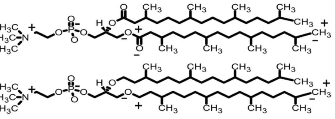
Figure 1. Top shows DPhPC (or ester-PHPC) with the main partial charges contributing to the dipole potential are shown as
+
and-
on one chain of each lipid, Bottom shows ether-DPhPC where the ester group of DPhPC has been replaced by the ether group. Adapted from ref 34.
Figure 2(a) left and 2(b) right: left DPhPC (ester-DPhPC): DM 22.6D; black arrow showing dipole moment direction for DPhPC and phosphatidylcholine headgroup, red arrow showing dipole moment direction for ester group, and white arrow showing dipole potential direction for
the DPhPC membrane with the potential being largest at the centre of the lipid bilayer of the membrane. Right ether-DPhPC 26.2DM black arrow showing dipole moment direction for DPhPC and phosphatidylcholine headgroup, and white arrow showing dipole potential direction for the ether-DPhPC membrane with the dipole potential being largest at the centre of the lipid bilayer of the membrane.
Figure 3(a) left and 3(b) right: left DPhPC-Mannitol complex 25.9D, and right DPhPC-BCNU complex 21.3D, both drugs H bonded to the O atom of the phosphate P-O group of DPhPC
Figure 4(a) left and 4(b) right: left DPhPC-Maanitol complex 23.2D, and right DPhPC-BCNU complex 23.7D, both drugs H bonded to the O atom of the ester C=O group of DPhPC