© The Author 2012. Published by Oxford University Press. All rights reserved. For permissions, please e-mail: journals.permissions@oup.com
Characterization and Turnover of CD73/IP
3R3-positive Microvillar Cells in
the Adult Mouse Olfactory Epithelium
Sandra Pfister
1,2,*, Maren G. Dietrich
1,3,*, Corinne Sidler
1, Jean-Marc Fritschy
1, Irene Knuesel
1and Rebecca Elsaesser
1,41
Institute of Pharmacology and Toxicology, University of Zurich, Winterthurerstrasse
190, CH-8057 Zurich, Switzerland and
2Neuroscience Center Zurich, University of Zurich,
Winterthurerstrasse 190, CH-8057 Zurich, Switzerland
3Present address: Division of
Endocrinology, Diabetes and Clinical Nutrition, University Hospital Zurich, Raemistrasse 100,
CH-8091 Zurich, Switzerland
4Present address: Carl Zeiss Microscopy GmbH, Carl Zeiss Gruppe,
Carl-Zeiss-Promenade 10, 07745 Jena, Germany
Correspondence to be sent to: Irene Knuesel, Institute of Pharmacology and Toxicology, University of Zurich, Winterthurerstrasse 190, CH-8057 Zurich, Switzerland. e-mail: knuesel@pharma.uzh.ch
*These authors contributed equally to the work.
Accepted June 22, 2012
Abstract
The main olfactory epithelium consists of 4 major cell types: sensory neurons, supporting cells, microvillar cells, and basal progenitor cells. Several populations of microvillar olfactory cells have been described, whose properties are not yet fully understood. In this study, we aimed to clarify the classification of microvillar cells by introducing a specific marker, CD73. Furthermore, we investigated the turnover of CD73-microvillar cells during adult life. Using direct and indirect immunofluo-rescence in adult main olfactory epithelium, we first demonstrate that ecto-5′-nucleotidase (CD73) is a reliable marker for microvillar cells reported previously to express phospholipase C β2 (PLC β2) along with type 3 IP3 receptors (IP3R3) and
tran-sient receptor potential channels 6 (TRPC6), as well as for cells labeled by transgenic expression of tauGFP driven by the IP3R3
promoter. The ubiquitous CD73 immunoreactivity in the microvilli of these 2 cell populations indicates that they correspond to the same cell type (CD73-microvillar cell), endowed with a signal transduction cascade mobilizing Ca++
from intracellular stores. These microvillar cells respond to odors, possess a basal process, and do not degenerate after bulbectomy, suggesting that they contribute to cellular homeostasis in the olfactory epithelium. Next, we examined whether CD73-microvillar cells undergo turnover in the adult olfactory epithelium. By combining CD73 immunofluorescence and BrdU pulse labeling, we show delayed BrdU incorporation in a small fraction of CD73-positive microvillar cells, which persists for several weeks after BrdU administration. These findings indicate that CD73-microvillar cells likely differentiate from proliferating progenitor cells and have a slow turnover despite their apical position in the olfactory epithelium. These combined properties are unique among olfactory cells, in line with the possibility that they might regulate cellular homeostasis driven by extracellular ATP and adenosine.
Key words: BrdU, CD73, neurogenesis, neuronal homeostasis, olfaction
Introduction
The postnatal olfactory epithelium (OE) is a pseudostrati-fied epithelium containing several distinct cell types, notably olfactory sensory neurons, supporting cells, microvillar cells (MVCs), as well as 2 types of progenitor cells (horizontal and globose basal cells) and Bowman’s gland and duct cells. The nasal cavity is directly exposed to air-borne contaminants;
as a consequence, cells in the nasal epithelia are prone to damage. Therefore, olfactory cells typically have a short lifespan and get replaced from progenitor cells throughout adult life (Graziadei et al. 1978; Schwob 2002; Gulbransen
and Finger 2005). In the OE, the basal germinal zone, from
which regeneration occurs, contains 2 cell populations with stem cell characteristics, the globose (GBCs) and horizontal
basal cells (HBCs) (Mackay-Sim and Kittel 1991; Huard
et al. 1998). GBCs are multipotent and have the highest
pro-liferation rate in the OE (Caggiano et al. 1994; Goldstein et al. 1998; Jang et al. 2003; Chen et al. 2004). HBCs are postulated to represent relatively quiescent, multipotent progenitors; extensive injury of the neuroepithelium can induce proliferation of HBCs to replenish the pools of neu-ronal and nonneuneu-ronal cells (Carter et al. 2004; Leung et al. 2007; Iwai et al. 2008; Packard et al. 2011). Interestingly, the supporting cells arranged in a single layer at the apical sur-face possess the capability to self-renew in the unperturbed OE. Nevertheless, after massive damage to the OE they get replaced from progenitor cells, as well (Schwob et al. 1995;
Weiler and Farbman 1998). The continuous turnover of
olfactory neurons and their replenishment after damage is critical to maintain the functional integrity of the OE.
In this process, the role and the fate of MVCs are not well understood. Part of the difficulty arises from their incomplete morphological and functional characteriza-tion. Although MVCs are clearly distinct from supporting cells, which also possess microvilli, the nomenclature of the various subpopulations described in rodent and human OE (Moran et al. 1982a, 1982b; Rowley et al. 1989; Carr
et al. 1991; Menco and Jackson 1997a, 1997b; Braun and
Zimmermann 1998; Asan and Drenckhahn 2005; Elsaesser
et al. 2005; Hansen and Finger, 2008; Lin et al. 2008; Hegg
et al. 2010) is controversial. In previous work, we have
described 1 subpopulation of MVCs, endowed with an inositol-triphosphate (IP3)-mediated signal transduction cascade, including phospholipase C beta 2 (PLC β2), type-3 IP3 receptors (IP3R3), and transient receptor potential chan-nels 6 (TRPC6) (Elsaesser et al. 2005; Montani et al. 2006). These flask-shaped cells, representing about 5% of all olfac-tory cells, are evenly distributed throughout the OE and are situated at the most superficial layer intermingled with sup-porting cells. They most likely correspond Jourdan’s “type B cells” (Jourdan 1975), as well as MVCs described by Moran
et al. (1982a) and microvillous cells type 2/4 (Menco and
Jackson 1997a, 1997b; Menco and Morrison 2003).
PLC β2-MVCs are excitable and convert extracellular signals in a Ca++ response (Elsaesser et al. 2005; Montani
et al. 2006). Interestingly, they selectively express the neu-roproliferative factor neuropeptide Y (NPY), suggesting a role in the control of neural proliferation in the postna-tal OE by stimulus-induced release of NPY (Hansel et al. 2001; Montani et al. 2006; Jia and Hegg, 2012). In particu-lar, adenosine-5′-triphosphate (ATP) has been shown to be such a stimulus, as the release of NPY is triggered by ATP (Kanekar et al. 2009; Jia et al. 2011) and ATP increases the expression of NPY in MVCs in vivo (Jia and Hegg, 2010).
However, Hegg et al. (2010) recently reported that MVCs expressing tauGFP+ under transgenic control of the IP
3R3 gene promoter were immunonegative for PLC β2, despite many common features with the MVCs described in our studies. Therefore, 1 goal of this study was to clarify the
relationship between PLC β2-MVCs and IP3R3-MVCs. To this end, we investigated ecto-5′-nucleotidase (CD73) as a possible marker of both cell populations in the mouse OE. CD73 is a glycosyl phosphatidyl inositol (GPI-) linked, mem-brane bound glycoprotein that catalyzes the extracellular hydrolysis of 5′-AMP to adenosine, which may subsequently activate adenosine receptors (Zimmermann 1992) or be recy-cled after intracellular transport. Thereby, CD73 is involved in various physiological processes mediated by adenosine including hypoxia, inflammation, antinociception, epithelial ion transport, and modulation of blood–brain barrier func-tions (Koszalka et al. 2004; Thompson et al. 2004; Colgan et al. 2006; Mills et al. 2008; Niemelä et al. 2008; Sowa et al. 2010a, 2010b). CD73 has been reported in the rat OE to label dark/horizontal cells at the basal side, MVCs at the lumi-nal side, and Bowman’s duct cells (Braun and Zimmermann, 1998). Here, we used direct and indirect immunofluorescence for CD73 to demonstrate unambiguously that this marker is expressed in PLC-β2/IP3R3-MVCs of the adult mouse OE, thereby establishing CD73 as a useful tool to study this major population of MVCs.
As a first step toward their functional characterization, we addressed important open issues in the field of OE regeneration, namely whether CD73-MVCs get replaced in the adult animal, at which rate, and from which progeni-tor cells. To answer these questions, we moniprogeni-tored the fate of CD73-MVCs pulse-labeled with the thymidine analog 5-bromo-2′-deoxyuridine (BrdU) combined with immuno-fluorescence staining for CD73.
Material and methods
Animals
IP3R3
tm(tauGFP) transgenic mice in which the first exon of the
Itpr3 gene is replaced by the coding region of the fusion
pro-tein tau-eGFP (Hegg et al. 2010) were kindly provided by Dr Diego Restrepo (University of Colorado Denver, CO). Adult male IP3R3
+/IP 3R3
− tauGFP+ mice were used for immunohistochemistry. Additionally, immunohistochem-istry was performed in tissue from adult (6–8 week old) male C57BL/6J wild-type mice bred in our Institute. All experimental procedures were approved by the Cantonal Veterinary Office in Zurich. The mice were housed 3–6 ani-mals per cage in a 12-h light/dark cycle with food and water provided ad libitum.
Mouse olfactory epithelium tissue preparation
Mice were given a single intraperitoneal injection of 180 mg/ kg BrdU (Sigma-Aldrich, Switzerland, #B5002) dissolved in 0.9% NaCl with an injection volume of 6.6 mL/kg body weight. At various time spans after BrdU injection (1, 3, 5, 7, 10, 14, 21, 28, and 42 days), mice were deeply anesthetized with Nembutal (50 mg/kg, i.p.) and perfused transcardially
with phosphate buffered saline (PBS) followed by aldehyde fixation (4% paraformaldehyde, 15% saturated picric acid, 150 mM sodium phosphate buffer, pH 7.4). Thereafter, the noses were rapidly dissected and stored for 2 h in fixative at 4 °C. After washing, they were decalcified in 5% ethyl-enediamine tetraacetic acid (EDTA, pH 7.4) for 7 days at 4 °C. Next, the specimens were cryoprotected overnight in 30% sucrose in PBS and subsequently frozen and stored at –80 °C. The specimens were embedded with Neg-50 Frozen Section Medium (Richard-Allan Scientific, MI) and coro-nal sections of 20 µm thickness were cut and mounted on gelatine-coated slides.
Whole-mount preparation: Animals were decapitated and
the OE was dissected out and transferred into fixative. The tissue was fixed for 1 h at 4 °C. After washing, the tissue was processed for immunohistochemistry.
DNA denaturation for BrdU staining
The sections were air-dried before being washed in PBS and incubated in 0.2 N HCl at room temperature (RT) for 5 min. Thereafter, they were transferred into 4 N HCl and incubated at 37 °C for 30 min. After denaturation, the sections were washed 4 times in PBS and processed for immunohistochemistry.
Immunofluorescence staining
The primary antibodies used are listed in Table 1; they were diluted in PBS containing 5% normal serum and 0.2% Triton X-100. Thereafter, sections were incubated overnight at 4 °C, washed 3 times in PBS for 10 min and incubated for 45 min at RT with secondary antibodies raised in goat or donkey and conjugated to Alexa488 (Molecular Probes) or Cy3 (Jackson Immunoresearch, West Grove, PA). They were diluted in PBS containing 5% normal serum (Alexa488 1:1000; Cy3 1:500; Cy5 1:500). Direct immunofluorescence staining of CD73 was performed with Cy3-conjugated CD73 anti-bodies (1:1500). The sections were air-dried and coverslipped using DAKO fluorescence mounting medium (Dako North America, CA).
In order to detect CD73 and BrdU simultaneously, the immunofluorescence staining protocol was slightly adjusted. To this end, sections were incubated overnight at RT with rat-anti-CD73 antibody (1:1000) containing 5% normal serum and 0.2% Triton X-100. Following three 10-min washing steps, sections were incubated for 30 min at RT with biotinylated secondary antibody (1:500) diluted in PBS con-taining 5% normal serum and 0.02% Triton X-100. After washing, the slides were transferred into fixative for 12 min at 4 °C. This was followed by washing in PBS three times for 10 min each. DNA denaturation was applied by incu-bating the sections firstly for 5 min in 0.2 N HCl at RT and subsequently for 30 min in 4 N HCl at 37 °C. Next, sections were washed 3 times in PBS before incubating for 20 h at RT with FITC-conjugated anti-BrdU antibody (1:100) in PBS containing 5% NGS and 0.02% Triton X-100. Slides were rinsed in PBS and transferred into Cy3-conjugated strepta-vidin solution and incubated for 30 min at RT. Thereafter, they were washed, air-dried, and coverslipped with DAKO mounting medium.
Image processing and analysis
Sections processed for immunofluorescence were analyzed by confocal microscopy (LSM-710, Zeiss, Jena, Germany) using 40× (NA 1.3) or 63× (NA 1.4) and sequential acquisi-tion of separate channels. Z-stacks of consecutive secacquisi-tions (5–8; 1024 × 1024 pixel; spaced 1 µm in z) were acquired with the pinhole set at 1 Airy unit. For visual display, image stacks were projected in the z-dimension and merged using the image analysis software Imaris (Bitplane, Zurich, Switzerland).
Three animals per time point post-BrdU injection were used to quantify the number of CD73-MVCs and BrdU-positive CD73-MVCs, respectively. Random sampling fields containing CD73-immunoreactive cells were selected at 2 anteroposterior levels of the main sensory OE. The first level contained all ethmoid turbinates and the second level composed the ethmoid turbinates located below the rostral tip of the olfactory bulb. Four sampling fields in each area were acquired using a 40× objective (N.A., 1.2). Stacks of Table 1 List of antibodies
Antibody Specificity Manufacturer Description/Nr Dilution
ACIII Wei et al. 1998; Wong et al. 2000 Santa Cruz Biotechnology, Inc. Rabbit polyclonal; #sc-588 1:1000 BrdU: FITC Vanderlaan and Thomas, 1985; Kondo
et al. 2010 AbD Serotec, Oxford Rat monoclonal, IgG2a conjugated to FITC-liquid; #OBT0030F 1:100 CD73 Yamashita et al. 1998; Eliopoulos et al.
2005; Kobie et al. 2006 eBioscience, Inc.; San Diego Rat monoclonal, IgG1; #16-0731 1:1000/1:1500 IP3R3 Blondel et al. 1993; Leite et al. 2003 BD Bioscience Mouse monoclonal, IgG2a, #610312 1:500
OMP Baker et al. 1989; Cummings et al. 2000 Wako, Chemicals, Richmond Goat polyclonal, #544-10001 1:500 PLC β2 Ali et al. 1997; Perez et al. 2002 Santa Cruz Biotechnology, Inc. Rabbit polyclonal, Q-15, #sc-206 1:500
11–12 confocal layers were projected in 2D and used for cell counts. Cells were only considered as double-labeled if the BrdU+ nucleus was clearly visible.
Statistical analysis was performed by Kruskal–Wallis test using GraphPad Prism (GraphPad Software, Inc. Version 4.01). Statistical significance was set at P < 0.05.
Results
CD73 is a selective marker for microvillar cells in the adult mouse olfactory epithelium
In previous studies, we investigated PLC β2-MVCs as a major microvilli-bearing cell type in the OE characterized by expression of a phosphatidyl inositol-mediated signal transduction pathway and postulated their involvement in the control of neuronal proliferation in the postnatal OE
(Elsaesser et al. 2005). Here, we detected the
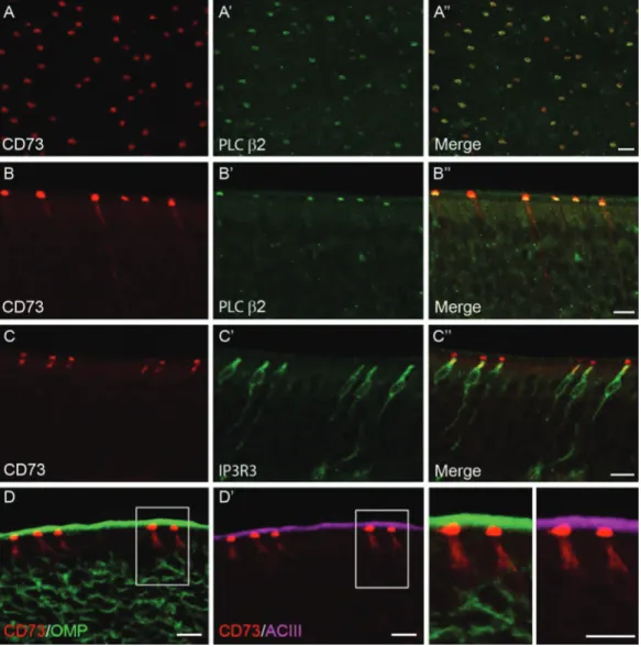
membrane-bound CD73 (ecto-5′-nucleotidase) as an unequivocal marker for PLC β2-MVCs. Using anti-CD73 antibodies in whole-mount specimen of adult mice revealed immu-noreactive cells that were evenly scattered throughout the sensory OE (Figure 1A). In double-labeling experi-ments with CD73 and PLC β2, CD73-immunoreactivity was colocalized with microvilli of PLC β2-positive cells
(Figure 1A). Staining of transverse sections of the OE
demonstrated that anti-CD73 antibodies labeled cells that strongly resembled PLC β2-MVCs (Elsaesser et al. 2005) based on their density, shape, and localization (Figure 1
B–D). The selectivity of CD73 as a marker for PLC β2-MVCs was confirmed by double immunofluorescence staining. These results show that these 2 proteins were extensively co-associated within the same cells throughout the OE (Figure 1A,1B), with 304 out of 330 cells (92%) from 5 animals being double-labeled for CD73 and PLC β2. Similar results were obtained for IP3R3 immunoreac-tivity. In wild-type mice, IP3R3 and CD73 were systemati-cally codetected in the same cells (Figure 1C). To confirm that CD73 is not expressed in olfactory neurons, parallel triple-labeling experiments were performed with olfactory marker protein (OMP) and adenylyl cyclase III (ACIII). As seen as high magnification, CD73 was never colocalized with either OMP or ACIII at the apical pole (Figure 1D), but was located underneath the layer of ACIII-positive cilia (Figure 1D′). In addition, as reported by Braun and
Zimmermann (1998) in the rat OE, CD73 may also be
expressed by Bowman’s duct/gland cells and dark/hori-zontal basal cells. However, we could not confirm these observations in adult mouse OE, due to nonspecific bind-ing of anti-rat IgGs (secondary antibodies) in the basal lamina and in the lamina propria. Moreover, using direct immunofluorescence for CD73, no staining was detectable in these structures. In conclusion, our data indicates that monoclonal anti-CD73 antibodies can be used as a reliable marker for PLC β2-MVCs.
IP3R3-eGFP microvillar cells are immunoreactive for CD73
Recently, Hegg et al. (2010) described MVCs expressing IP3R3 using a transgenic mouse strain. This cell type has several features in common with PLC β2-MVCs. Both of them bear microvilli at their apical protrusions, they possess axon-like processes that do not penetrate the basal lamina, and they do not degenerate after bulbectomy. Furthermore, they respond to odors with an increase of intracellular Ca2+ and do not express neuronal markers (Elsaesser et al. 2005;
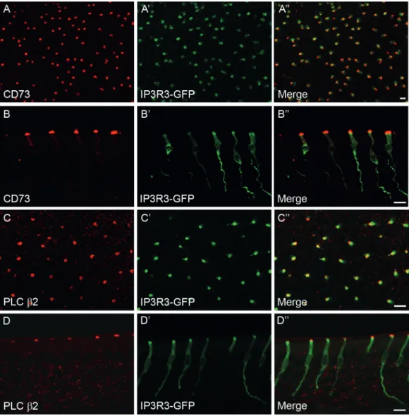
Hegg et al. 2010). In order to clarify whether they represent the same cell type, we examined CD73 immunoreactivity in IP3R3
+/IP 3R3
− tauGFP mice. Whole-mount preparations revealed that every IP3R3-tauGFP-positive cell in adult OE was capped by CD73 immunostaining (Figure 2A). Likewise, in transversally cut sections we detected that CD73-immunofluorescence was selectively co-associated with IP3R3-tauGFP-positive cells (Figure 2B). In a sam-ple of 400 randomly selected CD73-immunoreactive cells in whole mount and tissue sections from 4 adult mice, 98% were GFP-positive, suggesting systematic association of these 2 markers. Furthermore, we used PLC β2 anti-bodies to test the distribution of a marker of PLC β2-MVCs in IP3R3-tauGFP-positive cells. As expected, PLC β2 immu-noreactivity was present in virtually all tauGFP-positive cells in whole-mount preparation and in tissue sections of 3 animals (97% out of 300 GFP-positive cells; Figure 2C,2D). Therefore, these results indicate that these 2 populations of MVCs (PLC β2-MVCs and IP3R3-MVCs) correspond to the same cell type, and can be termed CD73-MVCs.
Microvillar cell turnover
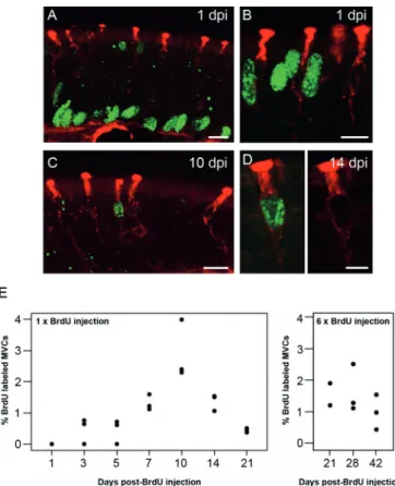
There are many open questions about the role and regula-tion of MVCs in the postnatal OE. In particular, it is not known whether differentiated MVCs undergo cell division and thereby self-renew or whether they originate from a population of olfactory progenitor cells in adult life. Here, we addressed these issues for CD73-MVCs, using an immu-nohistochemical protocol allowing the simultaneous detec-tion of CD73 and BrdU in the same tissue secdetec-tion. Mice were injected once with BrdU and the proportion of BrdU-labeled CD73-MVCs in the olfactory sensory epithelium was assessed at 1–21 days postinjection (dpi). We analyzed 3 mice per time point and counted CD73-MVCs cells bilat-erally in 4 sampling areas on 2 different sections resulting in 16 fields of view. As expected, many cells were immunoreac-tive for either CD73 or BrdU and only a small percentage was double-labeled. At 1 dpi, BrdU immunoreactivity was mainly visible in the basal germinative zone. The majority of BrdU-positive cells in this region likely constitute the pro-genitor cell population (Figure 3A). Nevertheless, there were also a few BrdU-immunoreactive cells detectable at the most superficial layer of the epithelium (Figure 3B). However, there were no BrdU/CD73 double-labeled cells detectable at 1 dpi (Figure 3B). The first BrdU-positive CD73-MVCs were
observed at 3 dpi. Thus, these cells seem to require 3 days between S-phase and the earliest detectable expression of CD73. Double-labeled cells were observed more frequently at 5, 7, and 10 dpi (Figure 3C,3D), clearly indicating that CD73-MVCs get replaced in the adult animal. At 10 dpi, the fraction of BrdU-positive CD73-MVCs was maximal with a mean value of 2.9% CD73-MVCs being double-labeled at this time point. Later on, the number of BrdU-labeled CD73-MVCs decreased to 0.5% at 21 dpi. These values were obtained from a total number of 583 CD73-positive cells at 10 dpi and 675 at 21 dpi. In line with this time-course, Kruskal–Wallis analysis yielded a significant overall time effect on the proportion of BrdU-positive CD73-MVCs (N = 3 per time point; H = 17.126; P = 0.008; Figure 3E).
To extent our observations to later time points (21, 28, and 42 dpi), we treated a separate cohort of mice with 6 BrdU injections (twice daily for 3 days). Under these conditions, BrdU-positive CD73-MVCs remained detectable even at 42 dpi, but without a significant overall time effect (Kruskal– Wallis; N = 2–3 per time point, H = 2.889, P = 0.2359; Figure 3E). This finding, together with the observation that a rela-tively low percentage of CD73-MVCs was BrdU-positive at any time point, indicated that MVCs represent a stable cell population with a long lifespan. In line with this con-clusion, quantification of the total number of CD73-MVCs revealed no significant differences in the 2 cohorts of mice analyzed (1 BrdU injection: H = 10.939; P = 0.090; 6 BrdU injections, N = 2–3 per time point, H = 5.556, P = 0.0662). Figure 1 CD73 as a marker of a major MVCs population in the olfactory epithelium. (A) Double immunofluorescence staining on whole-mount tissue
preparations using anti-CD73 antibody (red, left panel) and anti-PLC β2 antibody (green, middle panel, A′) revealed co-association of these 2 proteins in microvilli (merged, Aʺ). (B) Double immunofluorescence labeling against CD73 (red, left) and PLC β2 (green, B′) on coronal sections stained the same cells (merged, Bʺ). (C) Using an anti-IP3R3 antibody (green, middle panel, C′) stained cells that were co-associated with CD73 (red, left). The merged image is
shown on the right (Cʺ). (D) Triple-immunofluorescence staining on coronal sections using CD73 antibody (red, left and middle panel), OMP anti-body (green, left), and anti-ACIII antianti-body (magenta, middle). The OMP-immunoreactive receptor neurons were not co-associated with CD73-positive cells (D) nor did ACIII overlap with CD73 on cilia (D′). The boxed areas are enlarged on the right. Scale bars: 10 µm.
After 1 BrdU injection, we counted on average 240 ± 64 CD73-MVCs per mouse (mean ± SEM; N = 3), represent-ing 18 ± 2.8 CD73-MVCs per samplrepresent-ing field. These values are similar to those counted in mice examined after 6 BrdU injections (236 ± 38; 17.3 ± 1.8, respectively).
Discussion
The present results clarify the nomenclature of MVCs in the adult mouse OE by demonstrating that PLC β2-MVCs (Elsaesser et al. 2005; Montani et al. 2006) and IP3R3-MVCs (Hegg et al. 2010) represent the same cell type, character-ized in addition by prominent expression of CD73 at their apical pole. Accordingly, the failure to detect PLC-β2 in these MVCs in previous studies was likely due to techni-cal reasons, such as high sensitivity of antibodies to tissue
fixation. This cell population, which we propose to name CD73-MVCs, is endowed with multiple elements of the IP3-mediated signal transduction cascade, including IP3R3 and PLC β2. Furthermore, CD73-MVCs most likely also express Gq/11 and TRPC6, because Gq/11 has been reported in IP3R3-MVCs and TRPC6 in PLC β2-MVCs (Elsaesser
et al. 2005; Hegg et al. 2010). Conversely, it is highly unlikely that CD73-MVCs correspond to TrpM5-positive MVCs described by Hansen and Finger (2008) and Lin et al. (2008), because the latter cells have a clearly different morphology and do not express PLC β2, TRPC6, or IP3R3. Therefore, in adult mouse OE, there are at least 2 main, distinct types of MVCs. Further studies will help elucidating their specific role and determine possible species differences. In particu-lar, the relevance of CD73 for the function of MVCs will be an important topic of future investigations. There is an Figure 2 Identification of IP3R3-GFP-positive CD73-MVCs. Immunofluorescence staining was performed on tissue sections from IP3R3
+
/IP3R3 −
tauGFP+
mice. (A,B) CD73-immunoreactivity (red, left) is detected in IP3R3GFP-positive cells (green, A′,B′) as shown in whole-mount tissue preparations (A) and
coronal sections of the olfactory epithelium (B). (C,D) Immunofluorescence staining using anti-PLC β2 (red, left) antibody revealed its co-association with IP3R3-GFP MVCs (green, C’, D’) in whole-mount tissue (C) and coronal sections (D). Scale bars: A = 20 µm, B–D = 10 µm.
types of somatic cells that are exposed to noxious stimuli undergo a lifelong cycle of cell death and replacement. In the OE, the germinative zone lies adjacent to the basal lam-ina and contains multipotent cells. Olfactory cells, including sensory neurons, are continuously restored throughout life. This study showed that also CD73-MVCs undergo a turn-over. We detected the earliest BrdU-labeled CD73-MVCs at 3 dpi and observed a peak of newly generated CD73-MVCs at 10 dpi. This temporal profile is very similar to the matur-ation of other sensory cells. For example, olfactory sensory neurons need approximately 1 week to mature. BrdU-OMP double-labeled cells were reported to appear 7 days after BrdU injection (Miragall and Monti Graziadei 1982; Schwob et al. 1992; Kondo et al. 2010). Solitary chemosensory cells lying in the nonolfactory nasal epithelium express elements of their transduction cascade, α-gustducin, within 3 days after completing mitosis (Gulbransen and Finger 2005). In Type II taste vallate bud cells, α-gustducin and BrdU were coex-pressed starting from 2.5 dpi and reaching a peak at 6.5 dpi (Farbman 1980; Cho et al. 1998). The signaling molecule for taste transduction, PLC β2, has been shown to be expressed in taste bud cells from day 5 and reached maximum at day
12 (Hamamichi et al. 2006). The timing of CD73 expression
in CD73-MVCs after 3 days exiting S-phase indicates that MVCs might mature at a rate similar to other sensory cells.
The majority of epithelial cells is generated from a popu-lation of undifferentiated progenitor cells (Chen et al. 2004). These progenitor cells have the capacity to replace cells and maintain the epithelial homeostasis. Olfactory neurons are among the best-studied example of how regeneration from stem cells occurs. The cell lineage relationship has been extensively described; starting from undifferentiated pre-cursor cell types to mature neurons. Evidence indicates that olfactory neurons are generated by GBCs, at least in undam-aged tissue (Caggiano et al. 1994; Huard et al. 1998). As 1 exception, supporting cells have the capacity to proliferate and can replace themselves (Graziadei et al. 1979; Weiler and
Farbman 1998). Although self-renewal of CD73-MVCs
can-not be excluded at this stage, it would imply that both daugh-ter cells do not express detectable CD73 during the first days after exciting the S-phase, which appears unlikely.
Therefore, we suggest that CD73-MVCs do not divide after differentiation, but rather derive from a population of olfactory progenitor cells. The identity of this progeni-tor cell remains elusive. CD73-MVCs might originate from GBCs, like olfactory neurons. Other possible candidates are the HBCs, in keeping with the relatively rare occur-rence of BrdU-labeled CD73-MVCs. As a third possibility, Bowman’s gland/duct cells need to be considered, because Bowman’s gland/duct cells have the capacity to replenish supporting cells (Huard et al. 1998) and have been reported to express CD73 in the OE of young rats (Braun and
Zimmermann 1998). However, working with mouse tissue,
we could not confirm the presence of CD73 in cells of the Bowman’s gland/duct and we obtained no clear evidence for increasing evidence for purinergic signaling having a
promi-nent role in contributing to the control of progenitor cell proliferation in the adult OE (Hassenklöver et al. 2009; Jia et al. 2009; Jia and Hegg, 2010; Jia et al. 2011; Jia and Hegg 2012). CD73 catalyzes the hydrolysis of AMP to adeno-sine and, therefore, might contribute to rapid termination of purinergic signaling, notably on release of ATP from degenerating cells.
Adult stem cells are present in many tissues and have the ability to replace the majority of somatic cells. Several Figure 3 BrdU pulse-chase labeling combined with
immunofluores-cence staining for CD73 provides evidence for generation of adult-born CD73-MVCs. (A–D) Double immunofluorescence staining on coronal sec-tions using anti-CD73 antibody (red) and anti-BrdU antibody (green). (A) At 1 dpi, most of the BrdU-positive nuclei are present at the basal part of the olfactory epithelium. (B) BrdU-positive nuclei (green) were detected at the apical side of the epithelium at 1 dpi, but were not present in CD73-MVCs (red, apical part). (C,D) BrdU-labeled CD73-MVCs are detectable at 10 dpi (C) and 14 dpi (D). (E) Fraction of MVCs double-labeled for BrdU and CD73 at different time points after BrdU injection. Each dot represents the median value from an individual mouse. The left graph represents the data after a single BrdU injection, and the right graph after multiple BrdU injec-tions (see main text). In both experiments, there was no difference in the total number of CD73-MVCs (1× BrdU: median, 25th–75th percentile: 258, 198–297; 6× BrdU: 217, 164–273) counted at the various time points. The first double-labeled cells appeared at 3 dpi. The percentage of BrdU-labeled CD73-MVCs gradually increased, peaked at 10 dpi and thereafter declined. Kruskal–Wallis test confirmed a statistical effect of time (N = 3 for time point; H = 17.126; P = 0.008). After 1 BrdU injection, 240 ± 64 CD73-MVCs were counted on average per mouse and after 6 BrdU injections 236 ± 38, respectively. Scale bars: A–C = 10 µm, D = 5 µm.
BrdU-labeling in this organ, precluding a definitive conclu-sion about this possibility.
The proportion of BrdU-labeled CD73-MVCs reaches a maximum at 10 dpi and declines thereafter. This time course indicates either that some newly generated CD73-MVCs fail to complete differentiation and degenerate, or that some of their progenitors undergo several cycles of divi-sion prior to becoming postmitotic and differentiate. The first possibility would be in line with previous reports on the lifespan of newborn cells. For instance, roughly half of newly generated taste bud cells are eliminated within the first few days after birth (Hamamichi et al. 2006). The number of newly generated olfactory sensory neurons that express OMP increases gradually from 7 to 14 days and thereafter starts to decrease. Moreover, a large fraction of BrdU-positive cells underneath the OMP-positive cell layer dies within 14 days after birth (Kondo et al. 2010). In analogy to sensory neurons, which depend on signals from the olfactory bulb and from locally released factors within the OE (Mackay-Sim and Kittel 1991; Schwob et al.
1992; Carr and Farbman 1993; Kondo et al. 2010), newly
generated CD73-MVCs might need to receive trophic sup-port for their survival or be vulnerable to exposure to noxious stimuli.
It is the current understanding that the degree of exposure of a cell type to noxious stimuli is loosely correlated with its turnover; for example, airway epithelial cells lining the trache-obronchial tree have a significant longer turnover than cells in the nasal cavity (Basbaum and Jany 1990). Consequently, solitary chemosensory cells, located in a remarkably vulner-able location at the anterior end of the nasal cavity, have an estimated turnover of only 20 days (Gulbransen and Finger 2005). Likewise, olfactory sensory neurons in a slightly more protected position at the posterior part of the nasal cavity have a lifespan of 1–2 months (Moulton 1974; Graziadei and
Graziadei 1979; Graziadei et al. 1979; Miragall and Monti
Graziadei 1982; Mackay-Sim and Kittel 1991; Schwob et al.
1992; Schwob 2002). Exceptions are the olfactory supporting cells, involved in forming the apical layer of the OE, which are considered to have a long lifespan and be replaced at a low rate (Naguro and Iwashita 1992; Weiler and Farbman 1998). Because CD73-MVCs are intermingled with the sup-porting cells at the most superficial layer in close contact with the incoming airstream, they are prone to cellular dam-age, pointing to a short turnover. However, we detected only few double-labeled cells indicating that CD73-MVCs, like their neighboring supporting cells, have a slow turnover and a long lifespan. Indeed, even at 42 dpi, we still were able to detect BrdU-labeled CD73-MVCs in the OE.
In conclusion, in this study, we classified a major popu-lation of MVCs by demonstrating CD73 expression in PLC β2-MVCs and IP3R3-MVCs, which suggests that these 2 MVC types correspond to 1 population, termed CD73-MVCs. In addition, our results showed that CD73-MVCs undergo turnover and are, therefore, similar to
the majority of somatic cells that can be replaced through-out life, most likely to protect the organism against damage. Because of the postulated involvement of MVCs in olfactory neurogenesis, it will be essential to further investigate their function and their own regulation for a better understanding of the molecular and cellular pathways underlying progeni-tor cell proliferation and differentiation.
Funding
The work was supported by the Swiss National Science Foundation [National Research Program NRP63, 406340-128112 to R.E. and I.K.] and by the ‘Forschungskredit’ of the University of Zurich to R.E.
Acknowledgements
The authors thank all collaborators of the Animal Services of the Institute of Pharmacology and Toxicology for animal husbandry and care, and Dr Wolfgang Härtig (University of Leipzig) for CD73 antibody conjugation to Cy3.
References
Ali H, Fisher I, Haribabu B, Richardson RM, Snyderman R (1997) Role of phospholipase Cβ3 phosphorylation in the desensitization of cellular responses to platelet-activating factor. J Biol Chem 272:11706–11709. Asan E, Drenckhahn D. 2005. Immunocytochemical characterization of two
types of microvillar cells in rodent olfactory epithelium. Histochem Cell Biol. 123(2):157–168.
Baker H, Grillo M, Margolis FL (1989) Biochemical and immunocytochemi-cal characterization of olfactory marker protein in the rodent central nervous system. J Comp Neurol 285:246–261.
Basbaum C, Jany B. 1990. Plasticity in the airway epithelium. Am J Physiol. 259(2 Pt 1):L38–L46.
Blondel O, Takeda J, Janssen H, Seino S, Bell GI (1993) Sequence and functional characterization of a third inositol trisphosphate receptor subtype, IP3R-3, expressed in pancreatic islets, kidney, gastrointestinal tract, and other tissues. J Biol Chem 268:11356–11363.
Braun N, Zimmermann H. 1998. Association of ecto-5′-nucleotidase with specific cell types in the adult and developing rat olfactory organ. J Comp Neurol. 393(4):528–537.
Caggiano M, Kauer JS, Hunter DD. 1994. Globose basal cells are neu-ronal progenitors in the olfactory epithelium: a lineage analysis using a replication-incompetent retrovirus. Neuron. 13(2):339–352.
Carr VM, Farbman AI. 1993. The dynamics of cell death in the olfactory epithelium. Exp Neurol. 124(2):308–314.
Carr VM, Farbman AI, Colletti LM, Morgan JI. 1991. Identification of a new non-neuronal cell type in rat olfactory epithelium. Neuroscience. 45(2):433–449.
Carter LA, MacDonald JL, Roskams AJ. 2004. Olfactory horizontal basal cells demonstrate a conserved multipotent progenitor phenotype. J Neurosci. 24(25):5670–5683.
Chen X, Fang H, Schwob JE. 2004. Multipotency of purified, trans-planted globose basal cells in olfactory epithelium. J Comp Neurol. 469(4):457–474.
Cho YK, Farbman AI, Smith DV. 1998. The timing of alpha-gustducin expression during cell renewal in rat vallate taste buds. Chem Senses. 23(6):735–742.
Colgan SP, Eltzschig HK, Eckle T, Thompson LF. 2006. Physiological roles for ecto-5′-nucleotidase (CD73). Purinergic Signal. 2(2):351–360.
Cummings DM, Emge DK, Small SL, Margolis FL (2000) Pattern of olfactory bulb innervation returns after recovery from reversible peripheral deaf-ferentation. J Comp Neurol 421:362–373.
Eliopoulos N, Stagg J, Lejeune L, Pommey S, Galipeau J (2005) Allogeneic marrow stromal cells are immune rejected by MHC class I- and class II-mismatched recipient mice. Blood 106:4057–4065.
Elsaesser R, Montani G, Tirindelli R, Paysan J. 2005. Phosphatidyl-inositide signalling proteins in a novel class of sensory cells in the mammalian olfactory epithelium. Eur J Neurosci. 21(10):2692–2700.
Farbman AI. 1980. Renewal of taste bud cells in rat circumvallate papillae. Cell Tissue Kinet. 13(4):349–357.
Goldstein BJ, Fang H, Youngentob SL, Schwob JE. 1998. Transplantation of multipotent progenitors from the adult olfactory epithelium. Neuroreport. 9(7):1611–1617.
Graziadei PP, Graziadei GA. 1979. Neurogenesis and neuron regeneration in the olfactory system of mammals. I. Morphological aspects of differ-entiation and structural organization of the olfactory sensory neurons. J Neurocytol. 8(1):1–18.
Graziadei PP, Levine RR, Graziadei GA. 1978. Regeneration of olfactory axons and synapse formation in the forebrain after bulbectomy in neo-natal mice. Proc Natl Acad Sci USA. 75(10):5230–5234.
Graziadei PP, Levine RR, Monti Graziadei GA. 1979. Plasticity of con-nections of the olfactory sensory neuron: regeneration into the fore-brain following bulbectomy in the neonatal mouse. Neuroscience. 4(6):713–727.
Gulbransen BD, Finger TE. 2005. Solitary chemoreceptor cell proliferation in adult nasal epithelium. J Neurocytol. 34(1–2):117–122.
Hamamichi R, Asano-Miyoshi M, Emori Y. 2006. Taste bud contains both short-lived and long-lived cell populations. Neuroscience. 141(4):2129–2138.
Hansel DE, Eipper BA, Ronnett GV. 2001. Neuropeptide Y functions as a neuroproliferative factor. Nature. 410(6831):940–944.
Hansen A, Finger TE. 2008. Is TrpM5 a reliable marker for chemosensory cells? Multiple types of microvillous cells in the main olfactory epithe-lium of mice. BMC Neurosci. 9:115.
Hassenklöver T, Schwartz P, Schild D, Manzini I. 2009. Purinergic signal-ing regulates cell proliferation of olfactory epithelium progenitors. Stem Cells. 27(8):2022–2031.
Hegg CC, Jia C, Chick WS, Restrepo D, Hansen A. 2010. Microvillous cells expressing IP3 receptor type 3 in the olfactory epithelium of mice. Eur J Neurosci. 32(10):1632–1645.
Huard JM, Youngentob SL, Goldstein BJ, Luskin MB, Schwob JE. 1998. Adult olfactory epithelium contains multipotent progenitors that give rise to neurons and non-neural cells. J Comp Neurol. 400(4):469–486. Iwai N, Zhou Z, Roop DR, Behringer RR. 2008. Horizontal basal cells are
multipotent progenitors in normal and injured adult olfactory epithe-lium. Stem Cells. 26(5):1298–1306.
Jang W, Youngentob SL, Schwob JE. 2003. Globose basal cells are required for reconstitution of olfactory epithelium after methyl bromide lesion. J Comp Neurol. 460(1):123–140.
Jia C, Hegg CC. 2010. NPY mediates ATP-induced neuroproliferation in adult mouse olfactory epithelium. Neurobiol Dis. 38(3):405–413.
Jia C, Hegg CC. 2012. Neuropeptide Y and extracellular signal-regulated kinase mediate injury-induced neuroregeneration in mouse olfactory epithelium. Mol Cell Neurosci. 49(2):158–170.
Jia C, Cussen AR, Hegg CC. 2011. ATP differentially upregulates fibro-blast growth factor 2 and transforming growth factor alpha in neo-natal and adult mice: effect on neuroproliferation. Neuroscience. 177:335–346.
Jia C, Doherty JP, Crudgington S, Hegg CC. 2009. Activation of puriner-gic receptors induces proliferation and neuronal differentiation in Swiss Webster mouse olfactory epithelium. Neuroscience. 163(1):120–128. Jourdan F. 1975. Ultrastructure of the olfactory epithelium of the rat:
poly-morphism of the receptors. C R Acad Sci Hebd Seances Acad Sci D. 280:443–446.
Kanekar S, Jia C, Hegg CC. 2009. Purinergic receptor activation evokes neurotrophic factor neuropeptide Y release from neonatal mouse olfac-tory epithelial slices. J Neurosci Res. 87(6):1424–1434.
Kobie JJ, Shah PR, Yang L, Rebhahn JA, Fowell DJ, Mosmann TR (2006) T regulatory and primed uncommitted CD4 T cells express CD73, which suppresses effector CD4 T cells by converting 5’-adenosine monophos-phate to adenosine. J Immunol 177:6780–6786.
Kondo K, Suzukawa K, Sakamoto T, Watanabe K, Kanaya K, Ushio M, Yamaguchi T, Nibu K, Kaga K, Yamasoba T. 2010. Age-related changes in cell dynamics of the postnatal mouse olfactory neuroepithelium: cell proliferation, neuronal differentiation, and cell death. J Comp Neurol. 518(11):1962–1975.
Koszalka P, Ozüyaman B, Huo Y, Zernecke A, Flögel U, Braun N, Buchheiser A, Decking UK, Smith ML, Sévigny J, et al. 2004. Targeted disruption of cd73/ecto-5′-nucleotidase alters thromboregulation and augments vascular inflammatory response. Circ Res. 95(8):814–821.
Leite MF, Thrower EC, Echevarria W, Koulen P, Hirata K, Bennett AM, Ehrlich BE, Nathanson MH (2003) Nuclear and cytosolic calcium are regulated independently. Proc Natl Acad Sci U S A 100:2975–2980.
Leung CT, Coulombe PA, Reed RR. 2007. Contribution of olfactory neu-ral stem cells to tissue maintenance and regeneration. Nat Neurosci. 10(6):720–726.
Lin W, Ezekwe EA Jr, Zhao Z, Liman ER, Restrepo D. 2008. TRPM5-expressing microvillous cells in the main olfactory epithelium. BMC Neurosci. 9:114.
Mackay-Sim A, Kittel P. 1991. Cell dynamics in the adult mouse olfac-tory epithelium: a quantitative autoradiographic study. J Neurosci. 11(4):979–984.
Menco BM, Morrison EE. 2003. Morphology of the mammalian olfactory epithelium: from, fine structure, function and pathology. In: R.L. Doty and Marcel Dekker, editors. Handbook of olfaction and gustation. New York. p. 17–49.
Menco BP, Jackson JE. 1997a. Neuron-like cells on the apical sur-face of the developing rat olfactory epithelium. Neurosci Lett. 239(2–3):117–120.
Menco BP, Jackson JE. 1997b. Cells resembling hair cells in developing rat olfactory and nasal respiratory epithelia. Tissue Cell. 29(6):707–713. Mills JH, Thompson LF, Mueller C, Waickman AT, Jalkanen S, Niemelä
J, Airas L, Bynoe MS. 2008. CD73 is required for efficient entry of lymphocytes into the central nervous system during experi-mental autoimmune encephalomyelitis. Proc Natl Acad Sci USA. 105(27):9325–9330.
Miragall F, Monti Graziadei GA. 1982. Experimental studies on the olfac-tory marker protein. II. Appearance of the olfacolfac-tory marker protein during differentiation of the olfactory sensory neurons of mouse:
an immunohistochemical and autoradiographic study. Brain Res. 239(1):245–250.
Montani G, Tonelli S, Elsaesser R, Paysan J, Tirindelli R. 2006. Neuropeptide Y in the olfactory microvillar cells. Eur J Neurosci. 24(1):20–24. Moran DT, Rowley JC 3rd, Jafek BW. 1982a. Electron microscopy of human
olfactory epithelium reveals a new cell type: the microvillar cell. Brain Res. 253(1–2):39–46.
Moran DT, Rowley JC 3rd, Jafek BW, Lovell MA. 1982b. The fine structure of the olfactory mucosa in man. J Neurocytol. 11(5):721–746.
Moulton DG. 1974. Dynamics of cell populations in the olfactory epithe-lium. Ann N Y Acad Sci. 237:52–61.
Naguro T, Iwashita K. 1992. Olfactory epithelium in young adult and aging rats as seen with high-resolution scanning electron microscopy. Microsc Res Tech. 23(1):62–75.
Niemelä J, Ifergan I, Yegutkin GG, Jalkanen S, Prat A, Airas L. 2008. IFN-beta regulates CD73 and adenosine expression at the blood-brain barrier. Eur J Immunol. 38(10):2718–2726.
Packard A, Schnittke N, Romano RA, Sinha S, Schwob JE. 2011. DeltaNp63 regulates stem cell dynamics in the mammalian olfactory epithelium. J Neurosci. 31(24):8748–8759.
Perez CA, Huang L, Rong M, Kozak JA, Preuss AK, Zhang H, Max M, Margolskee RF (2002) A transient receptor potential channel expressed in taste receptor cells. Nat Neurosci 5:1169–1176.
Rowley JC 3rd, Moran DT, Jafek BW. 1989. Peroxidase backfills suggest the mammalian olfactory epithelium contains a second morphologically distinct class of bipolar sensory neuron: the microvillar cell. Brain Res. 502(2):387–400.
Schwob JE. 2002. Neural regeneration and the peripheral olfactory system. Anat Rec. 269(1):33–49.
Schwob JE, Szumowski KE, Stasky AA. 1992. Olfactory sensory neurons are trophically dependent on the olfactory bulb for their prolonged survival. J Neurosci. 12(10):3896–3919.
Schwob JE, Youngentob SL, Mezza RC. 1995. Reconstitution of the rat olfactory epithelium after methyl bromide-induced lesion. J Comp Neurol. 359(1):15–37.
Sowa NA, Taylor-Blake B, Zylka MJ. 2010a. Ecto-5′-nucleotidase (CD73) inhibits nociception by hydrolyzing AMP to adenosine in nociceptive circuits. J Neurosci. 30(6):2235–2244.
Sowa NA, Voss MK, Zylka MJ. 2010b. Recombinant ecto-5′-nucleotidase (CD73) has long lasting antinociceptive effects that are dependent on adenosine A1 receptor activation. Mol Pain. 6:20.
Thompson LF, Eltzschig HK, Ibla JC, Van De Wiele CJ, Resta R, Morote-Garcia JC, Colgan SP. 2004. Crucial role for ecto-5′-nucleotidase (CD73) in vascular leakage during hypoxia. J Exp Med. 200(11):1395–1405.
Vanderlaan M, Thomas CB (1985) Characterization of monoclonal antibod-ies to bromodeoxyuridine. Cytometry 6:501–505.
Wei J, Zhao AZ, Chan GC, Baker LP, Impey S, Beavo JA, Storm DR (1998) Phosphorylation and inhibition of olfactory adenylyl cyclase by CaM kinase II in Neurons: a mechanism for attenuation of olfactory signals. Neuron 21:495–504.
Weiler E, Farbman AI. 1998. Supporting cell proliferation in the olfactory epithelium decreases postnatally. Glia. 22(4):315–328.
Wong ST, Trinh K, Hacker B, Chan GC, Lowe G, Gaggar A, Xia Z, Gold GH, Storm DR (2000) Disruption of the type III adenylyl cyclase gene leads to peripheral and behavioral anosmia in transgenic mice. Neuron 27:487–497.
Yamashita Y, Hooker SW, Jiang H, Laurent AB, Resta R, Khare K, Coe A, Kincade PW, Thompson LF (1998) CD73 expression and fyn-dependent signaling on murine lymphocytes. Eur J Immunol 28:2981–2990. Zimmermann H. 1992. 5′-Nucleotidase: molecular structure and functional