HAL Id: hal-02662156
https://hal.inrae.fr/hal-02662156
Submitted on 30 May 2020
HAL is a multi-disciplinary open access
archive for the deposit and dissemination of
sci-entific research documents, whether they are
pub-lished or not. The documents may come from
teaching and research institutions in France or
abroad, or from public or private research centers.
L’archive ouverte pluridisciplinaire HAL, est
destinée au dépôt et à la diffusion de documents
scientifiques de niveau recherche, publiés ou non,
émanant des établissements d’enseignement et de
recherche français ou étrangers, des laboratoires
publics ou privés.
Juliette Martin, Leslie Regad, Hélène Lecornet, Anne-Claude Camproux
To cite this version:
Juliette Martin, Leslie Regad, Hélène Lecornet, Anne-Claude Camproux. Structural deformation
upon protein-protein interaction: A structural alphabet approach. BMC Structural Biology, BioMed
Central, 2008, 8 (12), 18 p. �10.1186/1472-6807-8-12�. �hal-02662156�
Open Access
Research article
Structural deformation upon protein-protein interaction: A
structural alphabet approach
Juliette Martin*
1,2, Leslie Regad
1, Hélène Lecornet
1and
Anne-Claude Camproux
1Address: 1Equipe de Bioinformatique Génomique et Moléculaire, INSERM UMRS726/Université Denis Diderot Paris 7, F-75005 Paris, France and 2Unité Mathématiques Informatique et Génome UR1077, INRA, F-78350 Jouy-en-Josas, France
Email: Juliette Martin* - [email protected]; Leslie Regad - [email protected]; Hélène Lecornet - [email protected]; Anne-Claude Camproux - [email protected]
* Corresponding author
Abstract
Background: In a number of protein-protein complexes, the 3D structures of bound and
unbound partners significantly differ, supporting the induced fit hypothesis for protein-protein binding.
Results: In this study, we explore the induced fit modifications on a set of 124 proteins available
in both bound and unbound forms, in terms of local structure. The local structure is described thanks to a structural alphabet of 27 structural letters that allows a detailed description of the backbone. Using a control set to distinguish induced fit from experimental error and natural protein flexibility, we show that the fraction of structural letters modified upon binding is significantly greater than in the control set (36% versus 28%). This proportion is even greater in the interface regions (41%). Interface regions preferentially involve coils. Our analysis further reveals that some structural letters in coil are not favored in the interface. We show that certain structural letters in coil are particularly subject to modifications at the interface, and that the severity of structural change also varies. These information are used to derive a structural letter substitution matrix that summarizes the local structural changes observed in our data set. We also illustrate the usefulness of our approach to identify common binding motifs in unrelated proteins.
Conclusion: Our study provides qualitative information about induced fit. These results could be
of help for flexible docking.
Background
Most of biochemical reactions inherent to the life of a cell are mediated by protein-protein interactions, e. g. the rec-ognition of a substrate by an enzyme, or an antigen by an antibody. Protein-protein interaction is influenced by sev-eral factors like the size and shape of the interface, shape complementarity between interacting proteins or
hydro-phobicity [1,2]. Interfaces between interacting proteins have been extensively studied for decades now [3,4]. It has been shown that they have distinct features when com-pared to non-specific interfaces observed in protein crys-tals [5-9], or when compared to the rest of the protein surface [10-16]. Different models have been proposed for the protein binding process. The first was the 'lock and Published: 28 February 2008
BMC Structural Biology 2008, 8:12 doi:10.1186/1472-6807-8-12
Received: 15 June 2007 Accepted: 28 February 2008 This article is available from: http://www.biomedcentral.com/1472-6807/8/12
© 2008 Martin et al; licensee BioMed Central Ltd.
This is an Open Access article distributed under the terms of the Creative Commons Attribution License (http://creativecommons.org/licenses/by/2.0), which permits unrestricted use, distribution, and reproduction in any medium, provided the original work is properly cited.
key' model, stating that interacting proteins bind to each other thanks to shape complementarity, without struc-tural modification. Another model has then been sug-gested: the induced fit, in which the protein structure is modified upon binding [17]. Finally, it is thought that unbound protein exist as an ensemble of conformations, some of them being more favorable for the interaction [18], this is the pre-existing equilibrium model. As the number of experimental 3D protein structures increases, some evidences of induced-fit and pre-existing equilib-rium are now available and are described in [19].
The prediction of protein-protein interactions is a current challenge. Some bioinformatic methods have been devel-oped in order to predict whether or not two proteins inter-act [20-25]. When it is known that two proteins interinter-act, docking methods are employed to predict the 3D struc-ture of the resulting complex, given the strucstruc-tures of inter-acting partners [26,27]. The performance of docking methods are monitored by Critical Assessment of Pre-dicted Interactions (CAPRI), a blind prediction experi-ment where structural biologists provide unpublished experimental complex structures as targets for docking programs [28]. Induced fit introduces a supplementary difficulty to the challenging task of docking. Slight modi-fications involve the rearrangement of side chains that change their conformations to accommodate the interac-tion with the interacting protein. Stronger modificainterac-tions can also alter the backbone conformation. Flexible pro-tein-protein docking methods are thus developed in order to account for these conformational changes (see for example [29] for a review of flexible docking methods). The extend of induced fit modification in protein-protein complexes has been previously studied. A study made by Betts and Sternberg in 1999 revealed that, in a dataset of 39 protein-protein complexes, a half exhibited substantial movements, when compared to pairs of similar proteins solved by different groups [30]. It has been later shown that the structural changes upon protein-protein binding correlate well with the theoretical displacements derived from normal mode analysis [31]. Recently, this was fur-ther explored on antibodies that bind different antigens [32]. The case of enzymes has also been addressed: the conformational modification induced by the binding appears to be small in most enzymes (less than 1 Å rmsd), but residues belonging to the binding site exhibit larger backbone motions [33]. Recently, Daily and Gray have used control sets to distinguish between enzyme induced fit modifications and experimental error or intrinsic flexi-bility of proteins [34]. They found that about 20% of the residues exhibit substantial conformational changes and noted a significant bias toward weakly constrained regions, e. g., loops.
In this study, we propose an investigation of structural changes in protein complexes, from a local point-of-view,
via a structural alphabet developed in our lab. We
con-sider a set of protein-protein complexes for which the crystallographic structures of both the complex and free partners are available, and quantify the structural changes in terms of structural letter modifications. We also use a control set of 14 protein pairs for which the structures has been independently determined by a different team, as in [34]. The correlation between global change and the number of local change is investigated. We then study the preference for particular letters in the interface regions, and analyze the structural letter substitutions that occur at the interfaces. We also use this new approach to detect common binding motifs in unrelated proteins.
Results and Discussion
Proteins from 62 complexes are represented as sequences of structural letters using our structural alphabet called HMM-SA [35-37]. We then analyze the differences between bound and unbound structural letter sequences. For clarity, we briefly present here the structural alphabet HMM-SA; more details can be found in [35-37].
HMM-SA, is a library of 27 structural prototypes of four residues, called structural letters, established using hidden Markov model formalism. Thanks to HMM-SA, the 3D structure of a protein backbone is simplified into a sequence of structural prototypes. The simplification relies on Cα positions only: each four-residue fragment of
the protein structure is described by four inter-Cα
dis-tances. The resulting distances are the input of a hidden Markov model, and the structure is translated as a sequence of structural letters. This encoding is made using the Viterbi algorithm [38] and takes into account both the similarity of the fragments with the 27 structural letters and the preferred transitions between structural letters. A protein structure of N residues is then encoded as a sequence of N - 3 structural letters. The model has been trained on 1429 X-ray structures of globular proteins, pre-senting less than 30% sequence identity with a resolution better than 2.5 Å and longer than 30 residues. These struc-tures were taken from the PDB, irrespective of their qua-ternary structures. They represent a total of 332,493 four-residue fragments.
The 27 structural letters, named [A-Z, a], are shown on Figure 1a. It has been shown previously [37], that four structural letters, [a,A,V,W] specifically describe α-helices, and five structural letters, [L,M,N,T,X], specifically describe β-strands. Letters [a] and [A] are the most regular, [a] being slightly shorter. It has been shown that linear helices are encoded by runs of [A], and curved helices are encoded by runs of [a] [39]. The 18 remaining structural letters describe loops. Letters [Z,B,C] form helix ends and
Presentation of HMM-SA
Figure 1
Presentation of HMM-SA. a) 3D structure of the 27 structural letters. Images are generated using pymol [54]. b)
hierarchi-cal clustering of the 27 structural letters using the rmsddev.
alpha− helix helix ends U Q O S R beta strands P Z B A a V W I C U O S E Q G M N T R X P K L D H Y F J 0.0 0.5 1.0 1.5 2.0 2. 5 rmsd dev W V a A C B Z E F I D H Y L N M T X J K G strand ends
a)
b)
letters [J,K] form strand ends. This alphabet allows a very precise decomposition of 3D structures. Some structural letters are structurally close, while others are more distant. This is quantified using the root-mean-square deviation between two structural letters (rmsddev). The rmsddev has been computed from 500 fragment pairs randomly cho-sen in the two structural letters [36]. It has been shown, that the rmsddev between two structural letters is always greater than the intrinsic variability of each structural let-ters, measured in the same way and called rmsdintra [36]. Figure 1b reports the hierarchical clustering of the 27 structural letters according to the rmsddev. Using a cut-off of 1 Å, the 27 structural letters are grouped into 8 groups: [Z,B,A,a,V,W], [I,C,U], [O,S], [E,Q], [G,M,N,T,R,X], [P,K,L,D,H,Y], [F], and [J].
Number of local changes
A set 14 protein pairs solved by different groups is used as a control set to assess the local structural changes observed in a data set of 68 protein-protein complexes, denoted as the complex set. In the control set, 1,128 structural letters are modified out of 4,072(28%). The total number of struc-tural letter pairs considered in the complex set is 32,356. Overall, 20,679 structural letters are unchanged (64%), and 11,677 are changed (36%). This proportion is signif-icantly greater than the proportion in the control set, as assessed by a Chi-square test (p-value < 2.10-16).
If we consider the 8 groups of structural letters with a 1 Å rmsddev threshold (described above and shown in Figure 1) by ignoring structural letter changes within the same group, we obtain the following results: 5% of the control
set is modified (216 modifications out of 4,072) and 11%
of the complex set is modified (3,666 modifications out of 32,356). These proportions are significantly different as assessed by a Chi-square test (p-value < 2.10-16).
The global compositions of bound and unbound chains, in terms of structural letters, are similar (data not shown), except for helical letters [A] and [a]: bound conformations have more [a] and less [A] than unbound conformations.
Number of changes in different classes
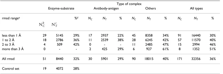
There are 3 classes of complexes in the complex set: enzyme-substrate, antibody-antigen, and others. Table 1 summarizes the number of changes in the different types of complexes.
Antibody-antigen complexes undergo 29% of structural letter modifications, a number similar to that obtained on the control set. Thus, on the limited number of structures available (10 antibody-antigen complexes), this class of proteins shows only moderate modifications upon pro-tein-protein binding. The 'other' class experiences the highest percentage of structural letter changes (40%). This class encompasses different kind of complexes (transport proteins, signaling proteins, viral capsid). The enzyme class has an intermediate behavior with 32% of modifica-tions. In their study, Daily et al found that 20% of the res-idues in enzymes are significantly modified upon binding [34]. Here, we find 32% of change in the structural letter sequences. As we show later, a part of these changes replace a structural letter by a similar one. Table 1 also reports the percentages of modified structural letters according to the root mean square deviation of the Cα (Cα rmsd). For all types of complexes, the global tendency
is a correlation between the percentage of modified struc-tural letters and the Cα rmsd, the exception being the
anti-body-antigen in which a low percentage of modified letter (29%) is obtained for high rmsd (more than 3Å) for 2 chains.
Table 1: Percentage of modified structural letters in the complex set
Type of complex
Enzyme-substrate Antibody-antigen Others All types
rmsd rangea %d N C NT % NC NT % NC NT % less than 1 Å 29 5145 29% 17 2937 22% 45 8358 34% 91 16440 30% 1 to 2 Å 18 2786 36% 11 2539 38% 28 6245 42% 57 11570 40% 2 to 3 Å 4 509 42% 0 - - 11 2485 47% 15 2994 46% more than 3 Å 0 - - 2 425 29% 6 927 61% 8 1352 51% All rmsd 51 8440 32% 30 5901 29% 90 18015 40% 171 32356 36% Control set 19 4072 28%
a: Cα root mean square deviation between bound and unbound conformations, b: number of protein chains involved, c: total number of structural
letters involved, d: percentage of structural letters that differ between bound and unbound conformation. NCb NTc
Local change versus global change
Some structures undergo minor global modifications, and other structures are significantly modified, as assessed by the Cα rmsd ranging from 0.2 to 14.0 Å. For comparison,
the Cα rmsd on the control set ranges from 0.20 to 0.38 Å,
with a mean value equal to 0.30 Å (same rmsd is obtained for allosteric and non-allosteric protein pairs). The per-centage of modified structural letters for different rmsd ranges is indicated in Table 1. As expected, the percentage of modified structural letters is higher on the structures with high rmsd.
To analyze in more details the relation between local and global modifications, we confront the Cα rmsd with the
percentage of structural letter modification in the complex
set. Figure 2a illustrates the correlation between the
number of structural letters that differ between bound and unbound conformations, and the Cα rmsd between
bound and unbound conformations, in the complex set. The first is a measure of local structural change, while the second is a measure of global change. It can be seen from Figure 2, that both measures are positively correlated: structures with great rmsd tend to have a high percentage of structural letters that differ between bound and unbound conformations. This confirms the results shown in Table 1. The Pearson correlation coefficient between both measures is 0.59 (p-value is equal to 2.10-15). How-ever, protein structures with a Cα rmsd lower than 1 Å
exhibit a great range of structural letter change proportion, from 10 to more than 70%. It indicates that the structural letter sequences bring an information about the structural changes that can not be evaluated by the Cα rmsd alone.
Two chains exhibit high Cα rmsd, around 5 Å, but a
mod-erate fraction of structural letter changes, around 30%. These two chains are chain A of receptor part in complex 2VIS (rmsd = 4.9 Å, percentage of change = 26% in a total of 207 structural letters) and chain B of receptor part of the same complex (rmsd = 5.0 Å, percentage of change = 32% in a total of 218 structural letters). The examination of these structures shows that they are made of two domains that undergo large motions upon binding, as can be seen on Figure 2b. The relative orientation of the two domains is significantly modified, hence leading to a high Cα rmsd. The local structures, however, remains similar,
as assessed by the moderate percentage of modified struc-tural letters. If they are superimposed by portion, the two domains have low Cα rmsd: 0.6 Å and 0.9 Å for domains
1–109 and 110–210 for chain A.
On the contrary, some structures exhibit slight global modifications but a high proportion of local changes: chains A and B of the receptor part of complex 1I4D (respective rmsd are 0.88 and 0.89 Å, respective percent-age of modified structural letters are 75 and 68%), and
chain B of the receptor part of complex 1F51 (rmsd equal to 1.45 Å, 69% of modified structural letters). These struc-tures, shown on Figure 2c, have a good conservation of there global structures, but the structural letter sequences capture some subtle differences in helix structures. The unbound helices are encoded by runs of helical letter [A], alternate with less regular letters [V] and [W], whereas bound helices are encoded by homogeneous runs of [a], suggesting a higher regularity of bound helices.
It thus appears that the structural alphabet approach offers a complementary approach to the global rmsd as a few local change can be associated to drastic global change, and inversely.
Comparison with local rmsd
The structural alphabet provides a simplified but detailed description of the protein backbone. As shown on Figure 1 some structural letters have very similar conformation, e.g., [a] and [A], whereas others are clearly different, e.g., [D] and [S]. This disparity has been quantified in [36], by the rmsddev: 0.15 Å between [A] and [a], and 1.6 Å between [D] and [S]. Furthermore, the structural letters have different intrinsic variability, as measured by the rmsdintra. The rmsdintra of the structural alphabet varies from 0.08 Å for letter [A], to 0.91 Å for letter [F] [36]. The consequences are that (i) different structural encoding can be observed for similar conformations (e. g. a run of [A] replaced by a run of [a]), and (ii) the same structural letter can encode relatively dissimilar fragments, e. g., the most variable letter [F]. It is then desirable to check for consist-ency between the structural alphabet approach and classi-cal external measures to assess the extend of the loclassi-cal deformations. The aim is to see if the structural alphabet, used for structure description, can also be used to detect significant local deformations.
Structural deformations between bound and unbound forms is usually assessed using classical rmsd computa-tion. In that section, we show a comparison between the assessment of deformation using the structural alphabet and classical rmsd. The measure of deformation using the structural alphabet is given by the rmsddev associated to the structural letter change between bound and unbound forms. For the measure using classical rmsd, we computed local Cα rmsd in a sliding window of four residues along
the protein. The reason why we choose a size of four resi-dues for the sliding window is because the structural let-ters are four-residue long. The results of this comparison on the complex set is shown in Figure 3. In case of identical structural letter in the bound and unbound structure (Fig-ure 3a), we consider the rmsdintra, instead of rmsddev, which is a measure of the intrinsic variability of each structural letter. A few cases of identical structural letters correspond to high local rmsd. These cases correspond to
Correspondence between global and local changes in bound/unbound chains
Figure 2
Correspondence between global and local changes in bound/unbound chains. a) Cα rmsd (x axis) versus percentage
of modified structural letters (y axis). Open circles: enzyme-substrate, plain squares: antigen-antibody, crosses: other. One chain of complex 1H1V, with a rmsd equal to 14 Å and 55% of modified structural letters is outside the range of this plot. The regression line shown on the plot is obtained by excluding chains with rmsd greater than 4.5 Å. Dashed line indicate the values obtained for the control set: 28% of modified letter, 0.30 Å rmsd. b) Superimposition of structures with high rmsd and low percentage of modified letters. Chain A of the receptor part of complex 2VIS (hemagglutinin from Influenza virus complexed with immunoglobulin): rmsd = 4.9 Å, 26% of local change. c) Superimposition of structures with low rmsd and many local changes, with corresponding structural letter encoding. Chain A of the receptor part of complex 1I4D (Rac1-GDP complexed with arfaptin from human), rmsd = 0.88 Å, 75% of local change, and chain B of the receptor part of complex 1F51 (transferase complex form bacillus subtilis) rmsd = 1.45 Å, 69% of local change. Orange: unbound conformations, green: bound conforma-tions. aaaVWAVWAAaaaaVWAVWAAZQGWZOVWVWB ZaaaaaaaaaaBAAAAAVWAAVWZZWaaaZZC YNGGWaaaaaaaaaaaaaaaaaaaaaaaaaaa CZWVWaaaaaaaaaaaaaaaaaBAAAVWZ−−− EaaaaaVWVWAVWVWBVWAAVWBVWaaaZEZW ZZaaVWAVWaaaaaaEZZZZZWZZZ aDORZaaaaaaaaVWVWaaaaaVWVWEDOGZW MXYCIMMTTTMT LKHZaaaaaaaaaaaaaaaaaaaaaaaaaaaa aaaaVZEWBQYXPILNTTTMNJYJTYGOWZEZ ZaaaVWaaaaaaaaaZQKPBQHRTTTTTMNGE QHFEPIMMNTTXTJJMFCGEEaEZaQLPGIGX region 30−59 218−245 region unbound encoding bound encoding bound encoding unbound encoding 1I4D chainA 1F51 chain B KXMGZZWAAAAAAVWAAAAAVWaaaaaaaaVW VWAVWVWAAAAAAAVWVWVWAZQGBCOVWVWA ABVWVWVWaaaaaaaaaaaaaaVWVWBVWZZC ZZWVWaaaVWAAAVWAAVWAVWAAAVWVWBaa aaVWVWVWAVWVWVWaaaaaaaBAAVWBZEZZ ZWZZZZZZaaaaaaaVWVWVWBZWA KPBVWaaVWAAVWBVWVWVWAAVWAAVWVWAA ADSGBVWAAVWAAAAAAAAAAAAAAVEDOGZW aVWBZZEZZCQGEIKNTTTMNJJKXKGOBZEW VWVWAAAAAAAAAAAZQKPZQYNXMMMTMNGE SPCQPIMNMNTTTJJLGZGBEZEa−−−PGITT TTYCITMTTMNN c) 0 1 2 3 4 5 10 20 30 40 50 60 70 80 rmsd
% of structural letter change
fragments that are surrounded by structural letter substitu-tions. For example, fragment 64–69 of the R chain of the ligand part of complex 1WQ1 is encoded by QFO in the unbound form and FFE in the bound form. The fragment encoded by the central F has a local rmsd equal to 1.9Å. The case of different structural letter between bound and unbound forms is called a structural letter substitution (Figure 3b and 3c). Here, we further introduce a distinc-tion between isolated substitudistinc-tions (Figure 3b) and sub-stitutions that appear in stretch (Figure 3c). An isolated substitution denotes a structural letter that is modified when one or both of its neighbors remain unchanged, e.g, ART → ABT or ART → ABG. Inversely, a stretched substi-tution denotes a structural letter change surrounded by modified structural letters, e. g., ART → CBG. 61% of the structural letter substitutions appear isolated, and 39% appear in stretch. An unexpected finding of this analysis is that some structural letter substitutions exhibit a high rmsddev but a low local rmsd (see Figure 3b and 3c). For example, we observe 534 cases of structural letter substitu-tions with an associated rmsddev greater than 1 Å and a local rmsd lower than 0.5 Å, out of 1,351 substitutions associated to rmsddev greater than 1 Å (39%). Among 598 isolated substitutions associated with rmsd dev greater than 1 Å, 418 correspond to local rmsd lower than 0.5 Å i. e., 70%. If we consider only stretched substitutions, this ratio is only 15% (116 out of 753). This effect is thus more fre-quently seen in isolated substitutions than in stretched substitutions. This can be globally assessed by the Pearson correlation coefficient between local rmsd and rmsddev: 0.50 for isolated substitutions and 0.87 for stretched
sub-stitutions. These cases correspond to fragments with low rmsd but encoded by highly dissimilar structural letters. They are due to the stochastic nature of the structural encoding using a HMM.
A consequence is that the rmsddev alone cannot be used to quantify the structural change. This analysis tells us that although the structural alphabet offers a unique original tool to detect and qualitatively describe structural defor-mation, this information has to be combined with the local rmsd in order to properly measure the deformation.
Analysis of interface regions
The interface regions are defined using Voronoi tessella-tions. Among the 32,356 residues in the complex set, 3,746 are thus defined as interface residues. Interface residues then represent 12% of the whole dataset (i. e. both surface and core residues).
Interface local structure
The local structure composition of the interface set is com-pared to the global composition of the complex set on Fig-ure 4a. The relative frequency of helical letters [A,a,V,W] and extended letters [L,N,M,T,X] is lower in the interface set than in the complex set. It is in agreement with the study of Ansari et al [40] who showed that coils are more abun-dant in protein-protein interfaces than in general. Letters that form helix ends [Z,B,C] are more abundant in the
interface set. The majority of coil letters are favored at the interface set [E,I,D,Q,O,H,Y,R,J,S,G] but some particular
letters are not: [U,F,P,K] (strand ends). The statistical
sig-Comparison of local rmsd and rmsddev
Figure 3
Comparison of local rmsd and rmsddev. a) Fragments that are encoded by the same structural letter in the bound and unbound forms. The rmsddev in this case is the rmsdintra, which measures the intrinsic variability of structural letters. b) Frag-ments that are encoded by different structural letters in the bound and unbound forms, in the case where the structural letter substitution is isolated or at the extremity of a stretch of substitutions. c) Fragments that are encoded by different structural letters in the bound and unbound forms, in the case where the structural letter substitution appears in a stretch of substitu-tion. The red line indicates the equality between local rmsd and rmsddev.
0.0
1.0
2.0
0.0
1.0
2.0
no subsitution
local rmsd rmsd_dev0.0
1.0
2.0
0.0
1.0
2.0
isolated subsitution
local rmsd rmsd_dev0.0
1.0
2.0
0.0
1.0
2.0
stretched subsitution
local rmsd rmsd_deva)
b)
c)
nificance of structural letter preference for interface regions is assessed by Z-scores computation, illustrated on Figure 4b. These Z-scores compare the structural letter proportion in the interface with the proportion in non-interface regions. The significance threshold, corrected by Bonferroni, is equal to 3.1. Figure 4b indicates that letters
[A,K,N,M,T] are significantly under-represented in the interface, while letters [B,C,E,O,H,Y,R,J,S] are significantly over-represented. The structural alphabet thus provides more information than the classical helix/strand/coil clas-sification: some structural letters found in the coil are not favored at the interface.
Local structure composition of the interface set
Figure 4
Local structure composition of the interface set. a) Percentage of each type of structural letter, in the complex set
(white) and the interface set (gray). b) Z-score to assess the significance of over- or under-representation of the structural let-ters in the interface. Dashed lines indicate the threshold for statistical significance (-3.1 and 3.1).
A a V W Z B C U E F I D Q O H Y R J S P K G L N M T X 02468 − 50 5 1 0
a)
b)
Number of local changes
A total of 3,746 structural letters are involved in the inter-face: 2,217 (59%) are unchanged and 1,529 (41%) are changed. If we consider the 8 groups of Figure 1, 604 structural letters are changed (16%). This is significantly greater than the results obtained for the whole structures (36% of structural letter changes and 11% with the 8 groups), as assessed by Chi-square tests (p-values < 2.10 -16).
To assess if each individual letter undergo more substitu-tions in the interface set than in the control set, we compute Z-scores (data not shown). All the structural letters, except [a], are more modified in the interface set than in the
con-trol set. The difference is significant for 19 letters out of 27,
particularly high for letters I, Q, J, and K.
Number of possible substitutions
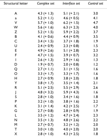
The number of possible substitutions (Nsub) for each type of structural letters are reported in Table 2. Nsub quantifies, for each structural letter, the mean number of structural letters it can be substituted with. In the control
set, helical letters [A,a,V,W,Z,B] experience the highest
Nsub, around 3–5. It means that these letters are replaced, on average, by three to five different letters. Extended
let-ter [L,N,M,T,X] have Nsub around 2. Among the structural letters that describe coils, only letters [E,F,Y,R,P] have a Nsub greater or equal to 2. It indicates that the expected variations in structural sequences caused by experimental error and natural protein flexibility affect preferentially helices, strands in a lesser extend, and a few coil letters. It is known that loop regions are more flexible that regular secondary structures. The variations observed in helix and strand structural sequences may be due to the fact that our structural alphabet offers a very detailed description of these regions.
The same global tendency is observed in the control and the interface sets: high Nsub for helical letters, some of the extended letters and a few coil letters. However, the Nsub computed from the the complex set are higher than the Nsub computed from the control set. The interface region analysis results, in a majority of cases, in higher Nsub than in the complex set, confirming that interface regions undergo more various structural changes. The Nsub are one to two points greater in the interface set than in the control set, except for letters [J] (+4.3), [R] (+2.9) and [E] (+2.8), resulting in Nsub greater than 5 for these letters. On the contrary, letter [D] has the lowest Nsub, equal to 2. This analysis thus reveals that some structural letters are particularly affected by the binding (i. e., [E,R,J]).
Severity of local deformations
The quantitative measurement of structural letter changes is assessed using the local rmsd. In the control set, 5% of the fragments show a local rmsd greater than 0.2 Å. We will then use 0.2 Å as a threshold to select significant local deformations. In the complex set, 25% of the fragments have local rmsd greater than 0.2 Å, and 35% if we restrict to the interface fragments. It thus appears that interface regions undergo more severe local changes than the rest of the structure.
Figure 5 presents an analysis of the importance of local deformations in the interface set depending on the struc-tural encoding. For each strucstruc-tural letter, we collect the local rmsd and display the number of structural letters affected by a local rmsd in a given range. The unbound form is taken as a reference for this analysis. Helical struc-tural letters [A,a,V,W,Z,B,C] experience very few local rmsd greater than 0.2 Å, indicating that α-helices are very stable local structures and are barely affected by the inter-action. Among them, letter [Z], which is found at helix borders, shows the highest proportion of local rmsd greater than 0.2 Å. Extended letters [L,N,M,T,X] exhibit higher rmsd, indicating that these local structures are more likely to be modified upon binding, especially letter [L]. The highest number of high local rmsd are observed in coil letters, in particular in letters [E,F,I,Q,J]. Some coil letters, like [U,D,G], are barely affected by the binding. Table 2: Number of possible substitutions (Nsub) in the different
data sets. The numbers between parentheses are the difference between the Nsub and the Nsub of the control set.
Structural letter Complex set Interface set Control set
A 4.3 (+ 1.3) 5.1 (+ 2.1) 3.0 a 5.2 (+ 1.1) 4.6 (+ 0.5) 4.1 V 5.7 (+ 1.0) 6.2 (+ 1.5) 4.7 W 5.6 (+ 1.6) 6.3 (+ 2.3) 4.0 Z 5.2 (+ 1.5) 5.9 (+ 2.2) 3.7 B 4.1 (+ 0.6) 4.4 (+ 0.9) 3.5 C 3.4 (+ 1.5) 3.7 (+ 1.8) 1.9 U 2.4 (+ 0.9) 2.3 (+ 0.8) 1.5 E 4.9 (+ 2.6) 5.1 (+ 2.8) 2.3 F 4.7 (+ 1.5) 3.9 (+ 0.7) 3.2 I 2.6 (+ 1.3) 2.9 (+ 1.6) 1.3 D 1.9 (+ 0.7) 2.0 (+ 0.8) 1.2 Q 2.7 (+ 1.1) 3.1 (+ 1.5) 1.6 O 3.3 (+ 1.7) 3.3 (+ 1.7) 1.6 H 2.7 (+ 0.9) 3.8 (+ 2.0) 1.8 Y 3.8 (+ 1.7) 3.5 (+ 1.4) 2.1 R 5.1 (+ 2.5) 5.5 (+ 2.9) 2.6 J 4.8 (+ 3.2) 5.9 (+ 4.3) 1.6 S 2.8 (+ 1.0) 3.4 (+ 1.6) 1.8 P 3.2 (+ 1.0) 3.8 (+ 1.6) 2.2 K 3.1 (+ 1.4) 4.2 (+ 2.5) 1.7 G 2.7 (+ 0.8) 2.8 (+ 0.9) 1.9 L 3.5 (+ 1.2) 4.7 (+ 2.4) 2.3 N 3.5 (+ 1.3) 4.8 (+ 2.6) 2.2 M 2.7 (+ 0.7) 3.2 (+ 1.2) 2.0 T 3.0 (+ 1.0) 4.0 (+ 2.0) 2.0 X 2.8 (+ 1.0) 4.3 (+ 2.5) 1.8
It thus appears that the severity of local deformation is not uniform among the structural letters, in particular among structural letters describing coils. Some structural letters are more likely to be affected by the formation of protein-protein complex.
Structural letter substitutions
Now that we have shown that some structural letters are preferentially affected upon binding, the next step is to analyze the resulting conformation after binding, namely the structural letter substitutions. Figure 6 is an illustra-tion of the probabilities of structural letter substituillustra-tion in the interface region. The unbound form is taken as the ref-erence for this computation. To take into account only sig-nificant changes, we restrict the analysis to the pairs of structural letters that correspond to a local rmsd greater than 0.2 Å. The number of structural letter pairs with local rmsd greater than 0.2 Å is 1309, including 488 cases of structural letter identity. Among the 729 possible substitu-tion probabilities (27 × 27), 312 are non-null and 139 are greater than 5%. It must be noted that the substitution probability matrix is highly asymmetrical.
For example, extended letters [A,a,V,W,Z,B,C] display high probabilities to be substituted into letter [Z,B] upon binding. The probability for letter [Z] to be transformed into [V] in the interface region upon binding is 8.8%, whereas it is 28.6% for the inverse transformation from [V] to [Z]. This arises from the normalization with respect to the unbound form needed for the probability compu-tation. The substitution count table is nearly symmetrical, as shown in additional file 1, but the number of structural letters in each class being unequal (see Figure 4), it results in asymmetry in the substitution probabilities. To facili-tate the global examination of Figure 6, let us separate the 27 structural letters into the 3 main groups associated to classical secondary structure elements: [a,A,V,W,Z,B,C] for helix and helix borders, [J,K,L,M,N,T,X] for strands and strand borders, and the remaining [D,E,O,S,R,Q,I,F,U,P,H,G,Y] for coils.
Many substitutions occur within the helical group [A,a,V,W,Z,B,C], and, in a lesser extend, in the extended group [L,N,M,T,X]. These substitutions are responsible for subtle modifications of regular secondary structures, like
Severity of local structural change for each structural letter in the interface set
Figure 5
Severity of local structural change for each structural letter in the interface set. Histogram of structural letter
counts in the unbound conformations, colored according to the severity of the local structural change occurring upon binding, measured by the local rmsd.
A a V W Z B C U E F I D Q O H Y R J S P K G L N M T X 0 5 0 100 150 200
0.2 < local rmsd < 0.5
1 < local rmsd < 1.5
local rmsd > 1.5
0.5 < local rmsd < 1
local rmsd < 0.2
illustrated in Figure 2. Inside the coil group, that gathers a variety of distinct conformations, significant substitutions occur from letters [Y,H,F,R,O] and toward letters [F,Q,S,G]. Eight substitutions with probability greater than 5% are observed from the helix group to the coil group, including 3 substitutions with probabilities greater than 10%: from [C] to [U] (11.9%), from [B] to [E] (18%) and from [W] to [E] (13%). In the same way, 12 substitu-tions with probability greater than 5% occur from the strand group to the coil group, one of them with probabil-ity greater than 10%: from [T] to [G] (10.5%). Four sub-stitutions with probabilities greater than 5% occur from the coil to the helix group (with probability to change from [E] to [B] equal to 15%) and 11 occur from coil to
strand group, including three substitutions with high probability: [I] to [J] (11%), [P] to [L] (15%) and [Y] to [K] (15.4%). No substitutions occur between strand and helix groups with high probabilities. This analysis tells us that the local structural modifications that occur upon binding are most of the time subtle substitutions between structural letters belonging to the same major groups. Conformational changes occur mainly following a con-tinuum between helices/coils and strands/coils and a few structural letters play key role in the modifications. The structural alphabet thus provides a new way to describe local structural changes as the substitution of a
Probabilities of structural letter substitution associated to local rmsd greater than 0.2Å in the interface set
Figure 6
Probabilities of structural letter substitution associated to local rmsd greater than 0.2Å in the interface set.
The unbound form is taken as the reference for the computation of substitution probabilities.
Bound conformation
Unbound conformation
A
a
V
W
Z
B
C
D
E
O
S
R
Q
I
F
U
P
H
G
Y
J
K
L
M
N
T
X
A a V W Z B C D E O S R Q I F U P H G Y J K L M N T X0
1
5
10
15
20
25
30
35
40
45
50
55
60
65
70
75
80
85
90
95
100
structural letter by another one. It is the first time, to our knowledge, that such a qualitative description is reported.
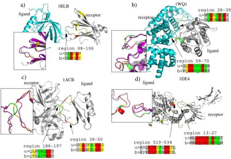
Graphical examples
Figure 7 illustrates some examples of structural letter sub-stitutions induced by protein-protein interaction. These four complexes are taken from the enzyme-substrate class (Figure 7a, b and 7d) and the other class (Figure 7c). In these four examples, drastic local changes (local rmsd greater than 1.5 Å) occur in the interface regions, within loops. We report the structural sequences of the modified regions, in bound and unbound forms. 1RLB, shown in Figure 7a, is a transthyretin/retinol binding complex. The chain A of transtyretin (the receptor) has a Cα rmsd equal
to 0.7 Å between bound and unbound forms and 47% of the structural letters are modified. A structural
modifica-tion of region 98–106 includes 2 substitumodifica-tions associated to local rmsd greater than 1.5 Å: [H] to [O] and [L] to [Q]. A less severe substitution (local rmsd between 1 and 1.5 Å) occurs from [I] and [D]. 1WQ1, shown in Figure 7b, is a ras GTPase/ras GAP complex. Ras GAP (the ligand) is modified in two distinct regions: in loop 28–39 and loop 58–70. Its global Cα rmsd is 1.1 Å and its percentage of
structural letter substitution is 51%. In region 28–39, a run of 7 successive structural letters undergo modifica-tions greater than 0.5 Å, whereas in region 58–70, a run of 8 successive structural letters are strongly modified. Region 58–70 involves letters [J,Q], which are over-mod-ified in interface regions. 1ACB, shown in Figure 7c, is a chymotrypsin/eglin C complex.
Examples of local structural changes induced by protein-protein binding
Figure 7
Examples of local structural changes induced by protein-protein binding. Proteins are colored according to the
rmsddev of the letter substitution unbound/bound form, using same color scheme as in Figure 5: white = local rmsd lower than 0.2Å, gray = local rmsd between 0.2 and 0.5 Å, yellow = local rmsd between 0.5 and 1 Å, green = local rmsd between 1 and 1.5 Å, red = local rmsd greater than 1.5 Å. The superimposition of bound and unbound structures (in magenta), is shown for the modified region. The structural encoding are shown for the interface region that are modified: > u: unbound encoding, > b: bound encoding.
a)
receptor ligand 1RLBb)
receptor ligand receptorc)
1ACB ligandd)
1DE4 ligand 1WQ1 receptor u>UIHRLN b>ZDOGQT region 98−106 u>MLLHFORJNKXT b>MMUQJPIKFGQM region 13−27 u>NUORJUQNX b>GQFSNLGFG u>GQFRJFQFOB b>PQPBVQFFEE region 58−70 region 38−50 u>UOIJFSGOQG b>USLNKHSLTP region 186−197 u>QLPOEQLYU b>QTLXJUESU u>KHQLPEIFQNKTL b>KPEOIPVOBAVSL region 519−534 region 28−39Structural modifications occur in both partners: region 186–197 in chymotrypsin and region 38–50 in eglin part. The global Cα rmsd for chymotrypsin (receptor) is 1.75 Å
and the percentage of structural letter substitution is 37%. For eglin (the ligand), global Cα rmsd is 1.5 Å and 45% of
the structural letters are modified. Both modifications involve letters [I,J,Q] which are significantly more modi-fied in the interface set than in the control set. 1DE4, shown in Figure 7d, is a complex between beta2-microglobulin and a transferrin receptor. The structural modification highlighted here occur in region 13–27 of the beta2-microglobulin and region 519–534 of the transferrin receptor (the ligand). The local structures of both partners are modified were the contact occurs. Beta2-microglobu-lin (the receptor) has a global Cα rmsd equal to 1.65 Å
and 49% of structural letter substitution. The transferrin receptor (ligand) has a global Cα rmsd equal to 1.6 Å and
41% of its structural letters are modified upon binding. Both regions involve letters [I,J,K].
A last illustration of the conformational change analysis using the structural alphabet is shown in Figure 8. In this analysis, we use the structural alphabet to detect common binding motifs in unrelated proteins. The question raised is: " do proteins with unrelated functions exhibit similar structural motifs at the interface ?" The objective is to identity, if any, such structural motifs that could be con-sidered as case of "local structural convergence" toward the same conformation, from unrelated proteins. We applied the following criteria to detect such cases:
• we look for structural motifs at least four structural letter long (i. e., seven residues);
• the motif should be present in the bound forms of at least two complexes from different classes. We consider the 3 classes from Table 3, namely enzyme/substrate, anti-body/antigen, and other;
• the motif should be located in totality at the protein-protein interfaces of the complexes;
• we do not consider runs of helical letters (A,a,V,W,Z,B,C) or extended letters (L,M,N,T,X,J,K). Heli-ces and strands being highly abundant in 3D structures, these motifs may be non significant;
• a significant local deformation should be seen, at the considered motif, in at least one complex;
• the local rmsd between the bound fragments covered by the motif should be lower than the local rmsd between unbound fragments.
Using these criteria, we extracted common bound motifs from proteins with unrelated function. With the rmsd cri-terion, we select cases where the conformational change induced by the binding put the bound structures closer than the unbound structures, what we call "local struc-tural convergence". Given the limited amount of data we have, and the stringent criteria we applied (in particular, we consider only 3 classes), we found only a few cases of local structural convergence. Two examples are illustrated
Examples of local conformational convergence
Figure 8
Examples of local conformational convergence. Red motifs in rectangles are the structural letter sequences of the
frag-ment highlighted in red in the structures. Numbers associated to blue arrows are the Cα local rmsd computed between the
red fragments. Numbers between brakets denote the region of the protein covered by the structural motif. Top row: unbound structures, bottom: bound structures. a) Motif GOIF is seen in the bound forms of complex 1AHW (antibody-antigene) and 1BVN (enzyme/substrate). b) Motif LLGI is seen in the bound forms of complex 1GRN (other) and 1UDI (enzyme/substrate).
receptor receptor
a)
unbound bound1.4
0.8
0.7
0.4
receptor ligand receptor ligand 1BVN[234−240] 1AHW [89−95]b)
ligand ligand receptor receptor ligand recptor1.9
2.2
0.9
0.8
1UDI [15−21] 1GRN [32−38] GOIJ GOIF SGRFGOIF LLGI LLGI
in Figure 8. Structural motif GOIF is seen in two unrelated complexes: 1AHW, an antibody/antigen complex, and 1BVN, an enzyme/substrate complex. The local Cα rmsd
for the corresponding fragment is 1.4 Å between unbound forms and 0.7 Å only between bound forms. Complex 1AHW undergoes only minor conformational change, as assessed by the rmsd equal to 0.4 Å, and a similar unbound structural motif: GOIJ. Complex 1BVN is modi-fied up to an amount of 0.8 Å, starting from a different structural motif: SGRF. The underlying amino-acid sequences are 'LQHGESP' (1AHW) and 'VIDLGGE' (1BVN). Structural motif LLGI is seen in one 'other' com-plex, 1GRN (complex between a G-protein and a GTPase activation domain) and one enzyme/substrate complex, 1UDI. Both complexes are significantly modified by the binding: 1.9 Å rmsd for 1GRN, from KPQL to LLGI, and 1UDI, in a lesser extend: 0.9 Å rmsd, from LNNG to LLGI. Local rmsd are equal to 2.2 and 0.8 Å before and after binding respectively. Underlying amino-acid sequences are 'YVPTVFD' (1GRN) and 'QLVIQES' (1UDI). These examples highlight the usefulness of the structural alpha-bet for further analysis studies using larger data sets.
Conclusion
This study reveals that the structural alphabet offers a new way to investigate local deformations induced by the pro-tein-protein interaction. Classical studies revealed that interface regions preferentially involve loops. Here, we show that two structural letters forming helix ends [B,C] are preferred at the interface and that only a part of the structural letters describing the loops, [O,H,Y,R,J,S], are preferred at the interface. Letters [E,R,J] are particularly affected by the binding (number of possible substitution greater than 5 versus 2 in the control set). Concerning the severity of the substitutions, letters [E,F,I,Q,J] are subject to major modifications.
It is the first time that local conformation changes can be qualitatively described in such a way. The main advantage of using the structural alphabet approach, compared to classical rmsd measure, is that it provides a description of bound and unbound conformations, and, in turn, a qual-itative description of the deformation. This feature opens the perspective for further studies, such as the classifica-tion of interface structural motifs and structural changes. The following questions could be addressed: are the
struc-tural modifications common to any type of complexes ? Can the same structural modifications be observed in unrelated proteins ? Could we use the qualitative descrip-tion of structural changes to make a classificadescrip-tion of bind-ing movements ? An example of such analysis is illustrated in Figure 8, in which we highlight two examples of com-mon binding structural motifs from unrelated proteins. Although the actual amount of data is insufficient to derive any conclusive remarks, the structural alphabet approach seems very promising to address such questions. The computation of structural letter substitution proba-bilities highlights some preferred substitutions. Such informations could be useful for flexible docking experi-ments and binding pocket detection at protein surfaces. Flexible docking strategies include the use of ensembles of alternate starting conformations -taken from molecular dynamic simulation [41-44] or other conformational sampling techniques [45]- and the explicit integration of conformational changes during the docking procedure via simulated annealing refinement [46] or multicopy mean-field approach [47]. In this framework, the structural letter substitution probabilities derived from the present study could be used in a conformational sampling technique. The structural letter substitution matrix could be used in a generative manner using a Markov process: starting from the unbound structural letter sequence, modifications are introduced using the matrix, to generate a potential bound structural letter sequence. It is then possible to re-build the bound backbone from the structural letter sequence [48,49]. This would probably require some external methods to predict which region is to be modi-fied. The strong transition rules between successive struc-tural letters [36] should also be taken into account in order to generate realistic structural letter sequences.
Methods
DatasetWe use the version 2.4 of the benchmark presented by Mintseris et al [50]: 83 crystallographic structures of pro-tein-protein complexes -the bound structures- accompa-nied by the crystallographic structures of the free ligands and receptors -the unbound structures. The Mintseris dataset consists in 23 enzyme-inhibitor complexes, 21 antibody-antigen complexes (11 of them are in bound/ unbound conformation) and 39 other type complexes. As Table 3: Description of the complex set
Type (number) Complexes PDB id
Enzyme-substrate (23) 1ACB, 1AVX, 1AY7, 1BVN, 1CGI, 1D6R, 1DFJ, 1E6E, 1EAW, 1EWY, 1EZU, 1F34, 1HIA, 1KKL, 1MAH, 1PPE, 1TMQ, 1UDI, 2MTA, 2PCC, 2SIC, 2SNI, 7CEI
Antibody-Antigen (10) 1AHW, 1BGX, 1BVK, 1DQJ, 1E6J, 1JPS, 1MLC, 1VFB, 1WEJ, 2VIS
Other (35) 1A2K, 1AK4, 1AKJ, 1ATN, 1B6C, 1BUH, 1DE4, 1E96, 1EER, 1F51, 1FC2, 1FQ1, 1FQJ, 1GCQ, 1GP2, 1GRN, 1H1V, 1HE1, 1HE8, 1I2M, 1I4D, 1IB1, 1IBR, 1IJK, 1KLU, 1KTZ, 1KXP, 1M10, 1ML0, 1N2C, 1QA9, 1RLB, 1SBB, 1WQ1, 2BTF
we are interested by structural changes upon binding, the 11 antibody-antigen complexes in bound/unbound con-formation are excluded from the analysis. Some ligands and receptors are multichains. The comparison between bound and unbound forms require a correspondence between the residue numbering of each form. This restric-tion leads to the exclusion of four complexes belonging to the 'other' class. When only the ligand (or the receptor) has inconsistent residue numbering, the receptor (or lig-and) is kept in the analysis. Similarly, when one chain of a multimer protein has inconsistent residue numbering, the others were kept in the analysis. 15 chains were then further removed.
The complete dataset of 68 complexes (containing 156 chains from 124 proteins) used in this study is described in Table 3. We will refer to this data set as the complex set. To distinguish the structural deformation induced by pro-tein-protein binding from the experimental uncertainty and the expected variations due to protein flexibility, a
control set is needed. We consider the control set of 14
pro-tein pairs used by Daily and coworkers [34]:
• 5 protein pairs independently crystallized by different groups: 2CBA/1CAM, 1VDQ/1HEL, 1UNE/1MKT, 1EY0/ 1STN, and 1TPH/1TPW.
• 9 pairs of allosteric proteins independently crystallized in the same form: 3CHY/1JBE, 1GDD/1BOF, 1GPB/ 8GPB, 4HHB/1A3N, 1T48/1T49, 1OIW/1YZK, 1VG8/ 1T91, 1XTS/1XTR, and 2TRT/2TCT.
Structural alphabet encoding
In this study, the ligand and receptor of each complex, in bound and unbound forms, are simplified into structural letter sequences using HMM-SA and the Viterbi algorithm [35,36]. Local conformational modifications between bound/unbound forms are studied through the structural letter sequences.
Comparison of bound and unbound structures
A total of 156 couples of bound/unbound chains are used to analyze the local structural changes induced by the pro-tein-protein binding. The principle of the study is illus-trated in Figure 9.
Classical measure of deformation
A classical measure of conformational change is the rmsd (root-mean square deviation), i. e., the mean deviation of atom positions after otimal superimposition of two struc-tures. The rmsd can be computed for the whole protein -a
global rmsd- or for a fragment of the protein -a local rmsd. In this study local and global rmsd are computed
using Cα atoms only, using the ProFit software [51].
Deformation assessment by the structural alphabet
As explained in the Results section, a general distance between two different structural letters is given by the
rmsddev, as defined in [36]. The rmsddev has been com-puted from 500 fragment pairs randomly chosen in the two structural letters. The rmsdintra, computed in the same way, measures the intrinsic variability of each structural letter.
The structural distance between two fragments of four res-idues can then be measured using the local rmsd or the rmsddev. Note that the difference between these two rmsd is that the local rmsd is computed for each pair of frag-ments using proFit, whereas the rmsddev is taken from a pre-computed table, by considering only the structural encoding of the fragments.
Structural letter substitution probabilities
The use of unbound/bound pairs allows to study the structural modifications as an oriented process: a protein evolves from the unbound state, toward the bound state. The probability to move from letter x to letter y is then given by:
where Nbound(x → y) denotes the number of structural let-ter x in the unbound form that are replaced by structural letter y in the bound form, and Nunbound(x) denotes the total number of structural letter x in the unbound form. When x = y, this quantity is the probability of being unchanged. Here, we consider that the unbound state is the starting state and the bound state is the final state. Then, the unbound state will be taken as a reference for the computation, and the resulting matrix might be asym-metrical.
Number of possible structural letter substitutions
The number of possible substitutions for each structural letter can then be computed from the substitution proba-bilities:
N sub(x) = eH(x)
were H(x) is the Shannon entropy for letter x:
A N sub equal to 1 indicates that structural letter x is inte-grally transformed into one structural letter (it can be itself). The maximum theoretical N sub is 27: it means that structural letter x is transformed into all the 27 structural letters, with equal probabilities.
P x y N bound x y N unbound x ( ) ( ) ( ) → = → H x P x k P x k k ( )= −
∑
( → ) ln ( → ).Definition of interface residues
The local modifications induced by protein-protein ing are studied in more details at the receptor-ligand bind-ing interface. Interfaces are detected usbind-ing Voronoi tessellations. Voronoi tessellations are a way to divide the space around a given set of points into cells. The Voronoi cell around a point contains all the points that are closer
to this point than the others. Voronoi tessellations are used to study contacts within proteins, without the use of threshold distance [52]. Here, Voronoi tessellations are used to identify the residues that make contacts between the receptor and the ligand. We use the PROVAT software [53] to compute the Voronoi cells around Cα, with default parameters. Two residues are in contact if their
Schematic description of the study
Figure 9
Schematic description of the study.
of structural encoding
Comparison
(count of structural changes,
type of changes, ...)
unbound conformation
bound conformation
bound structural encoding
FQKGIGWAVWBAAAAVZCCCCDSKPEZ
FQLGFGZWBaaaaaaVZCCZCDSKPBZ
unbound structural encoding
Voronoi cells share a surface with non-zero area. A struc-tural letter is a four residue fragment. The correspondence is made between a four residue fragment and its third Cα. The structural modifications are studied in more details in the interfaces. We will refer to this part of the data as the
interface set.
Zscore computation
To study the over-representation of the different structural letters in the interface regions, we compute Zscores defined by:
Nobs(x) denotes the observed number of letter x in the
interface set and Nexp(x) denotes the expected number of x in the interface if the compositions of interface and non-interface regions were similar:
were denotes the rela-tive frequency of x in non-interface region and Ninter the number of structural letter of any type in the interface set. Zscores are similarly computed to assess the over-modifi-cation of a given structural letter, with Nobs(x) the number of structural letter x that is modified upon binding in the
interface set, and
where denotes the probability for letter x to be modified in the control set, and denotes the number of letter x in unbound form in the interface set. Zscores are expected to follow a Gaussian distribution with mean equal to zero and standard deviation of 1. Sig-nificance thresholds are corrected to take the multiple tests into account.
Authors' contributions
JM and HL carried out the comparisons of structural sequences. JM and LR carried out the analysis of the results. ACC and JM conceived the study. All authors read and approved the final manuscript.
Additional material
Acknowledgements
We are grateful to INRA for awarding a Fellowship to JM and to Ministère de l'Enseignement Supérieur et de la Recherche for awarding a Fellowship to LR. We thank two anonymous referees for their remarks that helped us to improve the manuscript.
References
1. Chothia C, Janin J: Principles of protein-protein recognition.
Nature 1975, 256(5520):705-708.
2. Jones S, Thornton JM: Principles of protein-protein interac-tions. Proc Natl Acad Sci USA 1996, 93:13-20.
3. Janin J, Miller S, Chothia C: Surface, subunit interfaces and inte-rior of oligomeric proteins. J Mol Biol 1988, 204:155-164. 4. Janin J, Chothia C: The structure of protein-protein recognition
sites. J Biol Chem 1990, 265(27):16027-16030.
5. Ponstingl H, Henrick K, Thornton JM: Discriminating between homodimeric and monomeric proteins in the crystalline state. Proteins 2000, 41:47-57.
6. Valdar WS, Thornton JM: Conservation helps to identify biolog-ically relevant crystal contacts. J Mol Biol 2001, 313(2):399-416. 7. Mintseris J, Weng Z: Atomic contact vectors in protein-protein
recognition. Proteins 2003, 53(3):629-639.
8. Bahadur RP, Chakrabarti P, Rodier F, Janin J: A dissection of spe-cific and non-spespe-cific protein-protein interfaces. J Mol Biol 2004, 336(4):943-955.
9. Jefferson ER, Walsh TP, Barton GJ: Biological units and their effect upon the properties and prediction of protein-protein interactions. J Mol Biol 2006, 364(5):1118-1129.
10. Grishin NV, Phillips MA: The subunit interfaces of oligomeric enzymes are conserved to a similar extent to the overall protein sequences. Protein Sci 1994, 3(12):2455-2458.
11. Caffrey DR, Somaroo S, Hughes JD, Mintseris J, Huang ES: Are pro-tein-protein interfaces more conserved in sequence than the rest of the protein surface? Protein Sci 2004, 13:190-202. 12. Bordner AJ, Abagyan R: Statistical analysis and prediction of
protein-protein interfaces. Proteins 2005, 60(3):353-366. 13. Murakami Y, Jones S: SHARP2: protein-protein interaction
pre-dictions using patch analysis. Bioinformatics 2006, 22(14):1794-1795.
14. Bradford JR, Needham CJ, Bulpitt AJ, Westhead DR: Insights into protein-protein interfaces using a Bayesian network predic-tion method. J Mol Biol 2006, 362(2):365-386.
15. Burgoyne NJ, Jackson RM: Predicting protein interaction sites: binding hot-spots in protein-protein and protein-ligand interfaces. Bioinformatics 2006, 22(11):1335-1342.
16. Li M, XL W, Lin L, Liu T: Protein-protein interaction site predic-tion based on condipredic-tional random fields. Bioinformatics 2007, 23(5):597-604.
17. Koshland DE: Application of a Theory of Enzyme Specificity to Protein Synthesis. Proc Natl Acad Sci USA 1958, 44(2):98-104. 18. Tsai CJ, Kumar S, Ma B, Nussinov R: Folding funnels, binding
fun-nels, and protein function. Protein Sci 1999, 8(6):1181-1190. 19. Goh CS, Milburn D, Gerstein M: Conformational changes
associ-ated with protein-protein interactions. Curr Opin Struct Biol 2004, 14:104-109.
Zscore x N obs x N exp x N exp x
( ) ( ) ( )
( ) .
= −
Nexp( )x =finter( )x ×Ninter finter( )x
Nexp( )x =Pexp(x→x)×Ninterunbound( )x Pexp(x→x)
Ninterunbound( )x
Additional file 1
Structural letter substitution counts in the interface region. This figure
presents the counts of structural letter substitutions corresponding to local rmsd greater than 0.2Å, restricted to the interface region. The normaliza-tion with respect to the unbound form results in the substitunormaliza-tion probabil-ity matrix presented in Figure 6.
Click here for file
[http://www.biomedcentral.com/content/supplementary/1472-6807-8-12-S1.pdf]
Publish with BioMed Central and every scientist can read your work free of charge "BioMed Central will be the most significant development for disseminating the results of biomedical researc h in our lifetime."
Sir Paul Nurse, Cancer Research UK
Your research papers will be:
available free of charge to the entire biomedical community peer reviewed and published immediately upon acceptance cited in PubMed and archived on PubMed Central yours — you keep the copyright
Submit your manuscript here:
http://www.biomedcentral.com/info/publishing_adv.asp
BioMedcentral
20. Bock JR, Gough DA: Predicting protein-protein interactions from primary structure. Bioinformatics 2001, 17(5):455-460. 21. Chen H, Zhou HX: Prediction of interface residues in
protein-protein complexes by a consensus neural network method: test against NMR data. Proteins 2005, 61:21-35.
22. Martin S, Roe D, Faulon JL: Predicting protein-protein interac-tions using signature products. Bioinformatics 2005, 21(2):218-226.
23. Sato T, Yamanishi Y, Horimoto K, Kanehisa M, Toh H: Partial cor-relation coefficient between distance matrices as a new indi-cator of protein-protein interactions. Bioinformatics 2006, 22(20):2488-2492.
24. Friedrich T, Pils B, Dandekar T, Schultz J, Muller T: Modelling inter-action sites in protein domains with interinter-action profile hid-den Markov models. Bioinformatics 2006, 22(23):2851-2857. 25. Pitre S, Dehne F, Chan A, Cheetham J, Duong A, Emili A, Gebbia M,
Greenblatt J, Jessulat M, Krogan N, Luo X, Golshani A: PIPE: a pro-tein-protein interaction prediction engine based on the re-occurring short polypeptide sequences between known interacting protein pairs. BMC Bioinformatics 2006, 7:365-365. 26. Russell RB, Alber F, Aloy P, Davis FP, Korkin D, Pichaud M, Topf M,
Sali A: A structural perspective on protein-protein interac-tions. Curr Opin Struct Biol 2004, 14(3):313-324.
27. Gray JJ: High-resolution protein-protein docking. Curr Opin
Struct Biol 2006, 16(2):183-193.
28. Janin J, Wodak S: The third CAPRI assessment meeting Toronto, Canada, April 20–21, 2007. Structure 2007, 15(7):755-759.
29. Bonvin AMJJ: Flexible protein-protein docking. Curr Opin Struct
Biol 2006, 16(2):194-200.
30. Betts MJ, Sternberg MJ: An analysis of conformational changes on protein-protein association: implications for predictive docking. Protein Eng 1999, 12(4):271-283.
31. Tobi D, Bahar I: Structural changes involved in protein binding correlate with intrinsic motions of proteins in the unbound state. Proc Natl Acad Sci USA 2005, 102(52):18908-18913. 32. Keskin O: Binding induced conformational changes of
pro-teins correlate with their intrinsic fluctuations: A case study of antibodies. BMC Struct Biol 2007, 7:31-31.
33. Gutteridge A, Thornton J: Conformational changes observed in enzyme crystal structures upon substrate binding. J Mol Biol 2005, 346:21-28.
34. Daily MD, Gray JJ: Local motions in a benchmark of allosteric proteins. Proteins 2007, 67(2):385-399.
35. Camproux AC, Tuffery P, Chevrolat JP, Boisvieux JF, Hazout S: Hid-den Markov model approach for iHid-dentifying the modular framework of the protein backbone. Protein Eng 1999, 12(12):1063-1073.
36. Camproux A, Gautier R, Tuffery P: A hidden markov model derived structural alphabet for proteins. J Mol Biol 2004, 339(3):591-605.
37. Camproux AC, Tuffery P: Hidden Markov model-derived struc-tural alphabet for proteins: the learning of protein local shapes captures sequence specificity. Biochim Biophys Acta 2005, 1724(3):394-403.
38. Rabiner LR: A tutorial on Hidden Markov Models and selected applications in speech recognition. Proceedings of the IEEE 1989, 77:257-286.
39. Regad L, Guyon F, Maupetit J, Tuffery P, Camproux A: A Hidden Markov Model applied to the protein 3D structure analysis. Comput. Statist. Data Anal. 52(6):3198-3207.
http://www.sciencedi-
rect.com/sci-ence?....512776&md5=fe85bb9ccaeb85f02793b85904d88451 40. Ansari S, Helms V: Statistical analysis of predominantly
tran-sient protein-protein interfaces. Proteins 2005, 61(2):344-355. 41. Król M, Chaleil R, Tournier A, Bates P: Implicit flexibility in
pro-tein docking: cross-docking and local refinement. Propro-teins 2007, 69:750-757.
42. Smith GR, Sternberg MJE, Bates PA: The relationship between the flexibility of proteins and their conformational states on forming protein-protein complexes with an application to protein-protein docking. J Mol Biol 2005, 347(5):1077-1101. 43. Grunberg R, Leckner J, Nilges M: Complementarity of structure
ensembles in protein-protein binding. Structure 2004, 12(12):2125-2136.
44. van Dijk ADJ, de Vries SJ, Dominguez C, Chen H, Zhou HX, Bonvin AMJJ: Data-driven docking: HADDOCK's adventures in CAPRI. Proteins 2005, 60(2):232-238.
45. Zavodszky MI, Lei M, Thorpe MF, Day AR, Kuhn LA: Modeling cor-related main-chain motions in proteins for flexible molecu-lar recognition. Proteins 2004, 57(2):243-261. [Comparative Study].
46. Dominguez C, Boelens R, Bonvin AMJJ: HADDOCK: a protein-protein docking approach based on biochemical or biophys-ical information. J Am Chem Soc 2003, 125(7):1731-1737. 47. Bastard K, Prevost C, Zacharias M: Accounting for loop flexibility
during protein-protein docking. Proteins 2006, 62(4):956-969. 48. Tuffery P, Derreumaux P: Dependency between consecutive
local conformations helps assemble protein structures from secondary structures using Go potential and greedy algo-rithm. Proteins 2005, 61(4):732-740.
49. Tuffery P, Guyon F, Derreumaux P: Improved greedy algorithm for protein structure reconstruction. J Comput Chem 2005, 26(5):506-513.
50. Mintseris J, Wiehe K, Pierce B, Anderson R, Chen R, Janin J, Weng Z: Protein-Protein Docking Benchmark 2.0: an update. Proteins 2005, 60(2):214-216.
51. Martin ACR: [http://www.bioinf.org.uk/software/profit/].
52. Poupon A: Voronoi and Voronoi-related tessellations in stud-ies of protein structure and interaction. Curr Opin Struct Biol 2004, 14(2):233-241.
53. Gore SP, Burke DF, Blundell TL: PROVAT: a tool for Voronoi tessellation analysis of protein structures and complexes. Bioinformatics 2005, 21(15):3316-3317.
54. DeLano W: The PyMOL Molecular Graphics System. 2002 [http://www.pymol.org]. DeLano Scientific, San Carlos, CA, USA