HAL Id: inserm-00483484
https://www.hal.inserm.fr/inserm-00483484
Submitted on 14 May 2010
HAL is a multi-disciplinary open access
archive for the deposit and dissemination of
sci-entific research documents, whether they are
pub-lished or not. The documents may come from
teaching and research institutions in France or
abroad, or from public or private research centers.
L’archive ouverte pluridisciplinaire HAL, est
destinée au dépôt et à la diffusion de documents
scientifiques de niveau recherche, publiés ou non,
émanant des établissements d’enseignement et de
recherche français ou étrangers, des laboratoires
publics ou privés.
release of BDNF during spontaneous network activity.
Nicola Kuczewski, Christophe Porcher, Nadine Ferrand, Hervé Fiorentino,
Christophe Pellegrino, Richard Kolarow, Volkmar Lessmann, Igor Medina,
Jean-Luc Gaiarsa
To cite this version:
Nicola Kuczewski, Christophe Porcher, Nadine Ferrand, Hervé Fiorentino, Christophe Pellegrino,
et al.. Backpropagating action potentials trigger dendritic release of BDNF during spontaneous
network activity.. Journal of Neuroscience, Society for Neuroscience, 2008, 28 (27), pp.7013-23.
�10.1523/JNEUROSCI.1673-08.2008�. �inserm-00483484�
Development/Plasticity/Repair
Backpropagating Action Potentials Trigger Dendritic Release
of BDNF during Spontaneous Network Activity
Nicola Kuczewski,
1Christophe Porcher,
1Nadine Ferrand,
1Herve´ Fiorentino,
1Christophe Pellegrino,
1Richard Kolarow,
2Volkmar Lessmann,
2Igor Medina,
1and Jean-Luc Gaiarsa
11Institut de Neurobiologie de la Me´diterrane´e, Inserm Unite´ 901 and Universite´ de La Me´diterrane´e, 13273 Marseille Cedex 09, France, and2Institute of
Physiology, Otto-von-Guericke-University Magdeburg, 39120 Magdeburg, Germany
Brain-derived neurotrophic factor (BDNF) is a major regulator of activity-dependent synapse development and plasticity. Because BDNF
is a secreted protein, it has been proposed that BDNF is released from target neurons in an activity-dependent manner. However, direct
evidence for postsynaptic release of BDNF triggered by ongoing network activity is still lacking. Here we transfected cultures of
dissoci-ated hippocampal neurons with green fluorescent protein (GFP)-tagged BDNF and combined whole-cell recording, time-lapse
fluores-cent imaging, and immunostaining to monitor activity-dependent dendritic release of BDNF. We found that spontaneous
backpropagat-ing action potentials, but not synaptic activity alone, led to a Ca
2⫹-dependent dendritic release of BDNF-GFP. Moreover, we provide
evidence that endogenous BDNF released from a single neuron can phosphorylate CREB (cAMP response element-binding protein) in
neighboring neurons, an important step of immediate early gene activation. Therefore, together, our results support the hypothesis that
BDNF might act as a target-derived messenger of activity-dependent synaptic plasticity and development.
Key words: BDNF; synaptic plasticity; LTP; development; action potential; neurotrophins
Introduction
Ongoing neuronal activity generated by neuronal networks
con-tributes to the construction of cortical maps and circuits during
development, as well to reorganization of preexisting
connec-tions in the adult brain. In the last years, evidence has shown that
the morphogenic action of neuronal activity is, in part, mediated
through the control of neurotrophin synthesis and secretion
(Bonhoeffer, 1996; Lu and Figurov, 1997; Poo, 2001; Tyler et al.,
2002). In particular, brain-derived neurotrophic factor (BDNF)
has been reported to (1) modulate the strength of preexisting
synapses and the formation or functional maturation of new
syn-apses (Marty et al., 1997; Lu et al., 2005), (2) reverse the effects of
activity deprivation (McAllister et al., 1996; Rutherford et al.,
1997; Marty et al., 2000; Seil and Drake-Baumann, 2000), and (3)
contribute to the induction of several forms of
activity-dependent synaptic plasticity (Schuman, 1999; Huang and
Reichardt, 2001; Bramham and Messaoudi, 2005; Lu et al., 2005).
With the observation that BDNF can be targeted into
den-dritic secretory granules of the regulated pathway of secretion
(Lessmann et al., 2003), it was proposed that BDNF is released
from the postsynaptic target cells in an activity-dependent
man-ner to modulate synaptic development and plasticity. However,
to date, direct evidence for postsynaptic release of BDNF
trig-gered by physiological patterns of electrical activity is still lacking.
A calcium-dependent release of BDNF was demonstrated in
hip-pocampal and cortical neuronal cultures (Goodman et al., 1996;
Griesbeck et al., 1999; Hartmann et al., 2001; Balkowiec and Katz,
2002; Ga¨rtner and Staiger, 2002; Brigadski et al., 2005) and in
acute hippocampal slices (Griesbeck et al., 1999; Aicardi et al.,
2004) after the robust and sustained membrane depolarization
produced by high potassium concentration, bath-applied
gluta-mate agonist, or global tetanic stimulation. However, during
physiological network activity, membrane depolarization occurs
mainly as discrete events of short duration, i.e., EPSPs and/or
action potentials (APs). Therefore, the current challenge is to
determine whether ongoing network activity can lead to
den-dritic release of BDNF from a single neuron.
In the present study, we transfected cultures of dissociated
hippocampal neurons with green fluorescent protein
(GFP)-tagged BDNF and combined whole-cell recording time-lapse
flu-orescent imaging and immunostaining to monitor
activity-dependent dendritic release of BDNF. We found that
network-driven backpropagating action potentials (b-APs) trigger a
calcium-dependent dendritic release of BDNF-GFP.
Interest-ingly b-APs appeared to be necessary and sufficient to trigger
BDNF-GFP secretion during ongoing network activity. We
fur-ther show that APs evoked in single nontransfected neurons
trig-ger endogenous BDNF release that, in turn, induces the
phos-phorylation of the cAMP response element-binding protein
(CREB) in the neighboring neurons. These observations support
the view that BDNF might serve as a target-derived regulator of
activity-dependent synaptic plasticity.
Received Dec. 21, 2007; revised May 26, 2008; accepted June 2, 2008.
This work was supported by Inserm, Centre National de la Recherche Scientifique, Agence Nationale pour la Recherche (ANR), the Deutsche Forschungsgemeinschaft (SFB 553), Stiftung Rheinland-Pfalz, and the Schram-Stiftung. N.K. was the recipient of a Fondation pour la Recherche Me´dicale Fellowship and an ANR grant. We thank Drs. M. Colonnese and Y. Ben-Ari for critical reading of this manuscript.
Correspondence should be addressed to Nicola Kuczewski, Institut de Neurobiologie de la Me´diterrane´e, Inserm Unite´ 901, Parc Scientifique de Luminy, BP 13, 13273 Marseille Cedex 9, France. E-mail: kuczewski@inmed.univ-mrs.fr.
DOI:10.1523/JNEUROSCI.1673-08.2008
Materials and Methods
Cell cultures and transfections
All animal experiments have been performed in accordance with the European Communities Council Directive of 24 November 1986 (86/ 609/EEC). Neurons from postnatal day 0 rat hippocampus were dissoci-ated using trypsin and pldissoci-ated on coverslips codissoci-ated with poly-L-lysine at a
density of 70,000 cells/cm2in minimal essential medium (MEM)
supple-mented with 10% Nu-Serum (BD Biosciences), 0.8% glucose, 1 mM
so-dium pyruvate, 2 mMglutamine, and 10 IU/ml penicillin–streptomycin
as previously described (Krapivinsky et al., 2003) and incubated at 37°C, 5% CO2. On days 3, 7, and 10 of culture incubation, one-half of the medium was changed to MEM with 2% B27 supplement (Invitrogen).
Eleven days after plating [11 d in vitro (DIV)], neurons were trans-fected with cDNAs using a Magnetofection kit (OZ Biosciences) and Lipofectamine 2000 (Invitrogen) according to OZ Biosciences protocol (Buerli et al., 2007). After transfection, culture media was replaced with fresh MEM with 2% B27 supplement (Invitrogen), and the cultures were incubated at 30°C, 5% CO2. For most of the experiments, neurons were transfected with green fluorescent protein (pEGFP; BD Biosciences) or BDNF-GFP (Haubensak et al., 1998). The efficiency of transfection as well as the neuronal morphology of the BDNF-GFP-transfected neurons was similar to that of GFP-transfected neurons, suggesting that the over-expression of BDNF-GFP had no detectable effect on neuronal viability and dendritic development. All experiments were performed at 13–14 DIV.
Electrophysiological recordings
Electrophysiological recordings were performed at 13–14 DIV. Neurons were continuously perfused with artificial CSF (ACSF) containing the following (in mM): 140 NaCl, 2.5 KCl, 10 HEPES, 10D-glucose, and 2.0
CaCl2, and 1 MgCl2, pH 7.4. Recording electrodes (4 – 6 M⍀) were filled
with a solution containing the following for voltage-clamp recording (in mM): 100 cesium gluconate, 20 CsCl, 10 HEPES, 4 Mg-ATP, 0.4 Na-GTP,
10 sodium phosphocreatine, and 1 EGTA, pH 7.2; or the following for current-clamp recording (in mM): 135 potassium gluconate, 10 KCl, 10
HEPES, 4 Mg-ATP, and 0.3 Na-GTP. Recordings were made using an Axopatch-200A amplifier and pCLAMP acquisition software (Molecular Devices). Series resistance was⬍20 M⍀. Data were low-pass filtered at 2 kHz and acquired at 10 kHz, and analyzed using Clampfit software (Mo-lecular Devices). All electrophysiology experiments were performed at room temperature.
Time-lapse imaging
Real-time imaging was performed on an inverted epifluorescence micro-scope (Nikon Diaphot 300) using a 40⫻ objective. GFP fluorescence was detected with excitation (470 –510 nm) and emission (dichroic mirror: 495; 515–545 nm) bandpass filters. Illumination of the probe was con-trolled with an electronic shutter device (Uni-Blitz). Pictures were cap-tured with a digital CCD camera (Hamamatsu ORCA-ER). Frames for time-lapse imaging were acquired every 10 s using Simple PCI software (Hamamatsu). Fluorescence intensity was measured from dendritic re-gions containing clusters of BDNF-GFP with ImageJ software (National Institutes of Health; http://rsb.info.nih.gov/ij/) after subtraction of back-ground fluorescence. Clusters in which the fluorescence intensity varied during the 5 min control period by⬎5% were discarded from the anal-ysis. Fluorescence decreases caused by photobleaching and constitutive release were corrected by subtracting the extrapolation of the fluores-cence decrease in the first 5 min over the whole recording time. Values are plotted as the percentage of the fluorescence intensity of the last frame before stimulation. Percentage variation in the text and statistical analy-sis were calculated by comparing the relative fluorescence of the interval ⫺100 to 0 s (control) with that of 500–600 s (after stimuli). In the high-potassium experiments, 47.5 mMNaCl was substituted by 47.5 mM
KCl. In Figure 8, the release of BDNF-GFP was arbitrarily considered significant when the decrease in fluorescence intensity was⬎3% between 100 and 200 s after the stimuli.
Immunocytochemistry
Coverslips with transfected neurons were fixed (14 DIV) for 15 min (4% paraformaldehyde) at 4°C and rinsed several times in 0.1MPBS, pH 7.4.
Coverslips were then preincubated in a solution of PBS containing 3% normal goat serum and 0.1% Triton X-100 for 1 h at room temperature before the addition of primary antibody. The coverslips were then incu-bated overnight with antibodies raised against BDNF (rabbit; 1:1000; N-20; Santa Cruz Biotechnology), or trans-Golgi network 38 (mouse; 1:500; Abcam), or secretogranin II (rabbit; 1:500; Abcam), or MAP2 (mouse; 1:2000; Sigma), or synaptobrevin-2 (rabbit; 1:500; Synaptic Sys-tems). Immunoreactivity was detected with a Cy3-coupled anti-rabbit secondary antibody or Cy3-coupled anti-mouse secondary antibody (both 1:1000; FluoProbes). Control tissues for all immunohistochemical experiments were prepared in an identical manner, but primary or sec-ondary antibodies were omitted from the incubation solutions. Cover-slips were examined with a Zeiss confocal microscope using an oil-immersion 63⫻ 1.4 objective. Confocal micrographs shown here are digital composites of a Z-series scan of 10 –15 optical sections through a depth of 4 – 6m. Final images were constructed with ImageJ software.
Phospho-CREB activation and immunocytochemistry of cultured
hippocampal neurons
Twenty-four hours before stimulation (13 DIV), one-half of the culture medium was changed to MEM with 2% B27 supplement. We gave special attention to the standardizing of the neuronal densities and maintenance of culturing conditions because any change of any of the parameters affected CREB activation. To reduce the basal level of CREB phosphor-ylation, cultures were incubated for 4 h in TTX (1M); they were then
transferred in the recording chamber and perfused with ACSF containing 2,3-dihydroxy-6-nitro-7-sulfonyl-benzo[f]quinoxaline (NBQX; 5M)
andD-APV (40M). In the TrkB IgG condition, 5g/ml Ig was added
both during incubation and recording. Whole-cell recording of one neu-ron per coverslip was done in current-clamp configuration. For the stim-ulated group, four bursts (0.2 Hz) of 10 action potentials each (10 Hz) were produced by 10 ms current steps. Five to ten minutes after stimu-lation, the neurons were fixed, permeabilized, preincubated as described above, and coincubated with rabbit anti-phospho-CREB (pCREB; rab-bit; 1:1000; Millipore) and with mouse anti-MAP2 (Sigma). Immunore-activity for pCREB was detected with a Alexa 488-coupled anti-rabbit secondary antibody (1:500; FluoProbes). Immunoreactivity for MAP2 was detected with a Cy5-coupled anti-mouse secondary antibody (1:500; Jackson ImmunoResearch Laboratories). All procedures were performed in phosphate-free solution containing 140 mMNaCl, 5 mMKCl, and 10
mMHEPES-Na, pH 7.4. CREB immunostaining was performed in
par-allel cultures treated in the same condition as for pCREB experiment using a rabbit anti-CREB (CREB; rabbit; 1:2000; Cell Signaling Technology)
Surface GFP immunofluorescence staining
Surface-bound BDNF-GFP was detected by immunocytochemical detec-tion against GFP under nonpermeable condidetec-tions on neuronal cultures treated with TTX, 4-aminopyridine (4-AP), or glutamatergic receptor antagonists. Treated and control experiments were done in parallel from sister, low-density, neuronal cultures. Moreover, the number of trans-fected neurons in the different conditions and the average distance be-tween them was not significantly different: (control: 14.9⫾ 5.5 trans-fected neurons, average distance of 396⫾ 218m; 4-AP: 15.3 ⫾ 6.8 transfected neurons, average distance of 352⫾ 236m; TTX: 14.5 ⫾ 7.3 transfected neurons, average distance of 378 ⫾ 184m; p ⫽ 0.53, ANOVA). After washing in ACSF to remove unbounded BDNF-GFP, the still-living cultures were incubated at 4°C for 1 h in the presence of an anti-GFP antibody (10g/ml; Invitrogen). Because low temperature is known to inhibit endocytosis, BDNF-binding receptors remain on the surface during this incubation. After the incubation, the neurons were washed with ice-cold 0.1MPBS, pH 7.4, to remove the unbound anti-body, followed by fixation for 15 min with 4% paraformaldehyde/4% sucrose. After fixation, the neurons were exposed to a saturating concen-tration (10g/ml) of anti-rabbit secondary antibody coupled to either Cy3 (FluoProbes) or Cy5 (Jackson ImmunoResearch Laboratories) for 1.5 h under a nonpermeabilized condition. Surface immunocytochemi-cal detection of TrkB receptor was done on cultures subjected to the same experimental manipulation as for surface-bound BDNF-GFP by using the polyclonal rabbit TrkB antibody (1:2000; Abcam).
The coverslips were mounted on slides with an aqueous mounting medium (Gel Mount; Biomeda).
Image acquisition and analysis
Images were acquired with an Olympus Fluoview 500 confocal micro-scope using a 20⫻ objective and an oil-immersion 40 ⫻ 1.0 lens. For this, we first focused on rhodamine-filled neurons. Fluorescent images of
rhodamine, GFP, or MAP2 and pCREB were then acquired. Confocal micrographs shown here are digital composites of Z-series scans of 10 –15 optical sections through a depth of 4 – 6 m. Final images were constructed with ImageJ software.
pCREB analysis. For analysis of the intensity
of pCREB staining in neuronal cells, we first cre-ated a binary mask from MAP2-positive cells in the frame containing the rhodamine-filled cell and then analyzed pCREB intensity only in re-gions overlapping the binary mask. This proce-dure allowed avoiding detection of pCREB staining on non-neuronal cells. Acquisition pa-rameters were same for every set of experiments.
BDNF-GFP-binding receptor analysis.
Quan-tification of GFP-Cy3 material colocalized with BDNF-GFP was performed with ImageJ. Coex-istence of GFP-Cy3 and BDNF-GFP in trans-fected neuronal cells is expressed as the ratio between the mean of GFP-Cy3 and BDNF-GFP colocalized area and the mean BDNF-GFP area⫾ SEM as percentages. The sections were scanned by sequential acquisition of fluores-cence, first GFP and then Cy3, to avoid overlap of excitation and emission of fluorescence. Ver-ifications based on the observation of double-labeled details present in the cells close to the coverslip interface showed no visible shifts be-tween GFP and Cy3 fluorescence emissions. The density of GFP-Cy3 immunoreactivity colocal-ized with BDNF-GFP was indicated by vertical picture of the gray level histograms giving the amounts of immunoreactive material present. The intensity of each pixel, which ranged be-tween 0 and 255 gray levels, was proportional to the number of fluorescent photons emitted by the corresponding point in the section.
Statistical analysis
If not stated otherwise, all population data are expressed as mean⫾ SEM. Paired Student’s t test was used to compare statistical significance between pre- and post-depolarizing pulse (DP) or -AP values, and unpaired Student’s t test was used to examine the statistical significance of the differences between groups; ANOVA was used for multiple comparisons. Significantly differ-ent values ( p⬍ 0.05) are indicated in figures with asterisks.
Results
Activity-dependent release
of BDNF-GFP
We first studied the cellular distribution
and targeting of BDNF-GFP in primary
cultures of hippocampal neurons. As
al-ready reported (Haubensak et al., 1998;
Hartmann et al., 2001), a patchy
distribu-tion of the BDNF-GFP fluorescence was
observed in the neuronal processes,
whereas in neurons expressing GFP alone,
the fluorescence was always distributed uniformly throughout
the cytoplasm (Fig. 1a,b). Immunohistochemical labeling of the
neurons further showed that BDNF-GFP is targeted to dendritic
secretory granules localized to the vicinity of synaptic contacts
(supplemental Fig. 1, available at www.jneurosci.org as
supple-mental material).
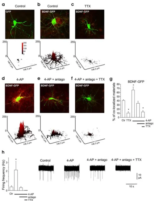
Figure 1. Spontaneous network activity triggers BDNF-GFP release. a–f, Overlapped images showing intracellular GFP fluo-rescence (green) and secreted BDNF-GFP detected using anti-GFP antibody (red) under nonpermeable conditions. a, Hippocam-pal neurons transfected with GFP alone. b–f, Neurons transfected with BDNF-GFP. b, Control condition. c, In the presence of TTX (1M). d, Stimulated with 4-AP (50M). e, Stimulated with 4-AP (50M) in the presence of NBQX (5M),D-APV (40M), and bicuculline (10M), indicated in the figure as “antago.” f, Stimulated with 4-AP (50M) in the presence of NBQX,D-APV, bicuculline, and TTX (1M). Top, Merged picture of both fluorescence channels of neurons transfected with the BDNF-GFP construct (green) and immunocytochemistry staining using an antibody against the GFP (red). Bottom, Quantification of the surface-bound BDNF-GFP on the transfected neuron (yellow signal); the plots are a three-dimensional representation of the mean gray level values. g, Quantitative analysis of surface-bound GFP comparing BDNF-GFP-transfected neurons in different ACSF conditions (n⫽ 4 different cultures, 4 neurons per culture in each condition). h, Average firing rate of the neurons in the different conditions, measured in cell-attached configuration (n⫽ 5 neurons in each condition). Right, Representative electro-physiological traces for each condition. The green signal produced by released BDNF-GFP is not visible in the images presented here because of the low laser intensity used to avoid saturation of the green signal in the transfected neurons. The red signal in the untransfected cell is attributable to the BDNF-GFP secreted by the transfected neurons that bound to membrane TrkB receptors of the neighboring cells. Antago in e, f, and h stands for NBQX (5M), APV (40M), and bicuculline (10M). Ctr, Control. In this and following figures, * indicates p⬍ 0.05 compared with control condition.
We then asked whether ongoing
net-work activity can cause BDNF secretion
and how the release is affected by the
mod-ification of the activity level of the cultures.
For this purpose, released BDNF-GFP was
detected by surface immunofluorescence
staining (see Materials and Methods).
Be-cause the surface-bound BDNF-GFP
de-pends not only on the quantity of the
re-lease but also on the density of the cells
surrounding the transfected neurons, we
compared the different conditions by
eval-uating exclusively the BDNF-GFP bound
on the extracellular membrane of the
transfected neurons. For this reason, we
use an antibody directed against GFP (red
signal) to identify BDNF-GFP that has
been released from the transfected cells. In
Figure 1, released BDNF-GFP that bound
to the extracellular membrane of
BDNF-GFP-expressing and nontransfected
neu-rons appears respectively as yellow and red
signal.
Yellow
staining
surrounding
BDNF-GFP-expressing neurons was
ob-served in control conditions (41
⫾ 2%
sur-face colocalized material) (Fig. 1b). In
con-trast, surface GFP immunofluorescence was
not detectable on GFP-only-expressing
neu-rons (3.5
⫾ 1.5% surface colocalized
mate-rial) (Fig. 1a). Adding TTX (1
M) to the
cul-ture medium for 3 h resulted in a marked
decrease in the surface-bound BDNF-GFP,
compared with control nontreated cultures
(Fig. 1c,h).
Ten minutes of 4-AP (50
M)
applica-tion led to an increase in postsynaptic
fir-ing activity (Fig. 1i) and dramatically
in-creased the density of surface-bound
BDNF-GFP (Fig. 1d,h). In the presence of
4-AP, the application of the ionotropic
glutamatergic and GABAergic receptor
an-tagonists NBQX,
D-APV, and bicuculline
decreases both neuronal firing (Fig. 1i) and
the density of surface-bound BDNF-GFP
(Fig. 1e,h) Finally, a further application of
TTX abolished neuronal firing (Fig. 1i)
and largely reduced density of
surface-bound BDNF-GFP (Fig. 1f,h).
Immunola-beling of the extracellular domain of the
TrkB receptors under nonpermeabilized conditions shows that
membrane TrkB receptor expression was not modified by the
different treatments (supplemental Fig. 2, available at www.
jneurosci.org as supplemental material), further supporting that
the modifications in surface-bound BDNF-GFP are
conse-quences of BDNF-GFP release changes.
Altogether, these observations show that, in hippocampal
cul-tures, spontaneous neuronal firing is required to trigger
BDNF-GFP release.
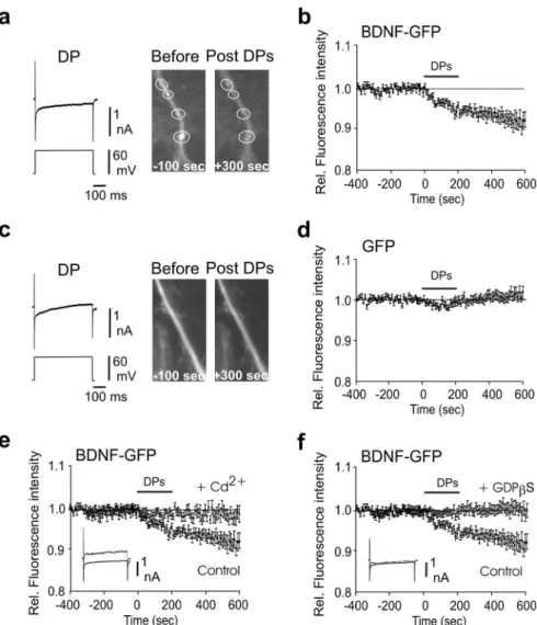
Repeated postsynaptic depolarization triggers a dendritic
Ca
2ⴙ-dependent release of BDNF-GFP
Next we combined whole-cell recording and time-lapse
fluores-cence imaging of transfected hippocampal neurons to directly
determine whether BDNF can be secreted from the dendrites
after short direct postsynaptic depolarization activating
voltage-dependent Ca
2⫹channels (VDCCs). To prevent possible effects
of excitatory synaptic activity, voltage-clamp whole-cell
record-ings were performed in the presence of the glutamatergic
recep-tor antagonists NBQX (5
M) and
D-APV (40
M). After a stable
control period, 20 DPs (50 – 60 mV amplitude, 500 ms duration
at 0.1 Hz) were applied to the cell through the recording pipette.
Each DP triggered an inward current (Fig. 2a). DPs applied to
BDNF-GFP-expressing neurons led to a significant decrease of
fluorescence intensity (⫺7.8 ⫾ 1.6% change 10 min after the
DPs; p
⫽ 0.0005 compared with pre-DPs; n ⫽ 11) (Fig. 2a,b).
When applied on GFP-only-expressing neurons, DPs only led to
a transient nonsignificant decrease in fluorescence (1
⫾ 1%
Figure 2. Depolarizing steps trigger dendritic secretion of BDNF-GFP. a, Decrease of fluorescence intensity from BDNF-GFP granules localized in the dendrites was produced by 20 depolarizing steps of 50 – 60 mV depolarization, 500 ms long, given at 0.01 Hz. Note that fluorescence decreased within the white circle and did not increase in the surrounding area, indicating that the decrease in fluorescence intensity was not attributable to lateral movements in the x–y-axes. Left, Representative traces of a DP; note the inward Ca2⫹current produced by the depolarization. Right, Example of dendritic BDNF-GFP granules (indicated by circles) before and after the DPs. b, Average time course of dendritic fluorescence change evoked by the DPs (n⫽ 11). c, d, DP failed to produce significant variation of fluorescence in GFP-only-transfected neurons (n⫽ 9). e, The effect of DPs on dendritic fluorescence was abolished by bath-applied Cd2⫹(200M; n⫽ 8); note the absence of Ca2⫹current in response to DP (superimposed control and Cd2⫹traces). f, Postsynaptic loading of GDP-S (0.6 mM), a G-protein inhibitor that blocks granular secretion, prevented the DP-induced decrease of fluorescence in the dendrites of BDNF-GFP-transfected cells (n⫽ 8); note that the Ca2⫹current in response to DP was not affected (superimposed control and Cd2⫹traces). All the experiments were per-formed in the presence of NBQX and APV. Rel., Relative.
change 10 min after DPs; p
⫽ 0.42 compared with pre-DPs; p ⫽
0.0001 compared with post-DPs in BDNF-GFP release
experi-ments; n
⫽ 9) (Fig. 2c,d), thus ruling out possible direct effect of
DPs on GFP fluorescence properties.
Because an intracellular Ca
2⫹influx through VDCCs is
nec-essary for dendritic BDNF release (Hartmann et al., 2001), we
investigated the effect of Cd
2⫹, a broad spectrum blocker of
VD-CCs. Bath-applied CdCl
2(100
M) blocked the DP-induced
in-ward currents (Fig. 2e, inset) and prevented the decrease of
fluo-rescence produced by the DPs (
⫺2 ⫾ 1.4% change; p ⫽ 0.33
compared with pre-DPs; p
⫽ 0.008 compared with post-DPs in
control group; n
⫽ 8) (Fig. 2e). The Ca
2⫹dependency of the
DP-induced decrease in fluorescence and the neuritic
accumula-tion of BDNF-GFP in secretory granule (supplemental Fig. 1,
available at www.jneurosci.org as supplemental material)
sug-gested that BDNF is released by membrane fusion and exocytosis.
To test this hypothesis, we investigated the effect of GDP
S, a
potent inhibitor of exocytosis (Zilberter et al., 1999). GDP
S (0.6
m
M) was dissolved in the pipette recording solution. In GDP
S-loaded cells, the DPs had no effect on the BDNF-GFP
fluores-cence intensity (0
⫾ 1.5%; p ⫽ 0.48 compared with pre DP; p ⫽
0.001 compared with control; n
⫽ 8) (Fig.
2f ). GDP
S had no effect on inward Ca
2⫹currents produced by the DPs (Fig. 2f ).
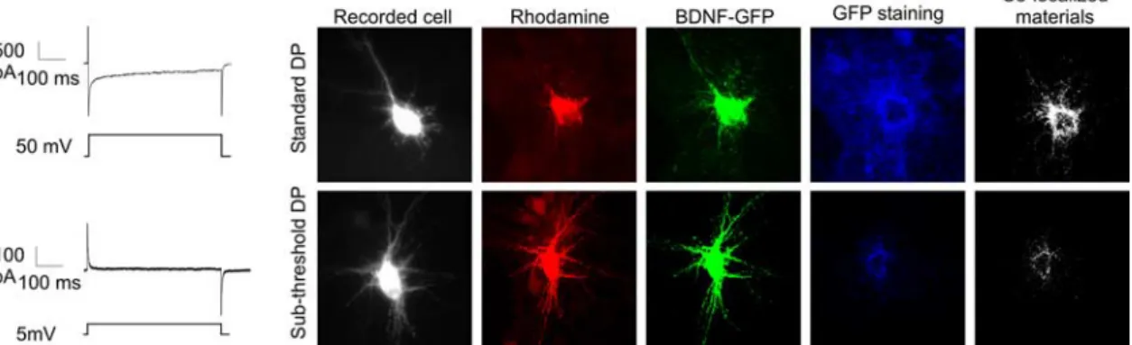
The release of BDNF-GFP by DPs was
confirmed by GFP surface
immunostain-ing (Fig. 3). Before recordimmunostain-ing, the culture
medium was washed three times, followed
by 3 h in TTX, to remove BDNF-GFP
re-leased by spontaneous activity. The
cul-ture was then transferred in the recording
chamber in the presence of NBQX (5
M)
and
D-APV (40
M). The recorded cell was
loaded with the fluorescent dye
rhoda-mine for post hoc identification.
BDNF-GFP-transfected cells were stimulated
ei-ther with the standard suprathreshold DPs
or with a subthreshold DPs (steps of 5 mV)
that did not produced inward Ca
2⫹cur-rents, and the culture was immediately
processed
for
immunohistochemistry
(Fig. 3). Suprathreshold DPs induced GFP
staining surrounding the
BDNF-GFP-expressing cells that spread to neighboring
nontransfected cells. With subthreshold
DPs, surface-bound GFP was restricted to
proximal BDNF-GFP expressers, possibly
because of constitutive release (Brigadski et al., 2005) (Fig. 3a).
Quantification of surface-bound BDNF-GFP on transfected
neu-rons show that suprathreshold DPs significantly increased
BDNF-GFP released (subthreshold DPs, 9
⫾ 2% of
colocaliza-tion; suprathreshold DPs, 56
⫾ 4% of colocalization; p ⫽
0.0001).
Altogether, these results show that repeated, short
postsynap-tic membrane depolarizations trigger a decrease in dendripostsynap-tic
flu-orescence in BDNF-GFP-expressing neurons that reflects a
Ca
2⫹-dependent release of BDNF.
Spontaneous backpropagating action potentials trigger
dendritic release of BDNF
As illustrated in Figure 4, both spontaneous and evoked somatic
action potentials travel back through the dendritic tree as b-APs
(n
⫽ 4; average distance, 100
M). We therefore investigated
whether spontaneous b-APs can lead to dendritic BDNF
secre-tion. Therefore, we performed time-lapse-imaging on
BDNF-GFP-expressing neurons recorded in current-clamp mode.
Dur-ing the control period, the cells were kept hyperpolarized at
Figure 4. Dendritic backpropagation of APs in cultured neurons. a, Example of paired somatic (s) and dendritic (d) whole-cell recording of a neuron in culture. b, APs elicited by somatic current injection backpropagate into the dendrite. c, Spontaneous AP generated in the soma backpropagates into the dendrite. Note the delay between somatic and dendritic AP.
Figure 3. Surface staining confirmed the BDNF-GFP secretion produced by the depolarizing steps. BDNF-GFP-expressing neurons received either 20 suprathreshold or 20 subthreshold DPs applied through the recording pipette. The intracellular solution contained rhodamine for post hoc identification. Surface staining of released BDNF-GFP was detected by immunocytochemical staining against GFP under nonpermeabilized conditions (blue staining).
around
⫺80 mV by steady current
injec-tion, to prevent action potential firing.
Af-ter the control period, neurons were
depo-larized to
⫺52 ⫾ 1 mV (n ⫽ 14; range,
⫺49 to ⫺60 mV). In five neurons, this
procedure led to the generation of
synap-tically driven firing activity; in these cases,
we observed a decrease in the dendritic
BDNF-GFP fluorescence (6
⫾ 1%
de-crease in fluorescence intensity 10 min
af-ter the first AP; n
⫽ 5; p ⫽ 0.002 compared
with prespike period) (Fig. 5a). On
aver-age, the cells fired 65
⫾ 22 APs at 0.5 ⫾
0.12 Hz (5.7
⫾ 1.4 Hz of maximum
instan-taneous frequency, quantified during the
first 2 min after the first AP). On the other
hand, in the neurons in which synaptic
ac-tivity did not reach the threshold for AP
generation (n
⫽ 5), or in which APs were
prevented by the Na
⫹channel blocker
5-N-(2,6-dimethylphenylcar-bamoylmethyl)triethylammonium
bro-mide (QX314; 5 m
M) in the pipette
solu-tion (n
⫽ 4), no variation in BDNF-GFP
fluorescence was observed (0.2
⫾ 0.9%
in-crease; n
⫽ 9; p ⫽ 0.7 compared with
con-trol; p
⫽ 0.003 compared with firing
group) (Fig. 5b).
A recent report has provided evidence
for BDNF secretion from a single neuron
produced by steady depolarization to
⫺40
mV in voltage-clamp recording (Magby et
al., 2006). To exclude possible direct
inter-action of QX314 with the process of BDNF
secretion, transfected neurons were
depo-larized from
⫺80 to ⫺40 mV in
current-clamp conditions, while the firing activity
was prevented by intracellular application
of QX314. This protocol produced
den-dritic release of BDNF-GFP not statistically different from that
induced by firing activity (4
⫾ 1% decrease in fluorescence
inten-sity; n
⫽ 6; p ⫽ 0.02 compared with control period; p ⫽ 0.24
compared with firing induced release) (Fig. 5c).
These observations show that backpropagating action
poten-tials are the principal trigger of dendritic BDNF-GFP release
in-duced by spontaneous network activity.
Spike-induced BDNF release required Ca
2ⴙfrom VDCC but
not from intracellular Ca
2ⴙstores
We next examined the minimal requirements for AP-induced
BDNF-GP secretion. First we investigated the contribution of
VDCCs for the spike-induced BDNF secretion. Because VDCCs
blockers can affect presynaptic transmitter release and thus,
in-directly, postsynaptic APs generated by ongoing synaptic activity,
intrinsic b-APs were directly induced by short (200 ms to 1 s)
current injections through the recording electrode. In control
conditions, the b-APs led to a decrease in dendritic BDNF-GFP
fluorescence (4.8
⫾ 1.2% decrease in fluorescence intensity; n ⫽
17; p
⫽ 0.01) (Fig. 6a). This decrease was abolished by bath
ap-plication of Cd
2⫹200
M(0.7
⫾ 0.8% decrease; n ⫽ 9; p ⫽ 0.42
compared with prespike period; p
⫽ 0.03 compared with control)
(Fig. 6b) or in zero extracellular calcium and 3 m
MEGTA (0.2
⫾
1% decrease; n
⫽ 4; p ⫽ 0.79 compared with prespike period;
data not shown) (see Hartmann et al., 2001).
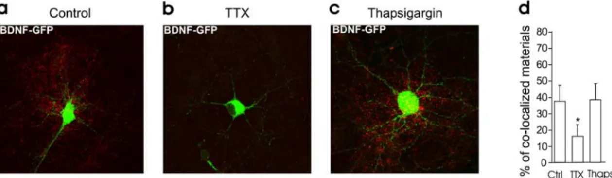
Several studies have stressed the important role of internal
Ca
2⫹stores in amplifying the initial Ca
2⫹influx for the release of
BDNF by high extracellular K
⫹or tetanic stimulation (Griesbeck
et al., 1999; Balkowiec and Katz, 2002; Kolarow et al., 2007). We
therefore investigated the possible contribution of intracellular
Ca
2⫹stores in BDNF release induced by b-APs. Twenty minutes
of preincubation of the neuronal cultures with 10
Mthapsigar-gin, an inhibitor of the endoplasmic reticulum Ca
2⫹-ATPase,
had no effect on dendritic BDNF-GFP release induced by b-APs
(5
⫾ 1% decrease in fluorescence intensity; n ⫽ 8; p ⫽ 0.002)
(Fig. 6c). Similarly, thapsigargin had no effect on BDNF secretion
occurring during spontaneous synaptic activity (Fig. 7).
Thapsi-gargin was, however, able to (1) produce a detectable rise in
rest-ing intracellular Ca
2⫹concentration (supplemental Fig. 3,
avail-able at www.jneurosci.org as supplemental material), (2) reduce
the amplitude of glutamate-induced Ca
2⫹rise (supplemental
Fig. 3, available at www.jneurosci.org as supplemental material),
and (3) as already reported (Kolarow et al., 2007), prevent
BDNF-GFP released induced by bath application of 50 m
Mpotassium
(KCl-induced decrease of fluorescence in control
⫽ 7.8 ⫾ 1.3%;
n
⫽ 11; p ⫽ 0.0002 compared with pre-KCl period; KCl-induced
decrease of fluorescence in the presence of thapsigargin
⫽ 2 ⫾
Figure 5. Synaptically driven b-APs trigger dendritic BDNF secretion through membrane depolarization. a, Synaptically in-duced b-APs, occurring when neurons were held at⫺52 ⫾ 1 mV, produced a dendritic BDNF-GFP secretion (n ⫽ 5); the arrow indicates the arrival of the first AP. Left, Representative trace. b, BDNF-GFP secretion did not occur when APs were prevented by QX314 in the recording pipette (n⫽ 9). Left, Representative trace. c, Membrane depolarization to ⫺40 mV, with QX314 in the pipette, produced a BDNF-GFP secretion even in the absence of APs (n⫽ 6). Left, Representative trace. Rel., Relative.
2%; n
⫽ 8; p ⫽ 0.3 compared with pre-KCl period; p ⫽ 0.02
compared with control) (supplemental Fig. 4, available at www.
jneurosci.org as supplemental material). We concluded that,
un-like in experiments with bath application of KCl, the release of
BDNF induced by b-APs does not require activation of other than
VDCC calcium sources.
We next investigated the minimum number of APs necessary
to produce a significant BDNF-GFP fluorescence loss. To control
the exact number of APs produced for each neuron, APs were
induced by short (10 ms) somatic current injections applied at a
frequency of 4 Hz in the presence of NBQX and APV. As shown in
Figure 8, increasing the number of APs increases the probability
of BDNF secretion reaching a plateau at eight APs.
Altogether, these observations show that backpropagating
ac-tion potentials trigger a dendritic, Ca
2⫹-dependent release of
BDNF-GFP through the activation of VDCCs.
b-APs trigger endogenous BDNF release
To date, experiments on activity-dependent release of BDNF in
cultured neurons have been performed on transfected cells that
overexpress BDNF (Goodman et al., 1996;
Griesbeck et al., 1999; Hartmann et al.,
2001; Balkowiec and Katz, 2002; Ga¨rtner
and Staiger, 2002; Brigadski et al., 2005).
However, such overexpression could have
modified the release properties of BDNF.
We therefore asked whether b-APs are able
to trigger secretion of native BDNF in
nontransfected cells. To this end, we used
pCREB as a sensor of BDNF release. Once
released, BDNF interacts with
high-affinity TrkB receptors to activate
down-stream signaling pathways. One of the
most common is the ERK (extracellular
signal-regulated protein kinase) pathway,
which leads to the phosphorylation of
CREB (Ghosh et al., 1994). We reasoned
that BDNF released from one single
stim-ulated neuron will bind to TrkB receptors
in the neighboring cells and induce the
phosphorylation of CREB.
Nontrans-fected cultures were incubated with TTX
for 4 h to reduce the basal levels of pCREB
and then transferred in the recording
chamber in the presence of CNQX and
D
-APV to prevent postsynaptic firing
gen-erated by spontaneous synaptic activity.
Four bursts of 10 APs (10 Hz; 5 s between
bursts) were produced by 10 ms current
step in one single neuron per culture, a
protocol that induces BDNF-GFP release
from transfected neurons (supplemental
Fig. 5, available at www.jneurosci.org as
supplemental material). The neuron was
loaded with rhodamine for post hoc
iden-tification. Five minutes after stimulation,
the cultures were fixed and processed for
immunocytochemistry. APs induced an
increase in pCREB level in MAP2-positive
cells (Fig. 9d–f,j). pCREB level was
signifi-cantly ( p
⫽ 0.001) lower in cultures in
which the cells were patched but not
stim-ulated (Fig. 9a– c,j). Finally, the increase in
pCREB induced by APs was prevented in the presence of the
BDNF scavenger TrkB-IgG (5
g/ml) in the culture medium,
showing that CREB phosphorylation results from the release of
BDNF (Fig. 9g–i,j). In all experiments, pCREB level was not
mod-ified in the MAP2-negative cells (i.e., glial cells) ( p
⫽ 0.973). No
differences in the total amount of CREB was observed for the
different conditions (mean gray values: control, 236
⫾ 18; n ⫽ 3
cultures, 20 cells per culture; TrkB, 230
⫾ 20; n ⫽ 3 cultures, 20
cells per culture; stimulated, 232
⫾ 17; n ⫽ 3 cultures, 20 cells per
culture; p
⫽ 0.32, ANOVA; data not shown).
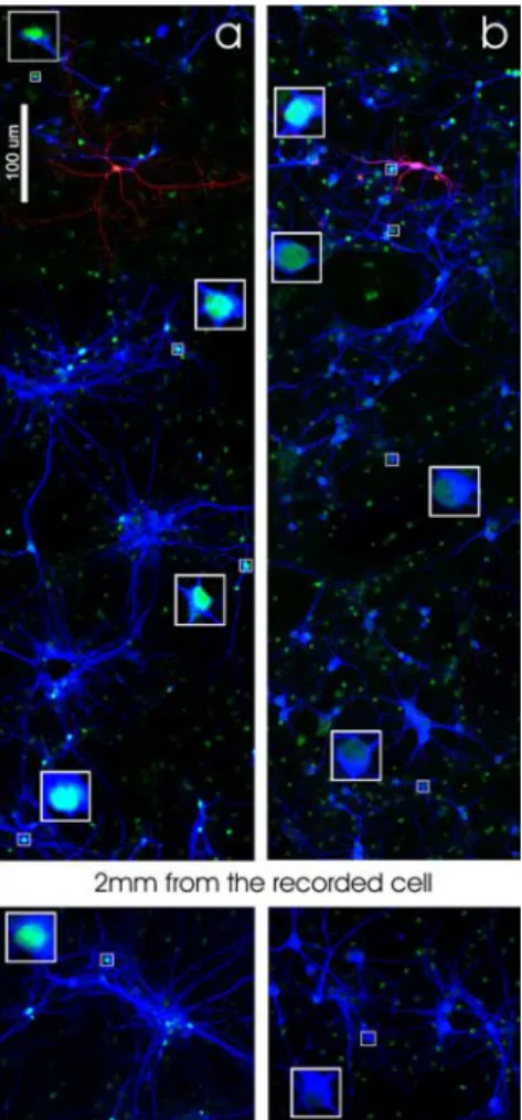
As illustrated in Figure 10, pCREB-positive somata are not
only restricted to the vicinity of the stimulated cell but can be
observed at long distance (up to 2
⫾ 0.5 mm; n ⫽ 3) (Fig. 10a).
One possible explanation for such broad distribution of
pCREB-positive neurons relies on the fact that both dendrites and axons
can release BDNF when stimulated (Lessmann et al., 2003) and
that BDNF can bind to TrkB receptors located on both axons and
dendrites of the “recipient” cells (Ginty and Segal, 2002;
Nagap-pan and Lu, 2005). Given the broad and complex dendritic and
axonal distribution of the cells in cultures (supplemental Fig. 6,
Figure 6. VDCC activation, but not intracellular Ca2⫹stores, is required for b-AP-induced BDNF secretion. a, BDNF-GFP secretion was induced by firing activity produced by somatic current injection. Left, Representative trace of step-induced firing activity (n⫽ 17). b, BDNF-GFP release was prevented by bath application of the VDCC blocker CdCl (200M; n⫽ 9). c, Thapsi-gargin (10M) did not prevent BDNF-GFP release elicited by b-APs (n⫽ 8). Left, Representative traces of step-induced firing activity. Arrows indicate the arrival of the first action potential. In a– c, Vh⫽ ⫺70 mV. Rel., Relative.
available at www.jneurosci.org as supplemental material),
pCREB-positive somata can be observed at a distance from the
stimulated cell. To restrict the release of BDNF to proximal
neu-rites, in another series of experiments, depolarizing pulses (20
DPs of 50 mV, 500 ms duration at 0.1 Hz) were applied through
the recording pipette, in the presence of TTX 1
M, to directly
activate proximal VDCCs while reducing the spread of the
depo-larization into distal neurites. In these conditions, DPs induced
an increase in pCREB level restricted to the neighboring
MAP2-positive cells (Fig. 10b) (radius from the soma of the stimulated
cell
⫽ 230 ⫾ 50
m; n ⫽ 3).
These experiments therefore show that spike firing in one
“donor” single cell can induce the release of endogenous BDNF
that activates TrkB receptors on “recipient” neurons
Discussion
Several studies have shown that impairing network activity
pro-duces structural and functional alterations that are reverted by
exogenous application of BDNF (McAllister et al., 1996;
Ruther-ford et al., 1997; Marty et al., 2000; Seil and Drake-Baumann,
2000), suggesting that activity-dependent secretion of BDNF can
act as a regulatory mechanism of network development and
plas-ticity. However, direct evidence for dendritic BDNF release
in-Figure 7. Intracellular Ca2⫹stores are not required for BDNF secretion produced by ongoing activity. a– c, Surface immunofluorescence staining on neuronal cultures in control condition (a) and
incubated in TTX (b) or thapsigargin (10M; c) for 3 h. d, Quantitative analysis of surface-bound GFP (yellow signal/green signal) in the different conditions (n⫽ 3 cultures, 4 neurons per cultures
in each condition). Thapsi, Thapsigargin.
Figure 8. The probability of BDNF-GFP release increases with the number of APs. The prob-ability to induce a dendritic BDNF-GFP secretion (fluorescence decrease⬎3%between100and 200 s after stimuli) is a function of the number of APs elicited by somatic current injection (at 3.96⫾0.5Hz).Inset,RepresentativetraceofAPs.Inparenthesesisthenumberofexperiments.
Figure 9. Endogenous BDNF is released by firing activity. Shown is activation of pCREB in nontransfected neuronal cells after electrophysiological stimulation. Left and middle, Images of pCREB (left) and MAP2 (middle), in neurons neighboring the patched rhodamine-filled cell (arrows). Right, Merged pictures of the rhodamine-filled cell immunofluorescence (red) in the presence of MAP2 and pCREB immunostaining (blue) for the same field of view. Scale bar, 45 m.a–c,Controlcondition;cellpatchedbutnotstimulated.d–f,Cellpatchedandstimulated to fire (40 spike at 10 Hz). g–i, Cell patched and stimulated to fire (40 spikes at 10 Hz) in the presence of the BDNF scavenger TrkB IgG. j, Histogram showing the pCREB level activation by measurement of fluorescence intensity (mean gray level values) (n⫽ 3 cultures in each condi-tion. The amount of pCREB was calculated through a distance of 250m from the recorded neuron. A total of 47, 56, and 49 neurons, respectively, were used for analysis of the intensity of pCREB in control, stimulated, and TrkB-IgG conditions). Electrophysiology was performed in ACSF supplemented with NBQX and APV. Stim, Stimulated.
duced by ongoing network activity was still lacking. Our study is
the first direct demonstration of cell autonomous, Ca
2⫹-dependent, dendritic release of BDNF in response to
spontane-ous activity. Moreover, we provide evidence that endogenspontane-ous
BDNF released from a single neuron can phosphorylate CREB in
neighboring neurons, an important step of immediate early gene
activation (Finkbeiner et al., 1997). Therefore, together, our
re-sults support the general hypothesis that BDNF can serve as a
target-derived messenger for activity-dependent development
and plasticity in response to single-neuron activation (Tanaka et
al., 2008).
b-APs are necessary and sufficient to trigger
activity-dependent dendritic release of BDNF
Our results show that b-APs are necessary and sufficient to trigger
activity-dependent dendritic release of BDNF. Thus, we found
that (1) ionotropic glutamatergic receptor antagonists were not
able to abolish activity-dependent release of BDNF (Fig. 1), (2) in
the presence of QX314, to block APs in the recorded cell,
synap-tically induced membrane depolarization was not able to
pro-duce any detectable release of BDNF-GFP (Fig. 5b), and (3)
evoked b-APs triggered in single cells induced dendritic release of
BDNF-GFP (Fig. 6a). Using phosphorylation of CREB in
neigh-boring neurons as a sensor for BDNF release, to avoid the possible
artifactual consequences of overexpressing tagged proteins, we
also show that APs induced in nontransfected neurons can lead to
the release of endogenous BDNF in the presence of ionotropic
glutamatergic receptor antagonists (Fig. 9).
Our results therefore suggest that depolarization mediated by
the activation of ionotropic glutamatergic transmission during
ongoing synaptic activity does not directly lead to BDNF
secre-tion. We, however, cannot exclude a contribution of
receptor-mediated depolarization on BDNF secretion in a more intact
system, for instance, during physiological or pathological
net-work oscillations. Thus, a dendritic release of BDNF-GFP evoked
by presynaptic tetanic stimulation of glutamatergic terminals in
neuronal cultures was blocked by AMPA and NMDA receptor
antagonists (Hartmann et al., 2001). This suggests that a
receptor-mediated depolarization of postsynaptic neurons,
caused by summation of glutamatergic postsynaptic potentials
and possibly the generation of APs, can trigger the postsynaptic
release of BDNF during strong presynaptic tetanic stimulation
(Hartmann et al., 2001). Therefore, although dendritic BDNF
release in the absence of neuronal firing can be triggered by
ex-perimentally induced membrane depolarization of sufficient
am-plitude (Fig. 5c) [but see also Magby et al. (2006) and Kolarow et
al., (2007)], during ongoing spontaneous network activity b-APs
generation is a required and sufficient step to trigger dendritic
BDNF release. In the present study, we show that as few as eight
b-APs at a frequency of 4 Hz are sufficient to produce a secretion
of BDNF. In a previous study, Ga¨rtner and Staiger (2002)
re-ported that a minimum of 100 electrical shocks induced at a
frequency of 50 Hz by field stimulation were required to detect
BDNF secretion. This difference could be explained by a lower
sensitivity of the ELISA technique used in this former study to
measure the extracellular BDNF. Interestingly, hippocampal
py-ramidal neurons fire spontaneous bursts of three to seven APs in
vivo (Ranck, 1973). Thus, our data describe that a physiological
pattern of neuronal firing can trigger dendritic secretion of
BDNF. This provides a new perspective on studies of the role of
ongoing neuronal activity in the maintenance of endogenous
BDNF levels in neuronal cultures and brain.
Several studies have stressed the important role of internal
Ca
2⫹stores in amplifying the initial Ca
2⫹influx for the release of
BDNF after electrical field stimulation (Balkowiec and Katz,
2002) or high K
⫹concentration (Griesbeck et al., 1999; Kolarow
et al., 2007). Here, we show that the inhibitor of the endoplasmic
reticulum Ca
2⫹-ATPase thapsigargin, which efficiently prevents
BDNF secretion triggered by high-potassium application
(Ko-larow et al., 2007; present study), does not prevent BDNF
secre-tion triggered by b-APs or ongoing synaptic activity. This result
suggests that the mechanism underlying Ca
2⫹-dependent
secre-tion of BDNF could differ depending on the stimulating
proto-col. The reason for this dissimilarity is presently unknown.
Dif-ference in kinetic and/or amplitude between KCl- or AP-induced
Ca
2⫹rises could explain why amplification of the Ca
2⫹signal by
intracellular stores is required in one case but not the other.
In-ternal stores might be more important for potassium-induced
release because of the inactivation of the VDCC after such
pro-longed depolarization, whereas this inactivation is absent after
repetitive (i.e., phasic) stimulation. It should be pointed out that
thapsigargin has been reported to increase the decay time course
but not the amplitude of the transient Ca
2⫹signal evoked by
b-APs, suggesting that internal Ca
2⫹stores participated in
den-dritic clearance of Ca
2⫹, but not in the Ca
2⫹rise (Markram et al.,
1995) (but see Sandler and Barbara, 1999).
Figure 10. Distribution of pCREB-positive neurons after single-cell stimulation. a, CREB phosphorylation produced by neuronal stimulation (40 spikes at 10 Hz) in current clamp is distributed in the culture and can be observed at a distance from the stimulated cell. b, CREB phosphorylation produced by neuronal stimulation (20 DP of 50 mV at 0.1 Hz) in voltage clamp in the presence of TTX is limited to the neurons surrounding the stimulated cell. Squares show nuclei at higher magnification.
Functional significance
Backpropagating APs in hippocampal and cortical pyramidal
cells evoke a transient dendritic increase in intracellular Ca
2⫹concentration, providing an associative signal for long-term
syn-aptic plasticity (Magee and Johnston, 1997; Markram et al.,
1997). The b-APs can also trigger a Ca
2⫹-dependent dendritic
release of different messengers, leading to short-term control of
synaptic transmission efficacy on both pyramidal cells and
inter-neurons (Zilberter et al., 2005). The observation that b-APs can
trigger a dendritic Ca
2⫹-dependent secretion of BDNF, and that
BDNF secretion from one single neuron can activate the
imme-diate early genes upstream regulator CREB in neighboring
neu-rons, provides another mechanism by which target neurons can
affect the development or efficacy of impinging synaptic
connec-tions. Accordingly, several forms of synaptic plasticity evoked by
postsynaptic firing (Gubellini et al., 2005) or correlated
presyn-aptic and postsynpresyn-aptic activity (Kang et al., 1997; Mu and Poo,
2006; Walz et al., 2006; Mohajerani et al., 2007) require the
pres-ence of endogenous BDNF. Moreover, BDNF released from
tar-get neurons locally promotes synapse formation (Ohba et al.,
2005; Kohara et al., 2007; Tanaka et al., 2008). Although
extrap-olations of data from neuronal cultures to a more intact system
are limited, our finding that b-APs or short postsynaptic
depo-larization triggers dendritic release of BDNF supports the notion
that BDNF can act as a target-derived messenger for synaptic
plasticity and development.
References
Aicardi G, Argilli E, Cappello S, Santi S, Riccio M, Thoenen H, Canossa M (2004) Induction of long-term potentiation and depression is reflected by corresponding changes in secretion of endogenous brain-derived neu-rotrophic factor. Proc Natl Acad Sci U S A 101:15788 –15792.
Balkowiec A, Katz DM (2002) Cellular mechanisms regulating activity-dependent release of native brain-derived neurotrophic factor from hip-pocampal neurons. J Neurosci 22:10399 –10407.
Bonhoeffer T (1996) Neurotrophins and activity-dependent development of the neocortex. Curr Opin Neurobiol 6:119 –126.
Bramham CR, Messaoudi E (2005) BDNF function in adult synaptic plasticity: the synaptic consolidation hypothesis. Prog Neurobiol 76:99 –125.
Brigadski T, Hartmann M, Lessmann V (2005) Differential vesicular target-ing and time course of synaptic secretion of the mammalian neurotro-phins. J Neurosci 25:7601–7614.
Buerli T, Pellegrino C, Baer K, Lardi-Studler B, Chudotvorova I, Fritschy J, Medina I, Fuhrer C (2007) Efficient transfection of DNA or shRNA vec-tors into neurons using magnetofection. Nat Protoc 2:3090 –3101. Finkbeiner S, Tavazoie SF, Maloratsky A, Jacobs KM, Harris KM, Greenberg
ME (1997) CREB: a major mediator of neuronal neurotrophin re-sponses. Neuron 19:1031–1047.
Ga¨rtner A, Staiger V (2002) Neurotrophin secretion from hippocampal neurons evoked by long-term-potentiation-inducing electrical stimula-tion patterns. Proc Natl Acad Sci U S A 99:6386 – 6391.
Ghosh A, Carnahan J, Greenberg ME (1994) Requirement for BDNF in activity-dependent survival of cortical neurons. Science 263:1618 –1623. Ginty DD, Segal RA (2002) Retrograde neurotrophin signaling: Trk-ing
along the axon. Curr Opin Neurobiol 12:268 –274.
Goodman LJ, Valverde J, Lim F, Geschwind MD, Federoff HJ, Geller AI, Hefti F (1996) Regulated release and polarized localization of brain-derived neurotrophic factor in hippocampal neurons. Mol Cell Neurosci 7:222–238.
Griesbeck O, Canossa M, Campana G, Ga¨rtner A, Hoener MC, Nawa H, Kolbeck R, Thoenen H (1999) Are there differences between the secre-tion characteristics of NGF and BDNF? Implicasecre-tions for the modulatory role of neurotrophins in activity-dependent neuronal plasticity. Microsc Res Tech 45:262–275.
Gubellini P, Ben-Ari Y, Gaı¨arsa JL (2005) Endogenous neurotrophins are required for the induction of GABAergic long-term potentiation in the neonatal rat hippocampus. J Neurosci 25:5796 –5802.
Hartmann M, Heumann R, Lessmann V (2001) Synaptic secretion of BDNF after high-frequency stimulation of glutamatergic synapses. EMBO J 20:5887–5897.
Haubensak W, Narz F, Heumann R, Lessmann V (1998) BDNF-GFP con-taining secretory granules are localized in the vicinity of synaptic junc-tions of cultured cortical neurons. J Cell Sci 111:1483–1493.
Huang EJ, Reichardt LF (2001) Neurotrophins: roles in neuronal develop-ment and function. Annu Rev Neurosci 24:677–736.
Kang H, Welcher AA, Shelton D, Schuman EM (1997) Neurotrophins and time: different roles for TrkB signaling in hippocampal long-term poten-tiation. Neuron 19:653– 664.
Kohara K, Yasuda H, Huang Y, Adachi N, Sohya K, Tsumoto T (2007) A local reduction in cortical GABAergic synapses after a loss of endogenous brain-derived neurotrophic factor, as revealed by single-cell gene knock-out method. J Neurosci 27:7234 –7244.
Kolarow R, Brigadski T, Lessmann V (2007) Postsynaptic secretion of BDNF and NT-3 from hippocampal neurons depends on calcium cal-modulin kinase II signaling and proceeds via delayed fusion pore opening. J Neurosci 27:10350 –10364.
Krapivinsky G, Krapivinsky L, Manasian Y, Ivanov A, Tyzio R, Pellegrino C, Ben-Ari Y, Clapham DE, Medina I (2003) The NMDA receptor is cou-pled to the ERK pathway by a direct interaction between NR2B and Ras-GRF1. Neuron 40:775–784.
Lessmann V, Gottmann K, Malcangio M (2003) Neurotrophin secretion: current facts and future prospects. Prog Neurobiol 69:341–374. Lu B, Figurov A (1997) Role of neurotrophins in synapse development and
plasticity. Rev Neurosci 8:1–12.
Lu B, Pang PT, Woo NH (2005) The yin and yang of neurotrophin action. Nat Rev Neurosci 6:603– 614.
Magby JP, Bi C, Chen ZY, Lee FS, Plummer MR (2006) Single-cell charac-terization of retrograde signaling by brain-derived neurotrophic factor. J Neurosci 26:13531–13536.
Magee JC, Johnston D (1997) A synaptically controlled, associative signal for Hebbian plasticity in hippocampal neurons [see comments]. Science 275:209 –213.
Markram H, Helm PJ, Sakmann B (1995) Dendritic calcium transients evoked by single back-propagating action potentials in rat neocortical pyramidal neurons. J Physiol 485:1–20.
Markram H, Lu¨bke J, Frotscher M, Sakmann B (1997) Regulation of synap-tic efficacy by coincidence of postsynapsynap-tic APs and EPSPs. Science 275:213–215.
Marty S, Berzaghi MdaP, Berninger B (1997) Neurotrophins and activity-dependent plasticity of cortical interneurons. Trends Neurosci 20:198 –202.
Marty S, Wehrle´ R, Sotelo C (2000) Neuronal activity and brain-derived neurotrophic factor regulate the density of inhibitory synapses in or-ganotypic slice cultures of postnatal hippocampus. J Neurosci 20:8087– 8095.
McAllister AK, Katz LC, Lo DC (1996) Neurotrophic regulation of cortical dendritic growth requires activity. Neuron 17:1057–1064.
Mohajerani MH, Sivakumaran S, Zacchi P, Aguilera P, Cherubini E (2007) Correlated network activity enhances synaptic efficacy via BDNF and the ERK pathway at immature CA3 CA1 connections in the hippocampus. Proc Natl Acad Sci U S A 104:13176 –13181.
Mu Y, Poo MM (2006) Spike timing-dependent LTP/LTD mediates visual experience-dependent plasticity in a developing retinotectal system. Neu-ron 50:115–125.
Nagappan G, Lu B (2005) Activity-dependent modulation of the BDNF re-ceptor TrkB: mechanisms and implications. Trends Neurosci 28:464 – 471.
Ohba S, Ikeda T, Ikegaya Y, Nishiyama N, Matsuki N, Yamada MK (2005) BDNF locally potentiates GABAergic presynaptic machineries: target-selective circuit inhibition. Cereb Cortex 15:291–298.
Poo MM (2001) Neurotrophins as synaptic modulators. Nat Rev Neurosci 2:24 –32.
Ranck JB Jr (1973) Studies on single neurons in dorsal hippocampal forma-tion and septum in unrestrained rats. I. Behavioral correlates and firing repertoires. Exp Neurol 41:461–531.
Rutherford LC, DeWan A, Lauer HM, Turrigiano GG (1997) Brain-derived neurotrophic factor mediates the activity-dependent regulation of inhi-bition in neocortical cultures. J Neurosci 17:4527– 4535.
Sandler VM, Barbara JG (1999) Calcium-induced calcium release contrib-utes to action potential-evoked calcium transients in hippocampal CA1 pyramidal neurons. J Neurosci 19:4325– 4336.
Schuman EM (1999) Neurotrophin regulation of synaptic transmission. Curr Opin Neurobiol 9:105–109.
Seil FJ, Drake-Baumann R (2000) TrkB receptor ligands promote activity-dependent inhibitory synaptogenesis. J Neurosci 20:5367– 5373.
Tanaka J, Horiike Y, Matsuzaki M, Miyazaki T, Ellis-Davies GC, Kasai H (2008) Protein synthesis and neurotrophin-dependent structural plastic-ity of single dendritic spines. Science 319:1683–1687.
Tyler WJ, Alonso M, Bramham CR, Pozzo-Miller LD (2002) From
ac-quisition to consolidation: on the role of brain-derived neurotrophic factor signaling in hippocampal-dependent learning. Learn Mem 9:224 –237.
Walz C, Ju¨ngling K, Lessmann V, Gottmann K (2006) Presynaptic plasticity in an immature neocortical network requires NMDA receptor activation and BDNF release. J Neurophysiol 96:3512–3516.
Zilberter Y, Kaiser KM, Sakmann B (1999) Dendritic GABA release de-presses excitatory transmission between layer 2/3 pyramidal and bitufted neurons in rat neocortex. Neuron 24:979 –988.
Zilberter Y, Harkany T, Holmgren CD (2005) Dendritic release of retro-grade messengers controls synaptic transmission in local neocortical net-works. Neuroscientist 11:334 –344.