HAL Id: hal-02991746
https://hal.archives-ouvertes.fr/hal-02991746
Submitted on 4 Dec 2020HAL is a multi-disciplinary open access archive for the deposit and dissemination of sci-entific research documents, whether they are pub-lished or not. The documents may come from teaching and research institutions in France or abroad, or from public or private research centers.
L’archive ouverte pluridisciplinaire HAL, est destinée au dépôt et à la diffusion de documents scientifiques de niveau recherche, publiés ou non, émanant des établissements d’enseignement et de recherche français ou étrangers, des laboratoires publics ou privés.
Unraveling the degradation of artificial amide bonds in
nylon oligomer hydrolase: from induced-fit to acylation
processes
Takeshi Baba, Mauro Boero, Katsumasa Kamiya, Hiroyuki Ando, Seiji
Negoro, Masayoshi Nakano, Yasuteru Shigeta
To cite this version:
Takeshi Baba, Mauro Boero, Katsumasa Kamiya, Hiroyuki Ando, Seiji Negoro, et al.. Unraveling the degradation of artificial amide bonds in nylon oligomer hydrolase: from induced-fit to acylation processes. Physical Chemistry Chemical Physics, Royal Society of Chemistry, 2015, 17 (6), pp.4492-4504. �10.1039/C4CP04419C�. �hal-02991746�
1
Unraveling the degradation of artificial amide bonds in Nylon
oligomer hydrolase: From induced-fit to acylation processes
*Takeshi Babaa,b, Mauro Boeroc, Katsumasa Kamiyad, Hiroyuki Andoa,
Seiji Negoroe, Masayoshi Nakanoa, *Yasuteru Shigetaf,g
a Department of Materials Engineering Science, Graduate School of Engineering Science, Osaka
University, 1-3 Machikaneyama, Toyonaka, Osaka 560-8531, Japan.
b Japan Society for the Promotion of Science (JSPS) Research Fellow.
c Institut de Physique et Chimie des Matériaux de Strasbourg, UMR 7504 CNRS and University of
Strasbourg, Strasbourg 23 rue du Loess, 67034 Strasbourg, France.
d Center for Basic Education and Integrated Learning, Kanagawa Institute of Technology, 1030
Shimo-Ogino, Atsugi, Kanagawa 243-0292, Japan.
e Department of Material Science and Chemistry, Graduate School of Engineering, University of
Hyogo, 2167 Shosha, Himeji, Hyogo 671-2280, Japan.
f Department of Physics, Graduate School of Pure and Applied Sciences, University of Tsukuba,
1-1-1 Tennodai, Tsukuba, Ibaraki 305-8571, Japan.
g JST, CREST, 4-1-8 Honcho, Kawaguchi, Saitama 332-0012, Japan.
2
ABSTRACT
To elucidate how the nylon oligomer hydrolase (NylB) acquires its peculiar degradation activity towards non-biological amide bonds, we inspect the underlying enzymatic processes going from the induced-fit upon substrate binding to acylation. Specifically, we investigate the mutational effects of two mutants, Y170F and D181G, indicated in former experiments as crucial systems because of their specific amino acid residues. To this aim, by resorting on first-principles molecular dynamics complemented with metadynamics we provide a detailed insight into the underlying acylation mechanism. Our results show that while in the wild type (WT) the Tyr170 residue points the NH group towards the proton-acceptor site of an artificial amide bond, hence ready to react, in the Y170F this does not occur. The reason is ascribed to the absence of Tyr170 in the mutant, replaced by phenylalanine, unable to form hydrogen bonds with the amide bond, thus resulting in an increase of the activation barrier of more than 10 kcal/mol. Nonetheless, despite the lack of hydrogen bonding between the Y170F and the mutant, also in this case the highest free energy barrier for the induced-fit is similar to that of WT. This seems to suggest that, in the induced-fit process, kinetics is little affected by the mutation. On the basis of additional structural homology analyses on enzymes of the same family, we suggest that natural selection is responsible for the development of the peculiar hydrolytic activity of Arthrobacter sp. KI72.
3
1. Introduction
Nylon-6 is a synthetic fiber with a wealth of applications from textiles to electronics industry and, as such, increasingly produced by chemical companies worldwide. The fundamental reaction
exploited is the ring-opening polymerization of ε-caprolactams.1,2 However, this has a
non-negligible environmental impact, since it amounts to industrial-scale production of non-biological amide bonds in long fibers containing more than 100 units of 6-aminohexanoate (Ahx). In this respect, an unprecedented discovery dating back to the 1940’s is the existence of Nylon-oligomer hydrolases, found in the Arthrobacter sp. KI72, able to degrade the nylon-6. This discovery brought to the attention of the scientific community that this bacterium was able to adapt to the presence of these non-biological compounds and, through natural selection, to develop a new functionality suitable to degrade the artificial Nylon-6 fibers at least for a relatively short period. Nowadays, three different types of the Nylon-oligomer hydrolases have been identified, namely Ahx-cyclic-dimer hydrolase (NylA), Ahx-liner-dimer (Ald) hydrolase (NylB), and endo-type Ahx-oligomer hydrolase (NylC).3-8 Each one of these hydrolase has its own inherent substrate
specificity. Specifically, NylA degrades the amide bond present in a cyclic Ahx dimer to produce a 6-aminohexanoate-liner-dimer (Ald). NylB degrades the amide bond formed in a linear Ahx oligomer, especially Ald, from the N-terminal of the oligomer. NylC degrades amide bonds of both cyclic and linear oligomers. Moreover, NylC is also able to revert nylon-6 into Ahx oligomers.7,8 Among these three Nylon-oligomer hydrolases, NylB has received special attention in a series of biochemical experiments. According to the reported genetic analyses, NylB has a closely resembling enzyme, NylB*, characterized by a 88% sequence identity. Nonetheless, despite this
close similarity, the reactivity to the substrate (Ald) of NylB* is lower than that of NylB by about a factor of 1/200.3 In an attempt at enhancing the reactivity and to overcome the difficulty in their
crystallization, hybrid enzymes of both NylB and NylB* were made by replacing some amino acid
residues in NylB*.9 This resulted in a set of 24 different structures labeled from Hyb-1 to Hyb-24 which were subsequently structurally analyzed by X-ray crystallography or crystallization. A noticeable example of this extensive investigation is represented by Hyb-24; this specific hybrid enzyme contains five amino acid residue substitutions with respect to the pristine NylB*: T3A, P4R,
4 T5S, S8Q, and D15G. Analogously, in the recently obtained Hyb-24DN and Hyb-24DNY a double mutation (G181D and H266N) and a triple mutation (G181D, H266N, and D370Y) with respect to
Hyb-24 were realized by one of us.10-13 The specific mutational sites were deduced by a comparison
of the amino acid sequences of NylB and NylB* (Asp181 and Asn266 in NylB, as opposed to
Gly181 and His266 in NylB*).9,10 It is also worthy to note that polymerase chain reaction (PRC) applied in random mutagenesis experiments, accidentally resulted in the discovery of the D370Y mutation mentioned above; nonetheless, its role was a matter of debate. Only later12,13 kinetics analyses allowed to establish that the actual role of D370Y is a stabilization of enzyme substrate complex. At the present state of knowledge, Hyb-24DNY possesses the highest activity among all the known mutants.12, 13 For this reason, hereafter, we shall assume Hyb-24DNY as our reference system and indicate it as the “wild type” (WT) for simplicity. Judging from the similarities in the overall structure and the amino acid sequence, NylB belongs to the class C of the β-lactamase family, which is characterized by the presence of a catalytic triad, Ser, Tyr, and Lys, having the ability to promote the hydrolysis of amide and/or ester bonds.9 According to X-ray crystallographic
analyses,11,13 the WT can assume two different conformations, (i) a substrate-free form (open form)
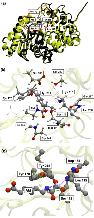
and (ii) a substrate-bound form (closed from) (see Fig.1 (a) for details). At variance with the other enzymes of the same family, the distinctive feature of NylB is the presence of a unique amino acid residue, Tyr170, near the catalytic triad, which forms a hydrogen bond (H-bond) with Ald, only in the closed form as sketched in Fig. 1(b).
Site-directed mutagenesis investigations were conducted on Hyb-24DN, the mutant in which the Tyr170 residue is replaced by a phenylalanine. These studies revealed that the hydrolytic activity of
Ald is nearly suppressed, becoming only about 1.4%, and its kinetics parameter values14,15, namely
the Michaelis constant (KM) and the rate constant (kcat) could not be determined because of such a low activity.11 This is an additional difficulty with respect to other mutants such as, for instance, Hyb-24Y (one mutation of D370Y from Hyb-24), Hyb-24DY (double mutation of G181D and D370Y from Hyb-24), Hyb-24DN, and Hyb-24DNY, for which the relevant kinetic parameters could be determined.13 This can again be ascribed to the fact that the Tyr170 residue is present both
5 is a crucial actor in the hydrolytic activity promoting the cleavage of non-biological amide bonds. Yet, the atomic-level elucidation of the underlying mechanism and the actual role of Tyr170 present still unclear issues. Part of these unresolved points could be recently addressed by our group16 by
resorting on hybrid quantum mechanics / molecular mechanics (QM/MM) dynamical simulations complemented with free energy sampling methods, allowing to unravel the reaction mechanism in NylB and addressing one of the possible roles of Tyr170 during the acylation process. The latter is the rate limiting step of the series of enzymatic reactions operated by NylB. We also noticed that the residue Asp181 does not contribute directly to the acylation process, because of its relatively distance from the active site (Fig. 1 (c)). Despite being a considerable step forward in the understanding of the uncommon enzymatic activity of NylB, several points remain obscure and a comprehensive picture of the whole mechanism leading to the degradation of amide bonds of non-biological origin has still to be reached. Indeed, several processes are involved, which can be schematically summarized in four principal steps: (1) substrate binding, (2) induced-fit, (3) catalytic (enzymatic) reaction, and (4) final detachment of the substrate. Since the introduction of mutations is expected to affect several processes at the same time, it is necessary to analyze at least the steps (2) and (3), representing the main core of the overall reaction and involving directly the amino acid residues present in the pristine system or substituted upon the mutations. What is left out in the present study are then the initial docking (1) and the final release of the substrate (4), which, however, do not involve chemical reactions. On the other hand, we focus here (i) on the induced-fit of a specific loop region in NylB to the substrate and (ii) on the acylation processes of both the WT and the Y170F mutant. Our purpose is to shed some light into the peculiar hydrolytic activity of NylB and to address the open question about the absence of such an activity in other peptidases and lactamases. Coming to the issue of the docking of the enzyme onto the substrate, we pay special attention here to the mutational effects of D181G on the stabilization of the enzyme-substrate
binding aimed at the enhancement of the substrate specificity in comparison with NylB and NylB*.
This paper is organized as follows. In section 2 we review the computational approaches used
for the reactive QM/MM Car-Parrinello dynamics (CPMD),17,18 for the Parallel Cascade Selection
6 the fragment molecular orbital (FMO) method.21-23 In the Section 3, we discuss in detail the results
of each simulation performed and, on the basis of structural homology analyses, we propose a computationally supported answer to the question concerning the origin of the hydrolytic activity of NylB. Finally, in Sec. 4 we draw our conclusions.
Fig.1
2. Computational details
2.1 Preparation of target systems
We started the preparation of the prototype structure for the WT enzyme-substrate complex from the X-ray coordinates available at the Protein Data Bank (PDB entry: 2zma). In this system the Ser112 residue is replaced by Ala to crystallize the complex. To revert back to the system targeted in this study, we replaced Ala112 with the original Ser112 residue and added all the missing amino acid residues to obtain an appropriate WT model. Analogously, the structure of the Y170F mutant was reconstructed by replacing Tyr170 with Phe170 in the WT modeled as explained above, whereas the Hyb-24 was obtained by substituting Asp181, Asn266, and Tyr370 with Gly181, His266, and Asp370 in WT, respectively. The target WT system used in the simulations consists of
the above enzyme-substrate complex solvated in water (described by a TIP3P force field)24 plus Na+
counter-ions to neutralize the system; such a model amounts to 41403 atoms.
The system was prepared by first equilibrating the solvated complex via classical molecular dynamics (MD) for about 10ns in an NPT ensemble, by setting the target temperature and pressure
at 300 K and 1 atm, respectively. To this aim, the PMEMD module (SPFP precision model) of the
AMBER25,26 program suite was used. The force field adopted for the amino acid residues was a standard Amber ff99, while for Ald the force field was generated ad hoc with the AMBER GAFF tool (see Ref. 27) for details using the Antechamber program of the AMBER package.25 The
solvated system is placed in a simulation cell of 77.57 x 76.62 x 69.30 Å3 where periodic boundary
condition are applied and the SHAKE algorithm28 was used all across MD simulations to satisfy
bond geometry constraints. The long-range interactions are evaluated by means of the particle mesh
7 residue are the ones originally determined by the group of J. H. Jensen30-33 and partly the ones
deduced by direct inspection of their local hydrogen bonding environments. In particular, Lys115, which is one of the fundamental amino acid residues in the active site, was set to be deprotonated (-NH2) to promote the hydrolysis.
2.2 Reactive QM/MM Car-Parrinello simulations for the acylation
The acylation reaction, which involves cleavage and formation of chemical bonds, was simulating within a reactive hybrid QM/MM approach34,35 complemented with metadynamics36-40
in which the QM driver is the Car–Parrinello molecular dynamics (CPMD)41 based on the density
functional theory.42, 43 The aim is the investigation of the reaction mechanism of the Y170F mutant and the estimation of the free energy difference between WT and Y170F. This approach has already
been extensively used and benchmarked many biological systems.44-51 In our specific systems, the
embedded QM region is composed of 103 atoms in the case of the WT and 102 atoms in the case of the Y170F mutant (Fig. 1 b). The total net charge for the QM subsystem equal to -1 e and monovalent hydrogen-like link atoms are used to saturate dangling bonds at the interface between the QM and the MM regions. Further details concerning the QM subsystems can be found in the supporting information (SI-1). The CPMD package52 was used for all these simulations. The exchange correlation functional used here is the one proposed by Hamprecht, Cohen Tozer and
Handy (HCTH),53 the core-valence interaction is described by Martins-Troullier pseudopotentials54
and valence electrons are represented in a plane wave basis set with an energy cut off of 70 Ry. All QM/MM CPMD simulations were done in a canonical NVT ensemble at a target temperature of 300 K. The fictitious electron mass for the integration of the CPMD electronic equations of motion was set to 400 a.u. and the time step adopted was 4 a.u. (0.096 fs). Classical MM interactions were
described by an Amber ff99 force field. Before moving to the reactive metadynamics36,37
simulations, the system was equilibrated via classical MD in an NVT ensemble set-up for 1 ns, then, subsequently, and unbiased QM/MM CPMD dynamics of the same type was performed to equilibrate also the newly added electronic degrees of freedom for about 2 ps. After the equilibration, metadynamics simulations were done with the purpose of understanding whether or
8 not the role of the catalytic triad Ser112-Lys115-Tyr215 is identical both in the WT and in the mutant. Although the reaction mechanism for the WT has already been reported elsewhere,16 we repeated this simulation using one single collective variable as explained later. On one hand, this provides a further and independent check of our former results and, on the other hand, the identical scheme applied to the Y170F mutant allows for a direct comparison of the relative reactivity of the two systems. To the benefit of the explanation of the parameters adopted, we recall that, in its Lagrangean formulation, the metadynamics scheme is summarized by the extension of the Car-Parrinello Lagrangean LCP to include the selected reaction coordinates, referred to as collective variables (CVs) hereafter, namely
, (2)
Concerning the single CV, s, used in the present case, we selected the distance between Ogatom
(OG) of Ser112 and carbonyl carbon (C1) of Ald393 (indicated by a green arrow in Fig. 1 c), which properly represents the nucleophilic attack of Ser112 to the amide carbon present in the substrate. The history dependent potential V t, s
(
a)
was constructed by accumulation of small Gaussian penalty functions having height width of 0.5 kcal/mol and 0.3 au and a new Gaussian function was added every 140 steps (13.44 fs), which will be referred to as one meta-step hereafter (Details of other parameters are shown in SI-2). The history-dependent term in principle convergesasymptotically to the free energy surface (FES) F s
( )
a which can then be computed as the average(
)
simul diff , 1 ) ( diff simul t t V s t dt t t s F a a , (3)to reduce statistical errors55, t
simul being the total simulation time and tdiff the time at which CVs start
diffusing in all the sampled phase space. For the present purpose, this can be assumed to be the completion of the acylation. Indeed, in this case, we followed the simulation until the first reaction occurs, since we focus specifically on the relative free energy barriers in WT and Y170F for this specific process. Because of the specific choice of collective variable, the system is unable to realize the reverse reaction unless reflecting walls able to reverse the momenta of the leaving atoms are
9 used (as e.g. in Ref. 45). Since this would result in a lack of momentum conservation, we avoided it to prevent any further biasing. However, this procedure is sufficient to estimate, at least approximately, free energy differences. Results were analyzed with our own program and visualized
by VMD.56
2.3 PaCS-MD simulation for induced-fit process
To investigate the difference between the WT and the Y170F mutant in the induced-fit processes, we resorted on the PaCS-MD approach, which generates reactive trajectories from a reactant (open form) to a product (closed form) by combining a series of short-time simulations.19, 20 Each one of the two structures was extracted from a 10 ns classical MD simulation. The induced-fit
mechanism concerning the WT has already been discussed in a former work20 and we refer to those
results for the ongoing discussion. Instead, for the Y170F mutant PaCS-MD was done for 25 cycles starting from 10 different initial configurations sampled on the basis of the same selection rule formerly adopted, specifically the root mean square displacement (RMSD) of the loop-segment (Asn166-Val177) except for all hydrogen atoms, invisible to X-ray crystallographic inspection, and Cα atoms of the 111th to 116th residues. This specific selection rule has been shown to efficiently
trigger the induced-fit for WT.20 To evaluate the free energy change due to the induced-fit, we
adopted the weighted histogram analysis method (WHAM)58, where 2000 reference structures (200
frames randomly selected selection in each one of 10 independent reactive trajectories) were used for the WHAM inspection. A biased umbrella potential with a coupling parameter k = 0.5 kcal/mol
Å2 was imposed on the loop-segment, apart from H atoms, the nitrogen atom of the amide bond of
Ald, and the Cα atoms of the 111th to 116th residues. The free energy obtained in this way was then
projected onto two specific distances: The separation between Cdatom (CD) of Glu168 and Cα
atom (CA) of Ser217, d1, and the one between Hz atom (HH) of Phe170 (replacing Ozatom
(OH) of Tyr170 in the WT) and nitrogen atom in amide bond (N3) of Ald393, d2, which clearly
correspond to the reaction coordinates previously adopted for the WT.20
10 To better quantify the interaction between ALD and the specific amino acid residues involved, namely Asp181 in Hyb-24 and Tyr170 in the Y170F mutant, we made use of an inter-fragment interaction energy (IFIE) analyses based on the fragment molecular orbital (FMO) method.21 The
total energy, E, in FMO framework is expressed as
E EI
I
å
(
EIJ EI EJ)
I >Jå
, (4) where EI and EIJ represent the fragment energies of I-th monomer and IJ-th dimer, respectively.53The IFIE value is defined as59
DEIJ EIJ' EI' EJ' + Tr ∆ P
[
IJVIJ]
, (5) where ∆ PIJis the matrix constructed by computing the difference between the IJ-th dimer and twoI-th and J-th monomers, while VIJ is the environmental electrostatic potential of the IJ-th dimer with
respect to all the other remaining fragments. The superscript on E'indicates the bare fragment energy
without the inclusion of the environmental electrostatic potential.
All FMO calculations were carried out as implemented in the ABINIT-MP program suite60 at the
FMO2 MP2/6-31G(d) level. To reduce the computational costs, we limited the calculations to one single snapshot extracted from a 10 ns trajectory obtained upon classical MD. Moreover, explicit solvent effect on the stability of the enzyme-substrate bound form was accounted for by the inclusion of all water molecules included in a region of 15 Å around the substrate.61, 62 The results obtained by
IFIE were visualized with the BioStation Viewer.63
3. Results &Discussion
Fig.2
Acylation process
The starting point of our analysis is the acylation process of the Y170F simulated via reactive
QM/MM CPMD metadynamics. The changes occurring in the chemical bonds upon acylation as provided by the metadynamics simulation are sketched in Figure 2. The specific bonds to which we
refer (shown in Fig.1 (c)) are r1: N3(Ald393)-C1(Ald393), r2: OG(Ser112)-C1(Ald393),
11 precisely, r1 represents the amide bond undergoing cleavage and dissociation at the end of the
acylation reaction, r2 and r3 represent the distance of the nucleophile from the substrate and that
from the donor proton to the substrate, respectively. Both these distances shrink during the process until they become proper chemical bonds once the acylation is realized. The change in r4 is useful to
monitor the nucleophilic character whenever a proton transfer attempt to the base happens during the process. In fact, on the basis of the variations of r4, we noticed that at three different stages
along the reactive trajectory a nucleophilic attack of Ser112 occurs (dotted circle regions in Fig. 2-(a)). Eventually, at the third and last attempt, the acylation occurred (meta-step 1240) as easily inferred from the changes in r1 and r3. In order to get a better insight into the failure of the former
two attempts, which did not result in the acylation, we inspected the configurations visited by the reactive trajectory schematically shown in Figs. 2 (b) and (c). Comparing (b) and (c) with (d), we noticed a change in r3 of different nature with respect to the one realized in the actual final acylation.
At the same time, r1 keeps constantly a distance close to the regular amide bond without any sign of
destabilization. This is deeply different from the case of Fig. 2 (d), where a hydrogen atom transfer from Ser112 to Lys115 (r4~1.0 Å) is realized and the nucleophilic attack of Ser112 can occur
resulting in the formation of a chemical bond with the carbonyl carbon of the amide bond of Ald (r2~1.2 Å). Accompanying these processes, the amide bond of Ald immediately dissociated after
Tyr215 protonates the nitrogen atom of the amide bond (r3~1.0 Å). Such a reaction mechanism for
the Y170F mutant is consistent with that of WT,16 (see SI-3) and the estimated free energy barrier is reported in Table 1. We recall that a typical error bar of 1-2 kcal/mol affects these values. Errors can be kept under control in the variational formulation of metadynamics used in our approach, which allows collective variables to oscillate around the “exact” solution in a way identical to the one in which the Car-Parrinello fictitious electron kinetic energy fluctuates around the exact BO trajectory. The dynamics of these extra degrees of freedom can be explicitly controlled by tuning the fictitious mass (kinetic term) and spring constant (harmonic term) and/or by suitable thermostats55 and, thus,
can be smoothened. Nothing, instead, can be done for the underlying DFT level.83
The most remarkable feature is the larger value of this barrier for the Y170F mutant with respect to the WT, namely 11 kcal/mol above the analogous barrier for the WT,16 in qualitatively good
12 agreement with experiments in which mutations of Y170F resulted in a dramatic decrease in the Ald degradation activity to about 1.4% with respect to WT.11
Table 1 Fig. 3
To elucidate the differences between the WT and its Y170F mutant during the acylation, the
structural changes, especially when Tyr215 provides the H atom to the amide N atom of the substrate, we compared the evolution of the mutant with respect to the WT system as sketched in the panels of Fig. 3. Before the acylation, the amide bond and the neighboring atoms are all located on an amide plane. Then, when the acylation occurs, the O atom belonging to the side chain of Ser112 attacks the amide bond forming an sp3 bonding configuration which includes the amide carbonyl C atom, resulting in the breaking of the initial planar symmetry mentioned above.
Simultaneously, the amide N atom also assumes an sp3 geometry. During this process, in the case of
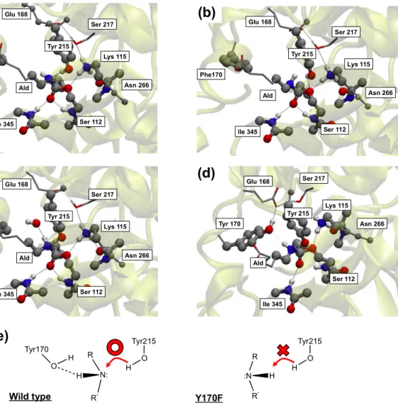
the WT, the Tyr170 contributes to the reaction with its own –OH group: This –OH forms one H-bond with the –NH moiety of the amide bond of Ald and this, in turn, forces the nitrogen atom in Ald to keep its lone pair oriented toward Tyr215. In such a configuration, the amide N pointing toward Tyr215 is ready to accept the H atom from Tyr215, as confirmed by the formation of a new and tight H-bond between the phenol OH and the amide NH group. At variance with the WT case, in the Y170F mutant (Fig. 3-(b)) the absence of Tyr170 and its H-bond does not allow the amide NH group to assume a configuration suitable to accept the H atom from Tyr215, thus suppressing the amide bond cleavage activity. This picture is perfectly consistent with the fact, recently demonstrated64, that the specific orientation of the lone pair of the nitrogen of the scissile amide
bond during catalysis is a crucial feature of enzyme catalyzed amide bond hydrolysis. After a few additional meta-steps, a water molecule from the surrounding solvent approaches to form an H-bond with the amide NH group as shown in Fig 3-(c), thus replacing the role of Tyr170 in the WT. From the point of view of the local H-bond environment only, this conformation resembles closely the one of the WT shown (Fig3-(d)). In addition, it is expected that hydrogen bond might contribute the acceptance tendency of hydrogen atom from Tyr215 as well as the control of direction of NH group considering the Fig. 3 (a). This indicates rather unambiguously that the formation of an
13 H-bond to the amide NH group is indispensable to trigger the H transfer from Tyr215 and that Tyr170 provides the fine control needed to keep the local geometry in a condition suitable to give rise to an efficient breaking of the amide bond. We can then infer that Tyr170 is the fundamental additional residue that must complement the catalytic triad to realize the acylation in this enzyme. Its absence, as in the case of the mutant, results clearly in the suppression of this specific activity or in a larger activation barrier that makes acylation highly improbable. For amines, two different sp3
configurations are in general allowed, as shown in Fig. 3-(e), and the conformational change between them is well known as an umbrella inversion, whose barrier is about 6 kcal/mol for an
ammonia molecule in the gas phase.65 By reasoning on these grounds, the order of magnitude of the
free energy difference between WT and the Y170F mutant in terms of activation barrier (~11 kcal/mol, according to our estimates) might be compatible with the results obtained via the umbrella inversion technique also in a protein environment.
Comparing our result with theoretical studies on general serine proteases,66-68 structures like the
one reported in Fig. 3-(e) were found, for instance, in Fig. 4 of Ref. 62 and discussed in the context of the stereochemistry of tetrahedral intermediates in Ref. 61. In the typical proteases, such as, for instance, chymotrypsin and trypsin, a substrate pocket able to bind to their specific substrate is always present. In the substrate pocket, the chymotrypsin and the trypsin recognize bulky aromatics and side chain in a peptide, respectively, enabling them to identify the site to be cleaved. Instead, the substrate-binding site of NylB is more like a cleft, and in principle can bind substrates so weakly that simple mechanical fluctuations in substrate and changes in the organization of water molecules around the substrates result in a decrease of the reactivity. Only after the induced-fit of Tyr170, allowing to smooth the fluctuations of the substrate in the vicinity of the catalytic triad and to prevent approaching of water molecules during the acylation phase, NylB can degrade the Nylon-oligomers. So far, focusing on the acylation processes, we established the different behavior of the WT and the Y170F mutant, providing a solid argument for the decrease in the hydrolytic activity of the latter. Nonetheless, a still unaddressed question is the actual role of the Tyr170 residue in the induced-fit to bind the substrate.
14
Induced-fit process
To complete the picture about the overall role of Tyr170, we investigated the induce-fit process. To this aim, the free energy surface (FES), projected onto two reaction coordinates d1 and d2, was
estimated by WHAM making use of a set of reactive trajectories generated via PaCS-MD simulations (Fig. 4 (a)). This analysis has shown that four minima (A)-(D) are located on this FES, where the distances in the reference structures are (d1, d2)=(10.89 Å, 14.11 Å) for the open-form
and (d1, d2)=(4.75 Å, 4.05 Å) for the closed-form, respectively. As shown in Fig. 4 (a), starting from
the open-form, both distances decrease from (d1, d2)=(10 Å, 14 Å) in the basin A to (d1, d2)=(9.0 Å,
7.5 Å) in the next local minimum B. In particular, the decrease along the d2 direction is relatively
larger than that along d1. Subsequently, the system reaches the minimum C at (d1, d2)=(7.1 Å, 5.5 Å),
while d2 decreases almost monotonically to a value close to the closed-form. From C to D,
characterized by distances (d1, d2) =(4.8 Å, 5.2 Å), d1 undergoes a noticeable decrease, whereas d2
remains almost constant. This point (D) is the global minimum of our FES landscape. Yet, the free energy difference between (C) and (D) is only ~0.2 kcal/mol, on the verge of the accuracy of these numerical methods, while the other minima are located at slightly higher values, which however are of the order of magnitude of 1 kcal/mol. These shallow minima separated by tiny barriers indicate that the loop region might fluctuate among them. The stabilization energy due to the induced-fit can be as the free energy difference between (A) and (D) and turns out to be (A) - (D) = 0.6 kcal/mol. The highest free energy barrier is the one separating (C) from (D) and its value is about 2.4 kcal/mol. Additional information can be found in the supporting information (SI-4).
Fig.4
To elucidate the mutational effects of Y170F on the induced-fit process (Fig. 4 (a)), we
compared the present results with the previous ones for the WT.20 Focusing on the FES landscape of
both systems, two major features are worthy of note. The first one is the number of (meta-) stable structures realized by the two systems, namely six in the case of the WT, as opposed to four for the Y170F mutant. The second one is the size of each basin where these local minima are located. In the case of the Y170F mutant, the areas of the free energy landscape around the minima, especially
15 (A) and (B), but also the other remaining two, are rather wide and characterized with not very steep slopes with respect to the WT. Although Glu168 is a residue common to both the WT and the mutant, in the first case it forms an H-bond with the substrate and Ser217; conversely fluctuation of Phe170 in the Y170F mutant prevents Glu168 from forming any H-bond with Ser217. Despite this important structural difference, the highest free energy difference in the FES of the Y170F mutant is nearly identical to that of the WT. These findings indicate that the mutational (structural) effect due to the induced-fit on the FES landscape is remarkable, while the stabilization energy is small from a kinetic point of view.
Substrate binding stability of Hyb-24 and Y170F mutants
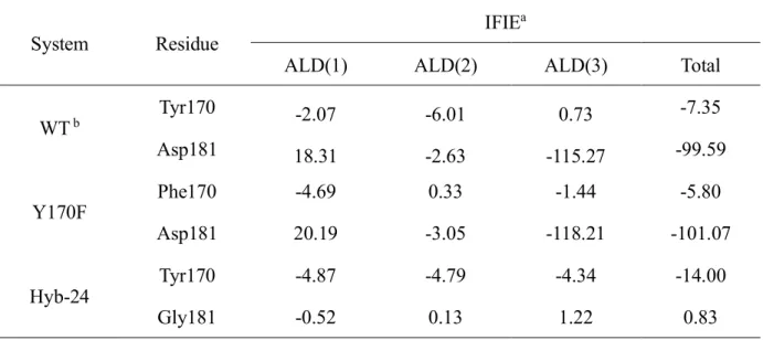
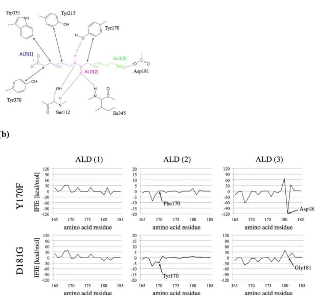
In an attempt at quantifying the effect of each residue on the overall stability of the enzyme-substrate bound form, such as Hyb-24, in particular the so-called D181G mutant, and the Y170F mutant, we performed IFIE analyses based on the FMO method. More precisely, we focused on the IFIE values between each ALD fragment and the most relevant amino acid residues, such as Tyr170 and Asp181 (see Fig. 5). In order to analyze contributions of IFIE in more detail, we further divided the substrate into three distinct fragments: the C-terminal (Ald(1)), the amide group (Ald(2)), and the N-terminal(Ald(3)), as shown in Fig 5. The result of this analysis is summarized in Table2.
The IFIE values for the WT have been already elsewhere56 and shown in SI-5. According to Table 2,
Asp 181 is characterized by an attractive interaction with Ald(3) stronger than that of Tyr170 with Ald(2). Conversely, in Hyb-24 the interaction between NylB and ALD(3) is lacking. Although the IFIE value to Ald(2) of Phe170 in the Y170F mutant becomes weaker than that of Tyr170 in the WT, the decrease of the IFIE is less pronounced than that to Ald(3) of the Hyb-24. These results suggest that Asp181 is quantitatively, energetically speaking, an amino acid residue more important than Tyr 170 for the specific substrate binding. Indeed, available kinetics data on Hyb-24DY, resulting from a
double mutation of G181D and D370Y from Hyb-24,
(kcat KM 0.35 s1mM1, KM 7.1 ±1.28mM1) and of Hyb-24Y, in which a single mutation of
D370Y from Hyb-24 is present, (kcat KM 0.016 s1nM1, KM 39.1 ± 6.73mM1),13 indicate
16 Fig.5
Homology analyses on NylB and possible molecular evolution route for Ald hydrolytic activity
Our computational results, corroborated by experiments, suggest that, from the evolutional point of view, the introduction of Y170 in the loop region is at the origin of the acquisition of the hydrolytic activity from NylB. To bring support to our assumption, we completed our study by investigating any homologous enzyme exists possessing a similar evolution for the substrate binding. To this purpose,
we performed structural homology searches for the WT (PDB id: 2zma) by using the DALI program69
and selected structures preserving the local configuration around Tyr170 similar to NylB. This search,
performed by using MultiSeq2.0 with the STAMP algorithm.70-72, brought to our attention the EstU1
system, having a high homology to NylB, as shown in Fig. 6 (a). The specific substrate of EstU1 is a cephalothin, which has bulky functional groups. Around the binding site of the substrate, EstU1 presents three amino acid residues, Tyr172, Phe174, and Phe190, which correspond to Glu168, Tyr170, and Asp181 in NylB, respectively. These three aromatic amino acid residues recognize the thiophene ring of cephalothin cooperatively by hydrophobic interactions (see Fig. 6 (b)). The remarkable difference between NylB and EstU1 is the location of the helix structure and the loop-segment evidenced in Fig. 6. In addition to the similarity in the structure, the side chain of Phe174 in the loop-segment of EstU1 undergoes a conformational change during the substrate binding process. This property is resembles closely the one of NylB. We can then infer that, upon acquiring Tyr170, NylB might have undergone a natural selection process to adapt to a nylon-oligomer-rich environment by skillfully using the loop region for the induced-fit to the substrate and by catalyzing the acylation via a decrease if the activation barrier.
Fig.6
Towards a rational design of enzymatic activities
Having unraveled the role of Tyr170 in the WT, which contributes mainly to the acylation step to regulate the direction of a hydrogen bond, we have now a convincing set of results allowing to proceed further. All our results concur to support the notion that NylB* needs not only the catalytic
triad Ser-Tyr-Lys, like the class C β-lactamase family, but also Tyr170 to degrade non-biological amide bonds present in nylon-oligomers. In particular, acquiring Tyr170 in NylB* is the key factor for
17 triggering the reaction by decreasing the activation barrier, in other words by decreasing the rate constant kcat. This is confirmed by a comparison with Hyb-24 (nearly identical to NylB*), which
possesses as well a weak reactivity to Ald. In the change from NylB* to NylB, Asp181 provides the
required substrate specificity, resulting in the decrease of kcat.
Recent advances in computer facilities and related theoretical methods allow nowadays for a
de novo design of functional proteins.73 In the last decade, researchers working in this field have succeeded in producing new enzymes called “theozymes”, which were entirely designed on a computer by inserting a suitably selected transition state structure into protein scaffolds.74-77
Unfortunately, for these theozymes, the resulting catalytic proficiency, defined as
Catalytic proficiency
(
kcat/ KM)
kuncat in M 1
(
)
, (6)was still inferior to the natural one. In this expression kuncat is the rate constant in the absence of the enzyme,78 while k
cat is the rate constant of the theozyme, lower than the natural one.
79 The lesson to
be retained is that looking just at the structure of the building block (transition state in this case) to be inserted inside a given scaffold is insufficient to obtain an enhancement of the catalytic activity. In this respect, in our study we have shown that Tyr170 and Asp 181 play an important role in direct and indirect decrease of kcat by stabilizing both the transition state and the substrate binding structure, thus allowing for an efficient degradation of the non-biological amide bond. Compared with the
strategy followed for the theozyme, the change realized in the passage from Hyb-24 (NylB* type) to
WT or Y170F (NylB type) could resemble the processes of the transition state design and the active site re-design briefly summarized above. Nonetheless, the theozyme was designed and realized in a laboratory and never underwent any natural selection process. Its absence in nature might be ascribed to its relative rigidity, which, contrary to other enzymes, implies a lack of dynamic motions of the flexible loop regions. We can then expect that taking into account also the response of the system to natural selection, as in the case of NylB, can be the key for a rational design of enzymatic reactions with appreciable catalytic proficiency, although we have to acknowledge that a major difficulty is still represented by the prediction of the possible conformational changes an enzyme could assume.80
18 Some hints could be obtained by the development of integrated schemes making use of coarse grained MD81 or Fluctuation Flooding Method82 jointly with quantum chemical transition state design not too far from the strategy adopted for the theozyme.
4. Conclusions
A combination of different theoretical methods was used to inspect in detail a series of enzymatic reaction representing the fundamental stages in the degradation of non-biological amide bonds operated by the nylon oligomer hydrolase NylB. To this aim a comparative study of the WT enzyme and its closely related mutant was conducted, unveiling the crucial role of two evolutionally important amino acid residues, Tyr170 and Asp181, in (1) the enzymatic reactions, (2) the induced-fit process, and (3) the stability of the enzyme-substrate binding. We found at which steps of this complex process Tyr170 and Asp181 are involved and which are their respective net effects on the overall process. Specifically, Tyr170 mainly contributes to the acylation step and plays a subsidiary role in the stabilization of the enzyme-substrate complex. According to the free energy changes during the acylation and the induced-fit steps upon the mutation, in the mutant the former process requires the overcoming of an activation barrier which is 11 kcal/mol higher with respect to the WT case. This is clearly a stumbling block to acylation that makes the reaction very unlikely for the Y170F system. Conversely, for the induced-fit process of the Y170F mutant, although the free energy landscape changes drastically with respect to the WT case, the free energy barrier is nearly unchanged, suggesting that the kinetics of the induced-fit is barely affected by the mutation or, at least, this mutation. Although the inter-fragment interaction energy (IFIE) of Phe170 in the Y170F mutant is lower than that of Try170 in the WT, such a decrement is smaller than that of Hyb-24. Based on all these results, we can conclude that Tyr170 is the essential crucial amino acid residue to promote the degradation of non-biological amide bonds by forming a novel type of a catalytic quad, as opposed to standard catalytic triads, responsible for both the stabilization of the transition state and the restraining of fluctuations in the substrate binding structure. On the other hand, Asp181 mainly contributes to the stabilization of the enzyme-substrate complex (substrate specificity), while the influence on the hydrolytic activity is negligible.
19
ACKNOWLEDGMENT
We thank Mr. Masanari Matsuoka for insightful discussions in the search for structural homology. This work is supported by Grant-in-Aid for Scientific Research of the Innovative Areas “Computics”, “Dynamic ordering and biofunction”, and Photosynergetics” (No. 25104716, 26102525, and 26107004), and a Grant-in-Aid for Young Scientists (B) (Grant No. 22740259) from Japan Society for the Promotion of Science (JSPS), and by the Strategic Programs for Innovative Research (SPIRE), MEXT, and the Computational Materials Science Initiative (CMSI), Japan. T.B. expresses his special thanks to the JSPS Research Fellowship for Young Scientists (No. 02503029). Theoretical calculations were partly performed at the Research Center for Computational Science, Institute for Molecular Science (Japan) and Center of Computational Materials Science, Institute for Solid State Physics, The University of Tokyo, (Japan). M.B. and T.B. acknowledge the Direction Informatique (Pôle HPC) of the University of Strasbourg for providing scientific support and access to computing resources. Part of the computing resources were funded by the Equipex Equip@Meso project. K.K. acknowledges the Cooperative Research Program of Institute for Protein Research, Osaka University
REFERENCES
[1] E. C. Horning, V. L. Stromberg, H. A. Lloyd, J. Am. Chem. Soc. 1952, 74, 5153-5155.
[2] M. Boero, T. Ikeshoji, C. C. Liew, K. Terakura, M. Parrinello, J. Am. Chem. Soc. 2004, 126, 6280-6286
[3] S. Kinoshita, S. Kageyama, K. Iba, Y. Yamada, and H. Okada, Agric. Biol. Chem., 1975, 39, 1219-1223.
[4] H. Okada, S. Negoro, H. Kimura, S. Nakamura, Nature, 1983, 306, 203-206.
[5] K. Kato, K. Ohtsuki, Y. Koda, T. Maekawa, T. Yomo, S. Negoro, I. Urabe, Microbiol., 1995, 141, 2585-2590.
[6] S. Negoro, Appl. Microbiol. Biotechnol., 2000, 54, 461-466.
[7] K. Nagai, Iida, K. Shimizu, R. Kinugasa, M. Izumi, D-I, Kato, M. Takeo, K. Mochiji, and S. Negoro, Appl. Microbiol. Biotechnol., 2014, DOI:10.1007/s00253-014-5885-2. –in press
20 [8] S. Negoro, N. Shibata, Y. Tanaka, K. Yasuhira, H. Shibata, H. Hashimoto, Y. Lee, S. Oshima, R. Santa, S. Oshima, K. Mochiji, Y. Goto, T. Ikegami, K. Nagai, D. Kato, M. Takeo, and Y. Higuchi, J. Biol. Chem., 2012, 287, 5079-5090.
[9] S. Negoro, T. Ohki, N. Shibata, N. Mizuno, Y. Wakitani, J. Tsurukame, K. Matsumoto, I. Kawamoto, M. Takeo, and Y. Higuchi, J. Biol. Chem., 2005, 280, 39644-39652.
[10] K. Kato, K. Fujiyama, H.S. Hatanaka, I.D. Priyambada, S. Negoro, I. Urabe, and H. Okada, Eur.
J. Biochem. 1991, 200, 165-169.
[11] S. Negoro, T. Ohki, N. Shibata, K. Sasa, H. Hayashi, H. Nakano, K. Yasuhira, D. Kato, M.Takeo, and Y. Higuchi, J. Mol. Biol., 2007, 370, 142-156.
[12] T. Ohki, N. Shibata, Y. Higuchi, Y. Kawashima, M. Takeo, D. Kato, and S. Negoro, Protein Sci.
2009, 18, 1662-1673.
[13] Y. Kawashima, T. Ohki, N. Shibata, Y. Higuchi, Y. Wakitani, Y. Matsuura, Y. Nakata, M. Takeo, D. Kato, and S. Negoro, FEBS J., 2009, 276, 2547-2556.
[14] P. H. Crowley, J. Theo. Biol., 1975, 50, 461-475.
[15] K. A. Johnson, and R. S. Goody, Biochem., 2011, 50, 8264-8269.
[16] K. Kamiya, T. Baba, M. Boero, T. Matsui, S. Negoro, and Y. Shigeta, J. Phys. Chem. Lett., 2014, 5, 1210.
[17] H. M. Senn, and W. Thiel. Angew. Chem. Int. Ed., 2009, 48, 1198-1229. [18] G. Dong, S. Shaik, and W. Lai, Chem. Sci., 2013, 4, 3624–3635
[19] R. Harada, A. Kitao, J. Chem. Phys. 2013, 139, 035103.
[20] T. Baba, R. Harada, M. Nakano, and Y. Shigeta, J. Comput. Chem., 2014, 35, 1240-1247. [21] S. Tanaka, Y. Mochizuki, Y. Komeiji, Y. Okiyama, and K. Fukuzawa, Phys. Chem. Chem.
Phys., 2014, 16, 10310-10344.
[22] K. Fukuzawa, C. Watanabe, I. Kurisaki, N. Taguchi, Y. Mochizuki, T. Nakano, S. Tanaka, and Y. Komeiji, Comput. Theor. Chem., 2014, 1034, 7-16.
[23] C. Watanabe, K. Fukuzawa, S. Tanaka, and S. Aida-Hyugaji, J. Phys. Chem. B, 2014, 118, 4993–5008.
21 [25] D.A. Case, V. Babin, J.T. Berryman, R.M. Betz, Q. Cai, D.S. Cerutti, T.E. Cheatham, III, T.A. Darden, R.E. Duke, H. Gohlke, A.W. Goetz, S. Gusarov, N. Homeyer, P. Janowski, J. Kaus, I. Kolossváry, A. Kovalenko, T.S. Lee, S. LeGrand, T. Luchko, R. Luo, B. Madej, K.M. Merz, F. Paesani, D.R. Roe, A. Roitberg, C. Sagui, R. Salomon-Ferrer, G. Seabra, C.L. Simmerling, W. Smith, J. Swails, R.C. Walker, J. Wang, R.M. Wolf, X. Wu and P.A. Kollman, 2014, AMBER 14, University of California, San Francisco.
[26] R. Salomon-Ferrer, A.W. Goetz, D. Poole; S. Le Grand, and R.C. Walker, J. Chem. Theory
Comput., 2013, 9, 3878-3888.
[27] T. Baba, K. Kamiya, T. Matsui, N. Shibata, Y. Higuchi, T. Kobayashi, S. Negoro, and Y. Shigeta,
Chem. Phys. Lett., 2011, 507, 157-161.
[28] J.-P. Ryckaert, G. Ciccotti, and H. J. C. Berendsen, J. Comput. Phys., 1977, 23, 327-341 [29] T. Darden, D. York, and L. Pedersen, J. Chem. Phys. 1993, 98, 10089.
[30] H. Li, A. D. Robertson, and J. H. Jensen, Proteins, 2005, 61, 704-721. [31] D. C. Bas, D. M. Rogers, and J. H. Jensen, Proteins, 2008, 73,765-783.
[32] M. H M. Olsson, C. R. Søndergard, M. Rostkowski, and J. H. Jensen, J. Chem. Theory Comput.,
2011, 7, 525-537.
[33] C. R. Søndergaard, M. H. Olsson, M. Rostkowski, and J. H. Jensen, J. Chem. Theory Comput.,
2011, 7, 2284-2295.
[34] A. Laio, J. VandeVondele, and U. Roethlisberger, J. Chem. Phys. 2002, 116, 6941-6947. [35] A. Laio, J. VandeVondele, and U. Roethlisberger, J. Phys. Chem. B 2002, 106, 7300-7307. [36] A. Laio, and M. Parrinello, Proc. Natl. Acad. Sci. U. S. A., 2002, 99, 12562–12566. [37] M. Iannuzzi, A. Laio, and M. Parrinello, Phys. Rev. Lett., 2003, 90, 238302.
[38] A. Barducci, M. Bonomi, and M. Parrinello, Comput. Mol. Sci., 2011, 826–843.
[39] M. Bonomi, D. Branduardi, G. Bussi, C. Camilloni, D. Provasi, P. Raiteri, D. Donadio, F. Marinelli, F. Pietrucci, R. A. Broglia, and M. Parrinello, Comput. Phys. Commun., 2009, 180, 1961–1972.
[40] F. Pietrucci , F. Marinelli , P. Carloni and A. Laio J. Am. Chem. Soc., 2009, 131, 11811–11818.
[41] R. Car, and M. Parrinello, Phys. Rev. Lett., 1985, 55, 2471–2474. [42] P. Hohenberg, and W. Kohn, Phys. Rev., 1964, 136, B864–B871.
22 [43] W. Kohn, and L.J. Sham, Phys. Rev., 1965, 140, A1133–A1138
[44] F. L. Gervasio, M. Boero, and M. Parrinello, Angew. Chem. Int. Ed., 2006, 45, 5606-5609. [45] M. Boero, T. Ikeda, E. Ito, and K. Terakura, J. Am. Chem. Soc., 2006, 128, 16798-16807.
[46] K. Kamiya, M. Boero, M. Tateno, K. Shiraishi, and A. Oshiyama, J. Am. Chem. Soc., 2007, 129, 9663-9673.
[47] M. Boero, J. Phys. Chem. B, 2011, 115, 12276.
[48] X. Biarnés, A. Ardevol, J. Iglesias-Fernández, A. Planas, and C. Rovira, J. Am. Chem. Soc., 2011,
133, 20301–20309
[49] F. Hofbauer, and I. Frank, Chem. Eur. J., 2012, 18, 16332–16338. [50] Rovira, C. Comput. Mol. Sci., 2013, 393-407.
[51] M. Boero, Lecture Notes in Physics, 2010, 795, 81-98.
[52] CPMD, http://www.cpmd.org/, Copyright IBM Corp 1990-2012, Copyright MPI für Festkörperforschung 1997-2001.
[53] F.A. Hamprecht, A.J. Cohen, D.J. Tozer, and N.C. Handy, J. Chem. Phys., 1998, 109, 6264–6271.
[54] N. Troullier, and J.L. Martins, Phys. Rev. B, 1991, 43, 1993–2006. [55] A. Laio and F. L. Gervasio, Rep. Prog. Phys. 2008, 71, 126601.
[56] W. Humphrey, A. Dalke, and K. Schulten, J. Mol. Graphics, 1996, 14, 33.
[57] A. Grossfield, WHAM: The Weighted Histogram Analysis Method, Version 2.07. Available at: http://membrane.urmc.rochester.edu/content/wham. Accessed September 22, 2014.
[58] D. Hankins, J. W. Moskowiz, F. H. Strillinger, J. Chem. Phys., 1970, 53 4544-4554.
[59] T. Nakano, T. Kaminuma, T. Sato, K. Fukuzawa, Y. Akiyama, M. Uebayasi, K. Kitaura, Chem.
Phys. Lett., 2002, 351, 475-480.
[60] Y. Mochizuki, K. Yamashita, K. Fukuzawa, K. Takamatsu, H. Watanabe, N. Taguchi, Y. Okiyama, M Tsuboi, T Nakano, S Tanaka, Chem. Phys. Lett., 2010, 493, 346-352.
[61] H. Ando, Y. Shigeta, T. Baba, C. Watanabe, Y. Okiyama, Y. Mochizuki, and M. Nakano, Mol.
Phys., 2014, DOI:10.1080/00268976.2014.941311. – in press
23
[63] BioStation Viewer, A. Kato, K. Fukuzawa, T. Nakano, Available at:
http://www.fsis.iis.u-tokyo.ac.jp/en/result/software/. September 22, 2014. [64] P.-O. Syrén, FEBS Journal 2013, 280, 3069-3083.
[65] K. S. Werpetinsk, and M. Cook. Phys. Rev., 1995, 52, R3397. [66] H. Lizbeth. Chem. Rev., 2002, 102, 4501-4524.
[67] V. Daggett, S. Schroeder, and P. Kollman, J. Am. Chem. Soc., 1991, 113, 8926-8935. [68] T. Ishida, and S. Kato. J. Am. Chem. Soc., 2003, 125, 12035-12048.
[69] L. Holm and P. Rosenström. Nucleic Acids Res., 2010, 38, W545–W549.
[70] S. S. Cha, Y. J. An, C. S. Jeong, M. K. Kim, J. H. Jeon, C. M. Lee, H. S. Lee, S. G. Kang, and J. H. Lee, Proteins: Struct. Funct. Bioinfo., 2013, 81, 2045-2051.
[71] E. Roberts, J. Eargle, D. Wright, and Z. Luthey-Schulten, BMC Bioinfo., 2006, 7,382. [72] R. B. Russell and G. J. Barton, PROTEINS: Struct. Funct. Genet., 1992, 14, 309-323. [73] D. J. Tantillo, C. Jiangang, and K. N. Houk, Curr. Opin. Chem. Biol., 1998, 2, 743-750.
[74] D. Röthlisberger, O. Khersonsky, A. M. Wollacott, L. Jiang, J. DeChancie, J. Betker, J. L. Gallaher, E. A. Althoff, A. Zanghellini, O. Dym, S. Albeck K. N. Houk, D. S. Tawfik, and D. Baker, Nature, 2008, 453, 190-195.
[75] H. K. Privett, G. Kiss, T. M. Lee, R. Blomberg, R. A. Chica, L. M. Thomas, D. Hilvert, K. N. Houk, and S. L. Mayo, Proc. Natl. Acad. Sci. USA, 2012, 109, 3790-3795.
[76] J. H. Mills, S. D. Khare, J. M. Bolduc, F. Forouhar, V. K. Mulligan, S. Lew, J. Seetharaman, L. Tong, B. L. Stoddard and D. Baker, J. Am. Chem. Soc., 2013, 135, 13393-13399.
[77] S. Bjelic, Y. Kipnis, L. Wang, Z. Pianowski, S. Vorobiev, M. Su, J. Seetharaman, R. Xiao, G. Kornhaber, J. F. Hunt, L. Tong, D. Hilvert, and D. Baker, J. Mol. Biol., 2014, 426, 256-271. [78] A. Radzicka, and R. Wolfenden, Science, 1995, 267, 90-93.
[79] W. S. Mak and J. B. Siegel Curr. Opin. Struct. Biol., 2014, 27 87-94.
[80] L. S. Mizoue, and W. J. Chazin. Curr. Opin. Struct. Biol., 2002, 12, 459-463.
[81] H. I. Ingólfsson, C. A. Lopez, J. J. Uusitalo, D. H. de Jong, S. M. Gopal, X. Periole, and S. J. Marrink, WIREs: Comput. Mol. Sci., 2014, 4, 225-248.
[82] R. Harada, Y. Takano, and Y. Shigeta. J. Chem. Phys, 2014, 140, 125103.
24
Figure Captions
Figure 1. Structure and main amino acid residues of NylB. (a) Induced-fit process: Superposition of
the NylB structures in the open form (black) and in the closed form (yellow). A large movement of the loop-segment N166-V177 toward the substrate occurs, indicated by the red arrow enclosed in the dotted red circle. (b) The QM region, located inside the red circle in (a), used in our hybrid QM/MM simulations. QM atoms are shown as balls and sticks, whose colors code, here and in the all the following figures, are gray for C, red for O, blue for N and white for H, and the yellow ribbon shows the secondary structure. (c) The reaction coordinate (red arrow) and other fundamental distances (green, blue and magenta) used to drive and monitor the acylation reaction. Details are given in the text and in Fig. 2.
Figure 2. Evolution of the reaction coordinate r1 and the most relevant distances during the
metadynamics simulation for the Y170F mutant: r1 = N3(Ald393)-C1(Ald393) (red), r2 =
OG(Ser112)-C1(Ald393) (green), r3 = HH(Tyr215)- N3(Ald393) (blue), and r4 =
HG(Ser112)-NZ(Lys115) (magenta). Panel (a) shows whole simulation, while panels (b), (c), and (d) are the magnifications of the three crucial events inside the dashed circles in (a).
Figure 3. Local structures and related differences between Y170F and WT relevant to the acylation
process. Panels (a), (b), and (c) correspond, respectively, to the events (b), (c), and (d) in Fig. 2. Panel (d) shows the WT for comparison. A schematic picture of the difference between Y170F and WT is shown in (e).
Figure 4. Free energy landscapes for the induced-fit process in the Y170F mutant(a) and the WT
(b), according to the results of Ref. 20.
Figure 5. (a) Schematic view of the interactions between the most relevant amino acid residues and
the substrate. For clarity, the ALD is divided into three fragments, the C-terminal, the peptide site, and the N-terminal, labeled as ALD(1) (blue), ALD(2) (pink), and ALD(3) (green), respectively. These fragments are the ones used in the FMO analysis shown in panel (b).
25
Figure 6. Result of the structural homology search (a) and details of the surrounding substrate (b)
Using the DALI program and selecting only structures preserving the local environment around Tyr170 in WT (pdb id 2ZMA), EstU1 (pdb id 4IVK) was discovered. For the structural aliment, we adopted the STAMP algorithm. The corresponding analogous main regions between WT and EstU1 are shown in the two adjacent panels in (a). The semi-transparent elliptical region on the top left of each structure indicates the two segments 179-189 of 4IVK, and 175-185 of 2ZMA, respectively. Each substrate is shown as yellow sticks.
Table 1. Maximum free energy differences in the acylation step for the WT and the Y170F mutant.
We recall that typical error bars of 1-2 kcal/mol from the underlying level of DFT adopted affect these estimations
System Maximum free energy difference DGa
WT 31
Y170F mutant 42
a unit of kcal / mol
Table 2. The computed IFIE values between the 170th and the 181st amino acid residues and each fragment in Ald.
System Residue
IFIEa
ALD(1) ALD(2) ALD(3) Total
WT b Tyr170 -2.07 -6.01 0.73 -7.35 Asp181 18.31 -2.63 -115.27 -99.59 Y170F Phe170 -4.69 0.33 -1.44 -5.80 Asp181 20.19 -3.05 -118.21 -101.07 Hyb-24 Tyr170 -4.87 -4.79 -4.34 -14.00 Gly181 -0.52 0.13 1.22 0.83
26 Tyr 170 Tyr 215 Ser 217 Ald Glu 168 Ile 345 Gly 267 Met 111 Ser 112 Gly 344 Lys 115 Asn 266 (b)
27
(a) (b)
(c) (d)
28 N: R H R' H O Tyr215 H O Tyr170 :N R R' H H O Tyr215 Phe170 Tyr 215 Ser 217 Ald Glu 168 Ile 345 Ser 112 Lys 115 Asn 266 Phe170 Tyr 215 Ser 217 Ald Glu 168 Ile 345 Ser 112 Lys 115 Asn 266 Phe170 Tyr 215 Ser 217 Ald Glu 168 Ile 345 Ser 112 Lys 115 Asn 266 Tyr 170 Tyr 215 Ser 217 Ald Glu 168 Ile 345 Ser 112 Lys 115 Asn 266
Wild type Y170F
(d)
(c)
(b)
(a)
(e)
29
(a) (b)
30
(a)
(b)
31
(a)
(b)
32
Table of contents
To unravel what provides the ability to degrade non-biological amide bond with Nylon oligomer
hydrolase, we investigate the process from induced-fit to acylation by a combination of different theoretical methods.