ARCGIVEs
MASSACHUSETS INSITUTEOF TECHNOWGY
SEP 172015
LIBRARIES
Determinants of protein-peptide interaction specificity in the
Bcl-2 and TRAF families
Glenna Wink Foight B.S. Biochemistry
North Carolina State University, 2009
SUBMITTED TO THE DEPARTMENT OF BIOLOGY IN PARTIAL FULFILLMENT OF THE REQUIREMENTS FOR THE DEGREE OF
DOCTOR OF PHILOSOPHY AT THE
MASSACHUSETTS INSTITUTE OF TECHNOLOGY
Signature of Author:
SEPTEMBER 2015
@ Massachusetts Institute of Technology. All rights reserved.
Signature redacted
-
'7,
Department of BiologySeptember 2015
Certified by:
Signature redacted
- Amy Keating Professor of Biology Thesis Supervisor
Accepted by:.
,Signature
redacted
Michael Hemann Associate Professor of Biology Co-Chair, Biology Graduate Committee
Determinants of protein-peptide interaction specificity in the Bcl-2 and TRAF families
by
Glenna Wink Foight
Submitted to the Department of Biology in partial fulfillment of the requirements for the degree of Doctor of Philosophy at the Massachusetts Institute of Technology
September 2015 Abstract
Protein-peptide interactions have important roles in the majority of cellular processes. There are many families of peptide recognition domains in which homologous members display differential binding preferences for peptide sequence features. Peptide binding specificity is critical for the functional roles played by each family member, which can be overlapping or distinct. The two peptide recognition domain families discussed in this work, Bcl-2 and TRAF proteins, have roles in cellular processes including apoptosis, inflammation, and immunity. Aberrant function of these proteins has been linked to a variety of diseases. There is great interest in understanding the mechanistic basis of protein-peptide binding specificity in these families and others. An improved understanding will enable models of binding preferences for interactome prediction and design of specific peptide reagents for the inhibition and study of protein-peptide interactions.
The anti-apoptotic Bcl-2 family members bind a-helical Bcl-2-homology 3 (BH3) motifs in pro-apoptotic Bcl-2 family members to prevent apoptosis. Kaposi Sarcoma herpesvirus and Epstein Barr herpesvirus express viral homologs of the anti-apoptotic Bcl-2 proteins, KSBcl-2 and BHRF 1, respectively, during viral replication to prevent host cell death. Because human Bel-2 proteins are important in preventing apoptosis in cancers, there is interest in targeting the viral homologs, as they may also have a role in herpesvirus-associated malignancies. I designed and screened libraries of BH3 peptide variants for binding specificity to KSBcl-2 and BHRF 1. From library screening and additional rational mutagenesis, I developed peptides that showed specific binding to KSBcl-2, BHRF 1, or the human homolog Ml- 1, and displayed large margins of specificity over the other human Bel-2 homologs.
TRAF proteins bind sequences in the unstructured regions of cell surface receptors and other adapter proteins in order to mediate downstream signaling events. TRAF-peptide binding preferences are relatively uncharacterized. I adapted a bacterial surface display system for screening peptides for TRAF binding. Using this system, I explored the binding preferences of TRAFs 2, 3, and 5 to single and double mutant libraries of two peptide interaction partners from CD40 and TANK. Comparison of the enriched peptide sequences reveals a surprising degree of difference between these three close TRAF homologs, yielding hypotheses relevant to TRAF function and inhibition.
Thesis Supervisor: Amy Keating Title: Professor of Biology
Acknowledgements
I would like to thank my supervisor, Amy Keating for all of her support and valuable training over the years. I have learned a lot from observing her highly rigorous approach to science. I have also benefited from her attention to the development of her students on both scientific and professional levels.
I would also like to thank my committee members Bob Sauer and Thomas Schwartz. Yearly committee meetings with them have provided valuable re-focusing of my projects. I would especially like to thank them for providing me with many letters for applications over the years. Thanks to Joshua Kritzer for serving on my defense committee.
Thanks to Carla Mattos, my undergraduate mentor, who taught me to be a good structural biochemist and gave me many opportunities that got my research career off to a great start.
Thanks to the many members of the Keating lab past and present: Sanjib Dutta for valuable training in library display technology, and lots of great, unsolicited advice over the years. Joe DeBartolo, Raheleh Rezaei-Araghi and Justin Jenson for valuable discussions on Bcl-2s. Jenifer Kaplan for being a great lab mate and for reading many paper and thesis chapter drafts. Christos Kougentakis for making the lab a fun place to work and for being an exceptional technician who was always volunteering to do work on the weekends.
I would also like to thank members of the MIT Koch flow cytometry core for assistance with sorting. Thanks to the other core facilities for much assistance: the MIT Biomicro Center, MIT Koch Biopolymers, and the Biophysical Instrumentation Facility. Special thanks to Bob Grant in the crystallography core for help and advice on screening crystals.
Thanks to the Biograd class of 2009. The coffee breaks, game nights, and Building-68-lunches have been invaluable in getting me through.
Finally, thanks to my family: My parents for supporting me in following my interests, and helping me keep things in perspective. My husband, Dillon for everything.
Table of Contents
List of Figures ... 9
List of Tables ... 11
Chapter 1. Introduction ... 12
Interaction determ inants in the cell... 14
Post-translational m odifications ... 14
Gene expression...16
M ethods for determ ining peptide interaction m otifs ... 17
Experim ental m ethods ... 17
Com putational m ethods ... 21
Binding specificity m echanism s ... 25
A hierarchy of binding specificity mechanisms within PRD families...25
The use of multiple motifs for specificity in large PRD families...26
The use of motif context residues for specificity in smaller PRD families ... 29
Applications of specificity prediction ... 38
Interactom e prediction ... 38
Inhibitor design ... 40
Research approach ... 42
References ... 44
Chapter 2. Designed BH3 peptides with high affinity and specificity for targeting M cl-I in cell...53
Introduction ... ... 54
Results and D iscussion ... 58
Selection of M cl-i-specific peptides ... 58
Specificity m echanism s ... 68
Cellular BH 3 profiling assays... 72
M ethods ... 79
Library construction and sorting ... 79
Fluorescence anisotropy assays ... 81
Fitting protocol for fluorescence anisotropy curves ... 82
Cellular BH 3 profiling assays ... 83
A cknow ledgem ents...85
References...86
Chapter 3. Locating herpesvirus Bcl-2 homologs in the specificity landscape of anti-apoptotic Bcl-2 proteins...90
Abstract ... 91
Introduction ... 92
Results ... 98
Comparison of eight Bcl-2 homologs based on sequence identity, structure, and binding preferences ... 98
Peptide libraries targeting K SBcl-2 and BHRF 1 specificity ... 109
Binding of library-derived peptides to 8 Bcl-2 hom ologs ... 120
Specificity mechanisms underlying receptor binding similarity patterns ... 124
Designed peptides with improved specificity for KSBcl-2 and BHRF 1 vs. Mcl-I...131
M aterials and M ethods ... 139
Sequence identity, hom ology m odeling and SiteM A P ... 139
Clustering ... 141
Expression and purification of Bcl-2 proteins ... 141
Binding affinity m easurem ents by fluorescence anisotropy ... 145
SPOT arrays ... 146
M odels used for library design ... 146
Library design ... 147
K SBcl-2 library ... 147
BH RF1 library ... 149
Library construction ... 150
Library Sorting ... 152
Illum ina sequencing and data processing ... 155
A cknow ledgem ents ... 158
References ... 159
Chapter 4. Peptide binding preferences of TRA Fs 2, 3 and 5 ... 166
Introduction ... 167
Results and D iscussion ... 174
Comparison of TRAFs by sequence identity of the MATH domain and peptide-binding site...174
Comparison of TRAFs by physicochemical properties of the peptide-binding site ... 175
Development of a surface display system for TRAF binding ... 177
Screening of single and double mutant libraries of CD40 and TANK ... 181
Structural hypotheses for peptide binding preferences ... 188
Specificity features in the CD40 library results ... 192
Conclusions ... 194
M ethods ... 195
SiteM A P and sequence identity analysis ... 195
Protein constructs and purification ... 196
Bacterial surface display constructs and library assem bly ... 197
Library sorting and FA CS analysis ... 215
Illum ina sam ple preparation ... 216
Sequencing data processing ... 217
References ... 218
Chapter 5. Conclusions and future directions...223
Characterization and m odeling of peptide binding preferences ... 225
Design and screening of peptide libraries to achieve specific peptides ... 229
A pplications of specific peptides ... 233
List of Figures
Figure 1.1. Deep mutational scanning provides a metric of function for mutations to all 20 amino
acids at each protein position ... 20
Figure 1.2. How a STATIUM model is built for a protein:peptide complex structure ... 25
Figure 1.3. Three large PRD families that use different motifs for specificity ... 27
Figure 1.4. Three peptide recognition domain families utilizing motif context for specificity...31
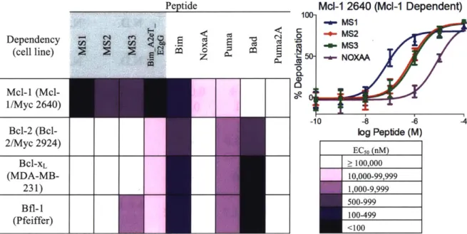
Figure 2.1. Mcl- 1-specific peptides labeled with fluorescein binding to five human Bcl-2 h om o lo g s ... 62
Figure 2.2. Competition fluorescence anisotropy binding experiments ... 64
Figure 2.3. Comparison of Bim BH3 position 2e in structures of Bfl-1, Bcl-xL, and Ml-I...70
Figure 2.4. BH3 profiling of cell lines using engineered and native BH3 peptides ... 74
Figure 2.5. Heat map of the EC50 values (peptde concentration in nM) for mitochondrial depolarization induced by engineered and native BH3 peptides in four cell lines ... 76
Figure 2.6. Engineered peptides do not cause mitochondrial membrane depolarization in B ax/B ak negative cells...78
Figure 3.1. Sequence comparison of 5 human and 3 viral Bcl-2 homologs ... 99
Figure 3.2. Schematic describing the SiteMAP analysis process ... 101
Figure 3.3. Comparison of the physicochemical characteristics of human and viral Bcl-2 homolog BH3 binding groove structures using SiteMAP ... 102
Figure 3.4. Comparison of BH3 peptide binding profiles for eight Bcl-2 homologs ... 104
Figure 3.5. Bim BH3 and point-mutant peptides on SPOT arrays binding to 100 nM BHRF1 or K S B cl-2 ... 10 8 Figure 3.6. Bacterial surface display screen for selective binders of KSBcl-2 and BHRFI ... 110
Figure 3.8. FACS plots showing the binding of the viral Bcl-2 protein and four human Bcl-2 p ro tein s ... 1 15
Figure 3.9. Enrichment of residues in library sequences versus viral Bcl-2 and Mcl-I model sco res ... 12 0 Figure 3.10. Binding of human and viral Bcl-2 homologs to peptides identified from library screen in g ... 12 4
Figure 3.11. Specificity mechanisms employed by library peptides ... 128
Figure 3.12. Conformational flexibility at Bcl-xL helix 2 -helix 3 bend ... 130
Figure 3.13. Specificity mechanisms that disfavor M l- 1 binding ... 131
Figure 4.1 Sequence identities show similarities between TRAFs 1, 2, 3, and 5 ... 175
Figure 4.2. Comparison of the physicochemical characteristics of core and exosite regions of TRA F binding grooves by SiteM A P ... 177
Figure 4.3. Cell surface display system and peptide constructs ... 179
Figure 4.4. Binding of TRAFs 2, 3, and 5 to control peptides displayed on E. coli ... 180
Figure 4.5. Sorting schemes for the CD40 and TANK libraries ... 181
Figure 4.6. TRAFs 2, 3, and 5 binding to the final 10- pM CD40 and TANK library pools ... 183
Figure 4.7. Functional scores for peptide single mutants ... 185
Figure 4.8. Sequence logos and SPOT arrays of CD40 peptides ... 186
Figure 4.9. Sequence logos from TANK library pools...188
Figure 4.10. Structural environments of peptide positions of interest ... 190 Figure 4.11. Sequence logos of enriched sequences in the specificity pools from the CD40 library ... 1 9 3
List of Tables
Table 1.1. Protein recognition domains discussed and a subset of their binding motifs ... 14
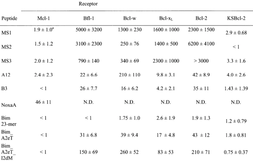
Table 2.1. Sequences of peptides used for fluorescence anisotropy and BH3 profiling assays .... 59
Table 2.2. Sequences of peptides used for fluorescence anisotropy and BH3 profiling assays .... 60
Table 2.3. Ki values (nM) for competition fluorescence anisotropy assays ... 65
Table 2.4. Affinities of native and designed BH3 peptides for six Bcl-2 homologs ... 66
Table 2.5. EC50 values (pM) for depolarization response ... 75
Table 3.1. Dissociation constants (nM) and 95% confidence intervals for BH3-like peptides in hum an p rotein s ... 105
Table 3.2 K SB cl-2 library design ... 112
Table 3.3. B H R F1 library design ... 112
Table 3.4. Conventionally sequenced clones and their frequencies ... 117
Table 3.5. Number of peptide sequences from Illumina sequencing of library pools ... 118
Table 3.6. Dissociation constants (nM) and 95% confidence intervals for the library peptides and m u tan ts ... 12 1 Table 3.7. Dissociation constants for Bel-2 homologs binding to mutants of Bim BH3 ... 126
Table 3.8. Dissociation constants for Bcl-2 homologs binding to peptides designed for increased specificity against M l-I...133
Table 3.9. Sequences of Bcl-2 constructs and primers ... 143
Chapter 1
Introduction
Protein-protein interactions are critical for essentially all cellular events. With -30,000 proteins expressed by the human genome, a large number of pairings are possible. Many protein-protein interactions (PPI) occur between the folded domains of protein-proteins, analogous to two puzzle pieces interlocking. Such domain-domain interactions have complex interfaces between tertiary structures, and we lack structural characterization of many of the known examples due to the relative difficulty of crystallizing protein complexes. An estimated 15-40% of PPIs occur between a folded domain of one protein and a linear polypeptide fragment of another protein
(Petsalaki and Russell, 2008). Domain-peptide interfaces are regarded as simpler than domain-domain interfaces as the peptide generally lacks secondary structure when unbound, and in many
cases, the domain surface does not change substantially upon peptide binding (London et al., 2010). Protein-peptide interactions are especially important for connecting proteins in signaling pathways. They also mediate the formation of complexes for a variety of processes such as the regulation of apoptosis or cell motility. Their essential biological roles and simple interfaces
relative to domain-domain interactions make protein-peptide interactions an important frontier for the advancement of our understanding of the protein interactome.
Many examples of peptide recognition domains (PRDs) have been identified, and the peptide binding preferences of several families of homologous PRDs have been extensively characterized in vitro (Liu et al., 2012). The peptide binding preferences of a PRD family are often represented as a motif. A sampling of PRDs and their known motifs are presented in Table 1.1. Motifs contain the residues that are most invariant in the known binders of a PRD. They can be inferred from multiple sequence alignments of cellular interaction partners, or established by experimental mutagenesis. Motifs are a simplistic representation of PRD binding preferences. The functional specificity of individual PRD family members arises in part from recognition of
different motifs, or tolerance of significant variation on motifs. Context residues, here defined as the residues not restricted in the motif ("x") or the residues terminal to the motif, also influence binding specificity.
Table 1.1. Protein recognition domains discussed and a subset of their binding motifs
Protein recognition domain Motifs recognizeda
PDZ (ST)x(ACVILF)>b, (VILFY)x(ACVILF)>, (DE)x(ACVILF)>
SH3 (RKY)xxPxxP, PxxPx(RK), xxx(PV)xxP, KPxx(QK)xxx, PxxDY
SH2 pYxN, pY(QDEVAIL)(DENPYHI)(IPVGAHS), pYxxQ,
pY(VLTFIC)xx
PTB x(ILVMFY)xNxx(FYpY)x, x(ILVM)LGxxPx
WW xx(p(ST))Px, PPxY, PPLP
GYF (QHR)xP(PL)PP(GS)H(RH)
Bcl-2 Dxxx(DxxD(GSA)DxD
TRAF (PSAT)x(QE)E, PxQxD, xxPxExx(FYWHDE)
EVH1 (FYWL)PxPP, (FYWL)PP(ALIVTFY)P, PPxF,
(FY)x(FW)5x(LMVIF)PxP(DE)
a Motifs are from the Eukatryotic Linear Motif database, except for the Bcl-2 motif.
b 4t> denotes the position of the C-terminus. "cD" denotes a hydrophobic residue.
This thesis addresses the topic of protein-peptide binding specificity, and the introduction covers the biological context of binding specificity, the binding specificity mechanisms of well-studied, and less-studied PRD families, as well as applicable methods and applications. It is important to first acknowledge that in the cell, the biophysical determinants of binding are only one of several factors that dictate whether two proteins will interact.
Interaction determinants in the cell
Post-translational inodifications
In vivo, protein-peptide interactions can be controlled by a variety of factors beyond their binding specificity such as post-translational modifications (PTMs), localization,
oligomerization, and coexpression. PTMs are required in the recognition motifs of several PRDs, including phosphorylation of serine, threonine, or tyrosine (bound by src-homolog domain 2 (SH2), phosphotyrosine-binding domain (PTB), forkhead-associated domain (FHA), 14-3-3 proteins, etc.), or acetylation or methylation of lysine or arginine (bromodomains and
chromodomains) (Pawson and Nash, 2003). Other PRD families have subsets of members with preferences for binding sites with PTMs-such as class IV WW domains, which bind
phosphorylated serine or threonine-and other subsets whose binding is inhibited by PTMs, such as class I WW domains, which bind a motif containing a tyrosine. Analysis of the co-localization of phosphosites and the motifs of SH3 (sre-homology 3), SH2, WW, and PDZ (PSD-95/Discs-large/ZO-1) domains in the human proteome found a statistically significant association (Akiva et al., 2012). Thus, PTMs act as switches to turn interactions with peptide motifs on or off, depending on the specificity of the PRD. There are even cases of "double switches" in which motifs for two different PRDs overlap, such as that of an SH2 domain and an SH3 domain; in this case, the SH2 domain binds to the site when it is phosphorylated, and the SH3 domain binds when the site is not phosphorylated (Akiva et al., 2012).
An example of a protein-peptide interaction that is heavily regulated by both PTMs and subcellular localization is the interaction between the B13 (Bel-2 homology 3) motif of the pro-apoptotic protein Bad and its anti-pro-apoptotic PRDs, Bcl-xL and Bcl-2. Bad promotes apoptosis by binding and inhibiting Bcl-xL and Bcl-2 when it is localized to the outer mitochondrial
membrane (Yang et al., 1995). Bad contains three serines that are phosphorylated by survival kinases, such as Akt (Danial, 2008). Two of these phosphorylated serines form interaction sites for 14-3-3 proteins, and the combination of phosphorylation and binding of 14-3-3 proteins results in the relocation of Bad to the cytoplasm. The pro-apoptotic activity of Bad is further
inhibited by phosphorylation of a serine in the BH3 binding motif, which directly blocks Bcl-xL and Bcl-2 binding (Danial, 2008). At another level of regulation, with an opposing effect, Bad is also a substrate for PRMT 1, a protein arginine methyltransferase. Methylation of Bad arginines that form the recognition site for Akt (outside of the BH3 motif) block Bad phosphorylation, promoting its apoptotic activity (Sakamaki et al., 2011). Bad is just one example of how PTMs
and localization can influence protein-peptide interactions. Further mapping of PTMs to motif sites in the proteome will continue to unveil complex levels of regulation for the formation of protein-peptide complexes.
Gene expression
Protein-protein interaction networks are also regulated at the levels of gene expression and protein abundance. Most models of protein interaction networks lack information about the dynamics of protein abundance or gene expression. Several studies have integrated temporal or conditional gene expression studies (e.g. different cell cycle stages or growth conditions) with protein interaction data from yeast and E. coli (de Lichtenberg et al., 2005; Hegde et al., 2008; Komurov and White, 2007; Tang et al., 2011). These analyses have created limited dynamic protein interaction networks, but the networks are enriched for the type of stable domain-domain interactions that are more easily identified in large yeast two-hybrid or affinity purification-mass spectrometry datasets. Transient interactions, such as many weak domain-peptide interactions, are often missed by yeast two-hybrid experiments (Vinayagam et al., 2009). Protein abundance
and signaling events also play a role in transient domain-peptide interactions, and these are generally not directly reflected in gene expression studies. For example, weak interactions such as those between tumor necrosis factor receptor-associated factors (TRAFs) and peptides in TNF receptors rely on avidity created by the oligomerization of TNF receptors in response to
extracellular signals (Pullen et al., 1999b). Therefore, significant advances are needed before gene expression, protein abundance, subcellular localization, and protein interaction data can be combined to create a comprehensive view of transient protein interaction networks. A clear accounting of possible protein interactions as determined in vitro is a necessary first step. As it would be an impossible task to directly measure all possible protein interactions, it is important to define the rules governing interactions. Determining the peptide motifs that enable interaction with each PRD family will enable modeling of protein interaction networks in the future.
Methods for determining peptide interaction motifs
Peptide interaction motifs can be identified through a variety of experimental and computational methods. In this section, a brief survey of classical experimental methods and a more in-depth description of a new technique, deep mutational scanning, will be followed by a
discussion of computational methods for motif and specificity determination. Experimental methods
Experimentally, peptide interaction sites are often initially found by mutagenesis, with alanine scanning or hydrophile scanning being used to identify the residues most important for binding (Boersma et al., 2008). More in-depth examination of PRD binding preferences requires
analysis of binding to many more peptides than are accessible by standard solution binding assays. Techniques such as SPOT arrays or protein microarrays can be used to
semi-quantitatively analyze binding to a modest number of peptides (hundreds to thousands) (Liu et al., 2012). Larger libraries of peptides are accessible by high throughput screening strategies such as phage display, cell surface display, and ribosome or mRNA display (Levin and Weiss, 2006). Phage, ribosome and mRNA display offer the capacity to screen very large libraries (routinely 1010 for phage and >1012 for ribosome and mRNA). Cell surface display techniques
have library sizes limited by the efficiency of getting the library DNA into cells, but offer the advantage of real-time, affinity-based resolution of binding partners when combined with fluorescence activated cell sorting (FACS). Yeast surface display libraries of 107 can be easily achieved, with larger libraries made possible by combining hundreds of transformations. With bacterial surface display in E. co/i, libraries of 1010 are easily achievable due to the higher transformation efficiency of gram negative bacteria relative to yeast (L6fblom, 2011). These library-screening techniques have been successful in screening large random peptide libraries or natural sequences and variants thereof. When combined with deep sequencing of the enriched pools, these techniques can greatly expand the binding sequence space beyond that of the known natural partners. These screens excel at providing peptide reagents of high affinity or specificity
for a PRD of interest. Screens can also be performed in the other direction - to find a variety of domain sequences that bind a given peptide (Chen et al., 2013; Ernst et al., 2010; Gold et al., 2013).
One disadvantage of these large library screens is that they are generally used to supply sequences of only the best leads and lack information on relative affinity or specificity of sequences of a broader spectrum. A recent method by Reich et al. utilizes the affinity
discrimination offered by FACS to sort a yeast surface display library of peptides into pools of different affinity (Reich et al., 2014). The relative affinity for each clone is computationally extracted from its distribution across the pools as determined by deep sequencing. The
combination of cell surface display methods, FACS, and deep sequencing has great potential to provide datasets containing sequence and affinity information from which to elucidate peptide-binding determinants.
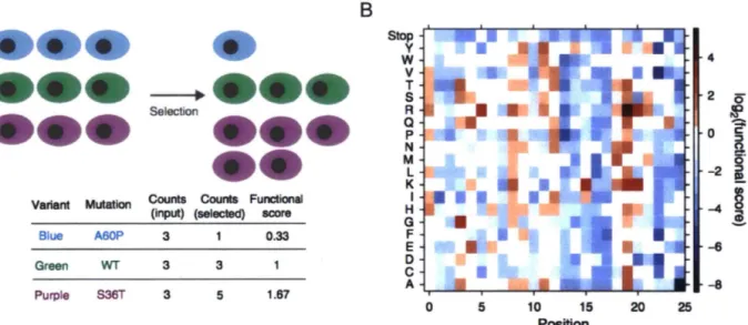
The power to systematically explore binding determinants has been greatly improved by the advent of a technique known as deep mutational scanning (Fowler et al., 2010). In its most general form, deep mutational scanning involves screening a comprehensive library of mutants of a protein of interest by an assay designed to measure some aspect of protein function.
Subsequent deep sequencing of the nafve and enriched library pools provides relative frequencies of sequences that can be used as a measure of the protein function assayed (Figure 1.1). This general framework has been applied to study sequence determinants of protein stability, enzyme
function, protein-ligand interactions, and more general conglomerates of overall protein function (Araya et al., 2012; Fujino et al., 2012; Melamed et al., 2013; 2015; Starita et al., 2015; Tinberg et al., 2013; Traxlmayr et al., 2012; Whitehead et al., 2012). Detailed information about the relative contributions to function, or fitness, of all 20 amino acids at each protein position has the potential to provide rich datasets for applications such as protein structure prediction, protein evolution and engineering, and the study of human disease genetics (Fowler and Fields, 2014).
Deep mutational scanning and related approaches have been applied to both protein-protein (antibody: antigen, RNA recognition motif:eIF4G 1, computationally designed influenza hemagglutinin binders) and protein-peptide interactions (WW and PDZ) (Forsyth et al., 2013; Fowler et al., 2010; Fujino et al., 2012; McLaughlin et al., 2012; Melamed et al., 2013; 2015; Whitehead et al., 2012). Any binding assay that can screen modestly sized libraries, such as phage display, cell surface display, ribosome or mRNA display, or two-hybrid assays can be utilized to enrich the low complexity libraries necessary for this technique. Analysis of the
correlation between binding affinity and enrichment in screening for binding is lacking, but small scale comparisons indicate a good correlation (Fowler et al., 2010; McLaughlin et al., 2012; Pil et al., 2006). The most comprehensive analysis compared dissociation constants measured by
fluorescence polarization to enrichment values from a bacterial two-hybrid screen for 86 PDZ domain mutants binding a peptide and found a good linear correlation over a range of Ko values
of0.1-200 µM (McLaughlin et al., 2012). However, when mutating an entire protein domain,
other parameters such as folding and stability will also influence the data. Researchers have used both experimental (e.g. initial selection for folded variants by binding of an antibody) and
computational strategies (e.g. integration of evolutionary conservation information) to identify mutations that affect the binding interface (Melamed et al., 2015; Pal et al., 2006).
A
Variant Mutation
Blue A60P
Green WT Purple S36T
Counts Counts Functional (input) (selected) score
3 0.33 3 3 3 5 1.67 B Stop y w v T s R a p N M L K I H G F E 0 c A 0 5 10 15 20 Position 4 2 0 i2
--=
0 : l c Sl 6' -2 : l !!.8
-4.!.
-6 -8 25Figure 1.1. Deep mutational scanning provides a metric of function for mutations to all 20 amino acids at each protein position. A) Next generation sequencing is used to quantify the number of copies of each variant in the naive library and the pool resulting from selection for some function of interest. B) The frequencies are converted to a functional score based on the frequencies of each variant in the naive and selected pools, and this is used to create a heat map of protein mutational tolerance. Figures are from Fowler et al., 2014.
The ability to leverage enriched mutations to engineer tighter binding partners also demonstrates that enrichment can be a good proxy for affinity (Fujino et al., 2012; Tinberg et al., 2013; Whitehead et al., 2012). One powerful application of deep mutational scanning of protein-protein interfaces is to use the datasets to inform design of combinatorial libraries of enriched mutations. These libraries can then be screened for variants that successfully combine the affinity
enhancing effects of several mutations to greatly increase binding affinity. This approach has been used to improve the affinity of computationally designed binders of influenza
hemagglutinin and antibodies that bind tumor necrosis factor-a receptor (Fujino et al., 2012; Whitehead et al., 2012).
To date, deep mutational scanning studies have mutated the protein side of protein-peptide interfaces. These data have been utilized to identify sequence determinants of binding specificity for peptide ligands. Deep mutational scanning of a PDZ domain was performed twice for binding to a cognate peptide ligand and a single point mutant of that ligand, and these
datasets were compared to find PDZ domain positions that governed peptide binding preferences (McLaughlin et al., 2012). When deeper mutation is performed, such that double mutants are present, information on epistasis or covariation between positions can be inferred by comparing single and double mutant enrichment (Araya et al., 2012; McLaughlin et al., 2012; Melamed et al., 2015; Whitehead et al., 2012). This analysis has been incomplete, as it is difficult to get complete coverage of double mutants in folded domains by the current methods used for library assembly. Double, and even higher-order mutations should be easier to cover in short contiguous peptide segments. Currently, the degree of covariation between positions in different linear peptide motifs is not known. This question needs to be answered in order to create fully parameterized computational models for prediction and design of peptide binding partners.
Computational methods
Computational models of protein-peptide interaction specificity can build on
experimental datasets and utilize features from sequence and structures of complexes. Peptide interaction motifs are often first identified on the sequence level in multiple sequence alignments of proteins of related function. Models of linear motifs such as position specific scoring matrices
(PSSMs), regular expressions, or hidden Markov models (HMMs) can be built based on lists of known examples and used to search for more proteins with a matching sequence (Dinkel et al., 2014; McLaughlin et al., 2011). Short linear motifs are difficult to establish without prior knowledge of binding sites, but several methods exist based on searching for motif over-representation in non-homologous sequences that are functionally related by some metric, such
as binding to a common partner in yeast two-hybrid datasets (Neduva and Russell, 2006; Neduva et al., 2005; Palopoli et al., 2015). In recent years, large library or array experiments have
generated long lists of natural and non-natural binders of PDZ, SH3, and Bcl-2 domains, among others (DeBartolo et al., 2012; Ernst et al., 2010; Tonikian et al., 2009; 2008; Xin et al., 2013). This increase in known binders has allowed the construction of powerful PSSMs and other models useful for scanning the proteome for more peptide binders of these domains (DeBartolo et al., 2014; Gfeller et al., 2011; Kim et al., 2012). Experimental generation of these datasets requires significant work, and the resulting models can only reflect the sequence space upon which they were built, limiting their utility for finding motifs in novel specificity classes. Though useful and fast, sequence-based methods for motif discovery and matching ignore an important source of information on interaction specificity-the physical interactions encoded in the complex structures.
Structure-based binding specificity prediction methods exist at varying levels of physical detail. All methods require a high resolution structure of an example protein-peptide complex and a set of experimentally validated peptide sequences upon which to build and test the model (London et al., 2013). I will discuss three examples of computational approaches spanning levels of physical detail and computational intensity including full physics-based models, a combined
complexes. As protein interaction modeling is an important goal in computational biology, these examples are only a sampling of the many published approaches.
At the most computationally intensive and physically detailed end of the spectrum lie models utilizing molecular dynamics simulations to model lists of complexes and molecular mechanics-generalized Born surface area (GBSA) to compute binding energies. MM-GBSA is used to calculate molecular interaction energy components for many complexes, which can be used to train a support vector machine to create a general model of the binding
preferences of a PRD. Wang and colleagues used this approach to create models of SH3, PDZ, PKA-AKAP, and chromodomain binding preferences, which they successfully used to identify peptide interactors from proteomes (Hou et al., 2012; 2011; 2009; Li et al., 2011; 2013). Because these models involve direct calculation of the energy contributions of each peptide and receptor residue, they can be used to identify the structural mechanisms behind different binding
specificities, such as that of class I versus class II SH3 binders (Hou et al., 2012).
Rosetta FlexPepBind is a second structural method for modeling of peptide binding specificity that scores peptide sequences using the combined statistical and physical Rosetta energy function as implemented in the protocol Rosetta FlexPepDock (Raveh et al., 2010). London et al. used this method to model the binding specificity of farnesyl transferase to its peptide substrates, and Bcl-2 to BH3 peptides (London et al., 2012; 2011). They found good predictive performance by simple threading of peptide sequences on a complex structure
followed by minimization and energy calculation. Treatment of peptide side-chain and backbone flexibility via minimization was necessary for good performance on a test set of known binding partners, and more extensive sampling of flexibility using Rosetta FlexPepDock improved performance, at the cost of increased computing time. The Rosetta FlexPepBind protocol
requires calibration on a set of known binding partners, and its performance is often improved with further parameterization based on known structural constraints of the protein-peptide system (London et al., 2011; 2012).
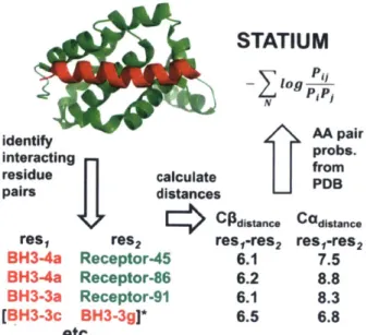
At a third and lowest level of complexity and computational intensity, lies STATIUM, a statistical model that relies only on a protein-peptide complex structure or homology model and a reference set of monomeric structures from the PDB (DeBartolo et al., 2012; 2014). STATIUM calculates the distances from the Ca and Cp atoms of each peptide residue to all receptor side-chain atoms within a given distance cutoff. It then tabulates the frequency with which all 20 amino acids are found in similar residue-residue pair geometries in the reference database and calculates a score based on that frequency normalized by the frequency of that amino acid in the reference protein set (Figure 1.2). Because the residue-residue distances in the reference set are pre-tabulated, scoring a sequence on a complex structure is essentially a "look-up procedure", allowing scoring of >106 sequences per second. STATIUM has been used to scan the human proteome for novel BH3 peptide sequences that bind the anti-apoptotic Bcl-2 homologs, and
shows good agreement with experiment-based models of BC-2:BH3 binding preferences
(DeBartolo et al., 2012; 2014). STATIUM is, in theory, applicable to any protein-protein interface, though it has yet to be tested extensively on other protein-peptide families.
STATIUM
log
identify AA pair
interacting probs.
residue calculate fro
pairs distances
C*Csne Cadistane
res, res2 res,-res2 res,-res2
BH3-4a Receptor-45 6.1 7.5 BH3-4a Receptor-86 6.2 8.8 BH3-3a Receptor-91 6.1 8.3
[BH3-3c BH3-3g]* 6.5 6.8
etc...
Figure 1.2. How a STATIUM model is built for a protein:peptide complex structure. Distances from each peptide Ca and CO to each receptor residue Ca and CP are calculated. The probability of finding each residue pair and distance are found in the reference PDB set and normalized against the frequency of each amino acid in the reference. The STATIUM score for a peptide is the sum of this value for each peptide residue. The more recent STATIUMsidechain program uses distances to all receptor side-chain heavy atoms. Figure is from DeBartolo et al., 2012.
Binding specificity mechanisms
A hierarchy of binding specificity mechanisms within PRD families
Detailed characterization of peptide binding for a few PRD families has uncovered mechanisms that differentiate the binding preferences of individual members within a PRD family. These differences can take the form of recognition of different motifs, variations on the same motif, or even recognition of completely different protein structures (e.g. binding the tertiary structure of another domain rather than a linear peptide). First, it will be helpful to understand the energetic basis of a motif. Stein and Aloy analyzed the complex structures of 30 different PRD families and evaluated the energetic contributions of peptide residues using the computational algorithm FoldX (Stein and Aloy, 2008). They found that residues in the motifs cited in the Eukaryotic Linear Motif (ELM) database contributed on average 80% of the binding
energy, whereas the more variable context residues only contributed 20%. Therefore, the
sequence features that make up a peptide motif can be regarded as the hotspot residues, i.e. those that contribute the most binding energy. A more general finding that sequence conservation in bound peptides correlates with per-residue binding energy contribution also held true in that residues that were fixed as one amino acid in the motif versus restricted to a set of amino acids had higher energetic contributions on average (Stein and Aloy, 2008). Given this concept of a motif, I will discuss how binding specificity is encoded on different levels, starting from the most diverged and ending with examples of fine-scale differences in binding specificity between
closely related members of the same PRD family.
The use of multiple motifs for specificity in large PRD
families
Nature utilizes similar protein forms for diverse purposes. One example of this concept in the realm of protein-ligand binding is the pleckstrin homology fold. This same fold is recycled in domains that bind a wide variety of ligands including phospholipids (pleckstrin homology (PH) domains), phosphopeptides (PTB domains), and polyproline peptides (Ena/VASP homology 1 domains (EVH 1)) (Pawson and Nash, 2003). Conversely, similar ligands (peptides containing polyproline type II helices) can bind similar interfaces (aromatic-rich) in domains with very
different folds (SH3, WW, EVH1, GYF, and profilin) (Li, 2005). This illustrates that there are certain structural solutions to recognition that are repeated in the protein interactome due to both divergent and convergent evolution. However, a high degree of specificity can be encoded within a given structural form by use of different residues.
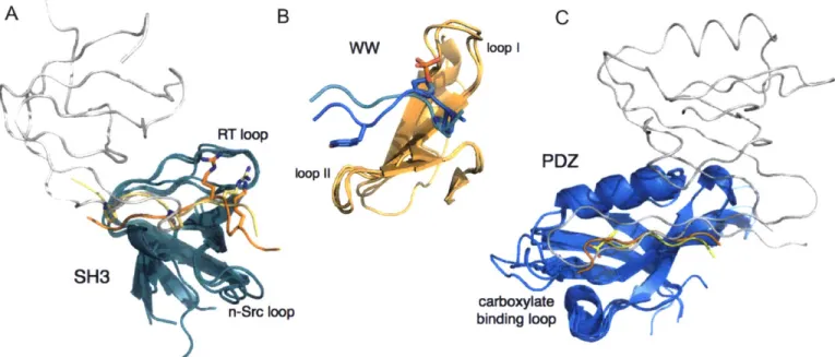
Within a PRD family, members can utilize highly diverged interface features to bind peptides or proteins of different structures. SH3 domains have multiplied in eukaryotes from 34 different SH3 domains in yeast to 958 in humans, according to the most recent estimates in the
SMART database (Letunic et al., 2015). Within such a large family, a diverse array of protein-binding modes has been realized over the course of evolution. Though the majority of SH3 domains bind peptides containing a PxxP motif, decades of work on the SH3 domains has uncovered examples that bind peptides with very different motifs (e.g. RxxK), peptides binding in 310- or a-helical conformations, and even interactions with tertiary structures of other domains (Figure 1.3A) (Li, 2005). The binding groove is defined by two loops of the SH3, the RT loop and the n-Src loop. Variations on the length and sequence content of these loops can change the shape and charge properties of the groove to favor peptides matching the RxxK versus the PxxP motif (Kaneko et al., 2011).
A B C
RT loop
PDZ loop 11,
SH3
n-Src loop binding loopcarboxylate
Figure 1.3. Three large PRD families that use different motifs for specificity. A) SH3 domains (teal) bound to a variety of partners: another SH3 domain (gray, 1GCQ (Nishida et al., 2001)), a peptide with a PxxPxR motif (yellow, 1GBQ (Wittekind et al., 1997)), and a peptide with an
RxxK motif (orange, 1H3H (Liu et al., 2003)). Motif residues are shown in sticks. B) WW domains (orange) bound to two peptides of different motifs: pSP (cyan, IFH8 (Verdecia et al., 2000)), PPxY (blue, lEGH (Huang et al., 2000)). C) PDZ domains (blue) bound to: a C-terminal peptide with C-terminal residue shown in sticks (yellow, 1RZX (Peterson et al., 2004)), an internal peptide (orange, 1X8S (Penkert et al., 2004)), and a -finger from another PDZ domain (gray, 1 QAV (Hillier, 1999)). Loops that play a critical role in specificity are labeled for each domain.
WW domains have many similarities to SH3 domains, and are also a very common domain with an estimated 247 examples in the human proteome (Letunic et al., 2015). WW domains are named after the two tryptophans that form the binding groove for an "xP" motif.
Several studies of WW domain binding specificity have grouped the binding preferences into 4-5 classes represented by different ligand motifs (Hu et al., 2004; Ingham et al., 2005). Two very different motifs are PPxY and p(S/T)P (phosphorylated serine or threonine) (Figure 1.3B). Like
SH3 domains, there is a common platform for binding the "xP motif', and different WW domains utilize variation in two loops to dictate specificity (Verdecia et al., 2000). The p(S/T)P motif, first identified in binders of the Pin 1 WW domain, is recognized by an arginine and serine on loop I that make direct contacts with the phosphorylated residue. In WW domains of the PPxY specificity class, a hydrophobic surface formed by loop II recognizes the tyrosine.
Another well-studied PRD family, the PDZ domain, also recognizes diverse motifs. PDZ domains predominantly bind to the C-termini of peptides, recognizing the C-terminal carboxylate via the backbone amides of the domain's carboxylate binding loop (Lee and Zheng, 2010). HoweMVer, %lik SHLIA31 dmnalis, PDZ domains can foVrm- Udin-dUMain1- interactions with other
PDZ domains and can also bind other peptide conformations, including internal peptide sequences (Figure 1.3C) (Chang et al., 2011; Hillier, 1999; Penkert et al., 2004). Among PDZ domains that bind C-terminal peptides, many different specificity classes have been identified that recognize distinct motifs. By selecting phage display libraries of random peptides for binding to 82 human and C. elegans PDZ domains, Tonikian et al. identified 16 specificity classes, into which the specificity profiles of 90% of the profiled domains could be grouped (Tonikian et al., 2008). These 16 different motifs, represented as position weight matrices
To analyze which PDZ domain residues governed specificity at each peptide position, Tonikian et al. determined the specificity profiles of 91 single mutants of the Erbin PDZ domain, varying
10 different PDZ positions. They found that the preferences at some peptide positions were primarily influenced by PDZ residues in direct contact with the peptide side chain, while other peptide positions were also influenced through non-direct contacts. This demonstrates that peptide binding specificity reflects a complex combination of direct residue-residue interactions and underlying structural features.
The use of motif context residues for specificity in smaller PRD families
PRD family members can bind peptides that match the same motif but still exhibit distinct preferences for their interaction partners. In the analysis of protein-peptide structures performed by Stein and Aloy, they looked at specificity by performing a peptide-swapping
experiment between structures of PRD families with at least 10 different domain-peptide pairs (cyclins, hormone receptors, MATH, PDZ, and class I and II SH3 domains) (Stein and Aloy, 2008). Even when controlling for similar peptide topology, they found that "context" peptide positions were more likely to have poor FoldX energies than motif positions when assessed in a non-cognate complex. Thus, PRD family members that can bind peptides of the same motif utilize the context residues for specificity. Context residues that are tolerated or even provide modest contributions to binding affinity in their cognate complexes can act as strong deterrents to binding non-cognate domains. This idea of "anti-motifs" present in the context residues has been observed in the binding specificity determinants of many PRD families (Liu et al., 2012). The large PRD families (SH2, SH3, PDZ, and WW) exhibit such context-dependent specificity mechanisms between closely related members. However, my discussion will focus on smaller PRD families, including Bcl-2, EVH1, and TRAF domains. Smaller PRD families often have
less diversity in motif preferences, and they are good examples of how fine-scale differences in
motif context can have substantial effects on binding specificity.
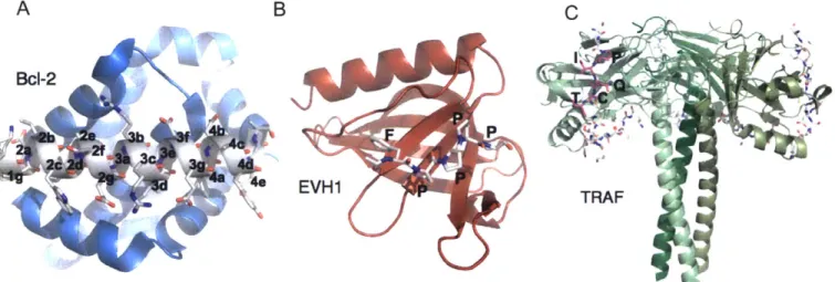
Bcl-2 proteins control apoptosis through interactions with helical BH3 motifs.
Pro-apoptotic Bcl-2 proteins can take one of three forms (Llambi et al., 2011). The effectors Bax and
Bak homo-oligomerize to form pores in the outer mitochondrial membrane, which release death
factors into the cytoplasm. This process is triggered by activator BH3-only proteins, such as Bid
and Bim. The third class of pro-apoptotic proteins are the sensitizer BH3-only proteins, such as
Bad, Noxa, Hrk, and Bik, which de-repress the function of the other two pro-apoptotic forms by
binding and inhibiting the anti-apoptotic Bcl-2 homologs. Anti-apoptotic Bcl-2
proteins-including Bcl-2, Bcl-xL, Bcl-w, Bfl- 1, Ml- 1, and Bcl-b in humans-bind the BH3 motif of the
BH3-only proteins and Bax and Bak to prevent apoptosis. The interaction between anti-apoptotic
Bcl-2 homologs and BH3 motifs has been extensively studied because the anti-apoptotic
homologs are widely overexpressed in human cancers to prevent apoptosis, making them
important drug targets.
TheLBJ mIIo~if, like other%.,L pi oifsUL1, is UOILn Luninunst-ructL-Lu-redre 1giOns
in BH3-only proteins, many of which are intrinsically disordered proteins (Rautureau et al.,
2010). BH3 motifs are also found folded as a-helices in the anti-apoptotic Bcl-2 and
pro-apoptotic effector homologs, and in these cases they are exposed for binding upon structural
rearrangements of the folded Bcl-2 domains (Lee et al., 2014). The BH3 motif binds to
anti-apoptotic Bcl-2 homologs as an amphipathic a-helix of-25 residues with a general motif
definition of DxxxxxD(GSA)Dx where "D" is a hydrophobic residue. Because the large
majority of contacts are mediated by residues within and immediately surrounding the motif, Bcl-2:BH3 interactions are usually studied with BH3 peptides of 20-30 residues. A nomenclature
of a heptad repeat (a-g) is often used to describe the peptide positions, with the buried
hydrophobic positions falling at positions a and d (see Figure 1.4A). Perhaps because this is a longer peptide interface than many short linear peptide-PRD interfaces that have been studied, and therefore has a larger number of peptide residues contributing to binding, there is substantial heterogeneity allowed at the hydrophobic positions (Dutta et al., 2010). The specificity of the six human anti-apoptotic Bcl-2 homologs for binding different BH3 sequences arises both from variations in the motif residues and the context residues. BH3-containing proteins, which include pro-apoptotic and pro-autophagic members, are differentially expressed and regulated in
response to apoptotic or autophagic stimuli. Thus, the BH3-binding preferences of the anti-apoptotic Bcl-2 homologs dictate their relative importance in responses to these stimuli (Chen et al., 2005). I will first summarize the work that has been done to study the binding preferences of anti-apoptotic Bcl-2 homologs and then discuss examples of specificity mechanisms relying on variations of motif and context residues.
A B
BcI-2
EVH1 TRAF
Figure 1.4. Three peptide recognition domain families utilizing motif context for specificity. A) A Bcl-2 homolog Mcl-I (blue) bound to the Bim BH3 peptide (gray) is shown with BH3
positions labeled according to heptad register (Fire et al., 2010). B) The EVHI domain of Mena is shown bound to one FPPPP motif of ActA (Prehoda et al., 1999). C) Trimerized TRAF3 is shown with monomers in shades of green. An extended peptide corresponding to the TANK TIM is in gray, with the 5-residue core site highlighted in magenta in one monomer (Li et al., 2002).
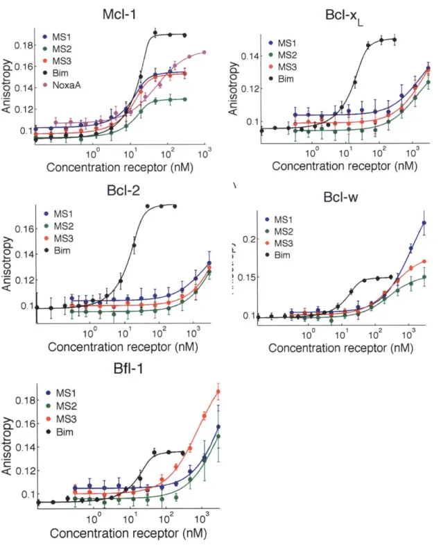
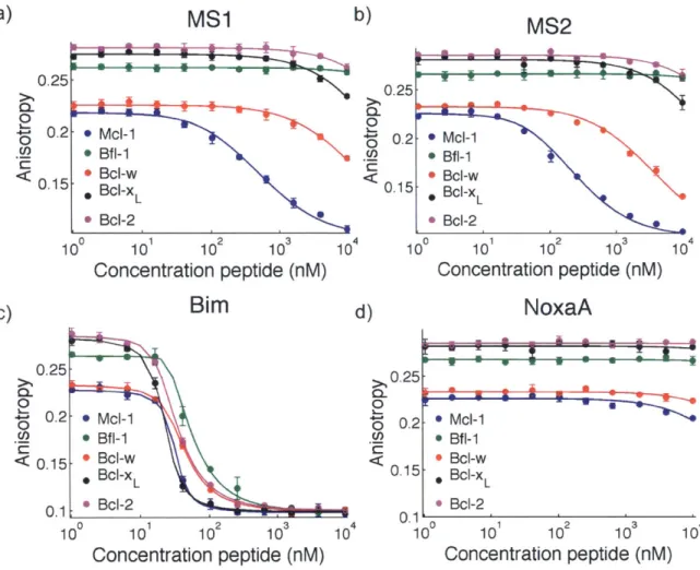
A number of high throughput approaches have been used to characterize the binding preferences of anti-apoptotic Bcl-2 homologs. All Bcl-2 homologs bind the BH3-only protein Bim with high affinity, and the Bim BH3 peptide has been used as a starting point to study mutational binding preferences of each Bcl-2 homolog. Dutta et al. synthesized peptides with 18
amino-acid variants (excluding cysteine and methionine) at 10 Bim BH3 positions on SPOT arrays, and measured binding of Mcl-I and Bcl-xL to the arrays (Dutta et al., 2010). Subsequent studies performed the same experiment with the other homologs Bcl-w, Bfl-1, and Bcl-2
(DeBartolo et al., 2012; London et al., 2012). These studies showed distinct preferences at subsets of peptide positions for each Bel-2 homolog, including both motif and context positions.
Larger combinatorial libraries of random peptides or variants of Bim BH3 have been screened for binding to Ml-1, Bcl-xL, and Bfl- 1. Lee et al. screened a random phage display library of 16-mer peptides for binding to Ml-I and largely recovered peptides that conform to the previously described BH3 motif (Lee et al., 2009a). Many of the recovered sequences
showed specificity for binding Ml- 1 over the other human homologs Bcl-xL, Bcl-2, and Bcl-w, despite the fact that the screen only selected for Mcl- 1 binding and not selectivity. Dutta et al. screened a library of Bim BH3 variants by yeast surface display for peptides showing specificity to Mcl-I or Bcl-xL or binding to both homologs (Dutta et al., 2010). Comparison of sequence logos from the three screening paths revealed specificity determinants for Mcl-I and Bcl-xL. More recent yeast display screens by Dutta et al. have found further specificity determinants for Bcl-xL and Bfl-1 (Dutta et al., 2013; 2014).
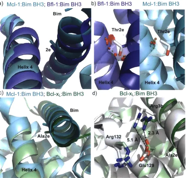
Bcl-2 homologs exhibit specificity differences at both motif and context positions in binding to BH3 peptides. One example of specificity from a motif residue is the preference for tyrosine or phenylalanine at the fourth hydrophobic position, 4a, exhibited by Bcl-xL. Mutation
of 4a to smaller residues including valine or asparagine is tolerated by Ml- 1, but drastically decreases binding to Bcl-xL (Dutta et al., 2010). This preference is encoded by the shape of the 4a binding pocket, which is enclosed in Bdl-xL, but open to the solvent in Mcl- 1 (Fire et al., 2010; Lee et al., 2009b). As another example, Bcl-xL and Bcl-2 have high sequence similarity and both bind the BH3-only protein Bad (Certo et al., 2006). The mutation of tyrosine at 4e to lysine in a designed B13 peptide does not decrease Bcl-xL affinity, but reduces binding to Bcl-2 by 7-fold (Dutta et al., 2014). This position is outside the core BH3 motif, near the C-terminus of the peptide. In a structure of Bcl-2 bound to a Bax BH3 peptide, the methionine at 4e is directed into the groove and largely shielded from the solvent, while in a structure of Bcl-xL bound to Bim, tyrosine at 4e is directed out from the groove with access to solvent (Ku et al., 2011; Lee et al., 2009b). The abundance of structural data for Bcl-2 homologs and the extensive mutational analysis of BH3 peptides has uncovered the structural basis of many other specificity
mechanisms employed by the anti-apoptotic Bcl-2 members.
EVH 1 (Ena/VASP homology 1) domains are present in four different families of
proteins: Homer, WASP, SPRED, and Ena/VASP (Peterson and Volkman 2009). These proteins bind to polyproline motifs to regulate actin cytoskeleton dynamics. The three human Ena/VASP proteins, Mena, Evl, and VASP, bind to actin regulatory factors like zyxin and vinculin with motifs of FPxDP, where (D is a hydrophobic residue (Figure 1.4B). The pathogen Listeria monocytogenes expresses the protein ActA, which contains four FPPPP sites. Ball et al. mapped the chemical shift perturbations caused by a longer ActA peptide, SFEFPPPPTEDEL on the surface of VASP (Ball et al., 2000). They found that the C-terminal leucine contacted a hydrophobic patch outside the core polyproline-binding site. This exosite interaction increases the affinity of the ActA peptide for VASP. Similar hydrophobic positions are not observed near
the FPxIP binding sites on zyxin, which do not bind as tightly as the ActA peptide. Thus, the pathogen uses a higher-affinity interaction to co-opt the host actin cytoskeleton for movement around the cell. It is not know whether any native Ena/VASP EVH1 binders form interactions outside of the core binding site, and Evl, Mena, and VASP have identical residues forming the binding site for the FPxDP sequence. However, differences between the three homologs can be seen in the loops surrounding the identified exosite, which lies towards the C-terminus of the peptide (Ball et al., 2000; Fedorov et al., 1999; Prehoda et al., 1999). Notably, Mena forms a different type of interaction with the folded LIM3 domain of Tes, and this interface overlaps with the same exosite region bound on VASP by ActA (Boeda et al., 2007). Tes does not bind to Evl
or VASP, indicating that differences between the homologs in this region have functional importance. It remains to be seen whether Evl, Mena, and VASP have differential binding preferences for their native binding partners containing FPxDP sites, or whether variations in
their functions should be attributed to differences in their other domains or aspects of their regulation (Gertler and Condeelis, 2011).
TRAF (tumor necrosis factor receptor-associated factor) proteins contain a peptide-binding interface on their MATH domains (meprin and traf homology). The MATH domain binds peptides from TNF receptors, as well as Toll-like receptors, other signaling receptors, and a variety of cytoplasmic signaling proteins (Figure 1.4C) (Xie, 2013). The MATH domain, also known as the TRAF-C domain, is at the C-terminus of TRAFs 1-6, which trimerize via the TRAF-N coiled-coil domain. N-terminal to the TRAF domain are zinc-finger domains, and TRAFs 2-6 also contain N-terminal RING domains, which play a regulatory role in many
signaling pathways through their E3 ubiquitin ligase activity. TRAFs also act as adapter proteins between signaling receptors and downstream factors, which can interact with their coiled-coil
domain (e.g. cIAP1 and 2), or be recruited or activated by the K63-linked polyubiquitin chains created by the TRAF RING domains (Xie, 2013). TRAFs 1, 2, 3, and 5 are close homologs that are primarily found in vertebrates, while TRAFs 4 and 6 are more ancient proteins (Zapata et al., 2007). The binding preferences of the MATH domains reflect this evolutionary history in that TRAFs 1, 2, 3, and 5 share many of the same binding partners, recognizing a major
((P/S/A/T)x(Q/E)E) and minor (PxQxxD) motif (Ye et al., 1999). TRAF6, though it plays a role in many of the same pathways and binds to some of the same proteins, recognizes a different motif (PxExx(Aromatic/Acidic)) (Ye et al., 2002). Binding partners of TRAF4 have not been characterized in direct binding assays as peptides, and the current structures of TRAF4 are apo structures, but there is some evidence that it may compete with TRAF6 for binding the same sites on NOD2 and Actl (Marinis et al., 2011; Zepp et al., 2012). The TRAF6 and TRAF1/2/3/5 motifs have obvious similarities, but the peptides bind in different orientations on the MATH domains. Structures of TRAF6 bound to peptides from CD40 and RANK show the peptide bound in a
P-strand
conformation nearly parallel to a MATH domains-strand
and able to form backbone hydrogen bonds (Ye et al., 2002). In contrast, structures of peptides bound to TRAFs 2and 3 show the peptides in a polyproline type II helix conformation, bound at an angle of 40' in comparison with the TRAF6-bound peptides (Ni et al., 2000; Ye et al., 1999). This difference can be attributed at least in part to a change in the position of the pocket that binds the proline in each motif, which is determined by different positioning of the aromatic residues that form the pocket.
The MATH-peptide interactions of TRAFs are very weak; the tightest reported
monomeric KD, measured for TRAF3 binding to a peptide from the cytoplasmic protein TANK, is 24 ptM, and many interactions are in the hundreds of micromolar (Li et al., 2002). The weak
nature of the monomeric interactions is an important feature of TRAF function, as pathways are only activated upon trimerization of their binding partners, such as the trimerization of TNFR2 by its ligand TNFu. Trimerization, or higher-order oligomerization, allows for avidity to recruit the trimeric TRAFs (Pullen et al., 1999b). Their weak nature makes the TRAF-peptide
interactions challenging to study in vitro, and only a limited number of the affinities have been directly quantified. Therefore, relatively little is known about the binding determinants of TRAFs beyond the motifs that have been constructed from multiple sequence alignments of known binding partners.
The most detailed information on binding preferences comes from SPOT array analysis of the two TRAF binding sites in the cytoplasmic tail of the TNFR homolog CD40 (Pullen et al., 1999a). The analysis examined binding of TRAFs 1, 2, 3, and 6 to mutants (all 20 amino acids) of each site of the TRAF 1/2/3 and TRAF6 binding sites on CD40. The results primarily
emphasized the lack of tolerance of mutation at the conserved motif positions. However, SPOT arrays performed with CD40 peptides of differing length indicated that TRAF3 may also bind CD40 sequence C-terminal to the core-binding site defined as the motif region. The increased affinity supplied by these exosite interactions relaxes the binding preferences of TRAF3 in the core peptide region. This observation did not hold true for TRAF2, suggesting that extra interactions outside of the core motif may be one specificity mechanism employed by TRAF3.
Structures of TRAF3 with long peptides (>17 residues) from CD40, BAFF-R, TANK, and LT3R show extra interactions mediated by peptide regions beyond the core motif (Ely and Li, 2002; Li et al., 2002; Ni et al., 2000; 2004). All TRAF2 structures were solved with short peptides
encompassing primarily the motif, so it is not known whether other TRAFs can utilize interactions outside the core motif for enhanced affinity or specificity. The use of peptide
residues beyond the motif regions is a common specificity strategy for PRDs and an aspect of protein-peptide binding specificity that is often poorly understood due to the convention of solving structures with peptides corresponding to only the minimal motif (Stein and Aloy, 2008).
Another example of specificity between close TRAF homologs was detailed in a recent paper on the binding of TRAF3 to the adapter protein Cardif (also known as Mavs) (Zhang et al., 2012). TRAF3, but not TRAF5, is recruited by Cardif upon intracellular detection of viral double stranded RNA, leading to the induction of type I interferons (Saha et al., 2006). TRAF5 cannot bind the TRAF interacting motif (TIM) in Cardif. Zhang et al. demonstrated that this specificity was due in part to interactions between a tyrosine hydroxyl on TRAF3 and the main chain carbonyls of two prolines present in Cardif C-terminal to the core motif. TRAF5 has a
phenylalanine at the same position, and mutation of this residue to tyrosine allowed TRAF5 to bind to CARDIF. Thus, there is ample evidence that TRAF3 utilizes interactions beyond the core TIM to determine its binding specificity.
With many large families of PRDs and even more potential peptide binding partners, evolution has arrived at several methods of achieving the binding specificity necessary for proper functioning of cellular pathways. As described for SH3, PDZ, and WW domains, family
members can utilize different loops and binding site residues to recognize very different motifs. Other families of proteins with overlapping functions, like the anti-apoptotic Bcl-2s, recognize variations on the motif and context residues to achieve different binding profiles. Context
residues can include both the "x" residues between hotspot residues represented by the motif and more distant peptide sites that may bind exosites on the PRD surface, as is seen for TRAF3 and the EVH1 domains. This supports the idea of protein-protein interaction specificity as a