HAL Id: hal-02542297
https://hal.umontpellier.fr/hal-02542297
Submitted on 14 Apr 2020
HAL is a multi-disciplinary open access archive for the deposit and dissemination of sci-entific research documents, whether they are pub-lished or not. The documents may come from teaching and research institutions in France or abroad, or from public or private research centers.
L’archive ouverte pluridisciplinaire HAL, est destinée au dépôt et à la diffusion de documents scientifiques de niveau recherche, publiés ou non, émanant des établissements d’enseignement et de recherche français ou étrangers, des laboratoires publics ou privés.
Diabetic Glomerulosclerosis
Olivia Lenoir, Marine Milon, Anne Virsolvy, Carole Henique, Alain Schmitt,
Jean-Marc Massé, Yuri Kotelevtsev, Masashi Yanagisawa, David Webb,
Sylvain Richard, et al.
To cite this version:
Olivia Lenoir, Marine Milon, Anne Virsolvy, Carole Henique, Alain Schmitt, et al.. Direct Action of Endothelin-1 on Podocytes Promotes Diabetic Glomerulosclerosis. Journal of the American Society of Nephrology, American Society of Nephrology, 2014, 25 (5), pp.1050-1062. �10.1681/ASN.2013020195�. �hal-02542297�
Direct Action of Endothelin-1 on Podocytes Promotes
Diabetic Glomerulosclerosis
Olivia Lenoir,*†Marine Milon,*†Anne Virsolvy,‡Carole Hénique,*†Alain Schmitt,†§|
Jean-Marc Massé,†§|Yuri Kotelevtsev,¶** Masashi Yanagisawa,††
David J. Webb,¶
Sylvain Richard,‡ and Pierre-Louis Tharaux*†‡‡
*Paris Cardiovascular Research Centre, Institut National de la Santé et de la Recherche Médicale, Paris, France;
†Université Paris Descartes, Sorbonne Paris Cité, Paris, France;‡Physiologie et Médecine expérimentale du Cœur et
des Muscles, Institut National de la Santé et de la Recherche Médicale U1046, Université Montpellier 1, Université Montpellier 2, Montpellier, France;§Transmission Electron Microscopy Platform, Institut National de la Santé et de la Recherche Médicale
U1016, Cochin Institut, Paris, France;|Centre National de la Recherche Scientifique UMR81044, Paris, France;¶The Queen’s
Medical Research Institute, University of Edinburgh, Edinburgh, United Kingdom; **Pushchino State Institute for Natural Sciences, Pushchino, Moscow Region, Russian Federation;††University of Texas Southwestern Medical Center, Dallas, Texas;
and‡‡Nephrology Service, Georges Pompidou European Hospital, Assistance Publique Hopitaux de Paris, Paris, France
ABSTRACT
The endothelin system has emerged as a novel target for the treatment of diabetic nephropathy. Endothelin-1 promotes mesangial cell proliferation and sclerosis. However, no direct pathogenic effect of endothelin-1 on podocytes has been shown in vivo and endothelin-1 signaling in podocytes has not been investigated. This study investigated endothelin effects in podocytes during experimental diabetic nephropathy. Stimulation of primary mouse podocytes with endothelin-1 elicited rapid calcium transients mediated by endothelin type A receptors (ETARs) and endothelin type B receptors (ETBRs). We then generated mice with a podocyte-specific double deletion of ETAR and ETBR (NPHS2-Cre3Ednralox/lox3Ednrblox/lox[Pod-ETRKO]). In vitro, treatment
with endothelin-1 increased total b-catenin and phospho-NF-kB expression in wild-type glomeruli, but this effect was attenuated in Pod-ETRKO glomeruli. After streptozotocin injection to induce diabetes, wild-type mice developed mild diabetic nephropathy with microalbuminuria, mesangial matrix expansion, glomerular basement membrane thickening, and podocyte loss, whereas Pod-ETRKO mice presented less albuminuria and were completely protected from glomerulosclerosis and podocyte loss, even when uninephrectomized. Moreover, glomeruli from normal and diabetic Pod-ETRKO mice expressed substantially less total b-catenin and phospho-NF-kB compared with glomeruli from counterpart wild-type mice. This evidence suggests that endothelin-1 drives development of glomerulosclerosis and podocyte loss through direct activation of endo-thelin receptors and NF-kB and b-catenin pathways in podocytes. Notably, both the expression and function of the ETBR subtype were found to be important. Furthermore, these results indicate that activation of the endothelin-1 pathways selectively in podocytes mediates pathophysiologic crosstalk that influences mesangial architecture and sclerosis.
J Am Soc Nephrol 25: 1050–1062, 2014. doi: 10.1681/ASN.2013020195
Diabetic nephropathy (DN) is the major microvas-cular complication of diabetes and the leading cause of ESRD in industrialized countries.1Clinically, DN
is manifested by microalbuminuria, proteinuria, and progressive glomerular dysfunction. The main pathologic features of DN include podocyte loss, mesangial cell hypertrophy, glomerular base-ment membrane thickening, glomerulosclerosis, and tubulointerstitial fibrosis.2–4With the current
Received February 27, 2013. Accepted November 5, 2013. Published online ahead of print. Publication date available at www.jasn.org.
Correspondence: Dr Pierre-Louis Tharaux, Paris Cardiovascular Centre, Institut National de la Santé et de la Recherche Médicale, 56 rue Leblanc, Paris, France. Email: pierre-louis.tharaux@inserm.fr Copyright © 2014 by the American Society of Nephrology
standard therapies, including angiotensin-converting enzyme inhibitors and/or angiotensin receptor blockers, only partial renal protection is obtained.5–7Thus, it is of particular
impor-tance to understand more about the pathogenesis of DN and to identify novel therapeutic targets in order to develop new therapies that will prevent or delay the progression of DN.
The endothelin (ET) system has recently emerged as an in-teresting novel target for the treatment of DN. ET-1 is a powerful mitogen and vasoconstrictor that influences a wide variety of organ functions and has been implicated in several cardiovascular and renal pathologies.8,9ET-1 signals through two G protein–
coupled receptors (GPCRs), endothelin receptor type A (ETAR) and endothelin receptor type B (ETBR), and can lead to the activation of a variety of signaling cascades such as NF-kB,10
b-catenin, phosphoinositide 3-kinase, or mitogen-activated protein kinase.11–13 Both receptors are present in the
kid-ney.14–17It was early recognized that ET-1 displays proliferative
effects on mesangial cells that are mediated by ETAR.18,19At the
glomerular level, ET-1 promotes mesangial cell proliferation, sclerosis, and podocyte injury, although it is not demonstrated whether this latter effect is direct.8,18,20
Several lines of evidence suggest a specific role for the ET signaling pathway in the pathogenesis of DN. ET-1 expression is increased in kidneys with DN and higher ET-1 concen-trations are found in the circulation of patients with DN as well as in animal models of DN.21–25In diabetic db/db mice,
me-sangial matrix expansion was shown to be temporally and spatially associated with glomerular immunoreactivity for ET-1.26ET receptor (ETR) blockers have been shown to be
nephroprotective in diabetic animals in a BP-independent manner.27–32In patients with DN, results from clinical trials
of ETR blockers depend on the general cardiovascular status of patients modulating tolerance to sodium and water retention as well as on the drug used.33,34Nevertheless, recent clinical
studies are encouraging and suggest that ETAR antagonists are not only capable of promoting a regression of proteinuria, but may also limit glomerulosclerosis-related renal injury.31,32,35
ETAR antagonists were found to have anti-inflammatory and antifibrotic effects during experimental DN,29,36but the cell
types that promote DN under the influence of ET-1 are still not known. Furthermore, despite the major role of podocyte dysfunction in DN, the specific involvement of ET-1 signaling in podocytes has not been fully investigated.8Therefore, this
study aimed to investigate the ET-1 signaling pathway in po-docytes during diabetes-induced nephropathy.
In this study, we show that mice with a podocyte-specific double deletion of Ednar and Ednbr alleles are protected from diabetes-induced glomerulosclerosis and podocyte loss. We found the first evidence that ET-1 activation in the kidney drives development of diabetic glomerulosclerosis and podo-cyte loss with direct activation of ETRs and NF-kB and b-catenin pathways in podocytes. Surprisingly, the ETBR sub-type was found to be important both at the expression level and the functional level. Ednbr mRNA expression in primary podocytes was found to be two times higher than Ednar
expression, and a ETB agonist elicited calcium mobilization with b-catenin and NF-kB signaling. Furthermore, these re-sults indicate that selective activation of the ETR pathways in podocytes is involved in pathophysiologic cellular crosstalk that influences mesangial architecture and sclerosis.
RESULTS
Characterization of the Podocyte-Specific ETAR/ETBR Knockout Mice
To determine whether the ET pathway may be involved in podocyte pathogenesis during DN, we generated mice with a podocyte-specific double deletion of Ednar and Ednbr by using the NPHS2-Cre recombinase (Pod-Cre), which expresses Cre recombinase exclusively in podocytes starting from the capil-lary loop stage during glomerular development.37 This was
confirmed by RT-PCR of Ednar and Ednbr mRNA. RT-PCR showed a statistically significant reduction in Ednar and Ednbr mRNA levels in isolated podocytes of NPHS2-Cre Ednarlox/lox Ednbrlox/lox(Pod-ETRKO) mice compared with control wild-type (WT) mice (Figure 1A). The purity of primary podocyte culture was validated by nephrin and podocin immunostain-ing (Supplemental Figure 1). Likewise, Western blot analyses of ETBR expression showed a significant decrease in ETBR expression (approximately 64%) in glomeruli from Pod-ETRKO mice (Figure 1B), suggesting that ETBR expression in podocytes accounts for the majority of ETBR in normal glomeruli. Pod-ETRKO mice developed normally until at least 12 months and had no gross morphologic and physiologic abnormalities (data not shown) (Figure 1C, Table 1). Renal function of Pod-ETRKO mice in the basal state was not al-tered, as determined by urinary albumin excretion and BUN levels (Table 1). Histologic and electronic microscopy analyses of glomeruli did not reveal any abnormality in glomerular ultrastructure from Pod-ETRKO mice compared with WT mice at basal state (Figure 1, C and D).
Endothelin Signaling in Podocytes Is Mediated Mainly by ETBR
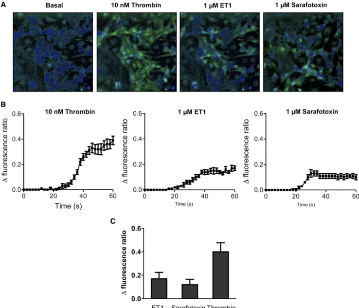
We analyzed ET-1 signaling in primary mouse podocytes by measuring the variations of intracellular Ca2+in cultured
po-docytes after stimulation with thrombin as a positive control or ET-1 and demonstrated functional receptors as ET-1 elici-ted rapid calcium transients (Figure 2). The ETBR-selective agonist sarafotoxin 6c induced a maximal calcium response similar to the one elicited by ET-1, thus showing that ETBR is functionally important in primary podocytes (Figure 2). Fur-thermore, the ETAR antagonist, BQ-123, as well as the ETBR antagonist, BQ-788, induced an approximately 65% decrease in ET-1–induced calcium transient, whereas a combination of both antagonists synergized this effect (Supplemental Figure 2). Taken together, these results show that both ETAR and ETBR are functional in podocytes, mediating ET-1–induced calcium release.
b-Catenin and NF-kB Pathways Are Downregulated in Podocytes from Pod-ETRKO Mice
We next investigated which signaling pathways could be activated downstream to the ETR in podocytes. Because
ETAR and ETBR can activate b-catenin and NF-kB pathways in nonglomerular cells,10,38–41we analyzed the activation
sta-tus of these pathways in glomeruli from Pod-ETRKO mice. We found that total b-catenin and phospho-NFkB expressions
Figure 1. ETAR and ETBR podocyte-specific deletions do not alter glomerular structure. (A) RT-PCR analysis of Ednar and Ednbr mRNA expression in isolated podocytes from 10-week-old WT and Pod-ETRKO mice. (B) Effective deletion of ETBR protein by NPHS2-Cre recombinase confirmed by immunoblotting analysis of isolated glomerular homogenates. Quantification of Western blot bands for ETBR normalized to tubulin band intensity. (C) Representative images of Masson’s trichrome–stained sections of glomeruli from 10-week-old WT and Pod-ETRKO mice. (D) Representative photomicrograph of transmission electron microscopy sections of podo-cytes from 10-week-old WT and Pod-ETRKO mice. Values are the mean6SEM from four mice. *P,0.05; **P,0.01. Scale bar, 50 mm in C; 1 mm in upper panel in D; 200 nm in lower panel in D.
Table 1. Animal phenotype
Parameter WT Mice Pod-ETRKO Mice
Control (n=9) Diabetes (n=15) Control (n=9) Diabetes (n=15) Blood glucose (mg/dl) 196.3616.4 547.4629.6a 171.467.6 537.6617.4a,b
Body weight (g) 32.161.9 25.361.2a 29.861.3 24.261.5a,b
Kidney to body weight ratio (%) 0.6060.02 0.8460.03a 0.5960.01 0.8660.02a,b
Urinary albumin to creatinine ratio (mg/mmol) 0.5460.14 3.1960.60a 0.7360.11 1.5260.39c
BUN (mmol/L) 21.161.7 27.361.6a 22.160.6 29.262a,b
aP,0.05 versus respective nondiabetic group with same genotype. bP,0.05 versus WT nondiabetic mice.
cP,0.05 versus diabetic WT mice.
Figure 2. Activation of the Ca2+pathway in podocytes stimulated by thrombin, ET-1, and sarafotoxin 6c. (A) Typical cell images
il-lustrating the change in fluorescence ratio induced after addition of ET-1 (1 mM), sarafotoxin 6c (1 mM), or thrombin (10 nM) compared with the basal fluorescence ratio. (B) Time course of the averaged responses to the compounds. (C) Summary of the maximal fluo-rescence ratios. Values are the mean6SEM of nine values representing three different experiments each performed in triplicate.
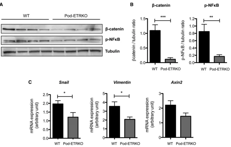
are reduced in glomeruli from Pod-ETRKO mice compared with WTmice in the basal state (Figure 3, A and B). b-Catenin and phospho-NF-kB expression levels are also diminished in primary podocyte cultures from Pod-ETRKO mice compared with WT mice (Supplemental Figure 3). Moreover, we ana-lyzed by RT-PCR the mRNA expression of the b-catenin target genes, snai1, vimentin, and axin2 and found that these three genes are also downregulated in glomeruli from Pod-ETRKO mice in the basal state (Figure 3C).
ET-1 Activates Glomerular b-Catenin and NF-kB Signaling Pathways In Vitro
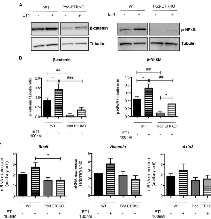
We then sought to determine whether ET-1 could directly activate b-catenin and NF-kB pathways in podocytes. Glo-meruli from WT and Pod-ETRKO mice were treated with ET-1 (100 nM) for 4 hours, after which we analyzed the acti-vation status of b-catenin and NF-kB signaling pathways. Im-munoblot analyses showed an increase in total b-catenin and phospho-NF-kB protein expression in glomeruli from WT mice treated with ET-1 (Figure 4, A and B). Interestingly, baseline as well as ET-1–stimulated b-catenin and phospho-NF-kB protein expressions in glomeruli from Pod-ETRKO
mice were significantly below levels in WT glomeruli (Figure 4, A and B). Moreover, maximum ET-1 stimulation of Pod-ETRKO glomeruli failed to trigger recruitment of b-catenin and phospho-NF-kB above baseline levels of WT glomeruli (Figure 4, A and B). Involvement of b-catenin activity by po-docyte ETRs was further confirmed because snail, vimentin, and axin2 mRNA expression induced by ET-1 in WT glomer-uli could not be achieved in glomerglomer-uli from Pod-ETRKO mice (Figure 4C). Furthermore, selective ETBR stimulation with sarafotoxin 6c mimicked ET-1 actions on b-catenin and NF-kB pathway activation (Supplemental Figure 4).
Podocyte-Specific Deletion of Ednar and Ednbr Protected Mice from Diabetes-Induced
Glomerulosclerosis
We then examined the role of the ET pathway in podocytes after diabetes mellitus (DM) induction by streptozotocin injection. Ten weeks after diabetes induction, mice presented polyuria and weight loss (data not shown) (Table 1). Pod-ETRKO DM and WT DM mice developed features of mild DN, as deter-mined by an increased kidney/body weight ratio and albumin-uria (Table 1). WT DM mice developed a higher urinary
Figure 3. At basal state, b-catenin and NF-kB signaling pathways are decreased in glomeruli of Pod-ETRKO mice. (A) Western blot analysis of b-catenin and phospho-NF-kB (p-NF-kB) expression in glomerular extracts from 10-week-old WT and Pod-ETRKO mice. Protein concentration is normalized to tubulin expression. (B) Quantification of Western blot bands for b-catenin and p-NF-kB nor-malized to tubulin band intensity. (C) RT-PCR analysis of Snai1, Vimentin, and Axin2 mRNA expression in glomerular extracts from 10-week-old WT and Pod-ETRKO mice. Values are the mean6SEM from at least six mice. *P,0.05; **P,0.01; ***P,0.001.
albumin excretion rate than Pod-ETRKO DM mice (P=0.03 for WT DM versus Pod-ETRKO DM) (Table 1). Histologic analyses showed tubular dilations and glomerular capillary dilations in the kidney of both WT diabetic and Pod-ETRKO diabetic mice (Figure 5, A and B). We analyzed the histologic
structure of the glomeruli from WT and Pod-ETRKO diabetic mice. More than half of the glomeruli from WT diabetic mice displayed mesangial thickening (approximately 58%), whereas glomeruli from Pod-ETRKO diabetic mice presented no such feature of glomerulosclerosis (Figure 5, B–D). This
Figure 4. In vitro, endothelin-1 activates b-catenin and NF-kB signaling pathways in glomeruli and its induction is decreased in Pod-ETRKO mice. (A) Western blot analysis of b-catenin and phospho-NF-kB (p-NF-kB) expression in glomeruli extracts from 10-week-old WT and Pod-ETRKO mice, treated or not with ET-1 at 100 nM for 4 hours. Protein concentration is normalized to tubulin expression. (B) Quantification of Western blot bands for b-catenin and p-NF-kB normalized to tubulin band intensity. (C) RT-PCR analysis of Snai1, Vimentin, and Axin2 mRNA expression in glomerular extracts from 10-week-old WT and Pod-ETRKO mice, treated or not with ET-1 at 100 nM for 4 hours. Values are the mean6SEM from at least six mice. *P,0.05 versus respective non-ET-1–treated mice;##
P,0.01 versus respective WT mice;###P,0.001 versus respective WT mice.
result was confirmed by electron microscopy (Supplemental Figure 5). Notably, preproendothelin-1 (Edn1) mRNA expres-sion was similarly upregulated in glomeruli from diabetic WT and Pod-ETRKO mice (Figure 5E). We next analyzed the ex-pression of two genes associated with sclerosis, Ctgf and Trpc6, and found that these two genes were significantly more upre-gulated in glomeruli from WT diabetic mice (+138% for Ctgf and +124% for Trpc6, WT DM versus WT) than in glomeruli from Pod-ETRKO diabetic mice (+43% for Ctgf and +37% for Trpc6, Pod-ETRKO DM versus Pod-ETRKO) (Figure 5E).
Podocyte-Specific Deletion of Ednar and Ednbr Protected Mice from Diabetes-Induced Podocytopathy
We next sought to investigate podocyte structure and number in diabetic mice. Podocalyxin and podocin staining showed weaker immunofluorescence in glomeruli from WT diabetic mice than in nondiabetic WT animals, thus demonstrating alterations in podocyte differentiation with diabetes. Podoca-lyxin and podocin immunostainings were strong and of similar intensity and pattern in WT and Pod-ETRKO nondiabetic mice. Diabetic Pod-ETRKO mice showed intermediate calyxin and podocin staining intensity, suggesting that podo-cyte alterations are less important in Pod-ETRKO diabetic mice (Figure 6A). Podocyte number per glomerulus, as determined by Wilms’ tumor antigen 1 (WT1) immunohistochemistry, was significantly decreased in WT diabetic mice (219% WT versus WT DM), whereas podocyte number in Pod-ETRKO diabetic kidneys remained similar to that measured in non-diabetic WT and Pod-ETRKO kidneys (Figure 6, B and C). Finally, electron microscopy analyses showed glomerular basement membrane thickening and podocyte foot process effacement in WT diabetic mice, whereas few ultrastructural defects were found in podocytes from Pod-ETRKO diabetic mice (Figure 6D).
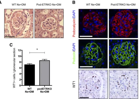
We then examined the role of the ET pathway in podocytes in a model of streptozotocin-induced diabetes after uninephrectomy, a more severe model of DN. Similar alleviation of glomerular damage was consistently found in Pod-ETRKO diabetic mice after uninephrectomy (Pod-ETRKO Nx+DM) compared with unin-ephrectomized WT diabetic mice (WT Nx+DM), with less glomerulosclerosis (Figure 7A) and maintenance of podocyte number (Figure 7, B and C).
Upon Diabetes, the b-Catenin Pathway Is Activated in Glomeruli by Podocyte ETRs
Because activation of the b-catenin pathways in glomeruli from diabetic mice was recently described,42,43we examined
the expression of the b-catenin target genes, snai1, vimentin, and axin2, in glomeruli from WT and Pod-ETRKO diabetic mice. Snai1, vimentin, and axin2 mRNA expression was sig-nificantly increased in glomeruli from diabetic WTmice com-pared with glomeruli from control WTmice: +347% for snai1, +202% for vimentin, and +236% for axin2 (Figure 8). Snai1, vimentin, and axin2 mRNA expression was less induced in Pod-ETRKO DM glomeruli (+169% for Snai1, +159% for
Vimentin, and +147% for Axin2, Pod-ETRKO DM versus Pod-ETRKO). Snail and Axin2 mRNA glomerular expression was significantly lower in glomeruli isolated from diabetic mice with selective ETR gene deletions compared with normal glomeruli (P,0.05), suggesting that podocyte ETRs signifi-cantly contribute to diabetes-induced activation of the b-catenin cascade (Figure 8).
DISCUSSION
In this study, we demonstrate that in streptozotocin-induced diabetes, mice with a podocyte-specific deletion of both Ednar and Ednbr (Pod-ETRKO) are protected from diabetes-induced glomerulosclerosis and podocyte loss, thus showing that ET signaling activation in podocytes increases susceptibility to de-velop DN. These results shed light on the pathophysiologic actions of the ET-1 system in DN and represent an example of the involvement of podocyte GPCRs in the pathophysiology of DN.
ET-1 activation in kidney tubules and glomeruli as well as in blood during DN is now well established,21–23but the targeted
cells of ET-1 and the mechanisms connecting ET-1 activation with the development of DN and particularly with podocyto-pathy development are still not completely elucidated. Sasser et al. demonstrated that the ETAR antagonist ABT-627 re-duced diabetes-inre-duced macrophage infiltration and TGF-b urinary excretion.29 In this study, the authors suggest that
ETAR activation in macrophages by increased renal produc-tion of ET-1 leads to macrophage recruitment in the kidney and TGF-b production by macrophages. Moreover, it has been shown that high glucose levels induce mesangial cell ET-1 pro-duction44and that ET-1 promotes mesangial cell contraction,
hypertrophy, and generation of inflammatory mediators, cy-tokines, and growth factors as well as the production of fibro-nectin in vitro. On the other hand, little is known about the mechanisms through which ET-1 might directly affect podo-cyte cell survival and/or function, particularly during diabetes. Disruption of the actin cytoskeleton has been observed after in vitro exposure to ET-1 or toxic compounds such as puromycin aminonucleoside or Shiga toxin in an ETAR-dependent man-ner in a mouse immortalized podocyte line.45–47Meanwhile,
expression (or lack of) of the ETBR was not searched for in this immortalized cell line. We observed high ETBR expres-sion in primary culture of podocytes and in freshly isolated glomeruli as reported in a seminal article in rat glomeruli.16
ETBR expression in podocytes was indeed described by im-munoelectron microscopy that allowed visualization of in-tense immunodeposits indicating ETBR in fully differentiated rat podocytes in situ, particularly in their foot processes.16
Other researchers found immunoreactive ETAR and ETBR in mesangial and glomerular capillary cells, respectively17;
var-iable staining for immunoreactive ETBR in glomeruli (in parietal epithelial cells and podocytes) has been reported in kidney biopsies from healthy controls and from patients with
IgA nephropathy,14 confirming the
find-ings reported by Karet et al.15 Here we
detected ETAR and ETBR expression in primary podocytes, along with novel func-tional data. ET-1 stimulates calcium tran-sients in podocytes in an ETAR- as well as ETBR-dependent fashion. This intriguing observation suggests that ETAR and ETBR may cosignal, potentially as hetero-dimers as recently suggested.48–50
For the first time, we further observed that sarafotoxin 6c, a selective agonist of ETBR, was sufficient to activate calcium and extracellular signal–regulated kinase signaling, thus showing that the ETBR sig-naling is functionally significant in primary podocytes. Moreover, sarafotoxin 6c stim-ulation in freshly isolated glomeruli also activated NF-kB and b-catenin, thus sug-gesting that chronic NF-kB and b-catenin activation in diabetic glomeruli is mediated at least in part by ETBR.
Surprisingly, therapy for CKD has fo-cused on ETAR antagonists only, based on previous experimental findings indicating that ETAR promotes mesangial sclerosis and glomerular capillary pressure.8,29,51–53
Indeed, the partial benefits of ETAR antag-onists for the treatment of DN have been extensively discussed after clinical data re-vealed that both ETAR and ETBR promote some degree of fluid retention in patients at an advanced stage.33,34,54 Here we point
out that the combination of ETAR antago-nists with ETBR antagoantago-nists may provide additional beneficial effects on glomerular remodeling and function in diabetes. In line with our mechanistic findings, recent clinical data indicate that dual endothelin receptor antagonism improves peripheral endothelial function in patients with type 2 diabetes and microalbuminuria.55
Figure 5. Deletions of Ednar and Ednbr specifically in podocytes protect glomeruli from diabetes-induced glomerulosclerosis. (A) Representative images of hematoxylin/ eosin-stained sections of renal cortex from 20-week-old WT control, WT diabetic, Pod-ETRKO control, and Pod-Pod-ETRKO diabetic mice. (B) Representative images of Masson’s trichrome–stained sections of glomeruli from 20-week-old WT control, WT diabetic, Pod-ETRKO control, and Pod-ETRKO diabetic mice. (C and D) Percentage of glo-meruli with mesangial thickening (C) in the renal cortex of 20-week-old WT control, WT diabetic, Pod-ETRKO control, and Pod-ETRKO diabetic mice. (D) RT-PCR analysis
of preproendothelin-1 (Edn1 gene), Ctgf, and trpc6 mRNA expression in glomerular extracts from 20-week-old WT control, WT diabetic, Pod-ETRKO control, and Pod-ETRKO diabetic mice. Values are the mean6SEM from at least six mice. *P,0.05 versus respective non-diabetic mice; #P,0.05 versus WT diabetic
mice; ***P,0.001 versus respective non-diabetic mice;###
P,0.001 versus WT diabetic mice. Scale bar, 50 mm.
We also identified that NF-kB and b-catenin are activated downstream of the ETR after ET-1 stimulation in cultured glomeruli. Moreover, ET-1 and b-catenin pathways are activated in glomeruli during progression of DN. Although glomerular prepro-ET-1 mRNA is overexpressed in Pod-ETRKO mice, b-catenin pathway acti-vation does not still occur. Taken together, these results strongly suggest that ET-1 sig-naling is activated in podocytes during diabetes and it exerts its detrimental re-modeling effects through activation of NF-kB and b-catenin pathways. It is note-worthy that Pod-ETRKO glomeruli stimu-lated with exogenous ET-1 showed reduced but still residual activation of NF-kB and b-catenin pathways, suggesting that resid-ual activation of NF-kB and b-catenin pathways elicited by ET-1 may have oc-curred in other glomerular cell types. It is well established that ET-1 can drive NF-kB and b-catenin pathway activation in several cell types; however, no information is avail-able thus far regarding the downstream effectors of the ETR in podocytes.10,38–41
NF-kB was recently implicated in glomer-ular aging, by promoting inflammation, coagulation, and fibrosis.56,57 A delicate
balance of b-catenin expression in podo-cytes was recently demonstrated to be crit-ical for podocyte cell adhesion and survival.43b-catenin overexpression in
docytes during DN or FSGS promoted po-docyte dysfunction and albuminuria, whereas blockade of the b-catenin pathway ameliorated kidney injury.42,43 Here we
provide evidence that ETR activation in podocytes during diabetes is a potent up-stream event leading to NF-kB and b-catenin activation and podocytopathy.
Surprisingly, we found that podocyte deletion of ETR protects not only from diabetes-induced loss of podocytes but also from mesangial matrix expansion. Recent studies indicated that isolated podocyte damage leads to glomerulosclerosis58and
that podocyte loss appears to be a requisite early event in DN.59Accordingly, podocyte
loss in individuals with type 2 diabetes pre-dicts nephropathy as well as mesangial expansion.60 Presumably, signals from
damaged podocytes to the mesangium, or the hemodynamic factors triggered by podocyte loss, could provide a stimulus to the mesangial cell to react by increases
Figure 6. ETAR and ETBR podocyte-specific deficiency protects podocytes from diabetes-induced podocyte loss. (A) Representative images of the expression of po-docalyxin (upper panel) and podocin (lower panel) by immunofluorescence in 20-week-old WT control, WT diabetic, Pod-ETRKO control, and Pod-ETRKO diabetic mice. Images are representative of at least six mice. (B) Representative images of the ex-pression of WT1 by immunohistochemistry in 20-week-old WT control, WT diabetic, Pod-ETRKO control, and Pod-ETRKO diabetic mice. Images are representative of at least six mice. (C) Quantification of the glomerular WT1-positive cell numbers in 20-week-old WT control, WT diabetic, Pod-ETRKO control, and Pod-ETRKO diabetic mice. Data are normalized to WT control and represent the mean6SEM from at least six mice. (D) Representative photomicrographs of transmission electron microscopy sections of podocytes from 20-week-old WT diabetic and Pod-ETRKO diabetic mice showing glomerular basement membrane thickening (asterisk) and foot process ef-facement (arrow) in WT DM mice. ***P,0.001 versus respective nondiabetic mice;
###
P,0.001 versus WT diabetic mice. Scale bar, 50 mm in A and B; 1 mm in upper panel in D; 200 nm in lower panel in D.
in extracellular matrix synthesis or decreases in extracellular matrix degradation. Our data suggest that modulation of the ET-1 pathway in podocytes influenced crosstalk between po-docytes and mesangial cells. Indeed, it was demonstrated that
cell culture medium conditioned by podo-cytes induced mesangial cell proliferation in vitro.61 Moreover, podocyte connective
tissue growth factor (CTGF) seems to have paracrine effects on mesangial cells to stim-ulate CTGF expression.62 Interestingly,
diabetic normal mice displayed a 3-fold increase in Ctgf glomerular mRNA expres-sion compared with their Pod-ETRKO counterparts. These results are compatible with the hypothesis that ETRs stimulate a paracrine factor secreted by podocytes, such as CTGF, that in turn may promote mesangial cells lesions during diabetes. Taken together, these studies identify the first GPCRs in podocytes that mediate progres-sion of DN and support the notion that persistent ETAR and ETBR activation in po-docytes is sufficient to promote glomerular injury with podocyte depletion through re-cruitment of the b-catenin pathway.
CONCISE METHODS Animals
Mice with podocyte-specific disruption of both Ed-nar and Ednbr genes (podocin-Cre+ Ednarlox/lox
Ednbrlox/lox mice) were generated by crossing
NPHS2-CRE+ mice37with Ednarlox/lox63 and
Ednbrlox/lox64mice. Mice are on a mixed C57BL/
6J/ 129/SV/ FVB/N genetic background. Animals homozygous for floxed Ednar and Ednbr genes but without Cre were used as controls in all stud-ies. For animals subjected to left uninephrectomy, animals were anesthetized by isoflurane inhala-tion and received 0.1 mg/kg buprenorphine twice daily for 2 days. The left renal artery and vein were ligated with 6-0 silk before kidney excision and the wound was closed in layers. Diabetes was induced after 7 days of recovery. All mice were given free access to water and standard chow. Experiments were conducted according to the French veteri-nary guidelines and those formulated by the Eu-ropean Commission for experimental animal use (L358-86/609EEC) and were approved by the Institut National de la Santé et de la Recherche Médicale (INSERM).
Induction of DM with Streptozotocin Twelve-week-old male mice were rendered di-abetic with streptozotocin (100 mg/kg in citrate buffer, pH 4.5) by intraperitoneal injection on 2 consecutive days. Control mice received citrate buffer alone. Mice with fasted glycemia.300 mg/dl were con-sidered diabetic. Mice were euthanized 10 weeks after diabetes
Figure 7. Pod-ETRKO mice are protected from diabetes-induced glomerulosclerosis and podocyte loss in a model of uninephrectomy followed by streptozotocin-diabetes induction. (A) Representative images of hematoxylin/eosin-stained sections of renal cortex from 16-week-old WT uninephrectomized diabetic and Pod-ETRKO unin-ephrectomized diabetic mice. (B) Representative images of the expression of podo-calyxin (upper panel), podocin (middle panel), and WT1 (lower panel) in 16-week-old WT uninephrectomized diabetic and Pod-ETRKO uninephrectomized diabetic mice. (C) Quantification of the glomerular WT1-positive cell numbers in 16-week-old WT uninephrectomized diabetic and Pod-ETRKO uninephrectomized diabetic mice. Data represent the mean6SEM from at least four mice. *P,0.05. DAPI, 49,6-diamidino-2-phenylindole. Scale bar, 50 mm.
Figure 8. The induction of b-catenin and NF-kB signaling pathways in diabetic glo-meruli is limited in Pod-ETRKO gloglo-meruli. RT-PCR analysis of Snai1, Vimentin, and Axin2 mRNA expression in glomerular extracts from 20-week-old WT control, WT diabetic, Pod-ETRKO control, and Pod-ETRKO diabetic mice. Values are the mean6SEM from at least six mice. *P,0.05 versus respective nondiabetic mice; **P,0.01 versus respective nondiabetic mice;##P,0.01 versus WT diabetic mice.
induction and uninephrectomized diabetic mice were euthanized 4 weeks after diabetes induction.
Assessment of Renal Function and Albuminuria
Urinary creatinine and BUN concentrations were quantified spec-trophotometrically using colorimetric methods. Urinary albumin excretion was measured using a specific ELISA assay for quantitative determination of albumin in mouse urine (BIOTREND Chemikalien GmbH).
Isolation of Glomerular and Podocyte Cultures
Decapsulated glomeruli were isolated as previously described.65,66
Briefly, freshly isolated renal cortex was mixed and digested by col-lagenase I (2 mg/ml; Gibco) in RPMI 1640 (Life Technologies) for 2 minutes at 37°C, and then collagenase I was inactivated with RPMI 1640 plus 10% FCS (Abcys). Tissues were then passed through a 70-mm cell strainer and a 40-70-mm cell strainer (BD Falcon) in PBS (Life Technologies) plus 0.5% BSA (Sigma-Aldrich). Glomeruli, adherent to the 40-mm cell strainer, were removed from the cell strainer with PBS plus 0.5% BSA injected under pressure, and then washed twice in PBS. Isolated glomeruli were then picked up in Phosphosafe extrac-tion buffer (Novagen) and frozen at 280°C for protein extracextrac-tion, or picked up in RLTextraction buffer (Qiagen) and frozen at 280°C for total RNA extraction or then processed for cell culture. For podocyte primary cell culture, freshly isolated glomeruli were plated in six-well dishes in RPMI 1640 plus 10% FCS and 1% penicillin/streptomycin (Life Technologies). Two days after seeding, glomeruli became adher-ent to the dish, and podocytes exited from glomeruli and started growing in the dish. Isolated podocytes were picked up in Phospho-safe extraction buffer and frozen at 280°C for protein extraction or picked up in RLTextraction buffer and frozen at 280°C for total RNA extraction.
Intracellular Calcium Measurements
Variations of intracellular Ca2+([Ca2+]i) were measured in cultured podocytes using the ratiometric fluorescence Ca2+indicator Fura-2 as previously described.67Cells subcultured for 4 days in chambered
coverglass (Lab-Tek) were loaded by incubation with 2.5 mmol/L Fura-2AM (TEFLabs) plus 0.02% pluronic acid F-127 (Molecular Probes, Inc.), in Locke buffer for 20 minutes at 37°C in a humidified air atmosphere. Cells were rinsed with buffer and mounted on a microscope stage (Axiovert, 320 objective; Zeiss) during a 15-minute waiting period for the de-esterification of Fura-2AM. Media and drugs (ET-1, sarafotoxin 6c, and thrombin) were then applied to the cells using a perfusion system. Images were captured digitally every 2 sec-onds with a cooled charge-coupled device camera (Photometrics, Roper Scientific). Cells were illuminated by excitation with a dual ultraviolet light source at 340 nm (Ca2+-bound) and 380 nm (Ca2+ -free) using a Lambda DG-4 excitation system (Sutter Instrument Company). Fluorescence emission was measured at 510 nm and an-alyzed using MetaFluor software (Universal Imaging Corporation). Changes in [Ca2+]i were deduced from variations of the F340/F380 ratio after correction for background and dark currents. Data were averaged with n representing the number of fields (at least 100 cells per recorded field).
Histopathology and Immunohistochemistry
Kidneys were immersed in 10% formalin and embedded in paraffin. Sections (4-mm thick) were processed for histopathology study or immunohistochemistry. Sections were stained with hematoxylin and eosin or Masson’s trichrome stain. The proportion of pathologic glo-meruli was evaluated by examination of at least 50 gloglo-meruli per section by an examiner (P.-.L.T.) who was blinded to the experimental conditions. For immunohistochemistry, we stained paraffin-embedded sections with the following primary antibodies: rabbit anti-WT1 (1:400; Santa Cruz Biotechnology, Inc.) and goat anti-podocalyxin (1:200; R&D Systems). For WT1 staining, specific staining was revealed using Histofine reagents (Nichirei Biosciences). For podocin immuno-fluorescence, fresh cryostat sections (4-mm thick) were immediately fixed in 4% paraformaldehyde and then incubated with a goat anti-podocin antibody (1:100; Santa Cruz Biotechnology, Inc.). For podocalyxin and podocin immunofluorescence, we used a secondary rabbit anti-goat IgG AF594-conjugated antibody (1:400; Invitrogen). The nuclei were stained using 49,6-diamidino-2-phenylindole. Photomicro-graphs were performed with an Axiophot Zeiss photomicroscope. Electron Microscopy
Small pieces of renal cortex were fixed in 2% glutaraldehyde, postfixed in 1% osmium tetroxide, and embedded in Araldite M (Sigma-Aldrich). Ultrathin sections were counterstained with uranyl acetate and lead citrate and examined using a transmission electron microscope. Western Blot Analyses
Glomerular and podocyte lysates were prepared using Phosphosafe extraction buffer. Equal amounts of proteins were loaded on SDS-PAGE gels for separation and transferred onto polyvinylidene difluoride membranes. After blocking with milk, the membranes were probed with the following antibodies: rabbit anti-ETBR (1:1000; Santa Cruz Biotechnology, Inc.), rabbit anti–b-catenin (1:1000; Cell Signaling Technologies), rabbit anti–phospho-NF-kB (p65 Ser536, 1:1000; Cell Signaling Technologies), and rat anti-tubulin (1:5000; Abcam). The results were visualized with horseradish peroxidase– conjugated secondary antibodies (1:2000; Cell Signaling Technolo-gies) and enhanced chemiluminescence (Supersignal West Pico). The LAS-4000 imaging system (FujiFilm) was used to reveal bands and densitometric analysis was used for quantification.
Real-Time RT-PCR
Total RNA extraction of mice glomeruli was performed using an RNeasy Microkit (Qiagen) and reverse transcribed with the QuantiTect Reverse Transcription Kit (Qiagen). Maxima SYBR Green/Rox qPCR Mix (Thermoscientific Fermentas) was used to amplify cDNA for 40 cycles on an ABI PRISM thermocycler. The comparative method of relative quantification (2-DDCT) was used to calculate the expression level of each target gene, normalized to glyceraldehyde 3-phosphate dehydro-genase. The oligonucleotide sequences are available upon request. Data are presented as the fold change in gene expression.
Statistical Analyses
Data are expressed as the mean6SEM. Statistical analyses were cal-culated using Prism 5.04 software (GraphPad Software, Inc.).
Between-group comparisons were performed using the two-tailed t test, whereas multiple-group comparisons between were performed using one-way ANOVA followed by the Newman–Keuls test. A P value,0.05 was considered statistically significant.
ACKNOWLEDGMENTS
The authors thank Elizabeth Huc and the ERI970 team for assistance in animal care and handling. They also thank Véronique Oberweiss, Annette De Rueda, Martine Autran, Bruno Pillard, and Philippe Coudol for excellent administrative support, as well as Kathleen Flosseau and Anna Chipont for technical assistance.
This study was supported by grants from the Institut National de la Santé et de la Recherche Médicale, the European Research Projects on Rare Diseases (E-Rare-2 JTC 2011 to P.-L.T.), and the French National Research Agency. O.L. held a postdoctoral fellowship from Région Ile-de-France (CORDDIM – Cardiovasculaire - Obésité - Rein - Diabète Initiative).
DISCLOSURES
None.
REFERENCES
1. Van Buren PN, Toto R: Current update in the management of diabetic nephropathy. Curr Diabetes Rev 9: 62–77, 2013
2. Ziyadeh FN, Hoffman BB, Han DC, Iglesias-De La Cruz MC, Hong SW, Isono M, Chen S, McGowan TA, Sharma K: Long-term prevention of renal insufficiency, excess matrix gene expression, and glomerular mesangial matrix expansion by treatment with monoclonal anti-transforming growth factor-beta antibody in db/db diabetic mice. Proc Natl Acad Sci U S A 97: 8015–8020, 2000
3. Ziyadeh FN: Mediators of diabetic renal disease: The case for tgf-Beta as the major mediator. J Am Soc Nephrol 15[Suppl 1]: S55–S57, 2004 4. Najafian B, Alpers CE, Fogo AB: Pathology of human diabetic
ne-phropathy. Contrib Nephrol 170: 36–47, 2011
5. Detournay B, Simon D, Guillausseau PJ, Joly D, Verges B, Attali C, Clement O, Briand Y, Delaitre O: Chronic kidney disease in type 2 di-abetes patients in France: Prevalence, influence of glycaemic control and implications for the pharmacological management of diabetes. Diabetes Metab 38: 102–112, 2012
6. Shepler B, Nash C, Smith C, Dimarco A, Petty J, Szewciw S: Update on potential drugs for the treatment of diabetic kidney disease. Clin Ther 34: 1237–1246, 2012
7. Ritz E: Limitations and future treatment options in type 2 diabetes with renal impairment. Diabetes Care 34[Suppl 2]: S330–S334, 2011 8. Fligny C, Barton M, Tharaux PL: Endothelin and podocyte injury in
chronic kidney disease. Contrib Nephrol 172: 120–138, 2011 9. Dhaun N, Goddard J, Kohan DE, Pollock DM, Schiffrin EL, Webb DJ:
Role of endothelin-1 in clinical hypertension: 20 years on. Hypertension 52: 452–459, 2008
10. Gerstung M, Roth T, Dienes HP, Licht C, Fries JW: Endothelin-1 induces NF-kappaB via two independent pathways in human renal tubular ep-ithelial cells. Am J Nephrol 27: 294–300, 2007
11. Sugden PH, Clerk A: Endothelin signalling in the cardiac myocyte and its pathophysiological relevance. Curr Vasc Pharmacol 3: 343–351, 2005
12. Bouallegue A, Daou GB, Srivastava AK: Endothelin-1-induced signal-ing pathways in vascular smooth muscle cells. Curr Vasc Pharmacol 5: 45–52, 2007
13. Sorokin A: Endothelin signaling and actions in the renal mesangium. Contrib Nephrol 172: 50–62, 2011
14. Lehrke I, Waldherr R, Ritz E, Wagner J: Renal endothelin-1 and endo-thelin receptor type B expression in glomerular diseases with pro-teinuria. J Am Soc Nephrol 12: 2321–2329, 2001
15. Karet FE, Kuc RE, Davenport AP: Novel ligands BQ123 and BQ3020 characterize endothelin receptor subtypes ETA and ETB in human kidney. Kidney Int 44: 36–42, 1993
16. Yamamoto T, Hirohama T, Uemura H: Endothelin B receptor-like im-munoreactivity in podocytes of the rat kidney. Arch Histol Cytol 65: 245–250, 2002
17. Wendel M, Knels L, Kummer W, Koch T: Distribution of endothelin receptor subtypes ETA and ETB in the rat kidney. J Histochem Cy-tochem 54: 1193–1203, 2006
18. Simonson MS: Endothelins: Multifunctional renal peptides. Physiol Rev 73: 375–411, 1993
19. Badr KF, Murray JJ, Breyer MD, Takahashi K, Inagami T, Harris RC: Mesangial cell, glomerular and renal vascular responses to endothelin in the rat kidney. Elucidation of signal transduction pathways. J Clin Invest 83: 336–342, 1989
20. Fukuda K, Yanagida T, Okuda S, Tamaki K, Ando T, Fujishima M: Role of endothelin as a mitogen in experimental glomerulonephritis in rats. Kidney Int 49: 1320–1329, 1996
21. Fukui M, Nakamura T, Ebihara I, Osada S, Tomino Y, Masaki T, Goto K, Furuichi Y, Koide H: Gene expression for endothelins and their receptors in glomeruli of diabetic rats. J Lab Clin Med 122: 149–156, 1993 22. Hargrove GM, Dufresne J, Whiteside C, Muruve DA, Wong NC:
Di-abetes mellitus increases endothelin-1 gene transcription in rat kidney. Kidney Int 58: 1534–1545, 2000
23. Itoh Y, Nakai A, Kakizawa H, Makino M, Fujiwara K, Kobayashi T, Kato T, Nagata M, Oda N, Katsumata H, Nagasaka A, Itoh M: Alteration of endothelin-1 concentration in STZ-induced diabetic rat nephropathy. Effects of a PGI(2) derivative. Horm Res 56: 165–171, 2001
24. Bruno CM, Meli S, Marcinno M, Ierna D, Sciacca C, Neri S: Plasma en-dothelin-1 levels and albumin excretion rate in normotensive, micro-albuminuric type 2 diabetic patients. J Biol Regul Homeost Agents 16: 114–117, 2002
25. Zanatta CM, Veronese FV, Loreto MS, Sortica DA, Carpio VN, Eldeweiss MI, da Silva VD, Lopes TG, Gross JL, Canani LH: Endothelin-1 and endothelin A receptor immunoreactivity is increased in patients with diabetic nephropathy. Ren Fail 34: 308–315, 2012
26. Mishra R, Emancipator SN, Kern TS, Simonson MS: Association be-tween endothelin-1 and collagen deposition in db/db diabetic mouse kidneys. Biochem Biophys Res Commun 339: 65–70, 2006
27. Ding SS, Qiu C, Hess P, Xi JF, Zheng N, Clozel M: Chronic endothelin receptor blockade prevents both early hyperfiltration and late overt di-abetic nephropathy in the rat. J Cardiovasc Pharmacol 42: 48–54, 2003 28. Cosenzi A, Bernobich E, Trevisan R, Milutinovic N, Borri A, Bellini G:
Nephroprotective effect of bosentan in diabetic rats. J Cardiovasc Pharmacol 42: 752–756, 2003
29. Sasser JM, Sullivan JC, Hobbs JL, Yamamoto T, Pollock DM, Carmines PK, Pollock JS: Endothelin A receptor blockade reduces diabetic renal injury via an anti-inflammatory mechanism. J Am Soc Nephrol 18: 143–154, 2007 30. Saleh MA, Boesen EI, Pollock JS, Savin VJ, Pollock DM: Endothelin receptor
A-specific stimulation of glomerular inflammation and injury in a strepto-zotocin-induced rat model of diabetes. Diabetologia 54: 979–988, 2011 31. Sarafidis PA, Lasaridis AN: Diabetic nephropathy: Endothelin
antago-nism for diabetic nephropathy. Nat Rev Nephrol 6: 447–449, 2010 32. Benz K, Amann K: Endothelin in diabetic renal disease. Contrib
Nephrol 172: 139–148, 2011
33. Kohan DE, Pollock DM: Endothelin antagonists for diabetic and non-diabetic chronic kidney disease. Br J Clin Pharmacol 76: 573–579, 2013
34. Ritz E, Wenzel RR: Endothelin antagonist as add-on treatment for proteinuria in diabetic nephropathy: Is there light at the end of the tunnel? J Am Soc Nephrol 22: 593–595, 2011
35. Kohan DE, Pritchett Y, Molitch M, Wen S, Garimella T, Audhya P, Andress DL: Addition of atrasentan to renin-angiotensin system blockade reduces albuminuria in diabetic nephropathy. J Am Soc Nephrol 22: 763–772, 2011
36. Gagliardini E, Corna D, Zoja C, Sangalli F, Carrara F, Rossi M, Conti S, Rottoli D, Longaretti L, Remuzzi A, Remuzzi G, Benigni A: Unlike each drug alone, lisinopril if combined with avosentan promotes regression of renal lesions in experimental diabetes. Am J Physiol Renal Physiol 297: F1448–F1456, 2009
37. Moeller MJ, Sanden SK, Soofi A, Wiggins RC, Holzman LB: Podocyte-specific expression of Cre recombinase in transgenic mice. Genesis 35: 39–42, 2003
38. Kapoor M, Liu S, Shi-wen X, Huh K, McCann M, Denton CP, Woodgett JR, Abraham DJ, Leask A: GSK-3beta in mouse fibroblasts controls wound healing and fibrosis through an endothelin-1-dependent mechanism. J Clin Invest 118: 3279–3290, 2008
39. Rosanò L, Cianfrocca R, Masi S, Spinella F, Di Castro V, Biroccio A, Salvati E, Nicotra MR, Natali PG, Bagnato A: Beta-arrestin links endothelin A receptor to beta-catenin signaling to induce ovarian cancer cell invasion and metastasis. Proc Natl Acad Sci U S A 106: 2806–2811, 2009 40. Rosanò L, Cianfrocca R, Tocci P, Spinella F, Di Castro V, Spadaro F,
Salvati E, Biroccio AM, Natali PG, Bagnato A: b-arrestin-1 is a nuclear transcriptional regulator of endothelin-1-induced b-catenin signaling. Oncogene 32: 5066–5077, 2013
41. Kao YS, Fong JC: A novel cross-talk between endothelin-1 and cyclic AMP signaling pathways in the regulation of GLUT1 transcription in 3T3-L1 adipocytes. Cell Signal 23: 901–910, 2011
42. Dai C, Stolz DB, Kiss LP, Monga SP, Holzman LB, Liu Y: Wnt/beta-catenin signaling promotes podocyte dysfunction and albuminuria. J Am Soc Nephrol 20: 1997–2008, 2009
43. Kato H, Gruenwald A, Suh JH, Miner JH, Barisoni-Thomas L, Taketo MM, Faul C, Millar SE, Holzman LB, Susztak K: Wnt/b-catenin pathway in podocytes integrates cell adhesion, differentiation, and survival. J Biol Chem 286: 26003–26015, 2011
44. Tsiani E, Lekas P, Fantus IG, Dlugosz J, Whiteside C: High glucose-enhanced activation of mesangial cell p38 MAPK by ET-1, ANG II, and platelet-derived growth factor. Am J Physiol Endocrinol Metab 282: E161–E169, 2002
45. Morigi M, Buelli S, Angioletti S, Zanchi C, Longaretti L, Zoja C, Galbusera M, Gastoldi S, Mundel P, Remuzzi G, Benigni A: In response to protein load podocytes reorganize cytoskeleton and modulate en-dothelin-1 gene: Implication for permselective dysfunction of chronic nephropathies. Am J Pathol 166: 1309–1320, 2005
46. Morigi M, Buelli S, Zanchi C, Longaretti L, Macconi D, Benigni A, Moioli D, Remuzzi G, Zoja C: Shigatoxin-induced endothelin-1 expression in cultured podocytes autocrinally mediates actin remodeling. Am J Pathol 169: 1965–1975, 2006
47. Ortmann J, Amann K, Brandes RP, Kretzler M, Münter K, Parekh N, Traupe T, Lange M, Lattmann T, Barton M: Role of podocytes for re-versal of glomerulosclerosis and proteinuria in the aging kidney after endothelin inhibition. Hypertension 44: 974–981, 2004
48. Evans NJ, Walker JW: Sustained Ca2+ signaling and delayed in-ternalization associated with endothelin receptor heterodimers linked through a PDZ finger. Can J Physiol Pharmacol 86: 526–535, 2008 49. Evans NJ, Walker JW: Endothelin receptor dimers evaluated by FRET,
ligand binding, and calcium mobilization. Biophys J 95: 483–492, 2008 50. Sauvageau S, Thorin E, Caron A, Dupuis J: Evaluation of endothelin-1-induced pulmonary vasoconstriction following myocardial infarction. Exp Biol Med (Maywood) 231: 840–846, 2006
51. Dhaun N, Macintyre IM, Melville V, Lilitkarntakul P, Johnston NR, Goddard J, Webb DJ: Blood pressure-independent reduction in
proteinuria and arterial stiffness after acute endothelin-a receptor an-tagonism in chronic kidney disease. Hypertension 54: 113–119, 2009 52. Neuhofer W, Pittrow D: Endothelin receptor selectivity in chronic
kid-ney disease: Rationale and review of recent evidence. Eur J Clin Invest 39[Suppl 2]: 50–67, 2009
53. Sorokin A, Kohan DE: Physiology and pathology of endothelin-1 in renal mesangium. Am J Physiol Renal Physiol 285: F579–F589, 2003 54. Rabelink TJ, Kohan DE: Endothelin receptor blockade in patients with
diabetic nephropathy. Contrib Nephrol 172: 235–242, 2011 55. Rafnsson A, Böhm F, Settergren M, Gonon A, Brismar K, Pernow J: The
endothelin receptor antagonist bosentan improves peripheral endo-thelial function in patients with type 2 diabetes mellitus and micro-albuminuria: A randomised trial. Diabetologia 55: 600–607, 2012 56. Wiggins JE, Patel SR, Shedden KA, Goyal M, Wharram BL, Martini S,
Kretzler M, Wiggins RC: NFkappaB promotes inflammation, coagulation, and fibrosis in the aging glomerulus. J Am Soc Nephrol 21: 587–597, 2010 57. Tilstra JS, Robinson AR, Wang J, Gregg SQ, Clauson CL, Reay DP, Nasto LA, St Croix CM, Usas A, Vo N, Huard J, Clemens PR, Stolz DB, Guttridge DC, Watkins SC, Garinis GA, Wang Y, Niedernhofer LJ, Robbins PD: NF-kB inhibition delays DNA damage-induced senes-cence and aging in mice. J Clin Invest 122: 2601–2612, 2012 58. Wharram BL, Goyal M, Wiggins JE, Sanden SK, Hussain S, Filipiak WE,
Saunders TL, Dysko RC, Kohno K, Holzman LB, Wiggins RC: Podocyte depletion causes glomerulosclerosis: Diphtheria toxin-induced podo-cyte depletion in rats expressing human diphtheria toxin receptor transgene. J Am Soc Nephrol 16: 2941–2952, 2005
59. Pagtalunan ME, Miller PL, Jumping-Eagle S, Nelson RG, Myers BD, Rennke HG, Coplon NS, Sun L, Meyer TW: Podocyte loss and progressive glomerular injury in type II diabetes. J Clin Invest 99: 342–348, 1997 60. Meyer TW, Bennett PH, Nelson RG: Podocyte number predicts
long-term urinary albumin excretion in Pima Indians with type II diabetes and microalbuminuria. Diabetologia 42: 1341–1344, 1999
61. Budde K, Neumayer HH, Salant DJ, Cybulsky AV, Coleman DL, Sterzel RB: Glomerular epithelial cell products stimulate mesangial cell pro-liferation in culture. Kidney Int 52: 733–741, 1997
62. Lee HS: Paracrine role for TGF-b-induced CTGF and VEGF in mesangial matrix expansion in progressive glomerular disease. Histol Histopathol 27: 1131–1141, 2012
63. Ge Y, Stricklett PK, Hughes AK, Yanagisawa M, Kohan DE: Collecting duct-specific knockout of the endothelin A receptor alters renal vaso-pressin responsiveness, but not sodium excretion or blood pressure. Am J Physiol Renal Physiol 289: F692–F698, 2005
64. Bagnall AJ, Kelland NF, Gulliver-Sloan F, Davenport AP, Gray GA, Yanagisawa M, Webb DJ, Kotelevtsev YV: Deletion of endothelial cell endothelin B receptors does not affect blood pressure or sensitivity to salt. Hypertension 48: 286–293, 2006
65. Schiwek D, Endlich N, Holzman L, Holthöfer H, Kriz W, Endlich K: Stable expression of nephrin and localization to cell-cell contacts in novel murine podocyte cell lines. Kidney Int 66: 91–101, 2004
66. Bollée G, Flamant M, Schordan S, Fligny C, Rumpel E, Milon M, Schordan E, Sabaa N, Vandermeersch S, Galaup A, Rodenas A, Casal I, Sunnarborg SW, Salant DJ, Kopp JB, Threadgill DW, Quaggin SE, Dussaule JC, Germain S, Mesnard L, Endlich K, Boucheix C, Belenfant X, Callard P, Endlich N, Tharaux PL: Epidermal growth factor receptor promotes glomerular injury and renal failure in rapidly progressive crescentic glomerulonephritis. Nat Med 17: 1242–1250, 2011 67. Gysembergh A, Lemaire S, Piot C, Sportouch C, Richard S, Kloner RA,
Przyklenk K: Pharmacological manipulation of Ins(1,4,5)P3 signaling mimics preconditioning in rabbit heart. Am J Physiol 277: H2458–H2469, 1999
See related editorial, “Endothelin Antagonists in Diabetic Nephropathy: Back to Basics,” on pages 869–871.
This article contains supplemental material online at http://jasn.asnjournals. org/lookup/suppl/doi:10.1681/ASN.2013020195/-/DCSupplemental.