HAL Id: hal-02936507
https://hal.archives-ouvertes.fr/hal-02936507
Submitted on 11 Sep 2020
HAL is a multi-disciplinary open access
archive for the deposit and dissemination of
sci-entific research documents, whether they are
pub-lished or not. The documents may come from
teaching and research institutions in France or
abroad, or from public or private research centers.
L’archive ouverte pluridisciplinaire HAL, est
destinée au dépôt et à la diffusion de documents
scientifiques de niveau recherche, publiés ou non,
émanant des établissements d’enseignement et de
recherche français ou étrangers, des laboratoires
publics ou privés.
Maïmouna Bocoum, Jean-Luc Gennisson, Alexander Grabar, François Ramaz,
Jean-Michel Tualle
To cite this version:
Maïmouna Bocoum, Jean-Luc Gennisson, Alexander Grabar, François Ramaz, Jean-Michel Tualle.
Reconstruction of bi-dimensional images in Fourier-Transform Acousto-Optic Imaging. Optics Letters,
Optical Society of America - OSA Publishing, 2020, 45, �10.1364/OL.396688�. �hal-02936507�
Reconstruction of bi-dimensional images in
Fourier-Transform Acousto-Optic Imaging
MAÏMOUNA
BOCOUM1, JEAN-LUC
GENNISSON2, ALEXANDER
A. GRABAR3, FRANÇOIS
R
AMAZ1,ANDJEAN-MICHEL
TUALLE4,*
1Institut Langevin, ESPCI Paris, PSL University, CNRS, 1 rue Jussieu, 75005 Paris, France
2Université Paris-Saclay, CEA, CNRS, INSERM, BioMaps, Service Hospitalier Frédéric Joliot, 4 Place du Général Leclerc, 91401 Orsay, France 3Physics Department, Uzhhorod National University, 88000 Uzhhorod, Ukraine
4Laboratoire de Physique des Lasers, CNRS UMR 7538, Université Sorbonne Paris Nord, 99 avenue J.-B. Clément, 93430 Villetaneuse, France *Corresponding author: [email protected]
Compiled July 24, 2020
We present a new method to perform acousto-optic imaging based on a spatio-temporal structuration of long-duration acoustic plane waves. This approach is particularly relevant when using detectors with long integration time. We show how it is possible to re-construct an image by measuring its two-dimensional Fourier components. A proof of concept is presented us-ing a photorefractive detection scheme, demonstratus-ing equal performances to direct imaging. The overall ac-quisition time is compatible with medical monitoring applications. © 2020 Optical Society of America
http://dx.doi.org/10.1364/ao.XX.XXXXXX
Acousto-Optic Imaging (AOI) is an in-depth optical imaging technique of highly scattering media [1–13]. It relies on the de-tection of photons which have been tagged by user-controlled ballistic ultrasounds (US). For an optical source of high tem-poral coherence, the light exiting the medium is composed of multiple speckle grains with a random phase relation. As US are turned on, the speckle pattern undergoes a weakly con-trasted modulation which in the spectral domain corresponds to a fraction of photons shifted by the US frequency. Various US spatio-temporal profiles can be used for tagging, among which pulsed [14] or continuous waves [2,15], spatially focused [14] or unfocused plane waves [16,17] and any of these combinations. For a given US profile, detection of tagged photons is then either performed from a single speckle grain [2,14,15] or by summing the contribution from multiple speckle grains. This is done for instance using photorefractive holography [8,10,18], spectral-hole burning [19,20] or camera-based detection [5], allowing to increase the signal-to-noise ratio (SNR) of an acquisition. Today, one of the main challenge of AOI is to demonstrate in-depth in vivo imaging. In this respect, it is crucial to work on the tagging-detection strategy which will allow mm spatial resolu-tion with maximum SNR, while remaining insensitive to the speckle decorrelation induced by living tissues.
The most straightforward way to make an image is to record the photon flux tagged with a pulsed Focused Wave (pFW) as it propagates along its collimated focal waist. A two-dimensional
image is then obtained by performing a line-by-line scan of a chosen plane. To maintain the axial resolution in the millimeter range, the pulse should not exceed the µs range in duration and its propagation should be sampled at a frequency higher than 1MHz. To do so, photorefractive holographic-based de-tection is ideal because the signal is temporally sampled with a photodiode. However, the response time of photorefractive crystals with sufficient gain reported in the literature is typically greater than a millisecond [8,10], which is not compatible with the decorrelation time inside living tissues (∼100-1000 µs) [21– 24]. This is a major limitation for in vivo imaging. Spectral-hole burning [19,20] is very promising because insensitive to speckle decorrelation, but in vivo imaging has yet not been demonstrated, in part because of the complexity and cumbersomeness of the cryogenic detection.
Long-exposure time camera-based detectors are a good al-ternatives to previously mentioned detection schemes because they can support short decorrelation time, provided that the latter remains greater than their exposure time [9,25,26]. The simultaneous detection of multiple speckle grains results from the superposition of the tagged photon field and an auxiliary reference field. Such detection requires however to adapt the US tagging strategy in order to account for their long integration time (typically≥µs) with respect to US period. For instance,
pFW imaging using CMOS detection was demonstrated with 5mm resolution by setting the camera integration time to 3 µs [9]. Increasing the exposure time by one or two orders of magnitude would significantly increase the SNR but drastically degrade the axial resolution. Alternatives have thus been proposed to recover longitudinal spatial resolution [4,25,26] while maintain-ing long camera exposure times. All techniques rely on either phase or amplitude modulation of both the US pulse and refer-ence arm. The resolution is no longer dependent on the pulse duration, but rather on the frequency bandwidth available to generate the modulation. For instance, periodic phase modu-lation was used to record the Fourier component of the image along US propagation direction [26]. This latter scheme is desig-nated as Fourier Transform acousto-optic imaging (FT-AOI).
In the present paper, we generalize FT-AOI to the two-dimensional case [27] using an acoustic transducer array, as
keep our approach as general as possible, we will theoretically present it for various detection schemes. The method will then be validated on experimental data using photorefractive holographic detection in order to provide simple comparison with pFW imaging.
Fig. 1.Illustrations of (a) One-dimension FT-AOI case re-ported in [26,28] and (b) Two-dimension FT-AOI investigated in the present work, with long pulses of duration T.
In one-dimension FT-AOI, the AC-voltage applied on a mono-element focusing transducer is phase-modulated as a function of time. As a result of US propagation, photons interact at all time with a pressure wave of complex amplitude P(z, t), as illustrated in Fig1(a). Note that the US carrier frequency is removed with the filtering of tagged photons, so we do not account for it within the scope of this article. Which ever the tagged photon detection, we define the signal Sirecorded from a single measurement i as:
Si=
Z
Mi(~r)I(~r)d3~r (1) where Mi(~r)is a complex tagging function associated to US propagation, and I(~r)is proportional to the local intensity of diffuse light, local absorption and scattering properties of the medium. Note that according to this definition, I(~r)is the image one would acquire performing pFW imaging. The tagging func-tion Mi(~r)depends (i) on the spatio-temporal envelop of the US pulse and (ii) on the processing choice of the tagged signal.
In a camera-based detection, the tagged signal is modulated by a real function hm(t). For off-axis heterodyne holography [28] this is done on the auxilary reference field with an acousto-optic modulator. In heterodyne lock-in CMOS detection [26] the mod-ulation is performed electronically on the detector chip. In both cases, the formalism is the same such that the tagging function for camera-based detection Mi(a)can be expressed as [28]:
M(a)i (~(r)) = |P|ˆ 2(~r) (2) where ˆP is the virtual amplitude defined as:
ˆ P(~r) = 1
T
ZT
0 hm(t)P(~r, t)dt (3) where T is the integration time of the camera.
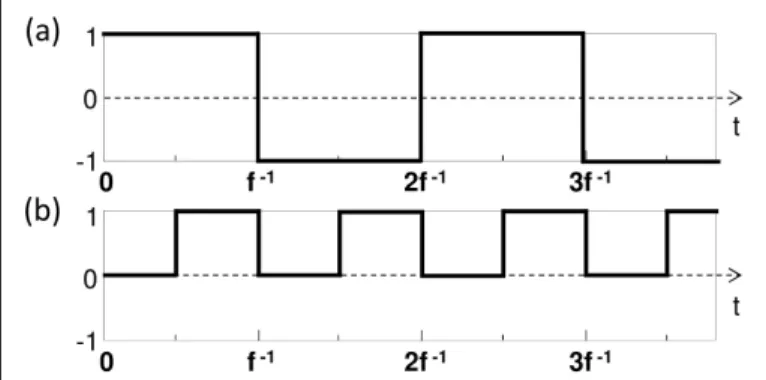
In one dimension, and neglecting diffraction of the US beam as it propagates, we can assume that P(~r, t) =P0h(t−c−1s z+τ), where h(t) is the pressure amplitude modulation function, cs≈1540 m/s is the US velocity and τ a fixed delay. We now cal-culate M(a)i using the f/2-periodic function h plotted in Fig2(a).
Mi(a)(z)∝ 1+cos(2πνzz−ϕ) (4)
where νz= f /csis a spatial frequency, and ϕ=2π f τ a phase offset which can be controlled independently of νzby changing the delay τ. The calculated tagging function is the sum of a constant term and a sine function with a period half that of P(z). Performing a linear combination of measurements Siacquired for ϕ = 0; π/2; π and 3π/2 we can numerically retrieve the complex Fourier component of the longitudinal profile corre-sponding to spatial frequency νz[17,26].
Fig. 2.Examples of possible modulation function h(t)of the acoustic amplitude. (a): for camera-based detection; (b): for photorefractive holographic detection.
FT-AOI can also be implemented from a temporally sampled signal of tagged photon based on photorefractive holographic-based detection or spectral hole burning detection, where signal is sampled with a photodiode. In this case, the tagging function M(b)i is obtained by post-prossessing of the acquired temporal trace, which allows to choose a complex function for hmbefore performing a numerical integration over time T. The expression of M(b)i is given by: Mi(b)(~r) = 1 T Z T 0 hm (t)|P|2(~r, t)dt (5) In this case, the 4-phase measurement is prevented by replac-ing the previous real function by hm(t) =exp(−2πj f t), while imposing that h(t)oscillates between 1 and 0 at a frequency f as shown in Fig.2(b). This corresponds to an amplitude rather than phase modulation of the pressure field. This is in particular what will be presented in the following experimental results, where detection is based on photorefractive holography. In this case, we easily calculate:
M(b)i (z)∝ j exp(−2πjνzz+jϕ) (6) Such a tagging function directly provides Fourier component of the longitudinal profile for ϕ= −π/2.
To generalize this to the two-dimensional case, we propose to use a long-duration US modulated plane wave (as opposed to focused), propagating along z direction. The substitution h(z, t) →h(x, z, t)is possible by controlling independently the temporal US emission at each position x. The amplitude modula-tion at posimodula-tion x is set to h[t+τm(x)]. The delay τm(x)depends linearly on the transverse position x such that:
τm(x) =τ−νxx
f =τ−
νxx
νzcs (7)
Still neglecting diffraction effects, we now have P(x, z, t) = P0h[t−c−1s z+τm(x)] as illutrated in Fig 1(b). The two-dimensional tagging functions simply result from the substitu-tion ϕ=2π f τ→2π f τm(x) =ϕ−2πνxx in respectively Eq.4 and6. Using the previous functions h(t)plotted respectively in Fig2(a) and Fig2(b) and the respective demodulation functions hm(t) =sin(π f t)and hm(t) =exp(−2πj f t), we calculate the following two-dimensional tagging functions:
M(a)(x, z)∝ 1+cos(2πνxx+2πνzz−ϕ) M(b)(x, z)∝ j exp(−2πjν xx−2πjνzz+jϕ) (8)
This constitutes a 2D generalization of previous tagging func-tions. As a result, 2D Fourier components of the AO image in the(x, z)-plane are obtained with a 4-phase measurement for camera based-detection and in a single measurement for tem-porally sampled AO signals. Let us note that the theoretical expressions in Eqs.8exhibit a sinusoidal behavior, thanks to the filtering of the square modulation by temporal averaging in Eqs.3and5. As a result, accurate extraction of Fourier com-ponents in the whole Fourier plane is possible, as opposed to previous work reported on unfocused US waves. In [16] for instance, tilted plane waves are emitted with limited angular span such that many Fourier components can not be measured. By structuring these waves laterally [17], more Fourier compo-nents can be reached but an error of reconstruction is made for modulations that aren’t purely sinusoidal. Since US transducer arrays do not generally allow for smooth modulation of the US wave, the harmonic content of the modulation leads to image degradation [17]. This effect is absent in our present approach thanks to the above-mentioned filtering. Furthermore, both of these methods work in the short-pulse regime, contrary to what we propose here.
We present here the experimental validation of 2D FT-AOI us-ing photorefractive holographic-based detection. This choice of detection might seem peculiar given that the imaging approach presented here is particularly interesting for camera-based detec-tion, potential candidates for in vivo imaging applications. From a fundamental point of view, this choice of detection is justified because it allows direct comparison with a pFW imaging, and is much simpler to implement.
Fig. 3.Experimental setup. (HWP): Half Wave Plate. (PBS): Polarising Beam Splitter.
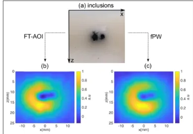
A scheme of the experiment is shown in Fig.3. The sam-ple is an Agar gel matrix with dimensions H×L×W = 5×5×2.5 cm3with 17% concentration of intralipid. The gel reduced scattering coefficient µ0s≈10 cm−1is representative of biological tissues [29]. Two absorbing inclusions of respectively 2 mm and 3 mm inner diameter and 5 mm length (along the transverse y direction), with a separation of 4 mm between their centers, were embedded in the middle of the sample thickness (y direction) at≈13 mm below the upper face. A photograph of these inclusions is shown in Fig.4(a) before being recovered by another layer of scattering gel. The sample is illuminated with a single-longitudinal mode laser centered at 780 nm. After the tapered amplifier (MOPA, Sacher Lasertechnik GmBH), the inci-dent power is about≈ 500 mW for a beam diameter of≈7 mm. The tagged light is collected in transmission through the sam-ple with a photorefractive-based detection scheme (SPS crystal doped with Te 1%) described elsewhere [8,10,13]. The tagged photon flux is recorded with a biased Si-photodiode (Thorlabs Det36A-10 MHz bandwidth, 13 mm2surface) coupled to a tran-simpedance amplifier (Femto GmBh), and sampled at 10 MHz with an acquisition board (Gage Digitizer). Our transducer ar-ray is a commercial US probe (SL10-2, Supersonic Imagine, Aix en Provence, France) with a fixed focal length in y-direction of 35 mm. The probe has 192 piezo-elements of 200µm width, resulting in a total width of 38.4 mm. We positioned it on the top of the sample, above a medical gel, and each piezo-element of the array is arbitrarily addressed to generate the structured acoustic plane wave. In particular, each element emits 100 µs-long US pulses centered at 3 MHz, modulated with the function h plotted in Fig.2(b), delayed according to the law given in Eq7, and at a repetition rate of 1 kHz. Note that the size of the piezo-elements is smaller than the US wavelength, ensuring a good sampling of the US. At all time, the US wavefront is encoded parallel to the emission plane so that US propagate in the z direction. With an excitation voltage of 15 V for the piezo-elements, the maximal pressure of the US wave was measured to be≈ 300 kPa. Such a pressure corresponds to a US peak intensity of≈3 W/cm2and, taking the duty cycle into account, to an average intensity of≈150 mW/cm2far below biomedical norms (720 mW/cm2) [30].
Fig. 4.(a): pictures of the inclusions, before being recovered by another layer of scattering gel; (b) normalized image obtained from FT-AOI; (c) normalized filtered image recorded with pFW.
traces are thus acquired for νx = mνx0and νz = nνz0, where
νx0and νz0are the fundamental frequencies. They are set to νx0 =0.026 mm−1and νz0 =1/(csT0) ≈0.033 mm−1, where T0 =20µs is the fundamental temporal modulation. This cor-responds to an image field of view of 30 mm in direction z and 38.4 mm in direction x. We chose m and n as integers such that−10 ≤ m ≤ 10, and 1 ≤ n ≤ 10. This adds up to 210 Fourier components in total, and the full acquisition was re-peated 10 times for signal averaging. This therefore corresponds to an overall acquisition time of 2.1s. To reconstruct an im-age, each averaged trace is multiplied by hm(t) =exp(−2πj f t), where f =csνz. Each(νx, νz)-Fourier component is then calcu-lated by integrating over time T. We arbitrarily set that value to T=3T0=60 µs so as to mimic an integration time typical of camera-based detectors compatible with in vivo decorrelation time scales. The components at νz=0 are extrapolated from the hypothesis that the AO image cancels close to the surface. The reconstructed image is shown on Fig4(b) where the two inclu-sions can be distinguished. We then wish to compare this image to that obtained using pFW waves limited to the same Fourier components. This reference image is performed by leaving the probe at the same position and scanning the image plane over 143 positions along the x axis with a focused pulse of 3 acoustic cycle in duration centered at 3 MHz. We then applied a low-pass filter on the image so as to match the Fourier plane explored in our 2D-FT imaging setup. The result is represented in Fig4(c), and is very similar to our previous image. This demonstrates that within the explored Fourier region, the image quality of both techniques are equivalent. At last, we would like to empha-size that for the pFW image, the nominal pressure was increased to 40V, so that the acoustic peak intensity can exceed 200 W/cm2 at the focal point. In addition, each line was averaged 500 times leading to an overall acquisition time of 71.5 s , that is to say 34 times higher than with FT-AOI. Although these parameters are only indicative, they illustrate the potential of FT-AOI for real life applications.
In conclusion, we have provided the first proof of concept for bi-dimensional Fourier-Transform Acousto-Optic Imaging. This method, which is an extension to that presented in [26], relies on the spatio-temporal modulation of an acoustic plane wave to extract the Fourier components of the acousto-optic image. It is based on a simple model where US diffraction is neglected. This model is validated by our experimental results, where the image of two inclusions obtained with FT-AOI compares well with that obtained with standard pFW for a given Fourier domain of exploration. So far, our result is important because 2D FT-AOI imaging relies on the use of long-duration acoustic pulses compatible with camera-based detection setups, potential can-didates for in vivo imaging. In addition, plane waves and long pulses allow to reduce the constraint on the acoustic peak-power in order to stay below biomedical norms. In future work we plan to reproduce these results using of an off-axis heterodyne holography detection schemes so as to pave the way towards in vivo imaging.
FUNDING INFORMATION
Plan Cancer ITMO-INSERM 2014-2019 (MALT-C16027HS); Défi Imag’In-CNRS (MALT); French LABEX WIFI (ANR-10-LABX-24 and ANR-10-IDEX-0001-02 PSL).
REFERENCES
1. J. Gunther and S. Andersson-Engels, Front. Optoelectronics10, 211
(2017).
2. W. Leutz and G. Maret, Phys. B: Condens. Matter204, 14 (1995).
3. M. Kempe, M. Larionov, D. Zaslavsky, and A. Z. Genack, J. Opt. Soc. Am. A14, 1151 (1997).
4. L. V. Wang and G. Ku, Opt. Lett.23, 975 (1998).
5. S. Lévêque, A. C. Boccara, M. Lebec, and H. Saint-Jalmes, Opt. Lett.
24, 181 (1999).
6. L. V. Wang, Opt. Lett.26, 1191 (2001).
7. S. Sakadži´c and L. V. Wang, Opt. Lett.29, 2770 (2004).
8. F. Ramaz, B. C. Forget, M. Atlan, A. C. Boccara, M. Gross, P. Delaye, and G. Roosen, Opt. Express12, 5469 (2004).
9. M. Atlan, B. C. Forget, F. Ramaz, A. C. Boccara, and M. Gross, Opt. Lett.30, 1360 (2005).
10. S. Farahi, G. Montemezzani, A. A. Grabar, J.-P. Huignard, and F. Ra-maz, Opt. Lett.35, 1798 (2010).
11. N. T. Huynh, D. He, B. R. Hayes-Gill, J. A. Crowe, J. G. Walker, M. L. Mather, F. R. Rose, S. P. Morgan, N. G. Parker, and M. J. Povey, J. Biomed. Opt.17, 1 (2012).
12. H. Ruan, M. L. Mather, and S. P. Morgan, Opt. Lett.37, 1658 (2012).
13. J.-B. Laudereau, E. B. à La Guillaume, V. Servois, P. Mariani, A. A. Grabar, M. Tanter, J.-L. Gennisson, and F. Ramaz, J. Biophotonics8,
429 (2015).
14. F. A. Marks, H. W. Tomlinson, and G. W. Brooksby, “Comprehensive approach to breast cancer detection using light: photon localization by ultrasound modulation and tissue characterization by spectral dis-crimination,” in Photon Migration and Imaging in Random Media and Tissues, , vol. 1888 (International Society for Optics and Photonics, 1993), pp. 500–510.
15. L. Wang, S. L. Jacques, and X. Zhao, Opt. letters20, 629 (1995).
16. J.-B. Laudereau, A. A. Grabar, M. Tanter, J.-L. Gennisson, and F. Ra-maz, Opt. Express24, 3774 (2016).
17. M. Bocoum, J.-L. Gennisson, J.-B. Laudereau, A. Louchet-Chauvet, J.-M. Tualle, and F. Ramaz, Appl. Opt.58, 1933 (2019).
18. T. W. Murray, L. Sui, G. Maguluri, R. A. Roy, A. Nieva, F. Blonigen, and C. A. DiMarzio, Opt. letters29, 2509 (2004).
19. Y. Li, H. Zhang, C. Kim, K. H. Wagner, P. Hemmer, and L. V. Wang, Appl. Phys. Lett.93, 011111 (2008).
20. C. Venet, M. Bocoum, J.-B. Laudereau, T. Chanelière, F. Ramaz, and A. Louchet-Chauvet, Opt. letters43, 3993 (2018).
21. M. Gross, P. Goy, B. Forget, M. Atlan, F. Ramaz, A. Boccara, and A. K. Dunn, Opt. letters30, 1357 (2005).
22. A. Lev and B. Sfez, JOSA A20, 2347 (2003).
23. Y. Liu, P. Lai, C. Ma, X. Xu, A. A. Grabar, and L. V. Wang, Nat. commu-nications6, 1 (2015).
24. M. M. Qureshi, J. Brake, H.-J. Jeon, H. Ruan, Y. Liu, A. M. Safi, T. J. Eom, C. Yang, and E. Chung, Biomed. optics express8, 4855 (2017).
25. M. Lesaffre, S. Farahi, M. Gross, P. Delaye, A. Boccara, and F. Ramaz, Opt. Express17, 18211 (2009).
26. K. Barjean, K. Contreras, J.-B. Laudereau, Éric Tinet, D. Ettori, F. Ra-maz, and J.-M. Tualle, Opt. Lett.40, 705 (2015).
27. J.-M. Tualle, F. Ramaz, J.-L. Gennisson, and M. Bocoum, patent appli-cationFR1856378 (2018).
28. K. Barjean, F. Ramaz, and J.-M. Tualle, J. Opt. Soc. Am. A33, 854
(2016).
29. S. L. Jacques, Phys. Medicine Biol.58, R37 (2013).
30. R. Phillips and G. Harris, “Information for manufacturers seeking mar-keting clearance of diagnostic ultrasound systems and transducers,” Tech. rep., Food and Drug Administration (2008).
FULL REFERENCES
1. J. Gunther and S. Andersson-Engels, “Review of current methods of acousto-optical tomography for biomedical applications,” Front. Opto-electronics10, 211–238 (2017).
2. W. Leutz and G. Maret, “Ultrasonic modulation of multiply scattered light,” Phys. B: Condens. Matter204, 14 – 19 (1995).
3. M. Kempe, M. Larionov, D. Zaslavsky, and A. Z. Genack, “Acousto-optic tomography with multiply scattered light,” J. Opt. Soc. Am. A14,
1151–1158 (1997).
4. L. V. Wang and G. Ku, “Frequency-swept ultrasound-modulated optical tomography of scattering media,” Opt. Lett.23, 975–977 (1998).
5. S. Lévêque, A. C. Boccara, M. Lebec, and H. Saint-Jalmes, “Ultra-sonic tagging of photon paths in scattering media:?parallel speckle modulation processing,” Opt. Lett.24, 181–183 (1999).
6. L. V. Wang, “Mechanisms of ultrasonic modulation of multiply scattered coherent light: a monte carlo model,” Opt. Lett.26, 1191–1193 (2001).
7. S. Sakadži´c and L. V. Wang, “High-resolution ultrasound-modulated optical tomography in biological tissues,” Opt. Lett.29, 2770–2772
(2004).
8. F. Ramaz, B. C. Forget, M. Atlan, A. C. Boccara, M. Gross, P. Delaye, and G. Roosen, “Photorefractive detection of tagged photons in ultra-sound modulated optical tomography of thick biological tissues,” Opt. Express12, 5469–5474 (2004).
9. M. Atlan, B. C. Forget, F. Ramaz, A. C. Boccara, and M. Gross, “Pulsed acousto-optic imaging in dynamic scattering media with heterodyne parallel speckle detection,” Opt. Lett.30, 1360–1362 (2005).
10. S. Farahi, G. Montemezzani, A. A. Grabar, J.-P. Huignard, and F. Ra-maz, “Photorefractive acousto-optic imaging in thick scattering media at 790 nm with a sn2p2s6:te crystal,” Opt. Lett.35, 1798–1800 (2010).
11. N. T. Huynh, D. He, B. R. Hayes-Gill, J. A. Crowe, J. G. Walker, M. L. Mather, F. R. Rose, S. P. Morgan, N. G. Parker, and M. J. Povey, “Application of a maximum likelihood algorithm to ultrasound modulated optical tomography,” J. Biomed. Opt.17, 1 – 13 (2012).
12. H. Ruan, M. L. Mather, and S. P. Morgan, “Pulse inversion ultrasound modulated optical tomography,” Opt. Lett.37, 1658–1660 (2012).
13. J.-B. Laudereau, E. B. à La Guillaume, V. Servois, P. Mariani, A. A. Grabar, M. Tanter, J.-L. Gennisson, and F. Ramaz, “Multi-modal acousto-optic/ultrasound imaging of ex vivo liver tumors at 790 nm using a sn2p2s6 wavefront adaptive holographic setup,” J. Biophoton-ics8, 429–436 (2015).
14. F. A. Marks, H. W. Tomlinson, and G. W. Brooksby, “Comprehensive approach to breast cancer detection using light: photon localization by ultrasound modulation and tissue characterization by spectral dis-crimination,” in Photon Migration and Imaging in Random Media and Tissues, vol. 1888 (International Society for Optics and Photonics, 1993), pp. 500–510.
15. L. Wang, S. L. Jacques, and X. Zhao, “Continuous-wave ultrasonic modulation of scattered laser light to image objects in turbid media,” Opt. letters20, 629–631 (1995).
16. J.-B. Laudereau, A. A. Grabar, M. Tanter, J.-L. Gennisson, and F. Ra-maz, “Ultrafast acousto-optic imaging with ultrasonic plane waves,” Opt. Express24, 3774–3789 (2016).
17. M. Bocoum, J.-L. Gennisson, J.-B. Laudereau, A. Louchet-Chauvet, J.-M. Tualle, and F. Ramaz, “Structured ultrasound-modulated optical tomography,” Appl. Opt.58, 1933–1940 (2019).
18. T. W. Murray, L. Sui, G. Maguluri, R. A. Roy, A. Nieva, F. Blonigen, and C. A. DiMarzio, “Detection of ultrasound-modulated photons in diffuse media using the photorefractive effect,” Opt. letters29, 2509–2511
(2004).
19. Y. Li, H. Zhang, C. Kim, K. H. Wagner, P. Hemmer, and L. V. Wang, “Pulsed ultrasound-modulated optical tomography using spectral-hole burning as a narrowband spectral filter,” Appl. Phys. Lett.93, 011111
(2008).
20. C. Venet, M. Bocoum, J.-B. Laudereau, T. Chanelière, F. Ramaz, and A. Louchet-Chauvet, “Ultrasound-modulated optical tomography in scattering media: flux filtering based on persistent spectral hole burning in the optical diagnosis window,” Opt. letters43, 3993–3996 (2018).
21. M. Gross, P. Goy, B. Forget, M. Atlan, F. Ramaz, A. Boccara, and A. K.
Dunn, “Heterodyne detection of multiply scattered monochromatic light with a multipixel detector,” Opt. letters30, 1357–1359 (2005).
22. A. Lev and B. Sfez, “In vivo demonstration of the ultrasound-modulated light technique,” JOSA A20, 2347–2354 (2003).
23. Y. Liu, P. Lai, C. Ma, X. Xu, A. A. Grabar, and L. V. Wang, “Optical focusing deep inside dynamic scattering media with near-infrared time-reversed ultrasonically encoded (true) light,” Nat. communications6,
1–9 (2015).
24. M. M. Qureshi, J. Brake, H.-J. Jeon, H. Ruan, Y. Liu, A. M. Safi, T. J. Eom, C. Yang, and E. Chung, “In vivo study of optical speckle decorrelation time across depths in the mouse brain,” Biomed. optics express8, 4855–4864 (2017).
25. M. Lesaffre, S. Farahi, M. Gross, P. Delaye, A. Boccara, and F. Ramaz, “Acousto-optical coherence tomography using random phase jumps on ultrasound and light,” Opt. Express17, 18211–18218 (2009).
26. K. Barjean, K. Contreras, J.-B. Laudereau, Éric Tinet, D. Ettori, F. Ra-maz, and J.-M. Tualle, “Fourier transform acousto-optic imaging with a custom-designed cmos smart-pixels array,” Opt. Lett.40, 705–708
(2015).
27. J.-M. Tualle, F. Ramaz, J.-L. Gennisson, and M. Bocoum, “Procédé d’imagerie acousto-optique par reconstruction de fourier en utilisant une onde plane comme onde porteuse de l’onde ultrasonore,” patent applicationFR1856378 (2018).
28. K. Barjean, F. Ramaz, and J.-M. Tualle, “Theoretical study of fourier-transform acousto-optic imaging,” J. Opt. Soc. Am. A33, 854–862
(2016).
29. S. L. Jacques, “Optical properties of biological tissues: a review,” Phys. Medicine Biol.58, R37–R61 (2013).
30. R. Phillips and G. Harris, “Information for manufacturers seeking mar-keting clearance of diagnostic ultrasound systems and transducers,” Tech. rep., Food and Drug Administration (2008).