HAL Id: hal-01780644
https://hal.archives-ouvertes.fr/hal-01780644
Submitted on 22 May 2018
HAL is a multi-disciplinary open access
archive for the deposit and dissemination of
sci-entific research documents, whether they are
pub-lished or not. The documents may come from
teaching and research institutions in France or
abroad, or from public or private research centers.
L’archive ouverte pluridisciplinaire HAL, est
destinée au dépôt et à la diffusion de documents
scientifiques de niveau recherche, publiés ou non,
émanant des établissements d’enseignement et de
recherche français ou étrangers, des laboratoires
publics ou privés.
isolated from a malnourished child using culturomics
El Hadji Seck, Mamadou Beye, Sory Ibrahima Traore, Saber Khelaifia,
Caroline Michelle, Carine Couderc, Souleymane Brah, Pierre-Edouard
Fournier, Didier Raoult, Fadi Bittar
To cite this version:
El Hadji Seck, Mamadou Beye, Sory Ibrahima Traore, Saber Khelaifia, Caroline Michelle, et al..
Bacil-lus kwashiorkori sp nov., a new bacterial species isolated from a malnourished child using culturomics.
MicrobiologyOpen, Wiley, 2018, 7 (1), pp.e00535. �10.1002/mbo3.535�. �hal-01780644�
MicrobiologyOpen. 2018;7:e535.
|
1 of 13 https://doi.org/10.1002/mbo3.535 www.MicrobiologyOpen.com Received: 28 April 2017|
Accepted: 15 August 2017 DOI: 10.1002/mbo3.535 O R I G I N A L R E S E A R C HBacillus kwashiorkori sp. nov., a new bacterial species isolated
from a malnourished child using culturomics
El hadji Seck
1| Mamadou Beye
1| Sory Ibrahima Traore
1| Saber Khelaifia
1|
Caroline Michelle
1| Carine Couderc
1| Souleymane Brah
2| Pierre-Edouard Fournier
1|
Didier Raoult
1,3| Fadi Bittar
1This is an open access article under the terms of the Creative Commons Attribution License, which permits use, distribution and reproduction in any medium, provided the original work is properly cited.
© 2017 The Authors. MicrobiologyOpen published by John Wiley & Sons Ltd. 1Aix-Marseille Univ, CNRS 7278, AP-HM, IRD 198, INSERM 1095, URMITE, IHU Méditerranée Infection, Marseille, France 2Hôpital National de Niamey, Niamey, Niger 3King Fahd Medical Research Center, Special Infectious Agents Unit, King Abdulaziz University, Jeddah, Saudi Arabia Correspondence Fadi Bittar, URMITE, IHU Méditerranée Infection, Marseille, France. Email: fadi.bittar@univ-amu.fr Funding information Fondation Méditerranée Infection
Abstract
Strain SIT6T was isolated from the fecal flora of a severely malnourished child as part of a broad “culturomics” study aiming to maximize the culture conditions for the in- depth exploration of the human microbiota. An analysis of the 16S rRNA gene se-quence showed that strain SIT6T shared 94.1% 16S rRNA gene sequence similaritywith Bacillus thermoamylovorans DKPT (NR_029151), the phylogenetically closest type
species. Colonies are creamy white, circular, 4–5 mm in diameter after cultivation at
37°C for 24 hr on 5% sheep blood- enriched Colombia agar. Growth occurs at tem-peratures in the range of 25–56°C (optimally at 37°C). Strain SIT6T is a gram- positive,
facultative anaerobic rod and motile by means of peritrichous flagella and sporulating; it is catalase and oxidase positive. The 2,784,637- bp- long genome, composed of 16 contigs, has a G+C content of 35.19%. Of the 2,646 predicted genes, 2,572 were protein- coding genes and 74 were RNAs. The major fatty acids are saturated species (15:0 iso, 16:0 and 17:0 anteiso). Of the 14 detected fatty acids, 11 are saturated, ei- ther linear or branched (iso and anteiso). Digital DNA–DNA hybridization (dDDH) es-timation and average genomic identity of orthologous gene sequences (AGIOS) of the strain SIT6T against genomes of the type strains of related species ranged between 18.6% and 38.3% and between 54.77% and 65.50%, respectively. According to our taxonogenomics results, we propose the creation of Bacillus kwashiorkori sp. nov. that
contains the type strain SIT6T (=CSUR P2452T, =DSM 29059T).
K E Y W O R D S
Bacillus kwashiorkori, culturomics, genome, taxonogenomics
1 | INTRODUCTION
Although the human intestinal flora is intrinsically associated with the host genotype and age, many external factors can affect and modify this microbiota, such as antibiotics, probiotics, and diet (Angelakis, Armougom, Million, & Raoult, 2012; Chen, He, & Huang,
2014; Moreno- Indias, Cardona, Tinahones, & Queipo- Ortuño, 2014). Recently, genomic and metagenomic advances have widely participated in describing the human microbiota, but culture isola-tion remains the only means and the first step to characterize the physiological and genomic properties of a given bacterium and to describe a potential new species (Vartoukian, Palmer, & Wade,
2010). For this reason, in our laboratory we have developed a new strategy called culturomics, which is based on the application of various culture conditions followed by rapid identification using matrix- assisted laser- desorption/ionization time- of- flight mass spectrometry (MALDI- TOF MS) to explore the bacterial composition (Lagier et al., 2012). This new concept has allowed us to significantly increase the bacterial species associated with the human digestive tract and to find many new species (Lagier et al., 2016). Using this strategy (i.e., culturomics), we were able to isolate a new species be-longing to the genus Bacillus.
This new isolate was described according to the new method that we have implemented (taxonogenomics) (Kokcha et al., 2012; Lagier, Elkarkouri, Rivet, Couderc, & Raoult, 2013; Seck et al., 2016). In brief, it involves using proteomic, fatty acid, and genomic features (Ramasamy et al., 2014; Welker & Moore, 2011; Seng et al., 2013), along with phe-notype and some conventional methods, such as 16S rRNA phylogeny and the G+C content. In this article, we describe the strain SIT6T (=CSUR P2452T, =DSM 29059T) isolated from the stool sample of a kwashiorkor patient.
2 | MATERIALS AND METHODS
2.1 | Organism information
The study and consent procedure were approved by the National Ethics Committee of Nigeria and the Ethics Committee of the Federative Research Institute 48 (Faculty of Medicine, Marseille, France) under the agreement number 09- 022. The stool sample was obtained from a 4- month- old Nigerian child suffering from acute mal- nutrition (kwashiorkor). The patient was not being treated with antibi-otics at the time of the sample collection and the sample was stored at −80°C. The stool sample was cultured in blood culture bottles supple-mented with sheep blood (BioMérieux, Marcy l’Etoile, France). During a 30- day preincubation period at 37°C in aerobic atmosphere, the liq-uid culture is then spread on Columbia agar with 5% sheep blood COS medium (BioMérieux, Marcy l’Etoile, France) and the isolated colonies are subsequently identified.
2.2 | Strain identification by MALDI- TOF MS and
16S rRNA sequencing
MALDI- TOF MS analysis of proteins was used to identify the bacte-ria. Each colony was deposited in duplicate on a MALDI- TOF MSP 96 target and then covered with 1.5 μl of a matrix solution (saturated solution of α- cyano- 4- hydroxycinnamic acid in 50% acetonitrile, 2.5% trifluoroacetic acid) to allow the crystallization of molecules. MALDI- TOF MS was performed using the LT Microflex spectrometer (Bruker Daltonics, Leipzig, Germany). All spectra were recorded in positive lin-ear mode for the mass range from 2,000 to 20,000 Da (parameters: ion source 1 [ISI], 20 kV; IS2, 18.5 kV lens, 7 kV). The generated spec-tra were then compared to the Bruker database, with the addition of new species found through the “culturomics” project. The resulting score dictates whether a tested species can be identified: a score ≥2
with a validly published species enables identification at the species level, a score ≥1.7 but <2 enables identification at the genus level, and a score <1.7 does not enable any identification.
Following three assays, unidentified colonies were identified using 16S rRNA gene sequencing as described previously (Bittar et al., 2014). The isolated colony was suspended in 200 μl distilled water for DNA extraction using an EZ1 DNA Tissue Kit with a BioRobot EZ1 Advanced XL (Qiagen, Courtaboeuf, France). The amplification of the 16S rRNA gene was performed using the universal primer pair fD1 and rP2 (Eurogentec, Angers, France) (Weisburg, Barns, Pelletier, & Lane, 1991). The PCR product was purified and sequenced using the BigDye Terminator v1.1 Cycle Sequencing Kit (PerkinElmer, Courtaboeuf, France) with the following internal primers: 536F, 536R, 800F, 800R, 1050F, and 1050R, and ABI Prism 3130xl Genetic Analyzer capillary sequencer (Applied Biosystems). 16S rRNA amplification and sequenc-ing were carried out as described previously by Morel et al. (2015). The 16S rRNA nucleotide sequences were assembled and corrected using CodonCode Aligner software (http://www.codoncode.com). Then, the BLASTn searches against the GenBank NCBI database (http://blast. ncbi.nlm.nih.gov.gate1.inist.fr/Blast.cgi) and EzBioCloud’s Identify Service (http://www.ezbiocloud.net/identify) (Yoon et al., 2017) were performed to determine the percentage of similarity with the closest bacteria. The MEGA 7 (Molecular Evolutionary Genetics Analysis) software (Kumar, Stecher, & Tamura, 2016) allowed us to construct a phyloge-netic tree. Sequence alignment of the different species was performed using CLUSTALW and the calculation of the evolutionary distance was done with the Kimura two- parameter model (Kimura, 1980; Thompson, Higgins, & Gibson, 1994).
2.3 | Growth conditions
In order to determine the ideal growth conditions for strain SIT6T, different growth temperatures (25°C, 28°C, 30°C, 37°C, 45°C, and 56°C) were tested under anaerobic and microaerophilic atmospheres using GENbag Anaer and GENbag microaer systems, respectively (BioMérieux, Marcy l’Etoile, France). The strain growth was also tested aerobically with and without 5% CO2. The growth of strain SIT6T was tested under different pH using a Columbia agar with 5% sheep blood COS medium (BioMérieux, Marcy l’Etoile, France) with NaCl, MgCl2, MgSO4, KCl, CaCl2, and glucose. The pH was modified by adding HCl to the medium and measured with a pH meter. The optimal pH for growth was determined by testing at different pH 5, 6, 6.5, 7, 7.5, 8, and 8.5. Growth at various NaCl concentrations (0.5%, 5%, 7.5%, 10%, 15%, and 200%) was investigated.
2.4 | Morphologic, biochemical, and antibiotic
susceptibility tests
Gram staining was performed and observed using a Leica DM 2500 photonic microscope (Leica Microsystems, Nanterre, France) with a 100× oil immersion lens. A thermal shock (80°C during 20 min) was applied on fresh colonies in order to test sporulation. The motility of
the strain was tested by observing fresh colonies using a DM1000 photonic microscope (Leica Microsystems) with a 40× objective lens. Catalase (BioMérieux) activity was determined in 3% hydrogen perox- ide solution and oxidase activity was assessed using an oxidase rea-gent (Becton- Dickinson, Le Pont- de- Claix, France).
Antibiotic susceptibility testing was performed using SIRscan Discs (i2a, Montpellier, France) on Mueller- Hinton agar according to
EUCAST 2015 recommendations (Matuschek, Brown, & Kahlmeter, 2014). The following antibiotics were tested: doxycycline (30 μg), ri-fampicin (30 μg), vancomycin (30 μg), erythromycin (15 μg), ampicillin (10 μg), ceftriaxone (30 μg), ciprofloxacin (5 μg), gentamicin (500 μg), penicillin (10 μg), trimethoprim/sulfamethoxazole (25 + 23.75 μg), imipenem (10 μg), metronidazole (4 μg), clindamycin (15 μg), colistin (50 μg), and oxacillin (5 μg). F I G U R E 1 Phylogenetic tree showing the position of Bacillus kwashiorkori SIT6T (red) relative to other phylogenetically close members of the family Bacillaceae. GenBank accession numbers are indicated in parentheses. Sequences were aligned using CLUSTALW, and phylogenetic inferences were obtained using (a) the maximum- likelihood method, (b) the neighbor-joining method and (c) the maximum parsimony method within the MEGA software. Numbers at the nodes are percentages of bootstrap values obtained by repeating the analysis 1,000 times to generate a majority consensus tree. Only values >70% were displayed. Bhargavaea
ginsengi ge14T (EF371375) was used as out- group
(a)
(b)
Using the commercially available biochemical API 20NE, API ZYM, and API 50CH strips, we investigated the biochemical characteristics of our strain according to the manufacturer’s instructions (BioMérieux). Negative staining was done in order to visualize the cell morphol-ogy. Cells were fixed with 2.5% glutaraldehyde in 0.1 mol/L cacodylate buffer for at least 1 hr at 4°C. A drop of cell suspension was depos-ited for approximately 5 min on glow- discharged formvar carbon film on 400 mesh nickel grids (FCF400- Ni, EMS). The grids were dried on blotting paper and the cells were negatively stained for 10 s with 1% ammonium molybdate solution in filtered water at room temperature. Electron micrographs were acquired with a Tecnai G20 Cryo (FEI) transmission electron microscope operated at 200 keV.
2.5 | FAME analysis by gas chromatography/mass
spectrometry
Cellular fatty acid methyl ester (FAME) analysis was performed by GC/MS. Two samples were prepared with approximately 2 mg of bacterial biomass each, harvested from five culture plates. Fatty acid methyl esters were prepared as described by Sasser (2006). GC/MS analyses were carried out as described previously (Dione et al., 2016). In brief, fatty acid methyl esters were separated using an Elite 5- MS column and monitored by mass spectrometry (MS) (Clarus 500- SQ 8 S, PerkinElmer, Courtaboeuf, France). A spectral database search was performed using MS Search 2.0 operated with the Standard Reference Database 1A (NIST, Gaithersburg, MD, USA) and the FAMEs mass spectral database (Wiley, Chichester, UK).
2.6 | Genomic DNA preparation
After pretreatment by a lysozyme (incubation at 37°C for 2 hr), the DNA of strain SIT6T was extracted on the EZ1 BioRobot (Qiagen) with the EZ1 DNA tissues kit. The elution volume was 50 μl. Genomic DNA (gDNA) was quantified by a Qubit assay with the high sensitivity kit (Life Technologies, Carlsbad, CA) to 55.8 ng/μl.
2.7 | Genome sequencing and assembly
Genomic DNA (gDNA) of B. kwashiorkori was sequenced on MiSeq Technology (Illumina Inc., San Diego, CA) with the mate pair strat-egy. The gDNA was barcoded in order to be mixed with 11 other projects with the Nextera Mate Pair sample prep kit (Illumina). gDNA was quantified by a Qubit assay with the high sensitivity kit (Thermo Fisher Scientific, Waltham, MA) to 66.2 ng/μl. The mate pair li-brary was prepared with 1 μg of gDNA using the Nextera mate pair Illumina guide. The gDNA sample was simultaneously fragmented and tagged with a mate pair junction adapter. The pattern of the fragmentation was validated on an Agilent 2100 Bioanalyzer (Agilent Technologies Inc., Santa Clara, CA) with a DNA 7500 LabChip. The DNA fragments ranged in size from 1 kb to 11 kb, with an optimal size at 3.927 kb. No size selection was performed and 505 ng of tagged fragments were circularized. The circularized DNA was me-chanically sheared to small fragments with an optimal size of 597 bp
on the Covaris device S2 in microtubes (Covaris, Woburn, MA). The library profile was viewed on a High Sensitivity Bioanalyzer LabChip (Agilent Technologies Inc.) and the final concentration library was measured at 59.2 nmol/L. The libraries were normalized at 2 nmol/L and pooled. After a denaturation step and dilution at 15 pmol/L, the pool of libraries was loaded onto the reagent cartridge and then onto the instrument along with the flow cell. An automated cluster gen-eration and sequencing run was performed in a single 39- hr run in a 2 × 251 bp. T A B L E 1 Classification and general features of Bacillus kwashiorkori strain SIT6T Property Term Current classification Domain: Bacteria Phylum: Firmicutes Class: Bacilli Order: Bacillales Family: Bacillaceae Genus: Bacillus Species: kwashiorkori Type strain: SIT6T Gram stain Positive Cell shape Rod shaped Motility Motile Sporulation Endospore forming Temperature range Mesophile Optimum temperature 37°C Optimum pH 7.5 Salinity 0.0–5.0 g/L Optimum salinity 0 g/L Oxygen requirement Facultative aerobic F I G U R E 2 Reference mass spectrum from Bacillus kwashiorkori SIT6T strain. Spectra from 12 individual colonies were compared and a reference spectrum was generated 0 2,000 4,000 6,000 8,000 Intens. (a.u.) 2,000 4,000 6,000 8,000 10,000 12,000 14,000 16,000 18,000 m/z
2.8 | Genome annotation and analysis
Open reading frames (ORFs) were predicted using Prodigal (http:// prodigal.ornl.gov/) with default parameters. However, the predicted ORFs were excluded if they spanned a sequencing gap region.
The predicted bacterial protein sequences were searched against GenBank and Clusters of Orthologous Group (COG) databases using BLASTP. The tRNAs and rRNAs were predicted using the tRNAScan- SE and RNAmmer tools, respectively. Signal peptides and numbers of transmembrane helices were predicted using SignalP (Nielsen,
F I G U R E 3 Gel view comparing Bacillus kwashiorkori SIT6T spectra with other members of the genus Bacillus. The gel view displays the raw spectra of all loaded spectrum files arranged in a pseudo- gel- like look. The x- axis records the m/z value. The left y- axis displays the running spectrum number originating from subsequent spectra loading. The peak intensity is expressed by a gray scale scheme code. The color bar and the right y- axis indicate the relation between the color a peak is displayed with and the peak intensity in arbitrary units. Displayed species are indicated on the right
F I G U R E 4 Gram staining of Bacillus kwashiorkori SIT6T
F I G U R E 5 Transmission electron microscopy of Bacillus
kwashiorkori SIT6T using a Morgani 268D (Philips) at an operating voltage of 60 kV. The scale bar represents 500 nm
Engelbrecht, Brunak, & von Heijne, 1997) and TMHMM (Krogh, Larsson, von Heijne, & Sonnhammer, 2001), respectively. Mobile genetic elements were predicted using PHAST (Zhou, Liang, Lynch, Dennis, & Wishart, 2011) and RAST (Aziz et al., 2008). ORFans were identified if their BLASTP E- value was lower than 1e- 03 for align-ment length >80 amino acids. If alignment lengths were <80 amino acids, we used an E- value of 1e- 05. Such parameter thresholds have already been used in previous work to define ORFans. Artemis (Carver, Harris, Berriman, Parkhill, & McQuillan, 2012) and DNA plotter (Carver, Thomson, Bleasby, Berriman, & Parkhill, 2009) were used for data management and visualization of genomic features, respectively. The mauve alignment tool (version 2.3.1) was used for multiple genomic sequence alignment (Darling, Mau, Blattner, & Perna, 2004). The mean level of nucleotide sequence similarity at the genome level between B. kwashiorkori and other Bacillus species was estimated using the Average Genomic Identity of Orthologous Gene Sequences (AGIOS) in- house software (Ramasamy et al., 2014). This software combines the functionality of other software programs: Proteinortho (Lechner et al., 2011) (detects orthologous proteins between genomes compared two by two, then retrieves the corresponding genes) and the Needleman–Wunsch global alignment algorithm (determines the mean percentage of nucleotide sequence identity among orthologous ORFs). Finally, the Genome- to- Genome Distance Calculator (GGDC) web server (http://ggdc.dsmz.de) was used to estimate the similarity between the compared genomes (Auch, Jan, Klenk, & Göker, 2010; Meier- Kolthoff, Auch, Klenk, & Göker, 2013).
Average nucleotide identity at the genome level be-tween B. kwashiorkori (CTDX00000000) and the other species
B. firmus (BCUY00000000), B. shackletonii (LJJC00000000), B. smithii
(BCVY00000000), B. aquimaris (LQXM00000000), B.
thermoamylo-vorans (CCRF00000000), B. coagulans (CP003056), B. alveayuensis
(JYCE00000000), B. sporothermodurans (LQYN00000000), B. acidicola (LWJG00000000), and B. ginsengihumi (JAGM00000000) was esti- mated using BLASTN and a home made software, following the algo-rithm described by Ouk, Chun, Lee, and Park (2016).
3 | RESULTS AND DISCUSSION
3.1 | Strain identification and phylogenetic analyses
Strain SIT6T was first isolated in May 2014 after a 30- day preincu-bation in a blood culture bottle with sheep blood and cultivation on 5% sheep blood- enriched Colombia agar in an aerobic atmosphere at 37°C. No significant MALDI- TOF score was obtained for strain SIT6T against the Bruker and URMITE databases, suggesting that the isolate was not a member of a known species. Strain SIT6T shared 94.1% 16S rRNA gene sequence similarity with B. thermoamylovorans DKPT (NR_029151) using GenBank NCBI database (reference RNA sequences). Although the 16S rRNA gene sequence of strain SIT6T showed 94.58% similarity with Bacillus kokeshiiformis MO- 04T and 94.57% similarity with Bacillus thermolactis R- 6488T by EzBioCloud’s identify server. Figure 1a, b, and c present the phylogenetic trees ofstrain SIT6T relative to other closest type species with a validly pub-lished name using maximum- likelihood, neighbor- joining, and maxi-mum parsimony methods, respectively. Consequently, as this 16S rRNA nucleotide sequence similarity was lower than the threshold of 98% recommended by Tindall, Rosselló- Mora, Busse, Ludwig, and Kämpfer (2010) to delineate a new species; it was classified as a new species called Bacillus kwashiorkori SIT6T (Table 1). Furthermore, this percentage of similarity comprised in the range of percentage similarity of Bacillus species (82.7–100%), confirming the new spe-cies status (Rossi- Tamisier, Benamar, Raoult, & Fournier, 2015). The reference spectrum for strain SIT6T was thus incremented in the URMITE database (http://www.mediterranee-infection.com/article. php?laref=256&titre=urms-database) (Figure 2) and then compared to other known species of the genus Bacillus. The differences exhibited are shown in the obtained gel view (Figure 3).
3.2 | Phenotypic description
Growth of strain SIT6T was observed between 25°C and 56°C on 5% sheep blood Colombia agar and optimal growth was achieved at 37°C after 24 hr incubation in aerobic conditions (37°C was the temperature at which this strain grows most rapidly). Poor growth occurred under microaerophilic and anaerobic conditions. Cells were motile and sporulating. Colonies were circular, white with a mean diameter of 5 mm on blood- enriched Colombia agar. Gram staining (Figure 4) showed gram- positive rods. Using electron mi-croscopy, the rods had a mean diameter of 1.8 μm and a length of 5.9 μm (Figure 5). Catalase and oxidase activities were positive for strain SIT6T. The major fatty acids are saturated species (15:0 iso, 16:0 and 17:0 anteiso). Of the 14 detected fatty acids, 11 are saturated, either linear T A B L E 2 Cellular fatty acid composition
Fatty acids IUPAC name
Mean
relative %a
15:0 iso 13- Methyl- tetradecanoic acid 19.6 ± 1.2 16:0 Hexadecanoic acid 19.5 ± 0.4 17:0 anteiso 14- Methyl- hexadecanoic acid 16.5 ± 1.3 18:1n12 6- Octadecenoic acid 12.7 ± 1.9 18:0 Octadecanoic acid 9.3 ± 0.4 17:0 iso 15- Methyl- hexadecanoic acid 6.9 ± 1.7 15:0 anteiso 12- Methyl- tetradecanoic acid 4.5 ± 0.1 18:2n6 9,12- Octadecadienoic acid 4.0 ± 0.2 16:0 iso 14- Methyl- pentadecanoic acid 3.9 ± 0.5 18:1n5 13- Octadecenoic acid 1.5 ± 0.1 14:0 Tetradecanoic acid TR 17:0 Heptadecanoic acid TR 15:0 Pentadecanoic acid TR 14:0 iso 12- Methyl- tridecanoic acid TR
aMean peak area percentage ± standard deviation (n = 3); TR, trace
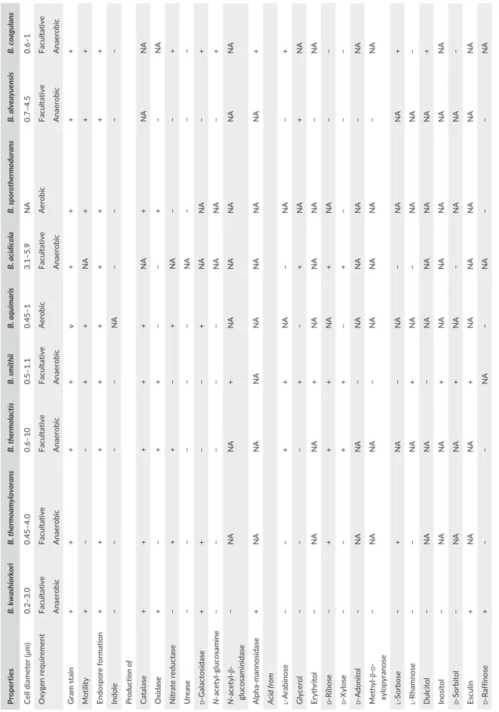
TABLE 3 Phenotypic charac terist ics of Bacillus kwashiorkori SIT6
T with other phylogenetically close
Bacillus strains a Properties B. kwashiorkori B. thermoamylovorans B. thermolactis B. smithii B. aquimaris B. acidicola B. sporothermodurans B. alveayuensis B. coagulans Cell diameter (μ m) 0.2–3.0 0.45–4.0 0.6–10 0.5–1.1 0.45–1 3.1–5.9 NA 0.7–4.5 0.6–1 Oxygen requirement Facultative Facultative Facultative Facultative Aerobic Facultative Aerobic Facultative Facultative Anaerobic Anaerobic Anaerobic Anaerobic Anaerobic Anaerobic Anaerobic Gram stain + + + + v + + + + Motility + − − + + NA + + + Endospore formation + + + + + + + + + Indole − − − − NA − − − − Production of Catalase + + + + + NA + NA NA Oxidase + − + + − − + − NA Nitrate reductase − + + − + NA − − + Urease − − − − − NA − − − d - Galactosidase + + − − + NA NA − + N - acetyl- glucosamine − − − − − NA NA − + N - β-glucosaminidase − NA NA + NA NA NA NA NA mannosidase + NA NA NA NA NA NA NA +
Acid from l- Arabinose
− − + + NA − NA − + Glycerol − − − + − + NA + NA Erythritol − NA NA + NA NA NA − NA d - Ribose − + + + NA + NA − − d - Xylose − − + + − + − − − d - Adonitol − NA NA − NA NA NA − NA β-d - xylopyranose − NA NA − NA NA NA − NA l - Sorbose − + NA − NA − NA NA + l - Rhamnose − − NA + NA − NA NA − Dulcitol − NA NA − NA NA NA NA + Inositol − NA NA + NA NA NA NA NA d - Sorbitol − NA NA + NA − NA NA − Esculin + NA NA + NA NA NA NA NA d - Raffinose + − − NA − NA − − NA (Continues)
Properties B. kwashiorkori B. thermoamylovorans B. thermolactis B. smithii B. aquimaris B. acidicola B. sporothermodurans B. alveayuensis B. coagulans d - Lyxose − NA NA − NA NA NA NA NA d - Fucose − NA NA − NA NA NA NA − l - Arabitol − NA NA NA NA − NA NA − d - Mannose + − − + − + − + + d - mannitol + − v + − + − − − d - Glucose + + − + + + + + + d - Fructose + + − + + + − + + d - Maltose + − + + + + + − + d - Lactose − − − + − v − − + Galactose − − − + − + − + + Habitat Human gut Oil Milk Milk Sea Acidic sphagnum Water, milk Sea Milk NA, not available; v, variable. aBacillus th ermoamy lovorans DK P T (Combet- Blanc et al., 1995), Bacillus th ermolactis R- 6488 T (Coorevits et al., 2011), Bacillus smith ii NR RL NR S- 173 T (Bae, Lee, & Kim, 2005), Bacillus aquimaris TF- 12 T (Yoon, Kim, Kang, Oh, & Park, 2003), Bacillus sporoth ermodurans M215 T (Heyndrickx et al., 2012), Bacillus acidicola 2 T (Albert, 2005), Bacillus alveayu ensis TM1 T (Bae et al., 2005), and Bacillus coagulans 2- 6 T (De Clerck et al., 2004) . TABLE 3 (Continued) F I G U R E 6 Graphical circular map of the chromosome. From outside to the center: Genes on the forward strand colored by clusters of orthologous groups (COG) categories (only gene assigned to COG), genes on the reverse strand colored by COG categories (only gene assigned to COG), RNA genes (tRNAs green, rRNAs red), G+C content and G+C skew T A B L E 4 Nucleotide content and gene count levels of the genome
Attribute Value % of totala
Genome size (bp) 2,784,637 100 DNA coding region (bp) 2,319,082 83.28 DNA G+C content (bp) 980,134 35.19 Total genes 2,646 100 RNA genes 74 2.79 tRNA genes 63 2.38 Protein- coding genes 2,572 97.20 Genes with function prediction 1,749 68.00 Genes assigned to COGs 1,715 66.68 Protein associated with hypothetical protein 487 18.93 Protein associated with ORFan 156 6.06 Genes with peptide signals 250 9.72 Genes with transmembrane helices 720 27.99 Genes associated with PKS or NRPS 6 0.2 Genes associated with mobilome 1,402 54.51 Genes associated with toxin/ antitoxin 84 3.26 aThe total is based on either the size of the genome in base pairs or the total number of protein- coding genes in the annotated genome.
or branched (iso and anteiso). The fatty acid composition of strain SIT6T is detailed in Table 2.
Table 3 shows the biochemical features of B. kwashiorkori SIT6T and the most closely related species.
Bacterial cells were resistant to metronidazole, but susceptible to imipenem, doxycycline, rifampicin, vancomycin, amoxicillin, ceftriax-one, gentamicin, trimethoprim/sulfamethoxazole, erythromycin, cip-rofloxacin, and gentamicin.
T A B L E 5 Number of genes associated with the 25 general COG functional categories
Code Value %a Description
J 130 5.05 Translation A 0 0 RNA processing and modification K 141 5.48 Transcription L 140 5.44 Replication, recombination, and repair B 1 0.04 Chromatin structure and dynamics D 18 0.70 Cell cycle control, mitosis, and meiosis Y 0 0 Nuclear structure V 43 1.67 Defense mechanisms T 88 3.42 Signal transduction mechanisms M 81 3.15 Cell wall/membrane biogenesis N 26 1.01 Cell motility Z 0 0 Cytoskeleton W 0 0 Extracellular structures U 26 1.01 Intracellular trafficking and secretion O 87 3.38 Posttranslational modification, protein turnover, chaperones C 120 4.67 Energy production and conversion G 137 5.33 Carbohydrate transport and metabolism E 138 5.37 Amino acid transport and metabolism F 39 1.52 Nucleotide transport and metabolism H 52 2.02 Coenzyme transport and metabolism I 67 2.61 Lipid transport and metabolism P 147 5.72 Inorganic ion transport and metabolism
Q 34 1.32 Secondary metabolites biosynthesis, transport, and catabolism R 241 9.37 General function prediction only
S 180 6.99 Function unknown − 857 33.32 Not in COGs
aThe total is based on the total number of protein- coding genes in the annotated genome.
T A B L E 6 Genomic comparison of Bacillus kwashiorkori with other Bacillus spp.
Species Strain
Genome accession
number Genome size (Mb) GC (%) Gene content
B. kwashiorkori SIT6T CTDX00000000 2.78 35.19 2,572
B. alveayuensis TM1T JYCE00000000 6.70 38.13 6,689
B. shackletonii LMG 18435T LJJC00000000 5.29 36.70 4,727
B. coagulans 2–6T CP003056 3.07 47.29 2,971
B. ginsengihumi Gsoil 114T JAGM00000000 3.92 35.85 3,832
B. firmus IAM 12464T BCUY00000000 4.97 41.45 4,922
B. aquimaris TF- 12T LQXM00000000 4.42 44.57 4,432
B. sporothermodurans M215T LQYN00000000 4.04 35.65 4,211
B. smithii NRRL NRS- 173T BCVY00000000 3.38 40.75 3,619
B. acidicola 105- 2T LWJG00000000 5.13 39.39 4,876
3.3 | Genome properties
The genome is 2,784,637 bp long with 35.19% G+C content (Figure 6). It is composed of 16 scaffolds, composed of 16 contigs. Of the 2,646 predicted genes, 2,572 were protein- coding genes, and 74 were RNAs (7 genes are 5S rRNA, 2 genes are 16S rRNA, 2 genes are 23S rRNA, and 63 genes are tRNA). A total of 1,749 (68%) were assigned as putative function (by COGs of NR blast). A total of 156 genes were identified as ORFans (6.07%). The remaining genes were annotated as hypothetical proteins (487 genes [18.93%]). Genome content is de-tailed in Table 4, while Table 5 presents the distribution of the genes into COG functional categories. The genome sequence has been deposited in GenBank under ac-cession number CTDX00000000.
3.4 | Comparison with other Bacillus spp. genomes
The draft genome of B. kwashiorkori (2.78 Mb) is smaller in size than those of Bacillus alveayuensis, Bacillus shackletonii, Bacillus coagulans,Bacillus ginsengihumi, Bacillus firmus, Bacillus aquimaris, Bacillus sporo-thermodurans, Bacillus smithii, Bacillus acidicola, and Bacillus thermoa-mylovorans (6.70, 5.29, 3.07, 3.92, 4.97, 4.42, 4.04, 3.38, 5.13, and
3.70 Mb, respectively) (Table 6). Bacillus kwashiorkori has a lower G+C content (35.19%) than those of B. alveayuensis, B. shackletonii, B.
co-agulans, B. ginsengihumi, B. firmus, B. aquimaris, B. sporothermodurans, B. smithii, B. acidicola, and B. sporothermodurans (38.13%, 36.70%,
47.29%, 35.85%, 41.45%, 44.57%, 35.65%, 40.75%, 39.39%, and 37.27%, respectively) (Table 6). The protein content of B. kwashiorkori (2,572) is lower than those of B. alveayuensis, B. shackletonii, B.
co-agulans, B. ginsengihumi, B. firmus, B. aquimaris, B. sporothermodurans, B. smithii, B. acidicola, and B. thermoamylovorans (6,689, 4,727, 2,971,
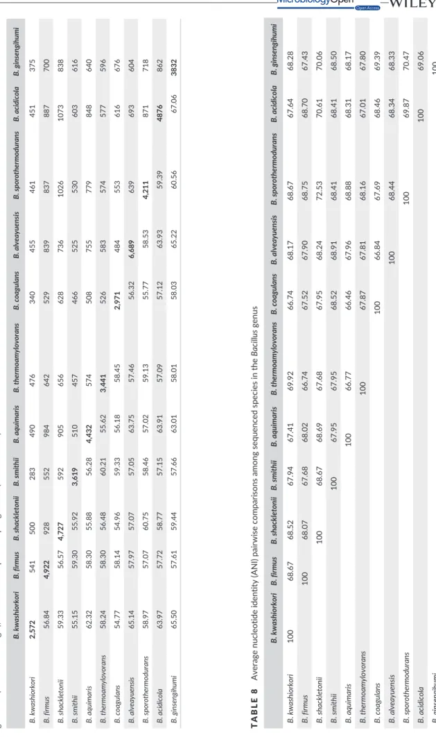
3,832, 4,922, 4,432, 4,211, 3,619, 4,876, and 3,441, respectively) (Table 6). However, the distribution of genes into COG categories is similar in all compared genomes (Figure 7). In addition, AGIOS values ranged from 54.77% to 67.06% among the Bacillus species compared (Table 7). The range of AGIOS varied from 54.77% to 65.50% between B. kwashiorkori and other compared Bacillus species
(Table 7). Moreover, B. kwashiorkori shares 455, 500, 340, 375, 541, 490, 461, 283, 451, and 476 orthologous genes with B. alveayuensis,
B. shackletonii, B. coagulans, B. ginsengihumi, B. firmus, B. aquimaris, B. sporothermodurans, B. smithii, B. acidicola, and B. thermoamylovorans,
respectively (Table 7). Of the species with standing in nomenclature, ANI values ranged from 66.46% between B. coagulans and B.
aquima-ris to 72.53% between B. sporothermodurans and Bacillus shackleto-nii. When comparing B. kwashiorkori to other species, the ANI value
ranged from 66.74% between B. kwashiorkori and B. coagulans to 69.92% between B. kwashiorkori and B. thermoamylovorans (Table 8). The low ANI values confirmed it as a new species because ANI val-ues bigger than 95 indicated that strains belong to the same species (Konstantinidis, Ramette, & Tiedje, 2006). Finally, digital DNA–DNA hybridization (dDDH) estimation of the strain SIT6T against the com-pared genomes confirmed its new species status, as it ranges between 18.6 and 38.3 (below the cutoff of 70%).
4 | CONCLUSION
Based on the phenotypic properties (Table 2), phylogenetic tree (Figure 1), MALDI- TOF analyses (Figure 3), and genomic comparison (taxonogenomics [Table 6 and Table 7] and GGDC results), we propose the creation of B. kwashiorkori sp. nov. represented by the strain SIT6T.
4.1 | Description of B. kwashiorkori sp. nov
Bacillus kwashiorkori (kwa.shi.or.ko’ri. L. adj. masc., in reference to
Kwashiorkor) species are gram- positive, facultative aerobic, short rods, 1.8–5.9 μm in size, and motile by means of peritrichous flagella and sporulating. Colonies are creamy white, circular, 4–5 mm in diam-eter after cultivation at 37°C for 24 hr on 5% sheep blood- enriched Colombia agar. Growth occurs at temperatures in the range of 25–56°C (optimally at 37°C). It is catalase and oxidase positive. Concerning the biochemical characteristics, the API 50CH strip showed positive reac-tions for d- glucose, d- fructose, d - mannose, arbutin, esculin ferric cit-rate, salicin, d- maltose, saccharose, d- trehalose, melezitose, d- raffinose,
F I G U R E 7 Distribution of functional
classes of predicted genes of Bacillus
kwashiorkori SIT6T with 10 members of
TABLE 7 Number of orthologous proteins shar ed between genomes (upper right triangle), average percentage similarity of nucleotides corresp onding to orth ologous protein shared between genomes (lower left triangle), and number of proteins per genome (bold numbers) B. kwashiorkori B. firmus B. shackletonii B. smithii B. aquimaris B. thermoamylovorans B. coagulans B. alveayuensis B. spor othermodur ans B. acidicola B. ginsengihumi B. kwashiorkori 2,572 541 500 283 490 476 340 455 461 451 375 B. firmus 56.84 4,922 928 552 984 642 529 839 837 887 700 B. shackletonii 59.33 56.57 4,727 592 905 656 628 736 1026 1073 838 B. smithii 55.15 59.30 55.92 3,619 510 457 466 525 530 603 616 B. aquimaris 62.32 58.30 55.88 56.28 4,432 574 508 755 779 848 640 B. thermoamylovorans 58.24 58.30 56.48 60.21 55.62 3,441 526 583 574 577 596 B. coagulans 54.77 58.14 54.96 59.33 56.18 58.45 2,971 484 553 616 676 B. alveayuensis 65.14 57.97 57.07 57.05 63.75 57.46 56.32 6,689 639 693 604 B. sporothermodurans 58.97 57.07 60.75 58.46 57.02 59.13 55.77 58.53 4,211 871 718 B. acidicola 63.97 57.72 58.77 57.15 63.91 57.09 57.12 63.93 59.39 4876 862 B. ginsengihumi 65.50 57.61 59.44 57.66 63.01 58.01 58.03 65.22 60.56 67.06 3832 TABLE 8 Average nucleotide iden tity (ANI) pai rwise comparisons among sequenced species in the Bacillus genus B. kwashiorko ri B. firmus B. shack letonii B. smithii B. aquimaris B. thermoamylo vo rans B. co agulans B. alv eayuensis B. sporothermodurans B. acidico la B. ginsengihumi B. kwashiorkori 100 68.67 68.52 67.94 67.41 69.92 66.74 68.17 68.67 67.64 68.28 B. firmus 100 68.07 67.68 68.02 66.74 67.52 67.90 68.75 68.70 67.43 B. shackletonii 100 68.67 68.69 67.68 67.95 68.24 72.53 70.61 70.06 B. smithii 100 67.95 67.95 68.52 68.91 68.41 68.41 68.50 B. aqu imaris 100 66.77 66.46 67.96 68.88 68.31 68.17 B. thermoamylovorans 100 67.87 67.81 68.16 67.01 67.80 B. coagulans 100 66.84 67.69 68.46 69.39 B. alveayuensis 100 68.44 68.34 68.33 B. sporothermodurans 100 69.87 70.47 B. acidicola 100 69.06 B. ginsengihumi 100 ANI values are in percentages. Strain s with ANI values over 95% are considered to belong to the same species.
and amidon. Negative reactions were recorded for glycerol, erythritol, d- arabinose, l- arabinose, d- ribose, d- xylose, l- xylose, d- adonitol, methyl- β- D- xylopyranoside, d- galactose, l- sorbose, l- rhamnose, dul-citol, inositol, d- mannitol, d- sorbitol, methyl- α- D- mannopyranoside, methyl- α- D glucopyranoside, N- acetyl- glucosamine, d- cellobiose, d- trehalose, inulin, glycogen, xylitol, gentiobiose, d- turanose, d- lyxose, d- tagatose, d- fucose, l- fucose, d- arabitol, l - arabitol, potassium gluco-nate, potassium 2- ketogluconate, and potassium 5- ketogluconate.
Using API 20NE, a positive reaction was obtained for d- maltose, d- glucose, d- mannitol, and esculin ferric citrate. But potassium nitrate, l- tryptophan, l- arginine, urea, nitrophenyl β- d- galactopyranoside, l- arabinose, N- acetyl- glucosamine, potassium gluconate, capric acid, adipic acid, malic acid, trisodium citrate, and phenylacetic acid were not assimilated.
API ZYM showed positive reactions for alkaline phosphatase, esterase (C4), cystine aminopeptidase, chymotrypsin, acid phospha-tase, phosphoamidase, galactosidase, and mannosidase, but negative results for esterase lipase (C8), lipase (C14), leucine aminopeptidase, valine aminopeptidase, trypsin, α- glucuronidase, glucosaminidase, and α- fucosidase. The major fatty acids are saturated species (15:0 iso, 16:0 and 17:0 anteiso). Strain SIT6T was resistant to metronidazole, but susceptible to imipenem, doxycycline, rifampicin, vancomycin, amoxicillin, ceftri- axone, gentamicin, trimethoprim/sulfamethoxazole, erythromycin, ci-profloxacin, and gentamicin. The G+C content of the genome is 35.19%. The 16S rRNA gene sequence and whole- genome shotgun sequence of B. kwashiorkori SIT6T are deposited in GenBank under accession numbers LK985393 and CTDX00000000, respectively. The strain SIT6T (=CSUR P2452T, =DSM 29059T) was isolated from the fecal flora of a Nigerian 4- month- old child suffering from acute malnutrition (kwashiorkor). Habitat is the human gut.
ACKNOWLEDGMENTS
The authors thank the Xegen Company (www.xegen.fr) for automat-ing the genomic annotation process. This study was funded by the Fondation Méditerranée Infection.
CONFLICTS OF INTEREST
The authors declare no conflicts of interest.
ORCID
Saber Khelaifia http://orcid.org/0000-0002-9303-3893
Fadi Bittar http://orcid.org/0000-0003-4052-344X
REFERENCES
Albert, R.-A., Archambault, J., Rosselló-Mora, R., Tindall, B.-J., & Matheny, M. (2005). Bacillus acidicola sp. nov., a novel mesophilic, acidophilic species isolated from acidic Sphagnum peat bogs in Wisconsin.
International Journal of Systematic and Evolutionary Microbiology, 55,
2125–2130.
Angelakis, E., Armougom, F., Million, M., & Raoult, D. (2012). The rela-tionship between gut microbiota and weight gain in humans. Future
Microbiology, 7, 91–109.
Auch, A. F., Jan, M., Klenk, H.-P., & Göker, M. (2010). Digital DNA- DNA hybridization for microbial species delineation by means of genome- to- genome sequence comparison. Standards in Genomic Sciences, 2, 117. Aziz, R. K., Bartels, D., Best, A. A., DeJongh, M., Disz, T., Edwards, R. A., …
Osterman A. L. (2008). The RAST server: Rapid annotations using sub-systems technology. BMC Genomics, 9, 75.
Bae, S. S., Lee, J.-H., & Kim, S.-J. (2005). Bacillus alveayuensis sp. nov., a thermophilic bacterium isolated from deep- sea sediments of the Ayu Trough. International Journal of Systematic and Evolutionary Microbiology,
55, 1211–1215.
Bittar, F., Keita, M. B., Lagier, J.-C., Peeters, M., Delaporte, E., & Raoult, D. (2014). Gorilla gorilla gorilla gut: A potential reservoir of pathogenic bacteria as revealed using culturomics and molecular tools. Scientific Report, 4, 7174. Carver, T., Harris, S. R., Berriman, M., Parkhill, J., & McQuillan, J. A.. (2012). Artemis: An integrated platform for visualization and analysis of high- throughput sequence- based experimental data. Bioinformatics (Oxford, England), 28, 464–469.
Carver, T., Thomson, N., Bleasby, A., Berriman, M., & Parkhill, J. (2009). DNAPlotter: Circular and linear interactive genome visualization.
Bioinformatics, 25, 119–120.
Chen, J., He, X., & Huang, J. (2014). Diet effects in gut microbiome and obesity. Journal of food science, 79, 442–451.
Combet-Blanc, Y., Ollivier, B., Streicher, C., Patel, B. K. C., Dwivedi, P. P., Pot, B., … Garcia, J.-L. (1995). Bacillus thermoamylovorans sp. nov., a mod-erately thermophilic and amylolytic bacterium. International Journal of
Systematic and Evolutionary Microbiology, 45, 9–16.
Coorevits, A., Logan, N. A., Dinsdale, A. E., Halket, G., Scheldeman, P., Heyndrickx, M., … De Vos, P. (2011). Bacillus thermolactis sp. nov., isolated from dairy farms, and emended description of Bacillus
ther-moamylovorans. International Journal of Systematic and Evolutionary Microbiology, 61, 1954–1961.
Darling, A.-C.-E., Mau, B., Blattner, F.-R., & Perna, N.-T. (2004). Mauve: Multiple alignment of conserved genomic sequence with rearrange-ments. Genome Research, 14, 1394–1403.
De Clerck, E., Rodriguez-Diaz, M., Forsyth, G., Lebbe, L., Logan, N. A., & DeVos, P. (2004). Polyphasic characterization of Bacillus coagulans strains, illustrating heterogeneity within this species, and emended de-scription of the species. Systematic and Applied Microbiology, 27, 50–60. Dione, N., Sankar, S. A., Lagier, J.-C., Khelaifia, S., Michele, C., Armstrong,
N., … Fournier, P.-E. (2016). Genome sequence and description of
Anaerosalibacter massiliensis sp. nov. New Microbes and new Infections, 10, 66–76.
Heyndrickx, M., Coorevits, A., Scheldeman, P., Lebbe, L., Schumann, P., Rodríguez-Diaz, M., … De Vos, P. (2012). Emended descriptions of
Bacillus sporothermodurans and Bacillus oleronius with the inclusion of
dairy farm isolates of both species. International Journal of Systematic
and Evolutionary Microbiology, 62, 307–314.
Kimura, M. (1980). A simple method for estimating evolutionary rates of base substitutions through comparative studies of nucleotide se-quences. Journal of Molecular Evolution, 16, 111–120.
Kokcha, S., Ramasamy, D., Lagier, J.-C., Robert, C., Raoult, D., & Fournier, P.-E. (2012). Non- contiguous finished genome sequence and description of Brevibacterium senegalense sp. nov. Standards in Genomic Sciences, 7, 233–245.
Konstantinidis, K., Ramette, A., & Tiedje, J.-M. (2006). The bacterial species definition in the genomic era. Philosophical transactions Royal Society B
Biological Sciences, 361, 1929–1940.
Krogh, A., Larsson, B., von Heijne, G., & Sonnhammer, E. L. (2001). Predicting transmembrane protein topology with a hidden Markov
model: Application to complete genomes. Journal of Molecular Biology,
305, 567–580.
Kumar, S., Stecher, G., & Tamura, K. (2016). MEGA7: Molecular evolution-ary genetics analysis version 7.0 for bigger datasets. Molecular Biology
and Evolution, 33, 1870–1874.
Lagier, J.-C., Armougom, F., Million, M., Hugon, P., Pagnier, I., Robert, C., … Raoult, D. (2012). Microbial culturomics: Paradigm shift in the human gut microbiome study. Clinical Microbiology and Infection, 18, 1185–1193.
Lagier, J.-C., Elkarkouri, K., Rivet, R., Couderc, C., & Raoult, D. (2013). Non contiguous- finished genome sequence and description of
Senegalemassilia anaerobia gen. nov., sp. nov. Standards in Genomic Sciences, 7, 343–356.
Lagier, J.-C., Khelaifia, S., Alou, M.-T., Ndongo, S., Dione, N., Hugon, P., … Raoult, D. (2016). Culture of previously uncultured members of the human gut microbiota by culturomics. Nature Microbiology, 1, 16203. Lechner, M., Findeiss, S., Steiner, L., Marz, M., Stadler, P. F., & Prohaska, S.
J. (2011). Proteinortho: Detection of (co- )orthologs in large- scale anal-ysis. BMC Bioinformatics, 12, 124.
Matuschek, E., Brown, D.-F.-J., & Kahlmeter, G. (2014). Development of the EUCAST disk diffusion antimicrobial susceptibility testing method and its implementation in routine microbiology laboratories. Clinical
Microbiology and Infection, 20, O255–O266.
Meier-Kolthoff, J.-P., Auch, A.-F., Klenk, H.-P., & Göker, M. (2013). Genome sequence- based species delimitation with confidence intervals and im-proved distance functions. BMC Bioinformatics, 14, 60.
Morel, A.-S., Dubourg, G., Prudent, E., Edouard, S., Gouriet, F., Casalta, J.-P., … Raoult, D. (2015). Complementarity between targeted real- time specific PCR and conventional broad- range 16S rDNA PCR in the syndrome- driven diagnosis of infectious diseases. European Journal of
Clinical Microbiology & Infectious Diseases, 34, 561–570.
Moreno-Indias, I., Cardona, F., Tinahones, F. J., & Queipo-Ortuño, M. I. (2014). Impact of the gut microbiota on the development of obesity and type 2 diabetes mellitus. Frontiers in microbiology, 5, 190. Nielsen, H., Engelbrecht, J., Brunak, S., & von Heijne, G. (1997). Identification
of prokaryotic and eukaryotic signal peptides and prediction of their cleavage sites. Protein Engineering, 10, 1–6.
Ouk, K.-Y., Chun, J., Lee, I., & Park, S.-C. (2016). OrthoANI: An improved algorithm and software for calculating average nucleotide identity.
International Journal of Systematic and Evolutionary Microbiology, 66,
1100–1103.
Ramasamy, D., Mishra, A.-K., Lagier, J.-C., Padhmanabhan, R., Rossi, M., Sentausa, E., … Fournier, P.-E. (2014). A polyphasic strategy incorporat- ing genomic data for the taxonomic description of novel bacterial spe-cies. International Journal of Systematic and Evolutionary Microbiology,
64, 384–391.
Rossi-Tamisier, M., Benamar, S., Raoult, D., & Fournier, P.-E. (2015). Cautionary tale of using 16S rRNA gene sequence similarity values
in identification of human- associated bacterial species. International
Journal of Systematic and Evolutionary Microbiology, 65, 1929–1934.
Sasser, M. (2006). Bacterial Identification by Gas Chromatographic Analysis
of Fatty Acids Methyl Esters (GC-FAME). Technical Note 101. Newark,
DE: MIDI Inc.
Seck, E., Sankar, S. A., Khelaifia, S., Croce, O., Robert, C., Couderc, C., … Fournier, P. (2016). Non contiguous- finished genome sequence and description of Planococcus massiliensis sp. nov., a moderately halo-philic bacterium isolated from the human gut. New Microbes and New
Infections, 10, 36–46.
Seng, P., Abat, C., Rolain, M., Colson, P., Lagier, J., & Gouriet, F. (2013). Laboratory: Impact of matrix- assisted laser desorption ionization – time of flight mass spectrometry. Journal of clinical microbiology, 51, 2182–2194. Thompson, J.-D., Higgins, D.-G., & Gibson, T.-J. (1994). CLUSTAL W:
Improving the sensitivity of progressive multiple sequence alignment through sequence weighting, position- specific gap penalties and weight matrix choice. Nucleic acids research, 22, 4673–4680.
Tindall, B.-J., Rosselló-Mora, R., Busse, H.-J., Ludwig, W., & Kämpfer, P. (2010). Notes on the characterization of prokaryote strains for taxo-nomic purposes. International Journal of Systematic and Evolutionary
Microbiology, 60, 249–266.
Vartoukian, S.-R., Palmer, R.-M., & Wade, W.-G. (2010). Strategies for cul-ture of ‘unculturable’ bacteria. FEMS microbiology letters, 309, 1–7. Weisburg, W.-G., Barns, S.-M., Pelletier, D.-A., & Lane, D.-J. (1991). 16S
Ribosomal DNA amplification for phylogenetic study. Journal of
bacte-riology, 173, 697–703.
Welker, M., & Moore, E.-R.-B. (2011). Applications of whole- cell matrix- assisted laser- desorption/ionization time- of- flight mass spectrometry in systematic microbiology. Systematic and applied microbiology, 34, 2–11.
Yoon, S.-H., Ha, S.-M., Kwon, S., Lim, J., Kim, Y., Seo, H., & Chun, J. (2017). Introducing EzBioCloud: A taxonomically united database of 16S rRNA and whole genome assemblies. International Journal of Systematic and
Evolutionary Microbiology, 67, 1613–1617.
Yoon, J.-H., Kim, I.-G., Kang, K. H., Oh, T.-K., & Park, Y.-H. (2003). Bacillus
marisflavi sp. nov. and Bacillus aquimaris sp. nov., isolated from sea
water of a tidal flat of the Yellow Sea in Korea. International Journal of
Systematic and Evolutionary Microbiology, 53, 1297–1303.
Zhou, Y., Liang, Y., Lynch, K. H., Dennis, J. J., & Wishart, D. S. (2011). PHAST: A fast phage search tool. Nucleic Acids Research, 39, W347–W352.
How to cite this article: Seck EH, Beye M, Traore SI, et al.
Bacillus kwashiorkori sp. nov., a new bacterial species isolated
from a malnourished child using culturomics. MicrobiologyOpen. 2018;7:e535. https://doi.org/10.1002/mbo3.535