HAL Id: hal-02108761
https://hal.archives-ouvertes.fr/hal-02108761
Submitted on 5 Nov 2020HAL is a multi-disciplinary open access
archive for the deposit and dissemination of sci-entific research documents, whether they are pub-lished or not. The documents may come from teaching and research institutions in France or abroad, or from public or private research centers.
L’archive ouverte pluridisciplinaire HAL, est destinée au dépôt et à la diffusion de documents scientifiques de niveau recherche, publiés ou non, émanant des établissements d’enseignement et de recherche français ou étrangers, des laboratoires publics ou privés.
squamous carcinomas of the vulva, penis and head and
neck
Marta Félez-Sánchez, Marleny Vergara, Silvia de Sanjosé, Xavier Castellsagué,
Laia Alemany, Ignacio Bravo
To cite this version:
Marta Félez-Sánchez, Marleny Vergara, Silvia de Sanjosé, Xavier Castellsagué, Laia Alemany, et al.. Searching beyond the usual papillomavirus suspects in squamous carcinomas of the vulva, penis and head and neck. Infection, Genetics and Evolution, Elsevier, 2016, 45, pp.198-204. �10.1016/j.meegid.2016.09.003�. �hal-02108761�
1
Searching beyond the usual papillomavirus suspects
in squamous carcinomas of the vulva, penis and head and neck
Marta Félez-Sánchez1,2, Marleny Vergara1, Silvia de Sanjosé1,2,3, Xavier Castellsagué1,2, Laia Alemany1,2 and Ignacio G. Bravo1,2,4* And VVAPO/RIS HPV TT study groups
1Infections and Cancer Laboratory, Catalan Institute of Oncology (ICO) L'Hospitalet de Llobregat, Barcelona, Spain; 2Bellvitge Institute of Biomedical Research (IDIBELL), L´Hospitalet de Llobregat, Barcelona, Spain; 3Centro de Investigación Biomédica en Red: Epidemiología y Salud Pública (CIBERESP). Instituto de Salud Carlos III, Madrid, Spain; 4MIVEGEC, National Center for Scientific Research (CNRS), Montpellier, France.
*Corresponding author contact information: Ignacio G. Bravo
Maladies Infectieuses et Vecteurs: Ecologie, Génétique, Evolution et Contrôle (MIVEGEC)
UMR CNRS 5290, IRD 224, UM
National Center for Scientific Research (CNRS) 911 Avenue Agropolis, BP 64501
34394 Montpellier Cedex 5 France
Tlf: +33 467 41 5123
2
Abstract
Human Papillomaviruses (HPVs) are involved in the etiology of anogenital and head and neck cancers
.
The HPV DNA prevalence greatly differs by anatomical site. Indeed, the high rates of viral DNA prevalence in anal and cervical carcinomas contrast with the lower fraction of cancer cases attributable to HPVs in other anatomical sites, chiefly the vulva, the penis and head and neck. Here we analyzed 2635 Formalin Fixed Paraffin Embedded surgical samples that had previously tested negative for the presence of HPVs DNA using the SPF10/DEIA procedure, in order to identify the presence of other PVs not explicitly targeted by standard molecular epidemiologic approaches. All samples were reanalyzed by using five broad-PV PCR primer sets (CP1/2, FAP6064/FAP64, SKF/SKR, MY9/MY11, MFI/MFII) targeting the main PV main clades. In head and neck carcinoma samples (n=1141), we recovered DNA from two BetaHPVs, namely HPV20 and HPV21, and from three cutaneous AlphaPVs, namely HPV2, HPV57 and HPV61. In vulvar squamous cell carcinoma samples (n=902), we found one of the samples containing DNA of one cutaneous HPV, namely HPV2, and 29 samples contained DNA from essentially mucosal HPVs. In penile squamous cell carcinoma samples (n=592), we retrieved the DNA of HPV16 in 16 samples. Our results show first that the SPF10/DEIA is very sensitive, as we recovered only 2.1% (55/2635) false negative results; second, that although the DNA of cutaneous HPVs can be detected in cancer samples, their relative contribution remains anyway minor (0.23%; 6/2635) and may be neglected for screening and vaccination purposes; and third, their contribution to malignancy is not necessarily warranted and need to be elucidated.Keywords: Cutaneous, HPVs, DNA, Broad-spectrum PCR, BetaPVs,
3
INTRODUCTION
Human Papillomaviruses (HPVs) are involved in the etiology of anogenital and oropharyngeal cancers (Forman et al., 2012). Within Papillomaviridae, HPVs belong into five different genera, namely Alpha-, Beta-, Gamma-, Mu- and NuPVs (Bernard et al., 2010). The large majority of HPVs, essentially Beta- and GammaPVs, cause asymptomatic infections and can be detected in healthy skin swabs and for some GammaPVs, also in mucosal rinses (Bottalico et al., 2011; Gottschling et al., 2009). AlphaPVs is a very heterogeneous clade regarding tropism and clinical manifestation of the disease. Although most infections by human AlphaPVs are clinically asymptomatic some of them cause productive cutaneous warts; other cause productive mucocutaneous warts; finally a number of human AlphaPVs with mucosal tropism can induce malignant transformation after decades of persistent infection and are identified as carcinogenic or possibly/probably carcinogenic for humans (Doorbar et al., 2012; IARC, 2007).
Careful retrospective investigations have shown that infections by human AlphaPVs are the most likely etiologic cancer agent, accounting for nearly 100% of cervical cancer cases (de Sanjose et al., 2010), 88% of anal cancer cases in both males and females (Alemany et al., 2015) and for 74% of cancers of the vagina (Alemany et al., 2014). These high rates of viral DNA prevalence contrast with the lower fraction of cancer cases attributable to HPVs in other anogenital sites, chiefly the vulva and the penis. In vulvar cancers, infections by HPVs have been associated with less than 30% of the cancer cases (de Sanjose et al., 2013) while HPV DNA is found in around 30% of penile cancer (Alemany et al., 2016). Finally, in head and neck (HN) cancers, the most consistent findings relate to oropharyngeal cancers, where HPV DNA has been detected in 25% of cancer cases in contrast with the rest of the oral cavity, where HPV DNA is found in less than 10% of the cases (Castellsague et al., 2016; D'Souza et al., 2007).
The development of PCR methods using general primers for amplification of a broad-range of HPVs had a major impact on the molecular epidemiology of viral-related infections. Many different primer sets targeting the L1 gene have been designed and used for the detection of a broad-range of HPVs, as this gene is the most conserved one at the nucleotide level (Mengual-Chulia et al., 2016). Given their overwhelming contribution to cancer, most of these consensus primers were designed to target oncogenic AlphaPVs. Among these are the single pair of consensus primers GP5/6 (Snijders et al., 1990) and its extended version GP5+/6+ (de Roda Husman et al., 1995); and the MY09/11 (Manos et al., 1989) and its extended version PGMY09/11
4
pair of degenerate primers (Gravitt et al., 2000). PCR methods using PGMY09/11 primers have been extensively used in epidemiologic studies of HPVs (Giuliano et al., 2001; Richardson et al., 2005; Richardson et al., 2003; Schiffman et al., 2005; Tabrizi et al., 2005). The MY-based method generates an amplicon of 450bp and targets a wide spectrum of HPVs, including all known possibly/probably/oncogenic AlphaPVs. The GP5+/6+ primer set has also been extensively used in many epidemiologic HPVs studies, either directly (Frisch et al., 1997; Hampl et al., 2006; Madsen et al., 2008; Skapa et al., 2007) or nested, after the MY primer amplification (Fox et al., 2005; Hampl et al., 2007; Piketty et al., 2003). This GP-based PCR generates an amplicon of 150bp and can amplify at least 20 mucosal AlphaPVs (de Roda Husman et al., 1995). The SPF10 primer set has been extensively used in epidemiological studies due to its high sensitivity and specificity. This method is able to amplify of 69 known AlphaPVs generating a small fragment (65bp) of the L1 gene (Kleter et al., 1998). Tests that rely on shorter fragments of the viral genome are considered to be more sensitive and usable for less preserved specimens. None of these widely used methods of HPVs detection are generally able to detect Beta- or GammaPVs. Another system widely used is the FAP primer set, which is very useful in identifying new PVs (Forslund et al., 2002; Forslund et al., 1999). This system is able to detect a broad-spectrum of PVs from both human and animal species (Antonsson et al., 2000; Antonsson and Hansson, 2002) and is usually the choice for detecting the presence of unknown, largely divergent PVs (Antonsson and McMillan, 2006; Bzhalava et al., 2014; Garcia-Perez et al., 2014)
The aim of the present study was to reanalyze samples from squamous cell carcinomas of the HN, penis and vulva that had previously tested negative for the presence of HPVs DNA using the SPF10/DEIA procedure in order to identify the presence of other PVs not targeted by standard epidemiologic approaches, covering mucosal as well as cutaneous HPVs.
5
MATERIALS AND METHODS
Sample Collection
Samples were obtained from a Formalin Fixed Paraffin Embedded (FFPE) repository from a retrospective cross-sectional study coordinated by the Catalan Institute of Oncology (ICO), Barcelona, Spain, designed and constructed for the assessment of the HPV contribution to a number of anogenital and HN human tumors (Alemany et al., 2016; Alemany et al., 2015; Alemany et al., 2014; Castellsague et al., 2016; de Sanjose et al., 2013; de Sanjose et al., 2010). All specimens were tested for the presence of tumour tissue as well as for the presence of HPV DNA using a two-step SPF10/DEIA/LiPA25 protocol (Kleter et al., 1999). Amplification products testing positive for the presence of HPVs DNA but that resulted negative for LiPA25 genotyping were Sanger-sequenced to identify the nature of the viral DNA amplified. The detailed protocols and results for cervical (de Sanjose et al., 2010), anal (Alemany et al., 2015), vaginal (Alemany et al., 2014), vulvar (de Sanjose et al., 2013), penile (Alemany et al., 2016) and HN cancers (Castellsague et al., 2016) are described elsewhere. For the purpose of this study, only squamous cell carcinoma samples from the vulva, penis and HN cancer that had tested negative for the presence of HPV DNA using the SPF10/DEIA protocol were included in the analyses. As positive controls, for each anatomical location we included further 5-10% samples of the same study that had tested positive for the presence DNA from a single HPV. The final dataset for the present work consisted of 1141 cases randomly chosen among all HPV DNA negative squamous head-and-neck cancers (380 cases from the larynx, 380 cases from the oral cavity and 381 cases from the pharynx) and 59 controls for squamous head-and-neck cancers, 902 cases and 83 controls for squamous vulvar cancers and 592 cases and 57 controls for squamous penile cancers.
PCR and sequencing
DNA was released from FFPE material by incubation of four 5µm slices with 250 µL of Proteinase K solution (10 mg/mL proteinase K, 50 mM Tris-HCl, pH 8.0) overnight at 56ºC. Samples were subsequently incubated at 95ºC for 8 minutes to inactivate proteinase K and stored at -80ºC until use. To serve as a control for the presence of input DNA, the human tubulin gene was targeted to generate an amplicon of 65 bp, the same length as the one generated by the SPF10 primers on the HPVs genomes (Alemany et al., 2015). For vulvar and penile cancer samples, the DNA solutions obtained after proteinase K treatment had been stored for several months at -80 ºC. For samples from these locations, to facilitate the release of DNA adsorbed to the
6
plastic walls, tubes were heated at 60ºC during 48h prior to aliquot withdrawing for PCR.
Samples were analyzed using different sets of previously described primers, listed in Table S1, designed to detect a broad range of mucosal and cutaneous PVs: i) CPI/CPII (Tieben et al., 1993); ii) FAP6085/FAP64 (Li et al.); iii) MY9/MY11 (Manos et al., 1989); iv) SKF/SKR (Sasagawa and Mitsuishi, 2012). Additionally, we designed a new set of broad-HPV primers by using CODEHOP (Rose et al 2003), (MFI/II) specifically targeting the E1 gene of cutaneous AlphaPVs. The MFI/II primer set was designed in order to complement the HPV detection of the SK primer set, as not all cutaneous AlphaPVs can be detected by using the SK primer set.
All PCRs reaction mixtures contained: 0.05 U/µL DNA Polymerase (Biotools), 1.0 x PCR reaction buffer (10X), 1.5 mM MgCl2, 0.4 mM dNTPs (Invitrogen), 0.2 µM of each primer (Biolegio) and 100 ng of DNA. Each PCR mixture was underwent 40 amplification cycles with different annealing temperatures for each primer set: 45ºC for SKF/R; 50ºC for CPI/II, FAP6084/64, MFI/II; and 47ºC for MY09/11. Finally, PCR products from those samples with an amplicon were sequenced at the Genoscreen facilities (Lille, France) in both strands, using the same primers used for amplification. For all samples previously classified as SPF10/DEIA-negative in which we identified by Sanger-sequencing the presence of DNA from an HPV included in the 69 AlphaPVs detected by DEIA, we additionally performed a fresh SPF10 PCR followed by the LiPA25 genotyping assay (Kleter et al., 1999).
7
RESULTS
We have tested a total of 2,635 FFPE surgical samples from squamous carcinomas of the HN, penis and vulva that had previously tested negative for the presence of HPV DNA using the SPF10/DEIA procedure.
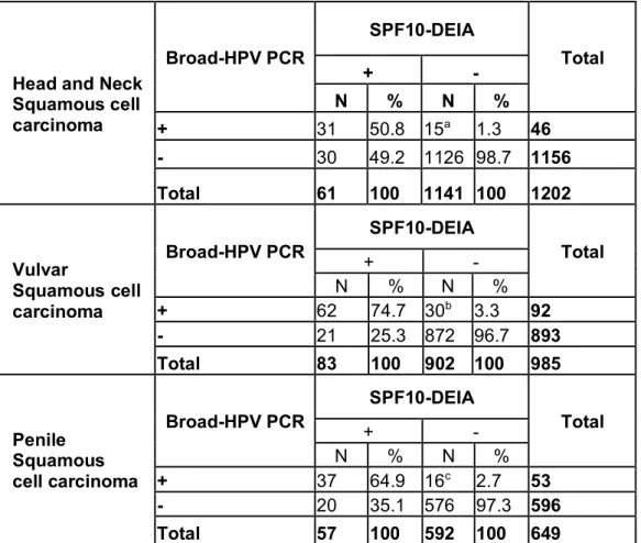
For squamous cancers of the HN, we were able to properly identify and genotype the HPV-DNA present in 31 out of 59 controls (53%) (Table 1). In all cases, the HPV hereby identified matched the one previously genotyped by LiPA25. We tested 1141 SPF10/DEIA-negative HN cancer samples, and we retrieved sequences specific for a one particular HPV in 15 out of these 1141 (1.3%) samples (Table 1, Table S2). In ten out of these 15 samples (66.7%), we detected DNA from mucosal HPVs, namely HPV16 (n=8), HPV51 (n=1) and HPV74 (n=1). Finally, we recovered DNA from two BetaHPVs, namely HPV20 and HPV21, and from three cutaneous AlphaPVs, namely HPV2, HPV57 and HPV61. Although ten from these 15 samples were expected to have tested positive for the initial SPF10/DEIA screening (i.e. those containing either HPV16, 51 or 74, targeted by the SPF10/DEIA) they resulted negative. When we performed a new SPF10/LiPA25 test, we recovered five of them (50%) as positive for HPV-DNA, matching in all cases the HPV genotype identified with the broad-spectrum primers, including HPV16 (n=3), HPV51 and HPV74.
For vulvar cancer samples, we were able to identify and genotype 62 out of 83 (75%) SPF10/DEIA-positive control samples. We tested further 902 SPF10/DEIA-negative samples and found HPV-DNA in 30 of them (3.3%) (Table 1, Table S3). One of the samples contained DNA of one cutaneous HPV, namely HPV2, while the remaining 29 samples contained DNA from essentially mucosal HPVs, namely HPV16 (n=18) HPV33 (n=1), HPV45 (n=2), HPV52 (n=1), HPV53 (n=1), HPV56 (n=1), HPV66 (n=1), HPV70 (n=1) and HPV74 (n=3). All these 29 samples should have tested positive for the initial SPF10/DEIA screening, and indeed 16 of them (55%) resulted positive when we performed a new SPF10/LiPA25 on them, the results matching those obtained with the broad primer sets. Specifically, we detected HPV16 (n=9), HPV33 (n=1), HPV45 (n=2), HPV52 (n=1), HPV70 (n=1) and HPV74 (n=2).
Finally, for penile cancer control samples, we successfully identified the presence of HPVs DNA in 37 out of 57 (65%). Among the 592 SPF10/DEIA-negative samples, we identified the presence of HPV DNA in 16 of them (2.7%), and in all cases the retrieved sequences corresponded to HPV16 (Table 1, Table S5). When we performed a new SPF10/LiPA25 genotyping on these 16 samples, four of them (25%) tested indeed positive and revealed the presence of HPV16 DNA.
8
DISCUSSION
We present here the largest study assessing the presence of DNA from cutaneous and mucosal HPVs in squamous cell carcinomas of the vulva, penis and HN, aiming at HPVs beyond those already identified as (probable/possible) oncogenic factors for these sites. We confirmed by multiple PCR tests the presence of DNA from a number of cutaneous HPVs in a very small number (6/2635) of cancer cases that seem to be truly negative for oncogenic HPVs. Nevertheless, it is important to keep in mind that the presence of viral DNA alone does not necessarily imply causation or relation to malignancy.
In the present study we have analyzed HPV-negative cancer cases of the HN, vulva and penis, previously tested by SPF10/DEIA/LiPA25 protocol. In HN samples, we detected HPV DNA in 15 out of 1141 (1.3%) samples previously diagnosed as HPV negative by SPF10/DEIA technology. Among them, five types corresponded to cutaneous HPVs: two belonging to BetaPVs (HPV20 and HPV21) and three belonging to AlphaPVs (HPV2, HPV57 and HPV61). All these samples came from the oral cavity except the HPV20, which was retrieved from a laryngeal cancer sample. In the same repository, we already confirmed the presence of two single infections by cutaneous HPVs (Table 3) (Castellsague et al., 2016). Studies on HPV prevalence demonstrated a high prevalence (more than 60%) of cutaneous HPVs in the oral cavity of healthy individuals (Bottalico et al., 2011; Lang Kuhs et al., 2013). Our results suggest that the contribution of these cutaneous types to cancer remains minor and may be neglected for screening and vaccination purposes. A prospective investigation (Agalliu et al., 2016) reported a positive association between infections with Beta and/or GammaPVs and increased head-and-neck cancer risk. Indeed, the authors reported that Beta1, Gamma11 and Gamma12 species were associated with 3.3 to 5.5-fold higher risk of HN carcinomas after adjusting for smoking, alcohol consumption and HPV16 detection. However, residual confounding cannot be ruled out as an explanation of these findings (Rollison and Gillison, 2016).
Other studies have also analyzed the presence of cutaneous HPVs in malignant head-and-neck tumours (Agalliu et al., 2016; Koskinen et al., 2003; Lindel et al., 2009; Paolini et al., 2012). Lindel and colleagues (Lindel et al., 2009) found a high prevalence of cutaneous HPVs in head-and-neck carcinomas samples (16/18 HPV-DNA positive tumours) by PCR amplification with multiple primers. The authors reported the presence of BetaPVs DNA as a single infection in eleven samples, and in two samples they found a multiple infection of two (or three) BetaPVs; finally, in three samples they found a multiple infection of a cutaneous BetaPV in combination with a low-risk
9
AlphaPV. Paolini and colleagues (Paolini et al., 2012) also demonstrated the presence of cutaneous BetaHPVs (HPV5, 14, 20, 21, 25, 36, 47, 100, 105 and 111) in HN squamous carcinomas samples (16/78 samples). However, they could not find cutaneous viral transcripts in any of these cancer samples.
We also analyzed penile SPF10/DEIA-negative cancer samples. Although in the analyzed samples, we did not find any cutaneous HPV type, the original description of the dataset already included the presence of cutaneous HPVs in two penile squamous carcinoma samples, namely HPV27 and HPV76 (Table3) (Alemany et al., 2016). Few published studies have examined the presence of cutaneous HPVs in penile carcinomas. Moreover, all have used varying methods and have focused on a single lesion type, making comparisons across studies difficult. By using PCR amplification with multiple primers, Wieland and colleagues (Wieland et al., 2000) described the presence of HPV8 (as single infection and as co-infection with a mucosal oncogenic type) in in situ squamous cell carcinomas of the penis (12/12 samples). In another study, the presence of HPV8 was found in penile squamous carcinoma samples, being that type the second most prevalent type (10/46 samples) as a single infection (6/10 samples) or as co-infection with a mucosal oncogenic type (4/10 samples) (Humbey et al., 2003). These authors also found other cutaneous BetaHPVs (HPV12, HPV17, HPV20 and HPV23) (Humbey et al., 2003).
Regarding vulvar squamous carcinomas, we detected the DNA of one cutaneous virus (HPV2) in one sample. De Koning and colleagues, by using PM-PCR reverse hybridization assay did not find any cutaneous HPV in a total of 39 vulvar cancer cases (de Koning et al., 2008). However, their study did not include squamous carcinoma samples in their set, making difficult the comparison of the results across the two studies.
Epidemiologic studies using standard methods of detection have demonstrated that other HPVs beyond high-risk HPVs can be involved in the malignization process (Guimera et al., 2013), although their prevalence is very low (Table 2). In the present study we describe the presence of HPV-DNA of cutaneous types in cancer samples. However, the mere presence of HPV-DNA in cancer samples is not sufficient to prove viral causation as it might simply reflect an ongoing viral infection unrelated to the carcinogenic process (Sarid and Gao, 2011). Thus, it is important to explore the expression patterns of other markers associated with HPV-induced carcinogenesis to assess the biological and oncogenic activities of HPVs identified in these cancers. Although the pathogenic pathway for malignant transformation in oncogenic AlphaPVs has been extensively studied (Bello et al., 2015; Duensing and Munger, 2004; Jones
10
and Munger, 1997; Moody and Laimins, 2010; Munger et al., 1989), the putative oncogenic mechanism of cutaneous HPVs remains unknown and may probably involves different molecular pathways than those well described for HPV16 and other oncogenic HPVs (Groves and Coleman, 2015; Rusan et al., 2015).
In SPF10/DEIA-negative samples, we detected the DNA of one of the 69 types detected by the SPF10/DEIA technology (Kleter et al., 1999). Among the 1,141 HN SPF10/DEIA-negative samples, ten samples contained an HPV type detected by DEIA. In five out of these ten samples, the type was confirmed by performing a LiPA25 analysis. In vulvar cancer samples 29/902 samples resulted positive for types detected by DEIA, and the LiPA25 confirmed the HPV type detected by PCRs methods in 16 of them. Finally, for penile samples the LiPA25 detected the HPV type reported by PCR in four out of 16/592 samples. Our results suggest that the SPF10/DEIA/LiPA25 methodology present very small rates of false-negative cases (ranging between .0.9-3.5%), which confirm its high sensitivity, especially for FFPE samples (Geraets et al., 2015).
The fraction of SPF10/DEIA-positive samples detected by the PCR methods used in this study ranged from 53% in HN to 65-75% in penile and vulvar cancer samples, respectively, demonstrating that our results might be underestimating the presence of cutaneous HPVs, as well the number of SPF10/DEIA false negative samples. Globally, the broad-spectrum primers sets used in this study are not optimized for retrieving small amplicons. Primer selection has been done taking into account the spectrum of the HPV types detected, in order to cover both mucosal and cutaneous HPVs; and their sensitivity in a single round assay. Using this primer selection, we aimed to covering both mucosal and cutaneous HPVs, detecting both E1 and L1 genes, with an amplicon range from 150bp to 450bp. Nevertheless, the amplicon length was larger than the 65bp targeted by the SPF10 primers (Kleter et al., 1998). This difference in amplicon size can easily explain the incompletely recovery of positive SPF10/DEIA samples.
Another technical factor that might also contribute to a hypothetical underestimation of the presence of cutaneous HPVs is the storage of the samples at -80ºC, which might compromise the DNA quality of the samples (Lee et al 2010). In order to minimize this effect we heated all the stored samples (including samples from the vulva and penis), during 48h at 60ºC. We found that this pretreatment may help recover DNA possibly adsorbed onto the plastic tube walls, as the positivity of control samples were higher in the localizations were the pretreatment were performed. However, this recovery
11
remained incomplete and still, samples with low DNA concentration may result in low DNA detection rates.
CONCLUSIONS
Our results show that previous estimates of HPVs involvement in vulvar, penile and HN cancers, based on SPF10/DEIA procedure do not suffer from significant underestimation biases. We show further that other HPVs beyond mucosal HPVs, typically detected by standard methods of detection may be present in carcinomas of the penis, vulva and HN, albeit their relative contribution remains minor and may have no public health impact whatsoever, and may be neglected for vaccination and cancer screening purposes. Nevertheless, our results brace previous reports with lower sample size, and mounting evidence suggests that certain cutaneous HPVs may be linked to a small number of cancer cases. Our results call for further studies to elucidate the pathogenic role of these “non-oncogenic” HPVs, the possible malignisation routes and mechanisms and the interplay between viral infection, host genetics and environment.
12
FUNDING
MFS is the recipient of an IDIBELL PhD scholarship. Partial support for this work was obtained from grants from the Instituto de Salud Carlos III (grants FIS PI081535, FIS PI1102096, FIS PI1102104, RCESP C03/09, RTICESP C03/10, RTIC RD06/0020/0095, RD12/0036/0056,), from the Agència de Gestió d’Ajuts Universitaris i de Recerca (2014SGR2016), from the Stichting Pathologie Ontwikkeling en Onderzoek (SPOO) foundation (the Netherlands), and from Sanofi Pasteur MSD and Merck & Co, Inc. None of the founders had any role in data analysis, interpretation, article writing or publishing.
Aknowledgements
We are grateful for the work of all the Institut Català d’Oncologia (ICO) team and the ICO International HPV in HN Cancer Study Group. The study is also part of the Human Papillomavirus Vulva, Vagina, Anus and Penis (HPV VVAP) international study coordinated at ICO, Barcelona, Spain.
13
BIBLIOGRAPHY
Agalliu, I., Gapstur, S., Chen, Z., Wang, T., Anderson, R.L., Teras, L.,
Kreimer, A.R., Hayes, R.B., Freedman, N.D., Burk, R.D., 2016. Associations
of Oral alpha-, beta-, and gamma-Human Papillomavirus Types With Risk
of Incident Head and Neck Cancer. JAMA Oncol.
Alemany, L., Cubilla, A., Halec, G., Kasamatsu, E., Quiros, B., Masferrer,
E., Tous, S., Lloveras, B., Hernandez-Suarez, G., Lonsdale, R., Tinoco, L.,
Alejo, M., Alvarado-Cabrero, I., Laco, J., Guimera, N., Poblet, E., Lombardi,
L.E., Bergeron, C., Clavero, O., Shin, H.R., Ferrera, A., Felix, A., Germar, J.,
Mandys, V., Clavel, C., Tzardi, M., Pons, L.E., Wain, V., Cruz, E., Molina, C.,
Mota, J.D., Jach, R., Velasco, J., Carrilho, C., Lopez-Revilla, R., Goodman,
M.T., Quint, W.G., Castellsague, X., Bravo, I., Pawlita, M., Munoz, N.,
Bosch, F.X., de Sanjose, S., 2016. Role of Human Papillomavirus in Penile
Carcinomas Worldwide. Eur Urol.
Alemany, L., Saunier, M., Alvarado-Cabrero, I., Quiros, B., Salmeron, J.,
Shin, H.R., Pirog, E.C., Guimera, N., Hernandez-Suarez, G., Felix, A.,
Clavero, O., Lloveras, B., Kasamatsu, E., Goodman, M.T., Hernandez, B.Y.,
Laco, J., Tinoco, L., Geraets, D.T., Lynch, C.F., Mandys, V., Poljak, M.,
Jach, R., Verge, J., Clavel, C., Ndiaye, C., Klaustermeier, J., Cubilla, A.,
Castellsague, X., Bravo, I.G., Pawlita, M., Quint, W.G., Munoz, N., Bosch,
F.X., de Sanjose, S., 2015. Human papillomavirus DNA prevalence and
type distribution in anal carcinomas worldwide. Int J Cancer.
Alemany, L., Saunier, M., Tinoco, L., Quiros, B., Alvarado-Cabrero, I.,
Alejo, M., Joura, E.A., Maldonado, P., Klaustermeier, J., Salmeron, J.,
Bergeron, C., Petry, K.U., Guimera, N., Clavero, O., Murillo, R., Clavel, C.,
Wain, V., Geraets, D.T., Jach, R., Cross, P., Carrilho, C., Molina, C., Shin,
H.R., Mandys, V., Nowakowski, A.M., Vidal, A., Lombardi, L., Kitchener, H.,
Sica, A.R., Magana-Leon, C., Pawlita, M., Quint, W., Bravo, I.G., Munoz, N.,
de Sanjose, S., Bosch, F.X., 2014. Large contribution of human
papillomavirus in vaginal neoplastic lesions: a worldwide study in 597
samples. Eur J Cancer 50, 2846-2854.
Antonsson, A., Forslund, O., Ekberg, H., Sterner, G., Hansson, B.G., 2000.
The ubiquity and impressive genomic diversity of human skin
papillomaviruses suggest a commensalic nature of these viruses. J Virol
74, 11636-11641.
Antonsson, A., Hansson, B.G., 2002. Healthy skin of many animal species
harbors papillomaviruses which are closely related to their human
counterparts. J Virol 76, 12537-12542.
Antonsson, A., McMillan, N.A., 2006. Papillomavirus in healthy skin of
Australian animals. J Gen Virol 87, 3195-3200.
Bello, J.O., Nieva, L.O., Paredes, A.C., Gonzalez, A.M., Zavaleta, L.R.,
Lizano, M., 2015. Regulation of the Wnt/beta-Catenin Signaling Pathway by
Human Papillomavirus E6 and E7 Oncoproteins. Viruses 7, 4734-4755.
Bernard, H.U., Burk, R.D., Chen, Z., van Doorslaer, K., zur Hausen, H., de
Villiers, E.M., 2010. Classification of papillomaviruses (PVs) based on 189
PV types and proposal of taxonomic amendments. Virology 401, 70-79.
Bottalico, D., Chen, Z., Dunne, A., Ostoloza, J., McKinney, S., Sun, C.,
Schlecht, N.F., Fatahzadeh, M., Herrero, R., Schiffman, M., Burk, R.D.,
2011. The oral cavity contains abundant known and novel human
14
papillomaviruses from the Betapapillomavirus and Gammapapillomavirus
genera. J Infect Dis 204, 787-792.
Bzhalava, D., Muhr, L.S., Lagheden, C., Ekstrom, J., Forslund, O., Dillner,
J., Hultin, E., 2014. Deep sequencing extends the diversity of human
papillomaviruses in human skin. Sci Rep 4, 5807.
Castellsague, X., Alemany, L., Quer, M., Halec, G., Quiros, B., Tous, S.,
Clavero, O., Alos, L., Biegner, T., Szafarowski, T., Alejo, M., Holzinger, D.,
Cadena, E., Claros, E., Hall, G., Laco, J., Poljak, M., Benevolo, M.,
Kasamatsu, E., Mehanna, H., Ndiaye, C., Guimera, N., Lloveras, B., Leon,
X., Ruiz-Cabezas, J.C., Alvarado-Cabrero, I., Kang, C.S., Oh, J.K.,
Garcia-Rojo, M., Iljazovic, E., Ajayi, O.F., Duarte, F., Nessa, A., Tinoco, L.,
Duran-Padilla, M.A., Pirog, E.C., Viarheichyk, H., Morales, H., Costes, V., Felix, A.,
Germar, M.J., Mena, M., Ruacan, A., Jain, A., Mehrotra, R., Goodman, M.T.,
Lombardi, L.E., Ferrera, A., Malami, S., Albanesi, E.I., Dabed, P., Molina, C.,
Lopez-Revilla, R., Mandys, V., Gonzalez, M.E., Velasco, J., Bravo, I.G.,
Quint, W., Pawlita, M., Munoz, N., Sanjose, S., Xavier Bosch, F., 2016. HPV
Involvement in Head and Neck Cancers: Comprehensive Assessment of
Biomarkers in 3680 Patients. J Natl Cancer Inst 108.
D'Souza, G., Kreimer, A.R., Viscidi, R., Pawlita, M., Fakhry, C., Koch, W.M.,
Westra, W.H., Gillison, M.L., 2007. Case-control study of human
papillomavirus and oropharyngeal cancer. N Engl J Med 356, 1944-1956.
de Koning, M.N., Quint, W.G., Pirog, E.C., 2008. Prevalence of mucosal and
cutaneous human papillomaviruses in different histologic subtypes of
vulvar carcinoma. Mod Pathol 21, 334-344.
de Roda Husman, A.M., Walboomers, J.M., van den Brule, A.J., Meijer,
C.J., Snijders, P.J., 1995. The use of general primers GP5 and GP6
elongated at their 3' ends with adjacent highly conserved sequences
improves human papillomavirus detection by PCR. J Gen Virol 76 ( Pt 4),
1057-1062.
de Sanjose, S., Alemany, L., Ordi, J., Tous, S., Alejo, M., Bigby, S.M.,
Joura, E.A., Maldonado, P., Laco, J., Bravo, I.G., Vidal, A., Guimera, N.,
Cross, P., Wain, G.V., Petry, K.U., Mariani, L., Bergeron, C., Mandys, V.,
Sica, A.R., Felix, A., Usubutun, A., Seoud, M., Hernandez-Suarez, G.,
Nowakowski, A.M., Wilson, G., Dalstein, V., Hampl, M., Kasamatsu, E.S.,
Lombardi, L.E., Tinoco, L., Alvarado-Cabrero, I., Perrotta, M., Bhatla, N.,
Agorastos, T., Lynch, C.F., Goodman, M.T., Shin, H.R., Viarheichyk, H.,
Jach, R., Cruz, M.O., Velasco, J., Molina, C., Bornstein, J., Ferrera, A.,
Domingo, E.J., Chou, C.Y., Banjo, A.F., Castellsague, X., Pawlita, M.,
Lloveras, B., Quint, W.G., Munoz, N., Bosch, F.X., 2013. Worldwide human
papillomavirus genotype attribution in over 2000 cases of intraepithelial
and invasive lesions of the vulva. Eur J Cancer 49, 3450-3461.
de Sanjose, S., Quint, W.G., Alemany, L., Geraets, D.T., Klaustermeier,
J.E., Lloveras, B., Tous, S., Felix, A., Bravo, L.E., Shin, H.R., Vallejos, C.S.,
de Ruiz, P.A., Lima, M.A., Guimera, N., Clavero, O., Alejo, M.,
Llombart-Bosch, A., Cheng-Yang, C., Tatti, S.A., Kasamatsu, E., Iljazovic, E., Odida,
M., Prado, R., Seoud, M., Grce, M., Usubutun, A., Jain, A., Suarez, G.A.,
Lombardi, L.E., Banjo, A., Menendez, C., Domingo, E.J., Velasco, J.,
Nessa, A., Chichareon, S.C., Qiao, Y.L., Lerma, E., Garland, S.M.,
Sasagawa, T., Ferrera, A., Hammouda, D., Mariani, L., Pelayo, A., Steiner,
I., Oliva, E., Meijer, C.J., Al-Jassar, W.F., Cruz, E., Wright, T.C., Puras, A.,
15
Llave, C.L., Tzardi, M., Agorastos, T., Garcia-Barriola, V., Clavel, C., Ordi,
J., Andujar, M., Castellsague, X., Sanchez, G.I., Nowakowski, A.M.,
Bornstein, J., Munoz, N., Bosch, F.X., 2010. Human papillomavirus
genotype attribution in invasive cervical cancer: a retrospective
cross-sectional worldwide study. Lancet Oncol 11, 1048-1056.
Doorbar, J., Quint, W., Banks, L., Bravo, I.G., Stoler, M., Broker, T.R.,
Stanley, M.A., 2012. The biology and life-cycle of human papillomaviruses.
Vaccine 30 Suppl 5, F55-70.
Duensing, S., Munger, K., 2004. Mechanisms of genomic instability in
human cancer: insights from studies with human papillomavirus
oncoproteins. Int J Cancer 109, 157-162.
Forman, D., de Martel, C., Lacey, C.J., Soerjomataram, I., Lortet-Tieulent,
J., Bruni, L., Vignat, J., Ferlay, J., Bray, F., Plummer, M., Franceschi, S.,
2012. Global burden of human papillomavirus and related diseases.
Vaccine 30 Suppl 5, F12-23.
Forslund, O., Antonsson, A., Edlund, K., van den Brule, A.J., Hansson,
B.G., Meijer, C.J., Ryd, W., Rylander, E., Strand, A., Wadell, G., Dillner, J.,
Johansson, B., 2002. Population-based type-specific prevalence of
high-risk human papillomavirus infection in middle-aged Swedish women. J
Med Virol 66, 535-541.
Forslund, O., Antonsson, A., Nordin, P., Stenquist, B., Hansson, B.G.,
1999. A broad range of human papillomavirus types detected with a
general PCR method suitable for analysis of cutaneous tumours and
normal skin. J Gen Virol 80 ( Pt 9), 2437-2443.
Fox, P.A., Seet, J.E., Stebbing, J., Francis, N., Barton, S.E., Strauss, S.,
Allen-Mersh, T.G., Gazzard, B.G., Bower, M., 2005. The value of anal
cytology and human papillomavirus typing in the detection of anal
intraepithelial neoplasia: a review of cases from an anoscopy clinic. Sex
Transm Infect 81, 142-146.
Frisch, M., Glimelius, B., van den Brule, A.J., Wohlfahrt, J., Meijer, C.J.,
Walboomers, J.M., Goldman, S., Svensson, C., Adami, H.O., Melbye, M.,
1997. Sexually transmitted infection as a cause of anal cancer. N Engl J
Med 337, 1350-1358.
Garcia-Perez, R., Ibanez, C., Godinez, J.M., Arechiga, N., Garin, I.,
Perez-Suarez, G., de Paz, O., Juste, J., Echevarria, J.E., Bravo, I.G., 2014. Novel
papillomaviruses in free-ranging Iberian bats: no virus-host co-evolution,
no strict host specificity, and hints for recombination. Genome Biol Evol
6, 94-104.
Geraets, D.T., Struijk, L., Kleter, B., Molijn, A., van Doorn, L.J., Quint, W.G.,
Colau, B., 2015. The original SPF10 LiPA25 algorithm is more sensitive
and suitable for epidemiologic HPV research than the SPF10 INNO-LiPA
Extra. J Virol Methods 215-216, 22-29.
Giuliano, A.R., Papenfuss, M., Abrahamsen, M., Denman, C., de Zapien,
J.G., Henze, J.L., Ortega, L., Brown de Galaz, E.M., Stephan, J., Feng, J.,
Baldwin, S., Garcia, F., Hatch, K., 2001. Human papillomavirus infection at
the United States-Mexico border: implications for cervical cancer
prevention and control. Cancer Epidemiol Biomarkers Prev 10, 1129-1136.
Gottschling, M., Goker, M., Kohler, A., Lehmann, M.D., Stockfleth, E.,
Nindl, I., 2009. Cutaneotropic human beta-/gamma-papillomaviruses are
rarely shared between family members. J Invest Dermatol 129, 2427-2434.
16
Gravitt, P.E., Peyton, C.L., Alessi, T.Q., Wheeler, C.M., Coutlee, F.,
Hildesheim, A., Schiffman, M.H., Scott, D.R., Apple, R.J., 2000. Improved
amplification of genital human papillomaviruses. J Clin Microbiol 38,
357-361.
Groves, I.J., Coleman, N., 2015. Pathogenesis of human
papillomavirus-associated mucosal disease. J Pathol 235, 527-538.
Guimera, N., Lloveras, B., Lindeman, J., Alemany, L., van de Sandt, M.,
Alejo, M., Hernandez-Suarez, G., Bravo, I.G., Molijn, A., Jenkins, D.,
Cubilla, A., Munoz, N., de Sanjose, S., Bosch, F.X., Quint, W., 2013. The
occasional role of low-risk human papillomaviruses 6, 11, 42, 44, and 70 in
anogenital carcinoma defined by laser capture microdissection/PCR
methodology: results from a global study. Am J Surg Pathol 37,
1299-1310.
Hampl, M., Sarajuuri, H., Wentzensen, N., Bender, H.G., Kueppers, V.,
2006. Effect of human papillomavirus vaccines on vulvar, vaginal, and
anal intraepithelial lesions and vulvar cancer. Obstet Gynecol 108,
1361-1368.
Hampl, M., Wentzensen, N., Vinokurova, S., von Knebel-Doeberitz, M.,
Poremba, C., Bender, H.G., Kueppers, V., 2007. Comprehensive analysis of
130 multicentric intraepithelial female lower genital tract lesions by HPV
typing and p16 expression profile. J Cancer Res Clin Oncol 133, 235-245.
Humbey, O., Cairey-Remonnay, S., Guerrini, J.S., Algros, M.P., Mougin, C.,
Bittard, H., Aubin, F., 2003. Detection of the human papillomavirus and
analysis of the TP53 polymorphism of exon 4 at codon 72 in penile
squamous cell carcinomas. Eur J Cancer 39, 684-690.
IARC, 2007. Monographs on the evaluation of carcinogenic risk to
humans. Human Papillomavirus. Lyon, IARC 90.
Jones, D.L., Munger, K., 1997. Analysis of the p53-mediated G1 growth
arrest pathway in cells expressing the human papillomavirus type 16 E7
oncoprotein. J Virol 71, 2905-2912.
Kleter, B., van Doorn, L.J., Schrauwen, L., Molijn, A., Sastrowijoto, S., ter
Schegget, J., Lindeman, J., ter Harmsel, B., Burger, M., Quint, W., 1999.
Development and clinical evaluation of a highly sensitive PCR-reverse
hybridization line probe assay for detection and identification of
anogenital human papillomavirus. J Clin Microbiol 37, 2508-2517.
Kleter, B., van Doorn, L.J., ter Schegget, J., Schrauwen, L., van Krimpen,
K., Burger, M., ter Harmsel, B., Quint, W., 1998. Novel short-fragment PCR
assay for highly sensitive broad-spectrum detection of anogenital human
papillomaviruses. Am J Pathol 153, 1731-1739.
Koskinen, W.J., Chen, R.W., Leivo, I., Makitie, A., Back, L., Kontio, R.,
Suuronen, R., Lindqvist, C., Auvinen, E., Molijn, A., Quint, W.G., Vaheri, A.,
Aaltonen, L.M., 2003. Prevalence and physical status of human
papillomavirus in squamous cell carcinomas of the head and neck. Int J
Cancer 107, 401-406.
Lang Kuhs, K.A., Gonzalez, P., Struijk, L., Castro, F., Hildesheim, A., van
Doorn, L.J., Rodriguez, A.C., Schiffman, M., Quint, W., Lowy, D.R., Porras,
C., Delvecchio, C., Katki, H.A., Jimenez, S., Safaeian, M., Schiller, J.,
Solomon, D., Wacholder, S., Herrero, R., Kreimer, A.R., 2013. Prevalence
of and risk factors for oral human papillomavirus among young women in
Costa Rica. J Infect Dis 208, 1643-1652.
17
Li, J., Pan, Y., Xu, Z., Wang, Q., Hang, D., Shen, N., Liu, M., Zhang, C.,
Abliz, A., Deng, Q., Cai, H., Ke, Y., 2013. Improved detection of human
papillomavirus harbored in healthy skin with FAP6085/64 primers. J Virol
Methods 193, 633-638.
Lindel, K., Helmke, B., Simon, C., Weber, K.J., Debus, J., de Villiers, E.M.,
2009. Cutaneous human papillomavirus in head and neck squamous cell
carcinomas. Cancer Invest 27, 781-787.
Madsen, B.S., Jensen, H.L., van den Brule, A.J., Wohlfahrt, J., Frisch, M.,
2008. Risk factors for invasive squamous cell carcinoma of the vulva and
vagina--population-based case-control study in Denmark. Int J Cancer
122, 2827-2834.
Manos, M.M., Ting, Y., Wright, D.K., Lewis, A.J., Broker, T.R., Wolinsky,
S.M., 1989. The use of polymerase chain reaction amplification for the
detection of genital human papillomaviruses. Cancer Cell 7, 209-214.
Mengual-Chulia, B., Bedhomme, S., Lafforgue, G., Elena, S.F., Bravo, I.G.,
2016. Assessing parallel gene histories in viral genomes. BMC Evol Biol
16, 32.
Moody, C.A., Laimins, L.A., 2010. Human papillomavirus oncoproteins:
pathways to transformation. Nat Rev Cancer 10, 550-560.
Munger, K., Phelps, W.C., Bubb, V., Howley, P.M., Schlegel, R., 1989. The
E6 and E7 genes of the human papillomavirus type 16 together are
necessary and sufficient for transformation of primary human
keratinocytes. J Virol 63, 4417-4421.
Paolini, F., Rizzo, C., Sperduti, I., Pichi, B., Mafera, B., Rahimi, S.S., Vigili,
M.G., Venuti, A., 2012. Both mucosal and cutaneous papillomaviruses are
in the oral cavity but only alpha genus seems to be associated with
cancer. J Clin Virol 56, 72-76.
Piketty, C., Darragh, T.M., Da Costa, M., Bruneval, P., Heard, I.,
Kazatchkine, M.D., Palefsky, J.M., 2003. High prevalence of anal human
papillomavirus infection and anal cancer precursors among HIV-infected
persons in the absence of anal intercourse. Ann Intern Med 138, 453-459.
Richardson, H., Abrahamowicz, M., Tellier, P.P., Kelsall, G., du Berger, R.,
Ferenczy, A., Coutlee, F., Franco, E.L., 2005. Modifiable risk factors
associated with clearance of type-specific cervical human papillomavirus
infections in a cohort of university students. Cancer Epidemiol
Biomarkers Prev 14, 1149-1156.
Richardson, H., Kelsall, G., Tellier, P., Voyer, H., Abrahamowicz, M.,
Ferenczy, A., Coutlee, F., Franco, E.L., 2003. The natural history of
type-specific human papillomavirus infections in female university students.
Cancer Epidemiol Biomarkers Prev 12, 485-490.
Rollison, D.E., Gillison, M.L., 2016. The Alpha, Beta, Gammas of Oral
Human Papillomavirus Infection and Head and Neck Cancer Risk. JAMA
Oncol.
Rusan, M., Li, Y.Y., Hammerman, P.S., 2015. Genomic landscape of human
papillomavirus-associated cancers. Clin Cancer Res 21, 2009-2019.
Sarid, R., Gao, S.J., 2011. Viruses and human cancer: from detection to
causality. Cancer Lett 305, 218-227.
Sasagawa, T., Mitsuishi, T., 2012. Novel polymerase chain reaction
method for detecting cutaneous human papillomavirus DNA. J Med Virol
84, 138-144.
18
Schiffman, M., Wheeler, C.M., Dasgupta, A., Solomon, D., Castle, P.E.,
2005. A comparison of a prototype PCR assay and hybrid capture 2 for
detection of carcinogenic human papillomavirus DNA in women with
equivocal or mildly abnormal papanicolaou smears. Am J Clin Pathol 124,
722-732.
Skapa, P., Zamecnik, J., Hamsikova, E., Salakova, M., Smahelova, J.,
Jandova, K., Robova, H., Rob, L., Tachezy, R., 2007. Human
papillomavirus (HPV) profiles of vulvar lesions: possible implications for
the classification of vulvar squamous cell carcinoma precursors and for
the efficacy of prophylactic HPV vaccination. Am J Surg Pathol 31,
1834-1843.
Snijders, P.J., van den Brule, A.J., Schrijnemakers, H.F., Snow, G., Meijer,
C.J., Walboomers, J.M., 1990. The use of general primers in the
polymerase chain reaction permits the detection of a broad spectrum of
human papillomavirus genotypes. J Gen Virol 71 ( Pt 1), 173-181.
Tabrizi, S.N., Stevens, M., Chen, S., Rudland, E., Kornegay, J.R., Garland,
S.M., 2005. Evaluation of a modified reverse line blot assay for detection
and typing of human papillomavirus. Am J Clin Pathol 123, 896-899.
Tieben, L.M., ter Schegget, J., Minnaar, R.P., Bouwes Bavinck, J.N.,
Berkhout, R.J., Vermeer, B.J., Jebbink, M.F., Smits, H.L., 1993. Detection
of cutaneous and genital HPV types in clinical samples by PCR using
consensus primers. J Virol Methods 42, 265-279.
Wieland, U., Jurk, S., Weissenborn, S., Krieg, T., Pfister, H., Ritzkowsky,
A., 2000. Erythroplasia of queyrat: coinfection with cutaneous
carcinogenic human papillomavirus type 8 and genital papillomaviruses in
a carcinoma in situ. J Invest Dermatol 115, 396-401.
19
TABLES
Table 1: Concordance between SPF10-DEIA and HPV broad-spectrum PCR (CPI/II, SKF/R, FAP6064/64, MY09/11 and MFI/II) in Head-and-neck (N=1200), Penile (N=649) and Vulvar (N=985) squamous cell carcinoma samples.
Head and Neck Squamous cell carcinoma Broad-HPV PCR SPF10-DEIA Total + - N % N % + 31 50.8 15a 1.3 46 - 30 49.2 1126 98.7 1156 Total 61 100 1141 100 1202 Vulvar Squamous cell carcinoma Broad-HPV PCR SPF10-DEIA Total + - N % N % + 62 74.7 30b 3.3 92 - 21 25.3 872 96.7 893 Total 83 100 902 100 985 Penile Squamous cell carcinoma Broad-HPV PCR SPF10-DEIA Total + - N % N % + 37 64.9 16c 2.7 53 - 20 35.1 576 97.3 596 Total 57 100 592 100 649
a HPVs detected: HPV20 (n=1, BetaPV), HPV21 (n=1, BetaPV), HPV2 (n=1, AlphaPV), HPV57
(n=1, AlphaPV), HPV61 (n=1, AlphaPV), HPV16 (n=8, AlphaPV), HPV51 (n=1, AlphaPVs), HPV74 (n=1, AlphaPVs).
b HPVs detected: HPV2 (n=1, AlphaPVs), HPV16 (n=18, AlphaPVs) HPV33 (n=1, AlphaPVs), HPV45 (n=2,
AlphaPVs), HPV52 (n=1, , AlphaPV), HPV53 (n=1, AlphaPV), HPV56 (n=1, AlphaPV), HPV66 (n=1, AlphaPV), HPV70 (n=1, AlphaPVs) and HPV74 (n=3, , AlphaPVs).
20
Table 2: HPV prevalence distribution found in anogenital and head and neck cancers. Information was obtained from the larger cross-sectional study on HPV prevalence distribution coordinated by the Catalan Institute of Oncology [9-14]. HPVs were classified according their phylogeny and clinical presentation: Mucosal oncogenic AlphaPVs, Mucocutaneous AlphaPVs causing genital warts, AlphaPVs causing cutaneous warts, Asymptomatic cutaneous BetaPVs and Asymptomatic GammaPVs.
Genus Clinical presentation
Percentage (%) Cervix (de Sanjosé et al. 2010) Vulva (de Sanjosé et al. 2013) Vagina (Alemany et al. 2013) HN (Castellsague et al. 2016) Penis (Alemany et al. 2016) Anus (Alemany et al. 2015) AlphaPVs Mucosal oncogenica 98.39 96.61 91.57 94.28 89.16 94.25 Mucocutaneous, genital wartsb 1.09 3.39 4.42 5.30 8.73 2.65 Cutaneous wartsc 0.01 0.00 0.40 0.00 0.30 0.44
BetaPVs Asymptomatic, Cutaneous 0.00 0.00 0.00 0.00 0.00 0.22
GammaPVs Asymptomatic, Cutaneous
0.00 0.00 0.00 0.00 0.00 0.00
Undetermined
0.50 0.00 3.61 0.42 1.81 2.65
aMucosal oncogenic AlphaPVs: AlphaPV species 5, AlphaPVs species 6, AlphaPVs species7, AlphaPVs species 9 and AlphaPVs species 11 bMucocutaneous genital warts AlphaPVs: AlphaPVs species 1, AlphaPVs species 8 and AlphaPVs species10
21
Table 3: Number of single HPV infections found in head and neck, penile and vulvar carcinomas. For each anatomical location, the first column corresponds to the number of single HPV infections found by SPF10-DEIA-LIPA25 methodology [12-14]. The second column corresonds to the number of single HPV infections found in the negative SPF10/DEIA/LIPA25 samples from the corresponding repository in the same anatomical location.
22
Squamous carcinoma
Head and Neck Penis Vulva
Clinical
presentation Specie Type
Castellsagué et al. 2016 (original n=3680) This study (retesting = 1141 previously negative) Alemany et al. 2016 (original n=1010) This study (retesting =592 previously negative) de Sanjosé et al. 2013 (original n=1709) This study (retesting =902 previously negative) Mu co sa l l es io n s, o n co g en ic p o te n ti al Alpha-5 HPV26 8 0 2 0 1 0 HPV51 4 1 2 0 2 0 HPV70 0 0 0 0 1 1 Alpha-6 HPV30 1 0 2 0 1 0 HPV53 2 0 2 0 3 1 HPV56 1 0 2 0 6 1 HPV66 1 0 2 0 1 1 Alpha-7 HPV18 9 0 4 0 17 0 HPV39 5 0 2 0 3 0 HPV45 6 0 9 0 13 2 HPV59 1 0 4 0 0 0 HPV68 3 0 1 0 2 0 Alpha-9 HPV16 339 8 194 16 318 18 HPV31 2 0 2 0 5 0 HPV33 11 0 2 0 25 1 HPV35 11 0 9 0 1 0 HPV52 5 0 4 0 8 1 HPV58 4 0 3 0 3 0 HPV67 2 0 0 0 0 0 Alpha-11 HPV73 0 0 0 0 1 0 Total 415 (97.19%) 9 (60%) 246(91.45%) 16 (100%) 411(96.93%) 26 (86.67%) Us u al ly b en ig n , p ro d u ct iv e mu co sa l l es io n s Alpha-1 HPV32 0 0 2 0 0 0 HPV42 0 0 1 0 0 0 Alpha-8 HPV40 0 0 1 0 0 0 HPV43 0 0 1 0 0 0 Alpha-10 HPV6 5 0 9 0 5 0 HPV11 2 0 4 0 2 0 HPV13 3 0 0 0 0 0 HPV74 0 1 2 0 3 3 Total 10 (2.34%) 1 (6.67%) 20 (7.43%) 0 (0.00%) 10 (2.36%) 3 (10.00%) Us u al ly b en ig n , pr oduc ti ve cu tan eo u s le si o n s Alpha-3 HPV61 0 1 0 0 2 0 HPV102 0 0 0 0 1 0 Alpha-4 HPV2 0 1 0 0 0 1 HPV27 0 0 2 0 0 0 HPV57 0 1 0 0 0 0 Alpha-14 HPV90 1 0 0 0 0 0 Total 1 (0.23%) 3 (20.00%) 2 (0.74%) 0 (0.00%) 3 (0.71%) 1 (3.33%) Us u al ly As ym p to ma ti c Cu ta n eo us Beta-1 HPV19 1 0 0 0 0 0 HPV20 0 1 0 0 0 0 HPV21 0 1 0 0 0 0 Beta-3 HPV76 0 0 1 0 0 0 Total 1 (0.23%) 2 (13.33%) 1 (0.37%) 0 (0.00%) 0 (0.00%) 0 (0.00%)