HAL Id: inserm-02264254
https://www.hal.inserm.fr/inserm-02264254
Submitted on 6 Aug 2019
HAL is a multi-disciplinary open access archive for the deposit and dissemination of sci-entific research documents, whether they are pub-lished or not. The documents may come from teaching and research institutions in France or abroad, or from public or private research centers.
L’archive ouverte pluridisciplinaire HAL, est destinée au dépôt et à la diffusion de documents scientifiques de niveau recherche, publiés ou non, émanant des établissements d’enseignement et de recherche français ou étrangers, des laboratoires publics ou privés.
reproductive outcomes in IVF with oral dydrogesterone
for luteal phase support?
Antoine Netter, Julien Mancini, Christophe Buffat, Aubert Agostini, Jeanne
Perrin, Blandine Courbiere
To cite this version:
Antoine Netter, Julien Mancini, Christophe Buffat, Aubert Agostini, Jeanne Perrin, et al.. Do early luteal serum progesterone levels predict the reproductive outcomes in IVF with oral dydrogesterone for luteal phase support?. PLoS ONE, Public Library of Science, 2019, 14 (7), pp.e0220450. �10.1371/jour-nal.pone.0220450�. �inserm-02264254�
Do early luteal serum progesterone levels
predict the reproductive outcomes in IVF with
oral dydrogesterone for luteal phase support?
Antoine NetterID1*, Julien ManciniID2, Christophe Buffat3, Aubert Agostini1,Jeanne Perrin1,4, Blandine Courbiere1,4
1 Department of Gynecology, Obstetrics and Reproductive Medicine, AP-HM La Conception, Poˆle femmes parents enfants, Marseille, France, 2 Aix-Marseille University, APHM, INSERM, IRD, SESSTIM, Public Health Department, La Timone Hospital, BIOSTIC, Marseille, France, 3 Laboratoire de Biochimie et de Biologie Mole´culaire, Hoˆpital de la Conception, Marseille, France, 4 Institut Me´diterrane´en de Biodiversite´ et d’E´ cologie marine et continentale (IMBE), Aix Marseille University, CNRS, IRD, Avignon University, Marseille, France
*antoine.netter@gmail.com
Abstract
Objective
We sought to determine whether the early luteal serum progesterone (P4) level predicts the
success of IVF treatment with oral dydrogesterone for luteal support.
Method
This retrospective monocentric cohort study included 242 women who underwent IVF treat-ment with fresh embryo transfer (ET) between July 2017 and June 2018. The population was unselected, and women were treated according to our unit’s usual stimulation proto-cols. For the luteal phase support (LPS), all women were supplemented with a 10 mg three-times-daily dose of oral dydrogesterone beginning on the day of oocyte pick-up (OPU). Blood sampling was performed on the day of ET (Day 2–3 after OPU) to determine the early luteal serum progesterone level.
Results
ROC curve analysis allowed us to determine two thresholds for the prediction of live birth using the early P4level. Women who had early luteal P4levels greater than 252 nmol/l had a
significantly higher live birth rate (27.1%) than women with early luteal P4between 115 and
252 nmol/l (17.2%) and women with early luteal P4below 115 nmol/l (6.0%; p = 0.011). After
a multiple regression analysis, an early luteal P4level greater than 252 nmol/l was still
asso-ciated with a higher chance of a live birth than a P4between 115 and 252 nmol/l (OR = 0.40
[0.18–0.91]; p = 0.028) or a P4below 115 nmol/l (OR = 0.10 [0.01–0.52]; p = 0.006).
Conclusions
Our study suggests a positive association between early P4levels and reproductive
out-comes in IVF using oral dydrogesterone for luteal support. The inconsistencies between our a1111111111 a1111111111 a1111111111 a1111111111 a1111111111 OPEN ACCESS
Citation: Netter A, Mancini J, Buffat C, Agostini A,
Perrin J, Courbiere B (2019) Do early luteal serum progesterone levels predict the reproductive outcomes in IVF with oral dydrogesterone for luteal phase support? PLoS ONE 14(7): e0220450.
https://doi.org/10.1371/journal.pone.0220450
Editor: Dragana Nikitovic, University of Crete,
GREECE
Received: May 26, 2019 Accepted: July 16, 2019 Published: July 30, 2019
Copyright:© 2019 Netter et al. This is an open access article distributed under the terms of the
Creative Commons Attribution License, which permits unrestricted use, distribution, and reproduction in any medium, provided the original author and source are credited.
Data Availability Statement: The underlying data
set has been publicly released via the following DOI:10.6084/m9.figshare.8187413(doi.org/10. 6084/m9.figshare.8187413).
Funding: The authors received no specific funding
for this work.
Competing interests: The authors have declared
results and those of other studies suggest that extrapolation is impractical. Further larger prospective cohort studies should be conducted to determine reliable thresholds that could be used to personalize luteal phase support.
Introduction
Over the past decades, increasing efforts have been made to identify best practices for con-trolled ovarian stimulation (COS) and embryo culture for In Vitro Fertilization (IVF) treat-ment [1]. In contrast, the luteal phase subsequent to the COS and oocyte retrieval have received less interest for a long time [2]. By modifying gene expression, progesterone guides the endometrial secretory transformation and therefore plays a fundamental role in implanta-tion and early embryologic development [3,4]. After COS, the endogenous secretion of proges-terone by the granulosa of the corpus luteum is insufficient [5]. The luteolytic effect of the GnRH analogs or GnRH antagonists used for the COS, inhibition of the hypothalamic-pitui-tary axis by the supra-physiological secretion of steroids, and aspiration of the granulosa cells during oocyte retrieval are the main hypotheses for this inadequate secretion [6]. Luteal phase support (LPS) compensates for this deficiency by supplementing with either progesterone or an hCG or GnRH agonist [7].
In recent years, clinical investigators have shown a renewed interest in the luteal phase sub-sequent to ART treatments. Evidence is accumulating regarding the existence of an optimal range of P4levels on the day of embryo transfer (ET) in frozen embryo transfer cycles with
hormone replacement therapy [8–10]. In 2018, Thomsen et al. published the results of a large prospective cohort study of 602 women who underwent IVF treatment and had serum P4
lev-els measured on the day of ET [11]. The authors determined that very low P4levels (< 60
nmol/l) on the day of fresh ET (Day 2–3 after oocyte pick-up (OPU)) were associated with decreased chance of success of the IVF treatment. This result allows us to consider a possible value of early luteal P4monitoring and luteal rescue by increasing the P4supplementation.
Thomsen et al. also reported that high P4levels (> 400 nmol/l) were associated with a
decreased chance of live birth after IVF treatment. If confirmed by other studies, these thresh-olds could encourage the cancellation of ET for women with high early luteal P4levels.
However, concerns have been raised regarding the statistical analysis of the aforementioned study [12], and further studies are needed to assess the reproducibility of the suggested thresh-olds before they can be used in common practice, in particular with different COS and LPS protocols.
A recent large phase III RCT demonstrated the noninferiority of oral dydrogesterone com-pared with micronized vaginal progesterone for LPS in terms of pregnancy rates and tolerabil-ity [13]. Thus, dydrogesterone was recently approved for LPS in IVF [14]. Furthermore, given the assumed preference for the oral route over vaginal route, dydrogesterone is presumed to become the new standard for LPS in IVF treatment according to some authors [15]. The early luteal phase P4levels have never been studied in the context of the use of dydrogesterone for
LPS [16].
Throughout this retrospective data analysis, we aimed to determine if early luteal P4levels
(on the day of fresh ET) could allow us to predict the success of IVF on an unselected popula-tion treated with oral dydrogesterone for LPS.
Materials and methods
Study design
We conducted a monocentric retrospective cohort study. Data were retrospectively and anon-ymously collected. Before the study began, it was approved by the ethics committee of Aix Marseille University. All patients were informed of the anonymous and retrospective use of their data and could refuse to participate by simple notification.
Study population
We enrolled couples who underwent IVF or ICSI treatments in our ART unit between July 2017 and June 2018 and who had early luteal phase serum P4levels measured on the day of
fresh ET (Day 2–3 after OPU). Women were eligible for inclusion regardless of the reason for the infertility or the number of previous unsuccessful IVF or ICSI cycles.
Ovarian stimulation protocols
The choices of COS protocols and gonadotrophin doses were made in accordance with the standards of our ART unit based on age, BMI, ovarian reserve, outcomes of previous IVF or ICSI cycles, and other comorbidities such as endometriosis or polycystic ovary syndrome (PCOS). GnRH antagonist protocols were generally used as the first choice, especially for patients with an expected low ovarian response and for women with PCOS; in contrast, GnRH long agonist protocols were used for patients with endometriosis or as a second choice for patients with an inadequate response to the previous GnRH antagonist protocol. Short GnRH agonist protocols were used for low responders who had already experienced several failures after previous stimulations.
Patients who were treated in a long GnRH-agonist protocol were downregulated using a unique IM injection of 3 mg triptorelin (Decapeptyl, Ipsen Pharma, France) on the 22ndday of the preceding cycle. Ovarian stimulation started after 14 days of downregulation after checking that the endometrial thickness was less than 4 mm. Final follicular maturation was induced by a single injection of 250μg of choriogonadotropin alpha (Ovitrelle, Merck Serono Europe Limited, United Kingdom) when two or more leading follicles reached a mean diameter of 17 mm.
If the GnRH antagonist protocol was used, ovarian stimulation started on cycle Day 2. Daily GnRH antagonist cotreatment was added from cycle Day 6. Final maturation could be induced by a single injection of 250μg of choriogonadotropin alpha (Ovitrelle, Merck Serono Europe Limited, United Kingdom) or by a dual trigger (250μg of choriogonadotropin alpha plus 0.3 mg of Triptorelin) when two or more leading follicles reached a mean diameter of 17 mm.
Ovarian stimulation was performed using either r-FSH, r-FSH/r-LH or hMG. Dose adjust-ments were performed according to ovarian response, as monitored by transvaginal ultra-sound during treatment. OPU was performed 36 h after the ovulation trigger. IVF or ICSI was performed according to normal clinical practice.
Luteal phase support
All patients received a 10 mg three-times-daily dose of oral dydrogesterone (Duphaston, Mylan Medical, France) starting on the day of OPU [14,15]. We systematically checked with the patients’ compliance with this treatment on the day of ET. No additional treatment (nei-ther hCG nor GnRH agonist bolus) was ever used during the luteal phase. Oral dydrogesterone
administration continued from the day of OPU until the day of pregnancy testing or until the seventh gestational week (in the case of pregnancy).
Hormone analysis
On day 2–3 after OPU, immediately before ET, blood samples were collected for P4and
estra-diol determination. We used an automated Cobas e411 instrument (Roche Diagnostics, Mann-heim, Germany) and the same assays for all hormone measurements during the entire study. Samples were tested by an electrochemiluminescence immunoassay for Progesterone III (Cobas 07092539 190). The intra- and inter-assay variation coefficients for the P determina-tions were 3.3% and 5.2%, respectively, and sensitivity was 0.2 ng/ml.
Endpoints
hCG serum level was measured 14 days after ET and considered positive if hCG > 10 UI/ml. Clinical pregnancy was defined as the presence of a live fetus within an intrauterine gestational sac upon ultrasound examination at gestational weeks 6–7. Early pregnancy loss was defined by positive hCG testing at Day 14 and the absence of a live fetus within an intrauterine gesta-tional sac upon ultrasound examination at gestagesta-tional weeks 6–7. Thus, this definition includes patients with decreasing hCG after the first test, patients with no intrauterine gestational sac during the first ultrasound, patients with ectopic pregnancy and patients with a visible embryo without cardiac activity. Clinical pregnancy loss was defined as the loss of a viable intrauterine pregnancy between the first ultrasound up to and including gestational weeks 24 + 0. A live birth was defined as the delivery of a live infant after gestational week 24 + 0. Clinical gesta-tional dating was performed using the day of OPU as gestagesta-tional week 2 + 0 [11].
Statistical analysis
Data are presented as percentages for categorical variables, as means and standard deviations for continuous parametric variables and as medians and ranges for continuous nonparametric variables.
First, a receiver operating characteristic (ROC) curve was used to evaluate the ability of early luteal P4levels to predict live birth and to determine thresholds that could discriminate
patients while maximizing sensitivity (Se) and specificity (Sp) with a good Youden index (Se + Sp– 1). Three P4groups were thus formed using the optimal thresholds found (< 115 nmol/
l, [115–252] nmol/l and > 252 nmol/l). ANOVA andχ2
tests were used, respectively, to com-pare continuous and categorical characteristics among the three P4groups.
Second, the multiple logistic regression model used by Thomsen et al. was applied to assess the differences in terms of reproductive outcomes among the aforementioned P4groups [11].
The model included the independent variables maternal age, maternal BMI, final follicle count on the day of trigger (>14 mm) and late follicular P4level (>4.77 nmol/l) to estimate positive
hCG levels, clinical pregnancy and live birth. For estimates of early pregnancy loss, adjust-ments were made for maternal age, maternal BMI, smoking, final follicle count and peak estra-diol level on the day of trigger.
Third, to compare our results to those of the Thomsen et al. study, we conducted a multiple logistic regression by retaining only patients who responded to the same inclusion criteria (age < 41, BMI < 35, and excluding the flare up protocols). We compared the same P4groups
(< 60 nmol/l; [60–100 nmol/l]; [100–400 nmol/l]; > 400 nmol/l) to determine the differences in terms of reproductive outcomes.
All statistical analyses were two-tailed, and results were considered significant when p-values < 0.05 were obtained. These analyses were performed using IBM SPSS Statistics 20.0 (IBM Inc., New York, USA).
Results
Study population and reproductive outcomes
During the selected time period, 242 women who were undergoing IVF treatment at our ART unit had a blood sample on the day of ET and were included in the study. Baseline characteris-tics of participants are provided inTable 1, and cycle characteristics with crude reproductive outcomes are presented inTable 2. The mean age of patients enrolled was 34.9± 4.7 years, and the mean BMI was 24.8± 5.3 kg/m2. After IVF treatment, 69 (28.5%) patients had a positive hCG level and 22 (9.1%) patients had early pregnancy loss, leaving 47 (19.4%) patients with clinical pregnancy. Overall, 43 (17.8%) patients gave birth to live children.
Threshold and P
4groups
The ROC curve for prediction of live birth based on early luteal P4levels (Fig 1) allowed us to
find two optimal thresholds and to identify three P4groups: patients (n = 50) who had early
luteal P4< 115 nmol/l had a significantly lower live birth rate (6.0%) than patients (n = 122)
with P4levels [115–252 nmol/l] (17.2%) and patients (n = 70) with P4 levels > 252 nmol/l
(27.1%; p = 0.011) (Table 2). Similarly, the rates of positive hCG (p = 0.043) and clinical preg-nancies (p = 0.009) significantly differed across the three groups. The rates of early pregnancy loss were 5/9 (55.6%), 11/33 (33.3%) and 6/27 (22.2%) respectively for the P4groups < 115
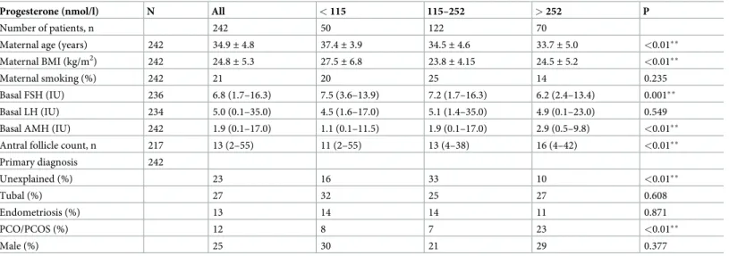
nmol/l, [115–252 nmol/l], > 252 nmol/l. The low number of observations did not allow us to find any significant difference across groups for this reproductive outcome (p = 0.182). Table 1. Baseline characteristics.
Progesterone (nmol/l) N All < 115 115–252 > 252 P
Number of patients, n 242 50 122 70
Maternal age (years) 242 34.9± 4.8 37.4± 3.9 34.5± 4.6 33.7± 5.0 <0.01��
Maternal BMI (kg/m2) 242 24.8± 5.3 27.5± 6.8 23.8± 4.15 24.5± 5.2 <0.01��
Maternal smoking (%) 242 21 20 25 14 0.235 Basal FSH (IU) 236 6.8 (1.7–16.3) 7.5 (3.6–13.9) 7.2 (1.7–16.3) 6.2 (2.4–13.4) 0.001��
Basal LH (IU) 234 5.0 (0.1–35.0) 4.5 (1.6–17.0) 5.1 (1.4–35.0) 4.9 (0.1–23.0) 0.549 Basal AMH (IU) 242 1.9 (0.1–17.0) 1.1 (0.1–11.5) 1.9 (0.1–17.0) 2.9 (0.5–9.8) <0.01��
Antral follicle count, n 217 13 (2–55) 11 (2–55) 13 (4–38) 16 (4–42) <0.01��
Primary diagnosis 242 Unexplained (%) 23 16 33 10 <0.01�� Tubal (%) 27 32 25 27 0.608 Endometriosis (%) 13 14 14 11 0.871 PCO/PCOS (%) 12 8 7 23 <0.01�� Male (%) 25 30 21 29 0.377
Descriptive data are presented as the mean± SD for continuous parametric data and as the median (range) for continuous nonparametric data. Categorical data are presented as percentages (%).
SI conversion factor for P4: nmol/l = 3.18 ng/ml.
��p< 0.01
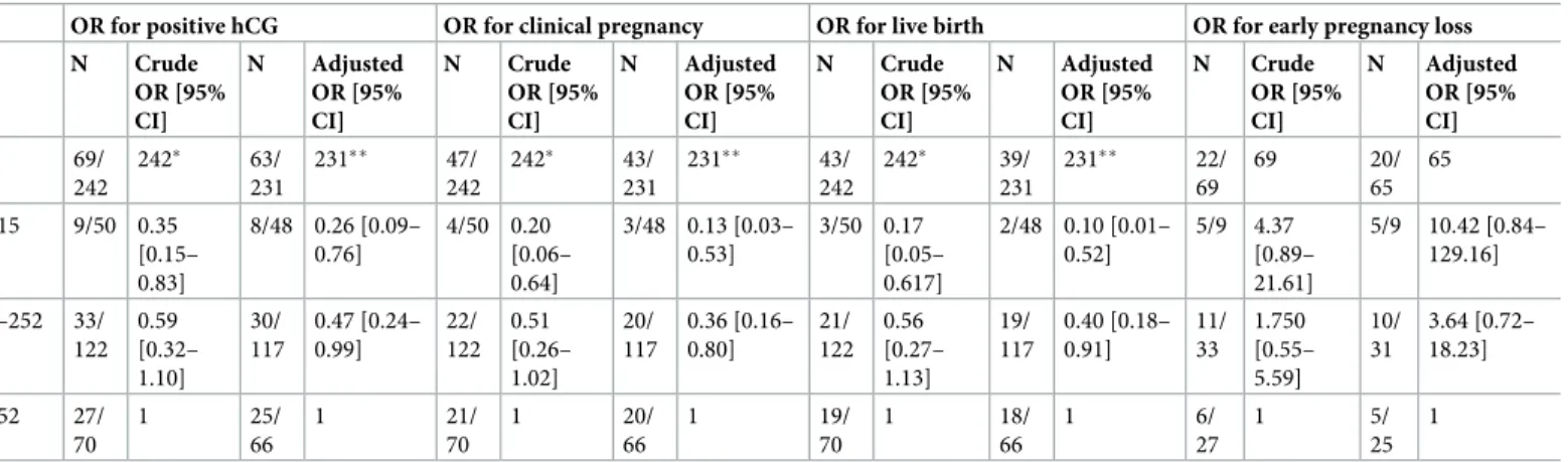
Multiple regression analysis
Due to missing data, the multiple regression analysis using the maternal age, the maternal BMI, the number of follicles > 14 mm and the late follicular P4levels > 4.77 nmol/l was only
conducted on 231 patients. The results of this analysis are presented inTable 3. Patients who had early luteal P4 levels < 115 nmol/l had the poorest chance of giving live birth (adjusted OR = 0.10 [0.02–0.52]) compared with patients with early luteal P4> 252 nmol/l. Patients with
early luteal P4[115–252 nmol/l] also had a significantly lower chance of giving live birth than
patients with P4> 252 nmol/l (adjusted OR = 0.40 [0.18–0.91]). The same significative
differ-ences were obtained for the positive hCG and the clinical pregnancies but not for the early pregnancy losses (Table 3).
Discussion
To our knowledge, our study is the first to study the effect of the early luteal phase during IVF with the use of oral dydrogesterone for the LPS. Lotus I, a large double-blinded RCT recently demonstrated the noninferiority of oral dydrogesterone compared with micronized vaginal progesterone for LPS in terms of pregnancy rates and tolerability [13]. Several other prospec-tive studies have shown the effecprospec-tiveness and safety of oral dydrogesterone for LPS during IVF or ICSI cycles and higher satisfaction rates for women compared with micronized vaginal Table 2. Descriptive data for controlled ovarian stimulation, oocytes, embryo transfer, and reproductive outcomes.
Progesterone (nmol/l) N All < 115 115–252 > 252 P
Number of patients, n (%) 242 50 (21%) 122 (50%) 70 (29%)
Protocol 242
Antagonist (%) 64 60 60 76 0.066
Long GnRH agonist (%) 28 22 33 21 0.449
Flare up (%) 8 18 7 3 0.011�
Total FSH dose (IU) 242 2925 (1000–7200) 3450 (1125–6750) 2850 (1125–7200) 2475 (1000–6750) <0.001��
Stim duration (days) 242 10.5± 1.7 10.3± 1.9 10.4± 1.7 10.7± 1.6 0.399 Final follicle count >14 mm on trigger day 240 8 (2–24) 5 (2–17) 8 (3–16) 10 (2–24) <0.001��
Mode of triggering for final oocyte maturation 242 0.353
hCG (%) 69 62 70 75
Dual trigger (%) 31 38 30 25
Number of mature oocytes retrieved (n) 242 8 (1–25) 4 (1–14) 8 (1–20) 12 (2–25) <0.001��
Number of fertilized oocytes (n) 242 5 (1–19) 3 (1–8) 5 (1–17) 6 (1–19) <0.001��
Single embryo transfer (%) 242 26 40 24 21 0.048�
Double embryo transfer (%) 242 74 60 76 79
At least one top quality embryo for transfer (%) 242 25 26 26 21 0.741 Positive hCG, n (%) 242 69 (28.5%) 9 (18.0%) 33 (27.0%) 27 (38.6%) 0.043�
Early pregnancy loss, n (%) 242 22/69 (31.9%) 5/9 (55.6%) 11/33 (33.3%) 6/27 (22.2%) 0.182 Clinical pregnancy, n (%) 242 47 (19.4%) 4 (8.0%) 22 (18,0%) 21 (30.0%) 0.009��
Live birth, n (%) 242 43 (17.8%) 3 (6.0%) 21 (17.2%) 19 (27.1%) 0.011�
Descriptive data are presented as the mean± SD for continuous parametric data and as the median (range) for continuous nonparametric data. Categorical data are presented as percentages (%).
SI conversion factor for P4: nmol/l = 3.18 ng/ml.
�p<0.05 ��p<0.01
progesterone [7,17–21]. Thus, according to some authors, dydrogesterone may become the new standard for LPS during fresh ET IVF cycles [15].
Our results suggest a positive association between early luteal P4levels and reproductive
outcomes in IVF treatment with oral dydrogesterone for the luteal support. Therefore, these results are not only inconsistent with the conclusion of Thomsenet al. but the complete oppo-site [11]. After applying the same exclusion criteria as in the Thomsenet al. study (age < 41, BMI < 35, and excluding the flare up protocols), only 179 patients were divided into the same P4groups <60 nmol/l; [60–100 nmol/l]; [100–400 nmol/l]; and > 400 nmol/l (respectively
n = 5; n = 12; n = 154; n = 8). Our results are compared to those of the Thomsen et al. study in
Fig 2. Although the low number of subjects in three of the four groups in this analysis did not allow us to find any significant differences among groups, the results of the two cohorts of patients are clearly inconsistent. The P4group [60–100 nmol/l] that was associated with the
best clinical pregnancy rate in the Thomsenet al. study had the poorest chance in our study. Similarly, the P4group > 400 nmol/l that was associated with the poorest live birth rate in the
aforementioned study was associated with the best live birth rate in our study.
Fig 1. Receiving operative characteristic (ROC) curve for the prediction of live birth based on early luteal progesterone levels during IVF. Area under ROC curve = 0.599 (0.507–0.691), p = 0.042.
The most plausible hypothesis for these discrepancies is that dydrogesterone interferes with the progesterone secretion by thecorpus lutei. Indeed, we measured P4levels only and do not
report the dydrogesterone levels, which would have required the use of an instrumental chro-matographic method [16,22]. Although the influence of dydrogesterone on the secretion of progesterone during the luteal phase has not been fully elucidated, its administration lowers the P4levels [23]. Two studies have also shown the efficacy of dydrogesterone administration
to prevent premature LH surges in the context of frozen-thawed ET, suggesting negative feed-back on the pituitary gland [23,24]. Although it is clearly impossible with the present study to estimate the respective weights of influence of exogenous dydrogesterone and endogenous progesterone on the outcomes of IVF treatment, our study clearly illustrates that the thresholds suggested by Thomsen et al. are not transposable when dydrogesterone is used for the LPS.
The present study has several limitations. The small sample size forced us to conduct the statistical analysis on early luteal P4cut-off values that were determined in a data-dependent
way. This method of analysis is known to bear the risk of finding an effect in the sample while no real difference exists in the population [12,25]. Although we noticed the same trends when the data were analyzed in an objective way–i.e. percentile groups–due to sample size, we were unable to show significant differences using this analysis. Comparability of our P4groups
could also be criticized, as there are significant differences between groups. To overcome this issue, we chose to use the exact same variables as Thomsenet al. for the multiple regression analysis [11]. They used a Directed Acyclic Graph to identify a minimum set of covariates to adjust for in the statistical analysis. However, in the present study, the sample size was too small to show significant differences between groups by using a classical selection process for the multiple regression. Therefore, we cannot formally exclude a confusion bias. Overall, in addition to the retrospective nature of the study, the lack of statistical power does not allow us to present an irreproachable conclusion and further larger prospective cohort studies must be conducted. Another obvious limitation of the present study is the absence of early luteal phase dydrogesterone level measurements. This missing information prevents us from fully conclud-ing on the pathophysiological mechanisms involved in the relationship between P4levels and
reproductive outcomes. Nevertheless, we successfully identified early luteal P4thresholds that–
if confirmed by prospective studies–may be used in clinical practice. Indeed, we determined Table 3. Reproductive outcomes in the different luteal progesterone groups.
OR for positive hCG OR for clinical pregnancy OR for live birth OR for early pregnancy loss N Crude OR [95% CI] N Adjusted OR [95% CI] N Crude OR [95% CI] N Adjusted OR [95% CI] N Crude OR [95% CI] N Adjusted OR [95% CI] N Crude OR [95% CI] N Adjusted OR [95% CI] N 69/ 242 242� 63/ 231 231�� 47/ 242 242� 43/ 231 231�� 43/ 242 242� 39/ 231 231�� 22/ 69 69 20/ 65 65 P4 < 115 nmol/l 9/50 0.35 [0.15– 0.83] 8/48 0.26 [0.09– 0.76] 4/50 0.20 [0.06– 0.64] 3/48 0.13 [0.03– 0.53] 3/50 0.17 [0.05– 0.617] 2/48 0.10 [0.01– 0.52] 5/9 4.37 [0.89– 21.61] 5/9 10.42 [0.84– 129.16] P4 115–252 nmol/l 33/ 122 0.59 [0.32– 1.10] 30/ 117 0.47 [0.24– 0.99] 22/ 122 0.51 [0.26– 1.02] 20/ 117 0.36 [0.16– 0.80] 21/ 122 0.56 [0.27– 1.13] 19/ 117 0.40 [0.18– 0.91] 11/ 33 1.750 [0.55– 5.59] 10/ 31 3.64 [0.72– 18.23] P4 > 252 nmol/l 27/ 70 1 25/ 66 1 21/ 70 1 20/ 66 1 19/ 70 1 18/ 66 1 6/ 27 1 5/ 25 1
�In the crude OR estimates, all 242 patients with embryo transfer are included.
��Due to missing data for the covariate late follicular phase in 7 patients, the final follicle count > 14 mm on trigger day for 2 patients, and the BMI for 2 patients, the
final adjusted regression model included 231 patients.
that 21% of our patients who had P4levels below 115 nmol/l had a dramatically poor live-birth
rate (6.0%). If confirmed, this finding would encourage physicians to cancel the ET for these patients. The effect of increasing the supplementation is more intangible because the dydroges-terone might induce negative feedback over the progesdydroges-terone secretion of thecorpus lutei [23]. Fig 2. Comparison of the reproductive outcomes of our retrospective cohort (in green, right section, 179 patients) with the Thomsenet al.
prospective cohort (in blue, left section, 389 patients).
Furthermore, increasing progesterone or dydrogesterone supplementation does not propor-tionally increase the serum levels, and uterine progesterone levels do not correlate well with serum levels [26–29]. These pharmacokinetic data add to the uncertainty regarding strategies to increase the LPS in the case of low early luteal P4levels.
Thus, our results should be interpreted with utmost precautions, and we only conclude that early luteal P4levels and the reproductive outcomes in IVF with oral dydrogesterone for luteal
support appear to be positively associated. The current state of knowledge on early luteal P4
monitoring is still too weak to allow for consideration of interventional studies or clinical applications. Further larger prospective cohort studies must be conducted to determine reli-able thresholds that could be used to personalize LPS.
Author Contributions
Conceptualization: Antoine Netter, Jeanne Perrin, Blandine Courbiere. Data curation: Julien Mancini.
Formal analysis: Antoine Netter, Julien Mancini. Investigation: Antoine Netter, Blandine Courbiere. Methodology: Antoine Netter, Blandine Courbiere. Project administration: Antoine Netter.
Supervision: Jeanne Perrin.
Validation: Christophe Buffat, Aubert Agostini, Jeanne Perrin, Blandine Courbiere. Writing – original draft: Antoine Netter, Blandine Courbiere.
Writing – review & editing: Antoine Netter, Blandine Courbiere.
References
1. Kushnir VA, Barad DH, Albertini DF, Darmon SK, Gleicher N. Systematic review of worldwide trends in assisted reproductive technology 2004–2013. Reprod Biol Endocrinol. 2017; 15: 6.https://doi.org/10. 1186/s12958-016-0225-2PMID:28069012
2. Yding Andersen C, Vilbour Andersen K. Improving the luteal phase after ovarian stimulation: reviewing new options. Reproductive BioMedicine Online. 2014; 28: 552–559.https://doi.org/10.1016/j.rbmo. 2014.01.012PMID:24656557
3. Carson DD. Changes in gene expression during the early to mid-luteal (receptive phase) transition in human endometrium detected by high-density microarray screening. Molecular Human Reproduction. 2002; 8: 871–879.https://doi.org/10.1093/molehr/8.9.871PMID:12200466
4. Wetendorf M, DeMayo FJ. The progesterone receptor regulates implantation, decidualization, and glan-dular development via a complex paracrine signaling network. Mol Cell Endocrinol. 2012; 357: 108– 118.https://doi.org/10.1016/j.mce.2011.10.028PMID:22115959
5. Fauser BC, de Jong D, Olivennes F, Wramsby H, Tay C, Itskovitz-Eldor J, et al. Endocrine profiles after triggering of final oocyte maturation with GnRH agonist after cotreatment with the GnRH antagonist ganirelix during ovarian hyperstimulation for in vitro fertilization. J Clin Endocrinol Metab. 2002; 87: 709–715.https://doi.org/10.1210/jcem.87.2.8197PMID:11836309
6. Child T, Leonard SA, Evans JS, Lass A. Systematic review of the clinical efficacy of vaginal progester-one for luteal phase support in assisted reproductive technology cycles. Reprod Biomed Online. 2018; 36: 630–645.https://doi.org/10.1016/j.rbmo.2018.02.001PMID:29550390
7. van der Linden M, Buckingham K, Farquhar C, Kremer JAM, Metwally M. Luteal phase support for assisted reproduction cycles. Cochrane Database Syst Rev. 2015; CD009154.https://doi.org/10.1002/ 14651858.CD009154.pub3PMID:26148507
8. Labarta E, Mariani G, Holtmann N, Celada P, Remohı´ J, Bosch E. Low serum progesterone on the day of embryo transfer is associated with a diminished ongoing pregnancy rate in oocyte donation cycles
after artificial endometrial preparation: a prospective study. Hum Reprod. 2017; 32: 2437–2442.https:// doi.org/10.1093/humrep/dex316PMID:29040638
9. Yovich JL, Conceicao JL, Stanger JD, Hinchliffe PM, Keane KN. Mid-luteal serum progesterone con-centrations govern implantation rates for cryopreserved embryo transfers conducted under hormone replacement. Reprod Biomed Online. 2015; 31: 180–191.https://doi.org/10.1016/j.rbmo.2015.05.005
PMID:26099447
10. Alsbjerg B, Thomsen L, Elbaek HO, Laursen R, Povlsen BB, Haahr T, et al. Progesterone levels on pregnancy test day after hormone replacement therapy-cryopreserved embryo transfer cycles and related reproductive outcomes. Reprod Biomed Online. 2018; 37: 641–647.https://doi.org/10.1016/j. rbmo.2018.08.022PMID:30385142
11. Thomsen LH, Kesmodel US, Erb K, Bungum L, Pedersen D, Hauge B, et al. The impact of luteal serum progesterone levels on live birth rates-a prospective study of 602 IVF/ICSI cycles. Hum Reprod. 2018;
https://doi.org/10.1093/humrep/dey226PMID:29955789
12. Venetis CA, Mol BW, Kolibianakis EM. Low as well as high serum P4 levels in the early and mid-luteal phase reduce the chance of a live birth following IVF treatment with fresh embryo transfer: where is the evidence? Hum Reprod. 2018; 33: 2312–2313.https://doi.org/10.1093/humrep/dey311PMID:
30325399
13. Tournaye H, Sukhikh GT, Kahler E, Griesinger G. A Phase III randomized controlled trial comparing the efficacy, safety and tolerability of oral dydrogesterone versus micronized vaginal progesterone for luteal support in in vitro fertilization. Hum Reprod. 2017; 32: 1019–1027.https://doi.org/10.1093/humrep/ dex023PMID:28333318
14. Abbott B.V. film-coated tablets 10 mg Summary of Product Characteristics. 2017.
15. Griesinger G, Blockeel C, Tournaye H. Oral dydrogesterone for luteal phase support in fresh in vitro fer-tilization cycles: a new standard? Fertil Steril. 2018; 109: 756–762.https://doi.org/10.1016/j.fertnstert. 2018.03.034PMID:29778368
16. Griesinger G, Tournaye H, Macklon N, Petraglia F, Arck P, Blockeel C, et al. Dydrogesterone: pharma-cological profile and mechanism of action as luteal phase support in assisted reproduction. Reprod Biomed Online. 2019; 38: 249–259.https://doi.org/10.1016/j.rbmo.2018.11.017PMID:30595525
17. Chakravarty BN, Shirazee HH, Dam P, Goswami SK, Chatterjee R, Ghosh S. Oral dydrogesterone ver-sus intravaginal micronised progesterone as luteal phase support in assisted reproductive technology (ART) cycles: results of a randomised study. J Steroid Biochem Mol Biol. 2005; 97: 416–420.https:// doi.org/10.1016/j.jsbmb.2005.08.012PMID:16213136
18. Patki A, Pawar VC. Modulating fertility outcome in assisted reproductive technologies by the use of dydrogesterone. Gynecol Endocrinol. 2007; 23 Suppl 1: 68–72.https://doi.org/10.1080/
09513590701584857PMID:17943542
19. Ganesh A, Chakravorty N, Mukherjee R, Goswami S, Chaudhury K, Chakravarty B. Comparison of oral dydrogestrone with progesterone gel and micronized progesterone for luteal support in 1,373 women undergoing in vitro fertilization: a randomized clinical study. Fertil Steril. 2011; 95: 1961–1965.https:// doi.org/10.1016/j.fertnstert.2011.01.148PMID:21333984
20. Tomic V, Tomic J, Klaic DZ, Kasum M, Kuna K. Oral dydrogesterone versus vaginal progesterone gel in the luteal phase support: randomized controlled trial. Eur J Obstet Gynecol Reprod Biol. 2015; 186: 49– 53.https://doi.org/10.1016/j.ejogrb.2014.11.002PMID:25622239
21. Saharkhiz N, Zamaniyan M, Salehpour S, Zadehmodarres S, Hoseini S, Cheraghi L, et al. A compara-tive study of dydrogesterone and micronized progesterone for luteal phase support during in vitro fertili-zation (IVF) cycles. Gynecol Endocrinol. 2016; 32: 213–217.https://doi.org/10.3109/09513590.2015. 1110136PMID:26486011
22. Abdel-Hamid ME, Sharaf LH, Kombian SB, Diejomaoh FME. Determination of Dydrogesterone in Human Plasma by Tandem Mass Spectrometry: Application to Therapeutic Drug Monitoring of Dydro-gesterone in Gynecological Disorders. Chroma. 2006; 64: 287–292. https://doi.org/10.1365/s10337-006-0035-3
23. Zhu X, Ye H, Fu Y. Duphaston and human menopausal gonadotropin protocol in normally ovulatory women undergoing controlled ovarian hyperstimulation during in vitro fertilization/intracytoplasmic sperm injection treatments in combination with embryo cryopreservation. Fertil Steril. 2017; 108: 505– 512.e2.https://doi.org/10.1016/j.fertnstert.2017.06.017PMID:28697910
24. Yu S, Long H, Chang HY-N, Liu Y, Gao H, Zhu J, et al. New application of dydrogesterone as a part of a progestin-primed ovarian stimulation protocol for IVF: a randomized controlled trial including 516 first IVF/ICSI cycles. Hum Reprod. 2018; 33: 229–237.https://doi.org/10.1093/humrep/dex367PMID:
25. Altman DG, Lausen B, Sauerbrei W, Schumacher M. Dangers of using “optimal” cutpoints in the evalua-tion of prognostic factors. J Natl Cancer Inst. 1994; 86: 829–835.https://doi.org/10.1093/jnci/86.11.829
PMID:8182763
26. Mol BW, Bossuyt PM, Sunkara SK, Garcia Velasco JA, Venetis C, Sakkas D, et al. Personalized ovar-ian stimulation for assisted reproductive technology: study design considerations to move from hype to added value for patients. Fertil Steril. 2018; 109: 968–979.https://doi.org/10.1016/j.fertnstert.2018.04. 037PMID:29935655
27. Paulson RJ, Collins MG, Yankov VI. Progesterone pharmacokinetics and pharmacodynamics with 3 dosages and 2 regimens of an effervescent micronized progesterone vaginal insert. J Clin Endocrinol Metab. 2014; 99: 4241–4249.https://doi.org/10.1210/jc.2013-3937PMID:24606090
28. Cicinelli E, de Ziegler D, Bulletti C, Matteo MG, Schonauer LM, Galantino P. Direct transport of proges-terone from vagina to uterus. Obstet Gynecol. 2000; 95: 403–406. PMID:10711552
29. Gibbons WE, Toner JP, Hamacher P, Kolm P. Experience with a novel vaginal progesterone prepara-tion in a donor oocyte program. Fertil Steril. 1998; 69: 96–101.https://doi.org/10.1016/s0015-0282(97) 00457-3PMID:9457941