HAL Id: hal-02445842
https://hal-amu.archives-ouvertes.fr/hal-02445842
Submitted on 22 Jan 2020
HAL is a multi-disciplinary open access
archive for the deposit and dissemination of
sci-entific research documents, whether they are
pub-lished or not. The documents may come from
teaching and research institutions in France or
abroad, or from public or private research centers.
L’archive ouverte pluridisciplinaire HAL, est
destinée au dépôt et à la diffusion de documents
scientifiques de niveau recherche, publiés ou non,
émanant des établissements d’enseignement et de
recherche français ou étrangers, des laboratoires
publics ou privés.
Zuzana Sekeyová, Monika Danchenko, Peter Filipčík, Pierre Edouard Fournier
To cite this version:
Zuzana Sekeyová, Monika Danchenko, Peter Filipčík, Pierre Edouard Fournier. Rickettsial infections
of the central nervous system. PLoS Neglected Tropical Diseases, Public Library of Science, 2019, 13
(8), pp.e0007469. �10.1371/journal.pntd.0007469�. �hal-02445842�
Rickettsial infections of the central nervous
system
Zuzana Sekeyova´
1, Monika Danchenko
1, Peter Filipčı´k
2, Pierre Edouard FournierID
3,4*
1 Institute of Virology, Slovak Academy of Sciences, Dubravska cesta, Bratislava, Slovakia, 2 Institute of
Neuroimmunology, Slovak Academy of Sciences, Dubravska cesta, Bratislava, Slovakia, 3 Aix-Marseille Univ, IRD, AP-HM, SSA, VITROME, IHU Mediterrane´ e-Infection, Marseille, France, 4 Centre National de Re´fe´rence des Rickettsia, Coxiella et Bartonella, IHU Mediterrane´e-Infection, Marseille, France
*pierre-edouard.fournier@univ-amu.fr
Abstract
As a result of migrations and globalization, people may face a possible increase in the
inci-dence of central nervous system rickettsial infections (CNS R). These diseases, caused by
Rickettsia species and transmitted to humans by arthropod bites, are putatively lethal.
How-ever, the diagnosis of CNS R is challenging and often delayed due to their nonspecific
clini-cal presentation and the strict intracellular nature of rickettsiae. Furthermore, transfer of
rickettsiae to the brain parenchyma is not yet understood. The aim of this review is to
ana-lyze and summarize the features and correlated findings of CNS R in order to focus attention
on these intriguing but frequently neglected illnesses. We also incorporated data on CNS
infections caused by Rickettsia-related microorganisms.
Introduction
Recent reports have warned about the impact of global warming in facilitating the
transmis-sion of certain vector-borne infectious diseases. However, due consideration must be taken of
the role played by other variables, such as the increase in international travel, migration, and
trade, with the risk of importing parasites and vectors with the goods [
1
]. Rickettsioses are
common tick-, flea-, or mite-borne bacterial illnesses with a clinical spectrum ranging from a
mild febrile illness to potentially life-threatening complications [
2
,
3
]. Rickettsial infections can
affect many organs, including the central nervous system (CNS) [
4
]. The most common
neu-rological manifestations reported in rickettsial infections include meningitis, encephalitis, and
acute disseminated encephalomyelitis [
5
]. However, unilateral facial nerve palsy [
6
], cerebral
infarction [
7
], or visual loss [
8
] have also been diagnosed. Therefore, it is critical to enhance
the awareness of physicians worldwide on CNS rickettsial infections (CNS R).
Few studies on the pathogenesis of rickettsiae for the CNS have been conducted. In neurons
cultured in vitro, rickettsiae have been demonstrated to cause a profound morphological
dete-rioration and intracellular decrease of ATP [
9
]. In addition, it was demonstrated that
rickett-siae can persist and reappear after a relatively long time in the CNS in immunocompromised
mice, causing a fatal neuroinflammation [
10
]. This suggests a potential neuropathic role of
rickettsiae. However, data on rickettsial infections in a context of degenerative CNS diseases,
which usually result from mixed neuropathologies and present with multiple symptoms, are
a1111111111
a1111111111
a1111111111
a1111111111
a1111111111
OPEN ACCESSCitation: Sekeyova´ Z, Danchenko M, Filipčı´k P, Fournier PE (2019) Rickettsial infections of the central nervous system. PLoS Negl Trop Dis 13(8): e0007469.https://doi.org/10.1371/journal. pntd.0007469
Editor: Abdallah M. Samy, Faculty of Science, Ain
Shams University (ASU), EGYPT
Published: August 29, 2019
Copyright:© 2019 Sekeyova´ et al. This is an open access article distributed under the terms of the
Creative Commons Attribution License, which permits unrestricted use, distribution, and reproduction in any medium, provided the original author and source are credited.
Funding: This work was supported by the following
projects: APVV-0280-12, “Sˇtefanik“ APVV-SK-FR-2017-0005, VEGA 2/0010/19, and the French National Research Agency under the program “investissements d’avenir,” reference ANR-10-IAHU-03. The funders had no role in study design, data collection and analysis, decision to publish, or preparation of the manuscript.
Competing interests: The authors have declared
scarce in the literature, and to date, no association between neurodegenerative diseases and
rickettsial infections has been demonstrated.
In this article, we systematically reviewed the literature reporting cases of CNS R to provide
scientists and healthcare providers with a summary of the current knowledge on and
chal-lenges posed by these diseases. We also included data on
Orientia, Anaplasma, and Ehrlichia
species, which are phylogenetically close to
Rickettsia species.
Literature search and selection criteria
We searched PubMed with the following Medical Subject Heading terms: “rickettsia” (9,504
results); “rickettsia” AND “central nervous system” (169 results); and “rickettsia” AND
“cen-tral nervous system” AND review (5 results). The search was limited to articles published in
English between January 1, 1960, and February 28, 2019. In addition, no online book or book
chapter in relation to this subject was identified.
We also searched PubMed with the Medical Subject Heading terms: “orientia” AND
“human” (945 results); “ehrlichia” AND “human” (1,421 results); “anaplasma” AND “human”
(1,385 results); “orientia” AND “human” AND “central nervous system” (26 results);
“ehrli-chia” AND “human” AND “central nervous system” (10 results); and “anaplasma” AND
“human” AND “central nervous system” (3 results).
Epidemiology of rickettsial diseases
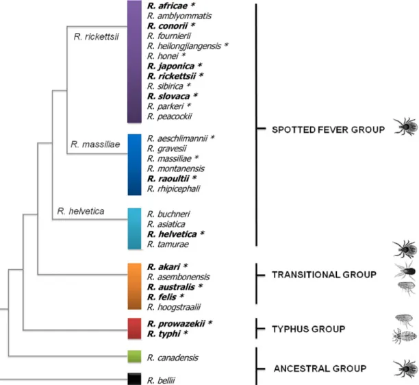
Rickettsiae are strictly intracellular bacteria that are vectored by various arthropods. To date,
18 species are recognized human pathogens, 12 of which cause CNS R (
Fig 1
). To these
patho-genic
Rickettsia species may be added O. tsutsugamushi and O. chuto that are phylogenetically
closely related and are still often referred to as rickettsiae. Rickettsioses are zoonotic diseases
that distributed worldwide (
Fig 2
) in foci of endemicity (
Table 1
), with sporadic and often
sea-sonal outbreaks [
11
]. However,
R. prowazekii has the potential to reemerge as outbreaks in
human populations living in close proximity and impoverished conditions [
12
] (
Fig 3
).
Clinical manifestations of rickettsial infections
Spotted fever rickettsioses typically present with a triad made of fever, inoculation eschar(s),
and generalized cutaneous rash. However, infected patients may develop other clinical
pic-tures, including nonspecific flu-like symptoms, isolated fever, myalgia, cough, generalized
lymphadenopathy, abdominal pain, and a variety of neurological symptoms that are described
next.
Most commonly, patients recover without sequellae. However, the clinical presentation
of rickettsial diseases may vary from mild to very severe, with the case-fatality rate for highly
virulent rickettsiae ranging from 2% to 30% [
13
]. The severity of rickettsial disease has been
associated with differences in pathogen virulence and host-related factors (e.g., age, delayed
diagnosis, hepatic and renal dysfunction, CNS and lung involvement) [
14
]. Regarding
species-specific variations in virulence, studies have suggested that these differences may be linked to
the degree of genomic degradation. In a comparison of 4 species exhibiting distinct virulence
profiles, the most virulent
Rickettsia sp. exhibited the most drastically reduced and degraded
genomes compared with closely related species of milder pathogenesis [
15
].
Mechanisms of bacterial entry into the CNS
The brain–blood barrier (BBB) at the level of brain microvessel endothelium is the major site
of blood–CNS exchange. The mechanisms of bacterial entry into the CNS are divided
according to whether they break the integrity of BBB cells (which consist of microvascular
endothelial cells of the brain, pericytes, and astrocytes) or interact with epithelial cells of the
blood–choroid barrier. Mechanisms used by pathogens to enter the CNS are usually classified
according to the involved cellular route [
16
,
17
]. These cellular entry routes are listed as
inter-cellular or parainter-cellular (passing between cells), transinter-cellular (the intrainter-cellular mode), or
leuko-cyte-facilitated in infected phagocytes (which is a “Trojan horse”-like mechanism) [
18
,
19
].
Rickettsial neuroinvasion occurs during the systemic phase of the disease and typically follows
bacterial dissemination via the bloodstream. As other intracellular microorganisms, rickettsiae
might use a transcellular approach, which is supported by the fact that bacterial replication
takes place within endothelial cells. Other studies have demonstrated that bacterial pathogens
(extra- and intracellular) can reach the BBB and are recognized by antigen-presenting cells
through binding to Toll-like receptors (
Fig 4
).
Fig 1. Phylogenetic tree ofRickettsia species inferred from the comparison of concatenated sequences from the gltA and sca4
genes.Rickettsia species are distributed into 4 groups: SFG, TRG, TG, and AG. SFG rickettsiae are mostly associated with ticks; TG rickettsiae with human body lice (R. prowazekii) and rat fleas (R. typhi); TRG rickettsiae with ticks, cat fleas, or mites; and AG rickettsiae with ticks. Asterisks mark pathogenic species, and bold letters indicate species causing CNS infections. AG, ancestral group; CNS, central nervous system; SFG, spotted fever group; TG, typhus group; TRG, transitional group.
They induce the activation of nuclear factor-kappa B- or mitogen-activated protein kinase
pathways and subsequently up-regulate leukocyte populations and express numerous proteins
involved in inflammation and the immune response [
16
,
18
]. However, the precise molecular
mechanisms of rickettsial entry into the CNS are not yet described.
Treatment of rickettsioses
The most efficient antibiotics to cure rickettsioses are tetracycline derivatives, primarily
doxy-cycline. The daily dose, 200 mg, is usually administered orally and is sufficient to cure
rickett-sioses. Treatment duration is 7 days or until 2 days after apyrexia is obtained [
20
,
21
]. In severe
forms, intravenous doxycycline may be used. Alternatively, chloramphenicol and macrolides
(azithromycin, clarithromycin, and roxytromycin) can be prescribed for treating rickettsial
diseases [
22
,
23
]. As a consequence, empirical therapy for CNS infections most often does not
include antibiotics active on rickettsiae.
Neuropathology in the framework of rickettsial diseases
Spotted fever group rickettsiae
In spotted fever group (SFG) rickettsioses, encephalitis is underdiagnosed due to
underrecog-nition and low sensitivity of serologies at the time of early symptoms [
24
]. Therefore, it is
Fig 2. Developmental cycle and host range ofRickettsia-infected ticks. Ticks are the main vectors and constitute a threat of rickettsial infection regardless of their development stage (larvae, nymphs, or adults). Many factors play a role in the epidemiology of tick-borne rickettsioses, including the prevalence and species diversity of rickettsiae in mammals [149–153] and the dispersion of infected ticks by migratory birds [154].
Fig 3. Body lice as the cause of typhus reemergence.R. prowazekii is classified as a category B bioterrorism agent. It is stable in dried louse feces and can be transmitted through aerosols. Detection of the pathogen in body lice is crucial for monitoring the transmission risk to humans. However, Brill–Zinsser disease, a relapsing form of epidemic typhus that may develop as sporadic cases up to 40 years after the initial acute infection, is unrelated to louse infestation but to stress or a waning immune system that initiates the reactivation of an earlier and latent infection. Patients developing Brill–Zinsser disease may, in turn, be the source of new outbreaks when conditions facilitate louse infestation and transmission [155]. The mechanism ofR. prowazekii latency has not been established. Brill–Zinsser disease should be considered as a possible diagnosis for acute fever in any patient who has lived in an area where epidemic typhus was endemic [156].
https://doi.org/10.1371/journal.pntd.0007469.g003
Table 1. Geographical distribution and arthropod vectors ofRickettsia and Orientia species causing CNS infections.
Species (type strain) Disease Vector Distribution Genome sequence accession number�
R. africae (ESF-5) ATBF Amblyomma and Rhipicephalus ticks
Sub-Saharan Africa, West Indies
NC_012633 R. akari (Hartford) Rickettsialpox L. sanguineus mites North America, Asia,
Europe
NC_009881
R. australis (Cutlack) QTT Ixodes ticks Australia NC_017058
R. conorii (Malish 7) MSF Rhipicephalus sanguineus ticks Europe, North Africa, Asia NC_003103 Rickettsia felis
(Marseille-URRWFXCal2)
Flea-borne rickettsiosis MostlyC. felis Worldwide NC_007109.1 Rickettsia helvetica (C9P9) Unnamed rickettsiosis Ixodes sp. Europe, Asia, North Africa NZ_CM001467 Rickettsia japonica (YH) Japanese/Oriental spotted
fever
Haemaphysalis ticks Asia NC_016050
Rickettsia raoultii (Khabarovsk) TIBOLA Dermacentor and Ixodes ticks Europe NZ_CP010969
Rickettsia rickettsii (Sheila Smith) RMSF MostlyDermacentor ticks North and Central America NC_009882
Rickettsia slovaca (13-B) TIBOLA Dermacentor ticks Europe, Asia NC_016639
R. prowazekii (Madrid E) Epidemic typhus Pediculus humanus humanus lice
Africa, Russia, South America
NC_000963
R. typhi (Wilmington) Murine/endemic typhus X. cheopis fleas Worldwide NC_006142
O. tsutsugamushi (Ikeda) Scrub typhus Leptotrombidium mites Asia NC_010793
�All genomes were imported from GenBank (http://www.ncbi.nlm.nih.gov/genome/)
Abbreviations: ATBF, African tick bite fever; CNS, central nervous system; MSF, Mediterranean spotted fever; QTT, Queensland tick typhus; RMSF, Rocky Mountain
spotted fever; TIBOLA, tick-borne lymphadenopathy
difficult for clinicians to recognize the clinical, biological, and neuroimaging features of this
type of encephalitis [
25
].
Rocky Mountain spotted fever
Rocky Mountain spotted fever (RMSF), caused by
R. rickettsii, frequently presents with
neuro-logical symptoms [
26
,
27
]. The CNS notably appears to be one of the major systems involved
during the latest stages of RMSF pathogenesis [
28
].
The onset of symptoms is usually abrupt, with severe headache, fever, chills, myalgias,
arthralgias, and prostration. Neurological symptoms are frequently a prominent feature.
Head-ache, restlessness, insomnia, and back stiffness are common. Delirium or coma alternating
with restlessness is present during the peak of fever [
26
,
27
,
29
–
31
]. Encephalitis, which may be
fatal, is a frequent manifestation in case of delayed diagnosis and treatment [
32
]. In addition,
several authors have described severe sequelae in patients who survived
R. rickettsii infection,
including hemiplegia, deafness, visual disturbance, slurred speech, mental confusion, and
cra-nial neuropathies that may persist for a few weeks following recovery [
33
–
37
].
Histological studies have highlighted that the pathologic changes in RMSF are most severe
in the skin, but the heart, lungs, and CNS also are involved. The brain is edematous, and
Fig 4. Mechanisms of bacterial penetration through the blood–brain barrier. (A) Intercellular or paracellular, described in
extracellular pathogens. The physical barrier formed by endothelial tight junctions is disturbed by bacterial penetration. (B) Transcellular, passing through cells, e.g., direct invasion of endothelial cells. In this scenario, blood-borne bacteria directly invade CNS endothelial cells. (C) By leukocytes, within infected macrophages, named “Trojan horse” mechanism. Infected leukocytes adhere to endothelial cells, allowing the spread of bacteria or, alternatively, leukocytes can transmigrate and deliver bacteria to the CNS parenchyma. CNS, central nervous system.
minute petechial hemorrhages are present. The characteristic microscopic lesions are small
round nodules composed of elongated microglia, lymphocytes, and endothelial cells
[
29
,
38
,
39
]. These are scattered diffusely through the brain in close relation to small vessels.
Vessels in the center of the lesions show severe degeneration. Endothelial cells are swollen,
and the lumen may be occluded. Areas of focal necrosis are common as the result of
thrombo-sis of small arteries. Some degree of perivascular infiltration without the presence of nodules
may be seen in both the meninges and brain parenchyma [
29
].
To the best of our knowledge, there is just one report of experimental in vitro rickettsial
infection of neuronal cells. Joshi and Kovacs [
40
] demonstrated that neurons are efficiently
infected by
R. rickettsii and that the infection causes significant neuronal apoptotic cell death.
A double-immunofluorescent staining technique was employed to identify infected neurons,
using anti-
R. rickettsii serum and a neuron-specific (antineuronal β-III isoform of tubulin
[anti-TuJ1]) antibody. The extent of apoptotic cell death of immunocytochemically identified
neurons was determined by terminal deoxynucleotidyl transferase-mediated
digoxigenin-dUTP nick-end labelling (TUNEL) assay. Primary cultures of rat cerebellar granular neurons
were infected (approximately 65% of neurons) during a 6-hour exposure to
R. rickettsii, and
the infection induced widespread neuronal degeneration. To verify that the observed neuronal
degeneration was
R. rickettsii-induced, primary cultures of rat cerebellar granular neuron
cul-tures were exposed to
R. rickettsii in the presence of tetracycline. The authors also included R.
rickettsii infection of endothelial cells as a control for infection and infection-induced
apopto-tic cell death [
40
].
The CNS is also a crucial target in other rickettsial diseases [
41
,
42
]. It has been
demon-strated by a specific immunohistochemistry technique, during the development of
mouse-model studies, that rickettsiae reach brain tissues when injected intravenously [
43
]. Such
studies demonstrate that rickettsiae are able to cross the blood–brain barrier to reach brain
tissues. The other rickettsial infections that may affect the nervous system exhibit
pathologi-cal and clinipathologi-cal changes similar to those of RMSF [
29
]. In rickettsial meningitis, the
cerebro-spinal fluid shows pleocytosis with lymphocyte predominance, hypoglycorrhachia, and
hyperproteinorrhachia.
Mediterranean spotted fever
Mediterranean spotted fever (MSF), also known as Boutonneuse fever and Marseilles fever, is
caused by
R. conorii. Although usually a benign and self-limiting exanthematous febrile illness
[
44
–
47
], MSF has a fatality rate of 2% to 7% among hospitalized patients [
48
]. In large MSF
case series from France and Spain, patients with severe and fatal forms of the disease developed
complications, including acute renal failure, thrombocytopenia, myocarditis, pneumonitis,
gastric hemorrhage, shock, and multiple organ failure [
44
–
47
,
49
]. Furthermore, a few reports
in the literature have described CNS involvement in the course of MSF, presenting as
meningi-tis [
50
,
51
], encephalitis [
45
,
47
,
49
,
50
,
52
,
53
], meningoencephalitis [
47
,
54
–
56
], or myelitis [
50
].
Among the patients who survived these severe forms, only 2 did not suffer sequelae. Sequelae
were severe, despite an appropriate doxycycline treatment of 200 mg every 12 hours [
54
,
57
].
Japanese spotted fever
Japanese spotted fever (JSF), caused by
R. japonica, was first reported in 1984 [
58
]. This
rickett-sial disease is characterized by high fever, rash, and inoculation eschar at the tick bite site [
58
].
JSF is clinically similar to scrub typhus, caused by
O. tsutsugamushi, but often with a more
severe course [
58
]. CNS involvement is rare, but cases complicated by pneumonia or
meningo-encephalitis have been reported [
59
–
61
]. There have been only 5 JSF reported cases with CNS
involvement [
62
–
64
]. The first fatal case was reported by Kodama and colleagues in 2002 [
65
].
All patients exhibited fever and mental disturbance, including 3 patients with nuchal rigidity
and 3 patients with seizures. All patients had aseptic meningitis [
66
].
Tick-borne lymphadenopathy
R. slovaca and R. raoultii cause a rickettsiosis that has successively been named tick-borne
lymphadenopathy (TIBOLA) [
67
–
69
],
Dermacentor-borne necrosis erythema and
lymphade-nopathy (DEBONEL) [
70
–
73
], and then scalp eschar and neck lymphadenopathy after tick
bite (SENLAT) [
74
]. The first
R. slovaca isolate was cultivated from a D. marginatus tick
col-lected in central Slovakia and was initially considered nonpathogenic [
67
,
75
,
76
]. However, in
1997,
R. slovaca was identified as the cause of a clinical entity combining scalp eschar and neck
lymphadenopathy after a tick bite [
76
]. After MSF, TIBOLA is nowadays the most prevalent
tick-borne rickettsiosis in Europe [
77
].
This syndrome is defined by the association of a tick bite and an inoculation eschar to the
scalp, surrounded by a circular erythema, and painful regional lymphadenopathies [
78
]. The
enlarged lymph nodes are located in the draining region of the tick bite, characteristically in
the occipital region and/or behind the sternocleidomastoidal muscle. Although rare, the most
frequent general symptoms are fever, nuchal lymphadenopathy, fatigue, dizziness, headache,
sweat, myalgia, arthralgia, and loss of appetite [
68
,
79
]. Symptoms suggestive of acute
encepha-litis with febrile relapses and a persistence of neurasthenic disorders have also been reported
[
68
,
80
].
Although
R. slovaca was the first demonstrated agent of TIBOLA [
76
] and was isolated
from a patient [
81
], other
Rickettsia species were suspected to be involved in the disease in
Spanish reports [
71
,
82
]. In 2008,
R. raoultii DNA was detected in a tick taken from the scalp of
a patient with TIBOLA [
83
]. In 2009, Parola and colleagues reported 6 more cases of
R. raoultii
infection [
84
]. To date,
R. raoultii has been described as causing a milder form of TIBOLA
than
R. slovaca [
84
].
African tick bite fever
African tick bite fever (ATBF), caused by
R. africae, is the most common rickettsiosis in
travel-ers to sub-Saharan Africa and also the most important rickettsiosis worldwide in terms of
numbers of cases per year [
85
–
87
]. The disease is most commonly mild and self-limiting
with-out any sequellae. However, in 2006, Jensenius and colleagues reported 6 patients with
evi-dence of long-lasting subacute neuropathy following ATBF contracted during a safari to
southern Africa [
88
]. Of these, 3 developed radiating pain, paresthesia, and/or motor weakness
of the extremities; 2 had hemifacial pain and paresthesia; and 1 developed unilateral
sensori-neural hearing loss.
R. helvetica infection
R. helvetica was first isolated from Ixodes ricinus ticks in Switzerland in 1979 [
89
], but its
description as a distinct species of the SFG was confirmed only in 1993 [
90
]. Initially, the
organism was considered as nonpathogenic. However, several patients with a mild and
self-limiting disease associating fever, headache, and myalgia had serological evidence of
R.
helve-tica infection [
91
]. Human cases have been reported in several European countries, e.g.,
Swe-den [
92
] (proven by PCR and DNA sequencing), France [
91
], Switzerland [
93
], and Italy [
94
]
(proven by serological findings). However, a more severe clinical disease has been reported
and may be associated with severe symptoms such as CNS disorders [
95
,
96
] (verified by PCR
in the cerebrospinal fluid, together with serologic evidence).
R. helvetica was even cultured
from a patient with subacute meningitis [
97
].
Typhus group rickettsiae
Epidemic typhus.
R. prowazekii, the agent of epidemic typhus, causes frequent
neurologi-cal manifestations. These include an agitated delirium that, when untreated, may progress to
death. A severe headache is almost always present. Neurological complications include
sei-zures, confusion, and coma. In severe cases, patients develop meningoencephalitis, with
meningism, tinnitus, and hyperacusis, followed by deafness, dysphoria, and agitation. In Brill–
Zinsser disease, the recurrent form of epidemic typhus that may occur months to years after
the initial infection, symptoms are similar to the primary infection, although generally milder
[
29
,
98
].
Murine typhus.
R. typhi, the causal agent of murine or endemic typhus, is transmitted to
humans by the rat flea
Xenopsylla cheopis. It is most often a mild illness, but severe forms have
been reported in refugee camps, and a fatal case in the United Kingdom was infected in Spain
[
99
]. There is a considerable variability in the frequency of neurological manifestations of
murine typhus, ranging from 2% to 20% of cases [
100
,
101
]. Headache is common, but
menin-gitis and encephalitis are occasionally reported [
102
]. Mental confusion, seizures, stupor, and
ataxia may occur infrequently [
101
,
103
–
105
]. In some cases, systematic signs and symptoms
may be minimal, and neurological manifestations may be the primary clinical manifestations
of
R. typhi infection. No neurologic sequellae have been reported in the past [
29
,
42
]. However,
recently, an abducens nerve palsy and meningitis induced by
R. typhi has been described
[
106
].
Transitional group rickettsiae
Rickettsialpox. Rickettsialpox was first described in 1946 in New York City, New York
[
107
]. The etiological agent is
R. akari, transmitted by the mite Liponyssoides sanguineus [
108
].
Rickettsialpox is still occurring in the state of New York [
109
,
110
], and European cases of
R.
akari infection were reported in Ukraine [
111
], Croatia [
112
], and Northern Europe [
113
].
Clinical signs of
R. akari infection develop about 7 to 10 days after an infected bite. At the
mite inoculation site, a vesicle appears, which dries up, leaving a scar. High fever (�39.4 ˚C)
occurs suddenly, along with severe headache and myalgias (especially in the back). A
general-ized papular rash develops, which can also be vesicular, resembling chickenpox. Other signs of
the disease include regional lymphadenopathy, nausea, and neurological symptoms such as
meningitis, photophobia, dizziness, eye movement, and neck stiffness. The disease is
self-limit-ing, usually without complications, but myalgias and headache may persist for 2 more weeks
after the rash [
114
].
Queensland tick typhus.
R. australis, the causative agent of Queensland tick typhus
(QTT), is transmitted by certain
Ixodes spp. ticks, which are predominantly present along the
eastern coast of Australia [
115
]. Originally described as a mild, self-limiting illness, recent case
reports demonstrated the occurrence of more severe forms with complications.
R. australis
shares many clinical features with rickettsialpox. Acute infections often begin with high-grade
fever, headache, myalgias, and a maculopapular rash after a short incubation period averaging
5 days [
116
,
117
]. Infection has the potential to progress to severe sepsis with multiorgan
fail-ure, requiring prolonged hospitalization and admission to intensive care [
118
]. Since
R.
austra-lis was first isolated in 1946, there has been one reported fatal QTT case [
119
]. Less common
manifestations of QTT include splenomegaly, abdominal pain, renal failure, dry cough, and
conjunctivitis [
120
]. Some reports documented confusion, seizures, and hallucinations as
fea-tures of the disease [
116
].
Flea-borne spotted fever. Flea-borne spotted fever is caused by
R. felis. It is transmitted to
humans by fleas, in particular
Ctenocephalides felis fleas. Patients often present with elevated
fever, headache, and myalgia. A cutaneous rash is inconstant. Other manifestations may
include abdominal pain, nausea, vomiting, cough, eschar, and photophobia. Rare cases of
neu-rological involvement have been reported, notably, hearing loss and subacute meningitis
[
121
,
122
].
Scrub typhus
The genus
Orientia contains two known species, O. tsutsugamushi and O. chuto [
123
], that
cause scrub typhus, which is still regarded by some experts as a rickettsiosis, and are closely
related to
Rickettsia species [
124
,
125
]. Scrub typhus is a common cause of febrile illness in
Asia. Fever and headache are the most common features of scrub typhus [
126
]. During World
War 2, scrub typhus was a well-recognized cause of lethal meningitis in the Asia–Pacific
region. Nowadays, only scarce data exist on the clinical burden of these pathogens in patients
with CNS disease, e. g., in Laos and Thailand, where meningitis and meningoencephalitis
occur as severe complications [
127
,
128
]. Isolated cases of acute encephalitis, abducens nerve
palsy, and acute transverse myelitis due to
O. tsutsugamushi were also recently described
[
129
,
130
].
Rickettsia-related organisms
Even though we have focused our review on members of the genus
Rickettsia, we have
searched the literature for CNS infection caused by the related organisms
Ehrlichia and
Ana-plasma species.
Neurological involvement in human monocytic ehrlichiosis, caused by
E. chaffeensis, is not
rare [
131
–
139
]. Severe headache and symptoms of meningeal irritation are present in as many
as 10% of cases [
140
]. Most cases present as aseptic meningitis, but complications such as
ataxia, cranial nerve paresis, meningoencephalitis, multiorgan failure, optic neuritis, seizures,
and demyelinating polyneuropathy have been reported [
137
,
138
,
141
–
143
]. In addition,
E.
canis was detected in a child with aseptic meningitis [
144
].
A. phagocytophilum appears to be a rare agent of CNS infections [
145
], but cases of
cere-bral infarction or brachial plexopathy associated to
A. phagocytophilum infection were
reported [
146
,
147
]. In addition,
A. phagocytophilum-infected neutrophils were described to
enhance transmigration of
Borrelia burgdorferi across the human blood–brain barrier in
vitro [
148
].
Conclusion
Through a literature review, we identified 13 rickettsioses, including scrub typhus, causing
CNS infections ranging from simple headache to lethal meningoencephalitis. In addition,
sur-viving patients may suffer incapacitating sequellae. To date, the precise mechanisms of
rickett-sial pathogenesis for the CNS are only partially known, but animal models have demonstrated
their pro-apototic effect for neurons. However, these bacteria are constantly susceptible to
tet-racyclines that may efficiently be used to cure patients. The remaining challenge is to integrate
rickettsioses more systematically in the differential diagnosis of CNS infections. Without this
effort, the diagnosis and treatment may be delayed, increasing the risk of severe forms.
References
1. Rossati A, Bargiacchi O, Kroumova V, Garavelli PL [Vector transmitted diseases and climate changes in Europe]. Infez Med. 2014; 22:179–92. PMID:25269959
2. Parola P, Paddock CD, Socolovschi C, Labruna MB, Mediannikov O, Kernif T, Abdad MY, Stenos J, Bitam I, Fournier PE, Raoult D. Update on tick-borne rickettsioses around the world: a geographic approach. Clin Microbiol Rev. 2013; 26:657–702.https://doi.org/10.1128/CMR.00032-13PMID: 24092850
3. Walker DH, Fishbein DB. Epidemiology of rickettsial diseases. Eur J Epidemiol. 1991; 7:237–45. PMID:1884775
4. Sahni SK, Narra HP, Sahni A, Walker DH. Recent molecular insights into rickettsial pathogenesis and immunity. Future Microbiol. 2013; 8:1265–1288.https://doi.org/10.2217/fmb.13.102PMID:24059918
Top five papers
• Context: Parola P, Paddock CD, Socolovschi C, Labruna MB, Mediannikov O, Kernif
T, Abdad MY, Stenos J, Bitam I, Fournier PE, and Raoult D (2013) Update on
tick-borne rickettsioses around the world: a geographic approach. Clin Microbiol Rev, 26,
657–702.
• Pathogenesis: Joshi SG and Kovacs AD (2007) Rickettsia rickettsii infection causes
apoptotic death of cultured cerebellar granule neurons. J Med Microbiol, 56, 138–141.
• Epidemiology: Sexton DJ and Kirkland KB (1998). Rickettsial infections and the
cen-tral nervous system. Clin Infect Dis,26, 247–248.
• Diseases: Bleck TP (1999) Central nervous system involvement in Rickettsial diseases.
Neurol Clin, 17, 801–81.
• Diseases: Araki M, Takatsuka K, Kawamura J, and Kanno Y (2002) Japanese spotted
fever involving the central nervous system: two case reports and a literature review. J
Clin Microbiol, 40, 3874–3876.
Key learning points
• Rickettsioses are arthropod-transmitted and zoonotic diseases with a worldwide
distribution.
• Rickettsioses represent a frequent threat for travelers to rural areas but are often
misdiagnosed.
• Eleven of the currently diagnosed rickettsioses cause CNS infections in humans.
• Rickettsial CNS infections range from meningitis to lethal encephalomyelitis.
5. Eldin C, Parola P. Rickettsioses as causes of CNS infection in southeast Asia. Lancet Glob Health. 2015; 3:e67–e68.https://doi.org/10.1016/S2214-109X(14)70379-1PMID:25617194
6. Kularatne SAM, Fernando R, Selvaratnam S, Narampanawa C, Weerakoon K, Wickramasinghe S, Pathirage M, Weerasinghe V, Bandara A, Rajapakse J. Intra-aural tick bite causing unilateral facial nerve palsy in 29 cases over 16 years in Kandy, Sri Lanka: is rickettsial aetiology possible? BMC Infect Dis. 2018; 18:418.https://doi.org/10.1186/s12879-018-3338-8PMID:30134848
7. Boulahri T, Taous A, Berri MA, Traibi I, Elbenaye J, Rouimi A. [Rickettsiosis associated with cerebral infarction: a new case study]. Pan Afr Med J. 2017; 26:80.https://doi.org/10.11604/pamj.2017.26.80. 11434PMID:28491211
8. Kahloun R, Gargouri S, Abroug N, Sellami D, Ben YS, Feki J, Khairallah M. Visual loss associated with rickettsial disease. Ocul Immunol Inflamm. 2014; 22:373–8.https://doi.org/10.3109/09273948.2013. 848907PMID:24171650
9. Bohacsova M, Filipcik P, Opattova A, Valarikova J, Quevedo DM, Skultety L, Novak M, Sekeyova Z. Survival of rat cerebrocortical neurons after rickettsial infection. Microbes Infect. 2015; 17:845–9. https://doi.org/10.1016/j.micinf.2015.09.024PMID:26432946
10. Osterloh A, Papp S, Moderzynski K, Kuehl S, Richardt U, Fleischer B. Persisting Rickettsia typhi Causes Fatal Central Nervous System Inflammation. Infect Immun. 2016; 84:1615–32.https://doi. org/10.1128/IAI.00034-16PMID:26975992
11. Azad AF, Beard CB. Rickettsial pathogens and their arthropod vectors. Emerg Infect Dis. 1998; 4:179–86. PMID:9621188
12. Raoult D, Woodward T, Dumler JS. The history of epidemic typhus. Infect Dis Clin North Am. 2004; 18:127–40.https://doi.org/10.1016/S0891-5520(03)00093-XPMID:15081509
13. Azad AF. Pathogenic rickettsiae as bioterrorism agents. Clin Infect Dis. 2007; 45 Suppl 1:S52–S55. https://doi.org/10.1086/518147PMID:17582570
14. Walker DH, Valbuena GA, Olano JP. Pathogenic mechanisms of diseases caused by Rickettsia. Ann N Y Acad Sci. 2003; 990:1–11.https://doi.org/10.1111/j.1749-6632.2003.tb07331.xPMID:12860594
15. Diop A, Raoult D, Fournier PE. Paradoxical evolution of rickettsial genomes. Ticks Tick Borne Dis. 2019; 10:462–9.https://doi.org/10.1016/j.ttbdis.2018.11.007PMID:30448253
16. Drevets DA, Leenen PJ, Greenfield RA. Invasion of the central nervous system by intracellular bacte-ria. Clin Microbiol Rev. 2004; 17:323–47.https://doi.org/10.1128/CMR.17.2.323-347.2004PMID: 15084504
17. Abbott NJ, Patabendige AA, Dolman DE, Yusof SR, Begley DJ. Structure and function of the blood-brain barrier. Neurobiol Dis. 2010; 37:13–25.https://doi.org/10.1016/j.nbd.2009.07.030PMID: 19664713
18. Barichello T, Fagundes GD, Generoso JS, Elias SG, Simoes LR, Teixeira AL. Pathophysiology of neo-natal acute bacterial meningitis. J Med Microbiol. 2013; 62:1781–9.https://doi.org/10.1099/jmm.0. 059840-0PMID:23946474
19. Kim KS. Mechanisms of microbial traversal of the blood-brain barrier. Nat Rev Microbiol. 2008; 6:625–34.https://doi.org/10.1038/nrmicro1952PMID:18604221
20. Rolain JM, Maurin M, Vestris G, Raoult D. In vitro susceptibilities of 27 rickettsiae to 13 antimicrobials. Antimicrob Agents Chemother. 1998; 42:1537–41. PMID:9660979
21. Rolain JM, Sthul L, Maurin M, Raoult D. Evaluation of antibiotic susceptibilities of three Rickettsial spe-cies including Rickettsia felis by a quantitative PCR DNA assay. Antimicrob Agents Chemother. 2002; 46:2747–51.https://doi.org/10.1128/AAC.46.9.2747-2751.2002PMID:12183224
22. Cascio A, Colomba C, Di RD, Salsa L, di ML, Titone L. Efficacy and safety of clarithromycin as treat-ment for Mediterranean spotted fever in children: a randomized controlled trial. Clin Infect Dis. 2001; 33:409–11.https://doi.org/10.1086/321864PMID:11438914
23. Colomba C, Saporito L, Polara VF, Rubino R, Titone L. Mediterranean spotted fever: clinical and labo-ratory characteristics of 415 Sicilian children. BMC Infect Dis. 2006; 6:60.https://doi.org/10.1186/ 1471-2334-6-60PMID:16553943
24. Bradshaw MJ, Venkatesan A. Emergency Evaluation and Management of Encephalitis and Myelitis in Adults. Semin Neurol. 2019; 39:82–101.https://doi.org/10.1055/s-0038-1676845PMID:30743295
25. Bradshaw MJ, Lalor KB, Vu N, Pruthi S, Bloch KC. Child Neurology: Rocky Mountain spotted fever encephalitis. Neurology. 2017; 88:e92–e95.https://doi.org/10.1212/WNL.0000000000003722PMID: 28289173
26. Horney LF, Walker DH. Meningoencephalitis as a major manifestation of Rocky Mountain spotted fever. South Med J. 1988; 81:915–8.https://doi.org/10.1097/00007611-198807000-00028PMID: 3393952
27. Walker DH, Raoult D. Rickettsia rickettsii and other spotted fever group rickettsiae (Rocky Mountain spotted fever and other spotted fever. In: Mandell, Douglas, Benett, eds. Principles and practice of infectious diseases. 2000; 2:2035–45. Churchill Livingstone, New York.
28. Walker DH, Raoult D, Dumler J S, Marrie T. Rickettsial diseases. In: Braunwald, Fauci, Kasper, Hau-ser, Longo, Jameson, eds. Harrison’s Principles of Internal medicine. 2001; 1:1056–73. McGraw-Hill Professional, New york.
29. Miller JR, Jubelt B. Bacterial infections. In: Rowland, ed. Merritt’s Textbook of Neurology ( 10th edn). 2018;125–7. lippincott, Williams & Wilkins, Philadelphia.
30. Helmick CG, Bernard KW, D’Angelo LJ. Rocky Mountain spotted fever: clinical, laboratory, and epide-miological features of 262 cases. J Infect Dis. 1984; 150:480–8.https://doi.org/10.1093/infdis/150.4. 480PMID:6491365
31. Kulkarni A. Childhood rickettsiosis. Indian J Pediatr. 2011; 78:81–7. https://doi.org/10.1007/s12098-010-0255-2PMID:20967515
32. Baganz MD, Dross PE, Reinhardt JA. Rocky Mountain spotted fever encephalitis: MR findings. AJNR Am J Neuroradiol. 1995; 16:919–22. PMID:7611074
33. Massey EW, Thames T, Coffey CE, Gallis HA. Neurologic complications of Rocky Mountain spotted fever. South Med J. 1985; 78:1288–90, 1303.https://doi.org/10.1097/00007611-198511000-00004 PMID:4071134
34. Steinfeld HJ, Silverstein J, Weisburger W, Rattner F. Deafness associated with Rocky Mountain spot-ted fever. Md Med J. 1988; 37:287–8. PMID:3367718
35. Kirk JL, Fine DP, Sexton DJ, Muchmore HG. Rocky Mountain spotted fever. A clinical review based on 48 confirmed cases, 1943–1986. Medicine (Baltimore). 1990; 69:35–45.
36. Archibald LK, Sexton DJ. Long-term sequelae of Rocky Mountain spotted fever. Clin Infect Dis. 1995; 20:1122–5.https://doi.org/10.1093/clinids/20.5.1122PMID:7619986
37. Sexton DJ, Kirkland KB. Rickettsial infections and the central nervous system. Clin Infect Dis. 1998; 26:247–8.https://doi.org/10.1086/517043PMID:9455578
38. Walker DH. Rickettsial and chlamydial disease. In: Kissane, ed. Anderson’s Pathology. 1990; 348–60. CV Mosby, St Louis.
39. Pai H, Sohn S, Seong Y, Kee S, Chang WH, Choe KW. Central nervous system involvement in patients with scrub typhus. Clin Infect Dis. 1997; 24:436–40.https://doi.org/10.1093/clinids/24.3.436 PMID:9114196
40. Joshi SG, Kovacs AD. Rickettsia rickettsii infection causes apoptotic death of cultured cerebellar gran-ule neurons. J Med Microbiol. 2007; 56:138–41.https://doi.org/10.1099/jmm.0.46826-0PMID: 17172530
41. Bleck TP. Central nervous system involvement in Rickettsial diseases. Neurol Clin. 1999; 17:801–12. PMID:10517929
42. Cowan G. Rickettsial diseases: the typhus group of fevers—a review. Postgrad Med J. 2000; 76:269– 72.https://doi.org/10.1136/pmj.76.895.269PMID:10775278
43. Rydkina E, Sahni SK, Santucci LA, Turpin LC, Baggs RB, Silverman DJ. Selective modulation of anti-oxidant enzyme activities in host tissues during Rickettsia conorii infection. Microb Pathog. 2004; 36:293–301.https://doi.org/10.1016/j.micpath.2004.01.002PMID:15120155
44. Raoult D, Weiller PJ, Chagnon A, Chaudet H, Gallais H, Casanova P. Mediterranean spotted fever: clinical, laboratory and epidemiological features of 199 cases. Am J Trop Med Hyg. 1986; 35:845–50. https://doi.org/10.4269/ajtmh.1986.35.845PMID:3728799
45. Walker DH, Herrero-Herrero JI, Ruiz-Beltran R, Bullon-Sopelana A, Ramos-Hidalgo A. The pathology of fatal Mediterranean spotted fever. Am J Clin Pathol. 1987; 87:669–72.https://doi.org/10.1093/ajcp/ 87.5.669PMID:3578143
46. Font CB, Espejo AE, Munoz ET, Uriz US, Bella CF, Segura PF. [Mediterranean boutonneuse fever. Study of 246 cases]. Med Clin (Barc). 1991; 96:121–5.
47. Dolado MA, Portocarrero SJ, Madridejo JR, Navarror PJC, Urra GD. Meningoencephalitis due to Rick-ettsia conorii. Etiopathological, clinical and diagnostic aspects. Neurologia. 1994; 9:72–5. PMID: 8204253
48. Duque V, Ventura C, Seixas D, Barai A, Mendonca N, Martins J, da Cunha S, Melico-Silvestre A. Med-iterranean spotted fever and encephalitis: a case report and review of the literature. J Infect Che-mother. 2012; 18:105–8.https://doi.org/10.1007/s10156-011-0295-1PMID:21879306
49. Amaro M, Bacellar F, Franca A. Report of eight cases of fatal and severe Mediterranean spotted fever in Portugal. Ann N Y Acad Sci. 2003; 990:331–43.https://doi.org/10.1111/j.1749-6632.2003.tb07384.x PMID:12860647
50. Ezpeleta D, Munoz-Blanco JL, Tabernero C, Gimenez-Roldan S. [Neurological complications of Medi-terranean boutonneuse fever. Presentation of a case of acute encephalomeningomyelitis and review of the literature]. Neurologia. 1999; 14:38–42. PMID:10079692
51. Tzavella K, Chatzizisis YS, Vakali A, Mandraveli K, Zioutas D, Alexiou-Daniel S. Severe case of Medi-terranean spotted fever in Greece with predominantly neurological features. J Med Microbiol. 2006; 55:341–3.https://doi.org/10.1099/jmm.0.46337-0PMID:16476800
52. Texier P, Rousselot JM, Quillerou D, Aufrant C, Robain D, Foucaud P. [Mediterranean bouton-neuse fever. Apropos of a fatal case in a newborn infant]. Arch Fr Pediatr. 1984; 41:51–3. PMID: 6721652
53. Benhammou B, Balafrej A, Mikou N. [Mediterranean boutonneuse fever disclosed by severe neurolog-ical involvement]. Arch Fr Pediatr. 1991; 48:635–6. PMID:1763933
54. De Klippel N, De Keyser J, Merckx H, Ebinger G. Meningoencephalitis caused by Rickettsia conorii. Clin Neurol Neurosurg. 1991; 93:345–7. PMID:1665772
55. Tikare NV, Shahapur PR, Bidari LH, Mantur BG. Rickettsial meningoencephalitis in a child—a case report. J Trop Pediatr. 2010; 56:198–200.https://doi.org/10.1093/tropej/fmp065PMID:19667037
56. Bougteba A, Basir A, Charradi N. [Meningoencephalitis caused by Rickettsia conorii in a young infant]. Rev Neurol (Paris). 2011; 167:173–6. PMID:20797742
57. Aliaga L, Sanchez-Blazquez P, Rodriguez-Granger J, Sampedro A, Orozco M, Pastor J. Mediterra-nean spotted fever with encephalitis. J Med Microbiol. 2009; 58:521–5.https://doi.org/10.1099/jmm.0. 004465-0PMID:19273650
58. Mahara F, Koga K, Sawada S, Taniguchi T, Shigemi F, Suto T, Tsuboi Y, Ooya A, Koyama H, Uchiyama T. [The first report of the rickettsial infections of spotted fever group in Japan: three clinical cases]. Kansenshogaku Zasshi. 1985; 59:1165–71. PMID:3938467
59. Kodama K, Noguchi T, Chikahira Y. [Japanese spotted fever complicated by acute respiratory failure]. Kansenshogaku Zasshi. 2000; 74:162–5. PMID:10741008
60. Kodama K, Senba T, Yamauchi H, Chikahira Y, Fujita H. Japanese spotted fever associated with mul-tiorgan failure. J Infect Chemother. 2001; 7:247–250. PMID:11810592
61. Honda E, Tanioka M, Honda T, et al. Nine cases of Japanese spotted fever, including 3 cases with DIC and MOF treated at ICU. Rinsho Hihu. 2003; 57:523–6.
62. Iwamoto K, Nishimura F, Yoshino Y, Mihara J, Okabe T, Kameda H, Kubagawa T. [A case of spotted fever with central nervous system involvement]. Kansenshogaku Zasshi. 1988; 62:1192–6. PMID: 3148016
63. Kodama K, Senba T, Yamauchi H, Chikahira Y, Fujita H. A patient with Japanese spotted fever compli-cated by meningoencephalitis. Kansenshogaku Zasshi. 2001; 75:812–4. PMID:11605191
64. Araki M, Takatsuka K, Kawamura J, Kanno Y. Japanese spotted fever involving the central nervous system: two case reports and a literature review. J Clin Microbiol 2002; 40:3874–6.https://doi.org/10. 1128/JCM.40.10.3874-3876.2002PMID:12354906
65. Kodama K, Senba T, Yamauchi H, Chikahira Y, Katayama T, Furuya Y, Fujita H, Yamamoto S. Fulmi-nant Japanese spotted fever definitively diagnosed by the polymerase chain reaction method. J Infect Chemother. 2002; 8:266–8.https://doi.org/10.1007/s10156-002-0185-7PMID:12373493
66. Nakata R, Motomura M, Tokuda M, Nakajima H, Masuda T, Fukuda T, Tsujino A, Yoshimura T, Kawa-kami A. A case of Japanese spotted fever complicated with central nervous system involvement and multiple organ failure. Intern Med. 2012; 51:783–6.https://doi.org/10.2169/internalmedicine.51.6214 PMID:22466839
67. Lakos A. Tick-borne lymphadenopathy—a new rickettsial disease? Lancet. 1997; 350:1006.https:// doi.org/10.1016/S0140-6736(05)64072-XPMID:9329524
68. Lakos A. Tick-borne lymphadenopathy (TIBOLA). Wien Klin Wochenschr. 2002; 114:648–54. PMID: 12422619
69. Lakos A, Korosi A, Foldvari G. Contact with horses is a risk factor for tick-borne lymphadenopathy (TIBOLA): a case control study. Wien Klin Wochenschr. 2012; 124:611–7.https://doi.org/10.1007/ s00508-012-0217-yPMID:22878792
70. Raoult D, Roux V. Rickettsioses as paradigms of new or emerging infectious diseases. Clin Microbiol Rev. 1997; 10:694–719. PMID:9336669
71. Oteo JA, Ibarra V, Blanco JR, Martinez dA V, Marquez FJ, Portillo A, Raoult D, Anda P. Dermacentor-borne necrosis erythema and lymphadenopathy: clinical and epidemiological features of a new tick-borne disease. Clin Microbiol Infect. 2004; 10:327–31.https://doi.org/10.1111/j.1198-743X.2004. 00782.xPMID:15059122
72. Ibarra V, Oteo JA, Portillo A, Santibanez S, Blanco JR, Metola L, Eiros JM, Perez-Martinez L, Sanz M. Rickettsia slovaca infection: DEBONEL/TIBOLA. Ann N Y Acad Sci. 2006; 1078:206–14.https://doi. org/10.1196/annals.1374.040PMID:17114711
73. Selmi M, Bertolotti L, Tomassone L, Mannelli A. Rickettsia slovaca in Dermacentor marginatus and tick-borne lymphadenopathy, Tuscany, Italy. Emerg Infect Dis. 2008; 14:817–20.https://doi.org/10. 3201/eid1405.070976PMID:18439371
74. Angelakis E, Pulcini C, Waton J, Imbert P, Socolovschi C, Edouard S, Dellamonica P, Raoult D. Scalp eschar and neck lymphadenopathy caused by Bartonella henselae after Tick Bite. Clin Infect Dis. 2010; 50:549–51.https://doi.org/10.1086/650172PMID:20070235
75. Brezina R, Rehacek J, Ac P, Majerska M. Two strains of rickettsiae of Rocky Mountain spotted fever group recovered from Dermacentor marginatus ticks in Czechoslovakia. Results of preliminary sero-logical identification. Acta Virol. 1969; 13:142–5. PMID:4389040
76. Raoult D, Berbis P, Roux V, Xu W, Maurin M. A new tick-transmitted disease due to Rickettsia slovaca. Lancet. 1997; 350:112–3.https://doi.org/10.1016/S0140-6736(05)61814-4
77. Oteo JA, Portillo A. Tick-borne rickettsioses in Europe. Ticks Tick Borne Dis. 2012; 3:271–8.https:// doi.org/10.1016/j.ttbdis.2012.10.035PMID:23177355
78. Raoult D, Lakos A, Fenollar F, Beytout J, Brouqui P, Fournier PE. Spotless rickettsiosis caused by Rickettsia slovaca and associated with Dermacentor ticks. Clin Infect Dis. 2002; 34:1331–6.https:// doi.org/10.1086/340100PMID:11981728
79. Rieg S, Schmoldt S, Theilacker C, de WK, Wolfel S, Kern WV, Dobler G. Tick-borne lymphadenopathy (TIBOLA) acquired in Southwestern Germany. BMC Infect Dis. 2011; 11:167.https://doi.org/10.1186/ 1471-2334-11-167PMID:21663601
80. Mittermayer T, Brezina R, Urvolgyi J. First report of an infection with Rickettsia slovaca. Folia Parasitol (Praha). 1980; 27:373–6.
81. Cazorla C, Enea M, Lucht F, Raoult D. First isolation of Rickettsia slovaca from a patient, France. Emerg Infect Dis. 2003; 9:135.https://doi.org/10.3201/eid0901.020192PMID:12533298
82. Ibarra V, Portillo A, Santibanez S, Blanco JR, Perez-Martinez L, Marquez J, Oteo JA. DEBONEL/ TIBOLA: is Rickettsia slovaca the only etiological agent? Ann N Y Acad Sci. 2005; 1063:346–8. https://doi.org/10.1196/annals.1355.056PMID:16481539
83. Mediannikov O, Matsumoto K, Samoylenko I, Drancourt M, Roux V, Rydkina E, Davoust B, Tarase-vich I, Brouqui P, Fournier PE. Rickettsia raoultii sp. nov., a spotted fever group rickettsia associated with Dermacentor ticks in Europe and Russia. Int J Syst Evol Microbiol. 2008; 58:1635–9.https://doi. org/10.1099/ijs.0.64952-0PMID:18599708
84. Parola P, Rovery C, Rolain JM, Brouqui P, Davoust B, Raoult D. Rickettsia slovaca and R. raoultii in tick-borne Rickettsioses. Emerg Infect Dis. 2009; 15:1105–8.https://doi.org/10.3201/eid1507.081449 PMID:19624931
85. Harrison N, Burgmann H, Forstner C, Ramharter M, Szell M, Schotta AM, Stanek G, Markowicz M. Molecular diagnosis of African tick bite fever using eschar swabs in a traveller returning from Tanzania. Wien Klin Wochenschr. 2016; 128:602–5.https://doi.org/10.1007/s00508-016-1047-0PMID: 27488618
86. Zammarchi L, Farese A, Trotta M, Amantini A, Raoult D, Bartoloni A. Rickettsia africae infection com-plicated with painful sacral syndrome in an Italian traveller returning from Zimbabwe. Int J Infect Dis. 2014; 29:194–6.https://doi.org/10.1016/j.ijid.2014.10.017PMID:25447725
87. Fujisawa T, Kadosaka T, Fujita H, Ando S, Takano A, Ogasawara Y, Kawabata H, Seishima M. Rick-ettsia africae infection in a Japanese traveller with many tick bites. Acta Derm Venereol. 2012; 92:443–4.https://doi.org/10.2340/00015555-1313PMID:22277948
88. Jensenius M, Fournier PE, Fladby T, Hellum KB, Hagen T, Prio T, Christiansen MS, Vene S, Raoult D, Myrvang B. Sub-acute neuropathy in patients with African tick bite fever. Scand J Infect Dis.2006; 38:114–8.https://doi.org/10.1080/00365540500321579PMID:16449002
89. Burgdorfer W, Aeschlimann A, Peter O, Hayes SF, Philip RN. Ixodes ricinus: vector of a hitherto unde-scribed spotted fever group agent in Switzerland. Acta Trop0. 1979; 36:357–67.
90. Beati L, Peter O, Burgdorfer W, Aeschlimann A, Raoult D. Confirmation that Rickettsia helvetica sp. nov. is a distinct species of the spotted fever group of rickettsiae. Int J Syst Bacteriol. 1993; 43:521–6. https://doi.org/10.1099/00207713-43-3-521PMID:8102245
91. Fournier PE, Grunnenberger F, Jaulhac B, Gastinger G, Raoult D. Evidence of Rickettsia helvetica infection in humans, eastern France. Emerg Infect Dis. 2000; 6:389–92. PMID:10905974
92. Nilsson K, Lindquist O, Pahlson C. Association of Rickettsia helvetica with chronic perimyocarditis in sudden cardiac death. Lancet. 1999; 354:1169–73.https://doi.org/10.1016/S0140-6736(99)04093-3 PMID:10513711
93. Baumann D, Pusterla N, Peter O, Grimm F, Fournier PE, Schar G, Bossart W, Lutz H, Weber R. [Fever after a tick bite: clinical manifestations and diagnosis of acute tick bite-associated infections in northeastern Switzerland]. Dtsch Med Wochenschr. 2003; 128:1042–7. PMID:12736854
94. Fournier PE, Allombert C, Supputamongkol Y, Caruso G, Brouqui P, Raoult D. Aneruptive fever asso-ciated with antibodies to Rickettsia helvetica in Europe and Thailand. J Clin Microbiol. 2004; 42:816– 8.https://doi.org/10.1128/JCM.42.2.816-818.2004PMID:14766859
95. Nilsson K. Septicaemia with Rickettsia helvetica in a patient with acute febrile illness, rash and myas-thenia. J Infect. 2009; 58:79–82.https://doi.org/10.1016/j.jinf.2008.06.005PMID:18649945
96. Nilsson K, Wallmenius K, Pahlson C. Coinfection with Rickettsia helvetica and Herpes Simplex Virus 2 in a Young Woman with Meningoencephalitis. Case Rep Infect Dis. 2011: 469194.https://doi.org/10. 1155/2011/469194PMID:22567472
97. Nilsson K, Elfving K, Pahlson C. Rickettsia helvetica in patient with meningitis, Sweden, 2006. Emerg Infect Dis. 2010; 16:490–2.https://doi.org/10.3201/eid1603.090184PMID:20202426
98. Marrie TJ, Raoult D. Rickettsial infections of the central nervous system. Semin Neurol. 1992; 12:213–24.https://doi.org/10.1055/s-2008-1041178PMID:1455109
99. Pether JV, Jones W, Lloyd G, Rutter DA, Barry M. Fatal murine typhus from Spain. Lancet. 1994; 344:897–8.
100. Allen A C, Saitz S. A comparative study of the pathology of scrub tyohus (Tsutsugamushi disease) and other rickettsial diseases. Am J Pathol. 1945; 21:681.
101. Bitsori M, Galanakis E, Gikas A, Scoulica E, Sbyrakis S. Rickettsia typhi infection in childhood. Acta Paediatr. 2002; 91:59–61. PMID:11883820
102. Silpapojakul K, Ukkachoke C, Krisanapan S, Silpapojakul K. Rickettsial meningitis and encephalitis. Arch Intern Med. 1991; 151: 1753–7. PMID:1888241
103. Gelston AL, Jones TC. Typhus fever: report of an epidemic in New York City in 1847. J Infect Dis. 1977; 136:813–21.https://doi.org/10.1093/infdis/136.6.813PMID:336803
104. Galanakis E, Gikas A, Bitsori M, Sbyrakis S. Rickettsia typhi infection presenting as subacute meningitis. J Child Neurol. 2002; 17:156–7.https://doi.org/10.1177/088307380201700217PMID: 11952082
105. Dumler JS, Taylor JP, Walker DH. Clinical and laboratory features of murine typhus in south Texas, 1980 through 1987. JAMA. 1991; 266:1365–70. PMID:1880866
106. Moy WL, Ooi ST. Abducens nerve palsy and meningitis by Rickettsia typhi. Am J Trop Med Hyg. 2015; 92:620–4.https://doi.org/10.4269/ajtmh.14-0559PMID:25548377
107. Huebner RJ, Stamps P, Armstrong C. Rickettsialpox; a newly recognized rickettsial disease; isolation of the etiological agent. Public Health Rep. 1946; 61:1605–14.
108. Brouqui P, Parola P, Fournier PE, Raoult D. Spotted fever rickettsioses in southern and eastern Europe. FEMS Immunol Med Microbiol. 2007; 49:2–12.https://doi.org/10.1111/j.1574-695X.2006. 00138.xPMID:17266709
109. Brettman LR, Lewin S, Holzman RS, Goldman WD, Marr JS, Kechijian P, Schinella R. Rickettsialpox: report of an outbreak and a contemporary review. Medicine (Baltimore). 1981; 60:363–72.
110. Kass EM, Szaniawski WK, Levy H, Leach J, Srinivasan K, Rives C. Rickettsialpox in a New York City hospital, 1980 to 1989. N Engl J Med. 1994; 331:1612–7.https://doi.org/10.1056/
NEJM199412153312403PMID:7969341
111. Eremeeva M, Balayeva N, Ignatovich V, Raoult D. Genomic study of Rickettsia akari by pulsed-field gel electrophoresis. J Clin Microbiol. 1995; 33:3022–4. PMID:8576365
112. Radulovic S, Feng HM, Morovic M, Djelalija B, Popov V, Crocquet-Valdes P, Walker DH. Isolation of Rickettsia akari from a patient in a region where Mediterranean spotted fever is endemic. Clin Infect Dis. 1996; 22:216–20.https://doi.org/10.1093/clinids/22.2.216PMID:8838175
113. Renvoise A, van’t Wout JW, van der Schroeff JG, Beersma MF, Raoult D. A case of rickettsialpox in Northern Europe. Int J Infect Dis. 2012; 16:e221–e222.https://doi.org/10.1016/j.ijid.2011.11.009 PMID:22257655
114. Raoult D, Roux V. Rickettsioses as paradigms of new or emerging infectious diseases. Clin Microbiol Rev. 1997; 10:694–719. PMID:9336669
115. Barker SC, Walker AR. Ticks of Australia. The species that infest domestic animals and humans. Zoo-taxa. 2014; 1–144.
116. Sexton DJ, Dwyer B, Kemp R, Graves S. Spotted fever group rickettsial infections in Australia. Rev Infect Dis. 1991; 13:876–86. PMID:1962102
117. Stewart A, Armstrong M, Graves S, Hajkowicz K. Rickettsia australis and Queensland Tick Typhus: A Rickettsial Spotted Fever Group Infection in Australia. Am J Trop Med Hyg. 2017; 97:24–9.https:// doi.org/10.4269/ajtmh.16-0915PMID:28719297
118. Stewart A, Armstrong M, Graves S, Hajkowicz K. Clinical Manifestations and Outcomes of Rickettsia australis Infection: A 15-Year Retrospective Study of Hospitalized Patients. Trop Med Infect Dis. 2017; 2(2).
119. Sexton DJ, King G, Dwyer B. Fatal Queensland tick typhus. J Infect Dis. 1990; 162:779–780.https:// doi.org/10.1093/infdis/162.3.779PMID:2388005
120. McBride WJ, Hanson JP, Miller R, Wenck D. Severe spotted fever group rickettsiosis, Australia. Emerg Infect Dis. 2007; 13:1742–4.https://doi.org/10.3201/eid1311.070099PMID:18217560
121. Renvoise A, Joliot AY, Raoult D. Rickettsia felis infection in man, France. Emerg Infect Dis. 2009; 15:1126–7.https://doi.org/10.3201/eid1507.090029PMID:19624937
122. Lindblom A, Severinson K, Nilsson K. Rickettsia felis infection in Sweden: report of two cases with sub-acute meningitis and review of the literature. Scand J Infect Dis. 2010; 42:906–9.https://doi.org/10. 3109/00365548.2010.508466PMID:20735330
123. Izzard L, Fuller A, Blacksell SD, Paris DH, Richards AL, Aukkanit N, Nguyen C, Jiang J, Fenwick S, Day NP, Graves S, Stenos J. Isolation of a novel Orientia species (O. chuto sp. nov.) from a patient infected in Dubai. J Clin Microbiol. 2010; 48:4404–9.https://doi.org/10.1128/JCM.01526-10PMID: 20926708
124. Tamura A, Ohashi N, Urakami H, Miyamura S. Classification of Rickettsia tsutsugamushi in a new genus, Orientia gen. nov., as Orientia tsutsugamushi comb. nov. Int J Syst Bacteriol. 1995; 45:589– 91.https://doi.org/10.1099/00207713-45-3-589PMID:8590688
125. Tshokey T, Stenos J, Durrheim DN, Eastwood K, Nguyen C, Vincent G, Graves SR. Rickettsial Infec-tions and Q Fever Amongst Febrile Patients in Bhutan. Trop Med Infect Dis. 2018; 3(1).
126. Jamil M, Lyngrah KG, Lyngdoh M, Hussain M. Clinical Manifestations and Complications of Scrub Typhus: A Hospital Based Study from North Eastern India. J Assoc Physicians India. 2014; 62:19–23.
127. Dittrich S, Rattanavong S, Lee SJ, Panyanivong P, Craig SB, Tulsiani SM, Blacksell SD, Dance DA, Dubot-Peres A, Sengduangphachanh A, Phoumin P, Paris DH, Newton PN. Orientia, rickettsia, and leptospira pathogens as causes of CNS infections in Laos: a prospective study. Lancet Glob Health. 2015; 3:e104–e112.https://doi.org/10.1016/S2214-109X(14)70289-XPMID:25617190
128. Olsen SJ, Campbell AP, Supawat K, Liamsuwan S, Chotpitayasunondh T, Laptikulthum S, Viriyaveja-kul A, Tantirittisak T, Tunlayadechanont S, Visudtibhan A, Vasiknanonte P, Janjindamai S, Boonluksiri P, Rajborirug K, Watanaveeradej V, Khetsuriani N, Dowell SF. Infectious causes of encephalitis and meningoencephalitis in Thailand, 2003–2005. Emerg Infect Dis. 2015; 21:280–9.https://doi.org/10. 3201/eid2102.140291PMID:25627940
129. Mittal M, Thangaraj JWV, Rose W, Verghese VP, Kumar CPG, Mittal M, Sabarinathan R, Bondre V, Gupta N, Murhekar MV. Scrub Typhus as a Cause of Acute Encephalitis Syndrome, Gorakhpur, Uttar Pradesh, India. Emerg Infect Dis. 2017; 23:1414–6.https://doi.org/10.3201/eid2308.170025PMID: 28726617
130. Dinesh KN, Arun BT, Vijayadevagaran V, Ananthakrishnan S, Kittu D. Clinical Profile of Scrub Typhus Meningoencephalitis among South Indian Children. J Trop Pediatr. 2018; 64:472–8.https://doi.org/ 10.1093/tropej/fmx096PMID:29272545
131. Grant AC, Hunter S, Partin WC. A case of acute monocytic ehrlichiosis with prominent neurologic signs. Neurology. 1997; 48:1619–23.https://doi.org/10.1212/wnl.48.6.1619PMID:9191777
132. Berry DS, Miller RS, Hooke JA, Massung RF, Bennett J, Ottolini MG. Ehrlichial meningitis with cere-brospinal fluid morulae. Pediatr Infect Dis J. 1999; 18(6):552–5. PMID:10391189
133. Lee YH, Yun YJ, Jeong SH. Isolated abducens nerve palsy in a patient with scrub typhus. J AAPOS. 2010; 14:460–1.https://doi.org/10.1016/j.jaapos.2010.06.016PMID:21035078
134. Yun JS, Song JS, Choi EJ, Hwang JH, Lee CS, Park EH. Successfully Managed Acute Transverse Myelitis Related to Scrub Typhus and Serial Image Findings. Am J Trop Med Hyg. 2017; 96:557–60. https://doi.org/10.4269/ajtmh.16-0524PMID:28115665
135. Marty AM, Dumler JS, Imes G, Brusman HP, Smrkovski LL, Frisman DM. Ehrlichiosis mimicking thrombotic thrombocytopenic purpura. Case report and pathological correlation. Hum Pathol. 1995; 26:920–5.https://doi.org/10.1016/0046-8177(95)90017-9PMID:7635455
136. Eng TR, Harkess JR, Fishbein DB, Dawson JE, Greene CN, Redus MA, Satalowich FT. Epidemio-logic, clinical, and laboratory findings of human ehrlichiosis in the United States, 1988. JAMA. 1990; 264: 2251–8. PMID:2214103
137. Harkess JR, Stucky D, Ewing SA. Neurologic abnormalities in a patient with human ehrlichiosis. South Med J. 1990; 83:1341–3.https://doi.org/10.1097/00007611-199011000-00031PMID:2237570
138. Ratnasamy N, Everett ED, Roland WE, McDonald G, Caldwell CW. Central nervous system manifes-tations of human ehrlichiosis. Clin Infect Dis. 1996; 23:314–9.https://doi.org/10.1093/clinids/23.2.314 PMID:8842270
139. Dredla B, Freeman WD. Ehrlichia Meningitis Mimicking Aneurysmal Subarachnoid Hemorrhage: A Case Study for Medical Decision-Making Heuristics. Neurohospitalist. 2016; 6:76–9.https://doi.org/ 10.1177/1941874415596743PMID:27053985
140. Davis LE, Paddock CD, Childs JE. Ehrlichiosis and the nervous system. In: Davis Kennedy, eds. Infec-tious diseases of the nervous system. 2000; 499–520. Butterworth-Heinemann, Oxford.
141. Carter N, Miller NR Fourth nerve palsy caused by Ehrlichia chaffeensis. J Neuroophthalmol. 1997; 17:47–50. PMID:9093962
142. Buzzard SL, Bissell BD, Thompson Bastin ML. Ehrlichiosis presenting as severe sepsis and meningo-encephalitis in an immunocompetent adult. JMM Case Rep. 2018; 5:e005162.https://doi.org/10. 1099/jmmcr.0.005162PMID:30425837
143. Lee MS, Goslee TE, Lessell S. Ehrlichiosis optic neuritis. Am J Ophthalmol. 2003; 135:412–3.https:// doi.org/10.1016/s0002-9394(02)01945-1PMID:12614774
144. Golden SE. Aseptic meningitis associated with Ehrlichia canis infection. Pediatr Infect Dis J. 1989; 8:335–7. PMID:2726324
145. Bakken JS, Krueth J, Wilson-Nordskog C, Tilden RL, Asanovich K, Dumler JS. Clinical and laboratory characteristics of human granulocytic ehrlichiosis. JAMA. 1996; 275:199–205. PMID:8604172
146. Horowitz HW, Marks SJ, Weintraub M, Dumler JS. Brachial plexopathy associated with human granu-locytic ehrlichiosis. Neurology. 1996; 46:1026–9.https://doi.org/10.1212/wnl.46.4.1026PMID: 8780084
147. Kim SW, Kim CM, Kim DM, Yun NR. Manifestation of anaplasmosis as cerebral infarction: a case report. BMC Infect Dis. 2018; 18:409.https://doi.org/10.1186/s12879-018-3321-4PMID:30119642
148. Nyarko E, Grab DJ, Dumler JS. Anaplasma phagocytophilum-infected neutrophils enhance transmi-gration of Borrelia burgdorferi across the human blood brain barrier in vitro. Int J Parasitol. 2006; 36:601–5.https://doi.org/10.1016/j.ijpara.2006.01.014PMID:16600247
149. Heglasova I, Vichova B, Kraljik J, Mosansky L, Miklisova D, Stanko M. Molecular evidence and diver-sity of the spotted-fever group Rickettsia spp. in small mammals from natural, suburban and urban areas of Eastern Slovakia. Ticks Tick Borne. 2018; Dis 9: 1400–6.https://doi.org/10.1016/j.ttbdis. 2018.06.011PMID:30207272
150. Lieskovska N, Minichova L, Sorf R, Gacikova E, Vrbova E, Kazimirova M, Sekeyova Z. Dogs as senti-nels for distribution of spotted-fever group rickettsiae in Slovakia. Travel Med Infect Dis. 2018; 26:64– 5.https://doi.org/10.1016/j.tmaid.2018.05.011PMID:29800723
151. Toledo RS, Tamekuni K, Filho MF, Haydu VB, Barbieri AR, Hiltel AC, Pacheco RC, Labruna MB, Dum-ler JS, Vidotto O. Infection by spotted fever rickettsiae in people, dogs, horses and ticks in Londrina, Parana State, Brazil. Zoonoses Public Health. 2011; 58:416–23.https://doi.org/10.1111/j.1863-2378. 2010.01382.xPMID:21824336
152. Hornok S, Sugar L, Fernandez de Mera IG, de la Fuente J, Horvath G, Kovacs T, Micsutka A, Gonczi E, Flaisz B, Takacs N, Farkas R, Meli ML, Hofmann-Lehmann R. Tick- and fly-borne bacteria in ungu-lates: the prevalence of Anaplasma phagocytophilum, haemoplasmas and rickettsiae in water buffalo and deer species in Central Europe, Hungary. BMC Vet Res. 2018; 14:98.https://doi.org/10.1186/ s12917-018-1403-6PMID:29554900
153. Eremeeva ME, Weiner LM, Zambrano ML, Dasch GA, Hu R, Vilcins I, Castro MB, Bonilla DL, Padgett KA. Detection and characterization of a novel spotted fever group Rickettsia genotype in Haemaphy-salis leporispalustris from California, USA. Ticks Tick Borne Dis. 2018; 9:814–8.https://doi.org/10. 1016/j.ttbdis.2018.02.023PMID:29545107
154. Klitgaard K, Hojgaard J, Isbrand A, Madsen JJ, Thorup K, Bodker R. Screening for multiple tick-borne pathogens in Ixodes ricinus ticks from birds in Denmark during spring and autumn migration seasons. Ticks Tick Borne Dis. 2019; 10:546–52.https://doi.org/10.1016/j.ttbdis.2019.01.007PMID:30709658
155. Bechah Y, Capo C, Mege JL, Raoult D. Epidemic typhus. Lancet Infect Dis. 2008; 8:417–26.https:// doi.org/10.1016/S1473-3099(08)70150-6PMID:18582834
156. Faucher JF, Socolovschi C, Aubry C, Chirouze C, Hustache-Mathieu L, Raoult D, Hoen B. Brill-Zinsser disease in Moroccan man, France, 2011. Emerg Infect Dis. 2012; 18:171–2.https://doi.org/10.3201/ eid1801.111057PMID:22261378