HAL Id: tel-00711284
https://tel.archives-ouvertes.fr/tel-00711284
Submitted on 23 Jun 2012HAL is a multi-disciplinary open access archive for the deposit and dissemination of sci-entific research documents, whether they are pub-lished or not. The documents may come from teaching and research institutions in France or abroad, or from public or private research centers.
L’archive ouverte pluridisciplinaire HAL, est destinée au dépôt et à la diffusion de documents scientifiques de niveau recherche, publiés ou non, émanant des établissements d’enseignement et de recherche français ou étrangers, des laboratoires publics ou privés.
Role of the Asf1-Rad53 interaction in genomic stability
in S.cerevisiae
Yue Jiao
To cite this version:
Yue Jiao. Role of the Asf1-Rad53 interaction in genomic stability in S.cerevisiae. Agricultural sciences. Université Paris Sud - Paris XI, 2011. English. �NNT : 2011PA112105�. �tel-00711284�
Université Paris-Sud 11
THÈSE
Pour obtenir le titre de
Docteur de l’Université Paris-Sud 11
L’Ecole Doctorale Gènes, Génomes, CellulesPrésentée et soutenue publiquement par
Yue JIAO
Le 4 Juillet 2011
RÔLE DE L’INTERACTION
ASF1-RAD53 DANS LA STABILITE
GENOMIQUE CHEZ S.CEREVISIAE
Jury
Dr. Benoî
t ARCANGIOLI
Rapporteur
Dr. Serge GANGLOFF
Rapporteur
Dr. Françoise OCHSENBEIN
Examinatrice
Dr. Carl MANN
Directeur de thèse
Pr. Herman VAN TILBEURGH Président du Jury
Thèse préparée au sein du laboratoire
Stabilité génomique
DSV/SBIGeM/ iBiTec-S/LSOC
CEA-Saclay
Acknowledgments
First of all, I would like to thank my supervisor Dr. Carl MANN for giving me the opportunity to work on this project and for his continual guidance and advice in every aspect of the work.
Thank you to my committee members for their valuable suggestions and feedback in evaluating my research. I would also like to sincerely thank Dr. Françoise Ochsenbein for always having time for me, her valuable help, collaboration and guidance.
A big thank you to all the members of our laboratory, both past and present members, who have provided support and humor during the last 3.5 years. In praticular I would like to thank Régis Courbeyrette for sharing his valuable experiences in yeast technologies and Michael Montblanc for his help with the GST-pull down experiment. Especial thanks are due to my dearest friends Sandrine Ragu, Kévin Contrepois Mayssa Nassour and Jean-Yves Thuret for their personal and professional support and so much encouragement. It was a pleasure to be a part of such a wonderful group in my daily work. Special thanks to my yeast-roomate Aeid Igbaria for the stimulating scientific and non-scientific discussions.
Additionally, I would like to acknowledge my collaborators for their excellent experimental contribution and scientific interest, especially Karsten Seeger, Albane Gaubert, Aurélie Lautrette, Armelle Lengronne and Philippe Pasero.
I am very grateful to my funding sources that allowed me to undertake this research: CEA-Irtelis (The International PhD Program) and ARC (Association pour la recherche sur le cancer).
Finally thanks to my mom LiLi Ma and my dad XiaoMing Jiao for encouraging and inspiring me throughout my life and their unconditional love. Thank you for believing in me even when I didn’t believe in myself. A massive thanks to Isabelle San Martin for always giving me a warm hug when I was tired. I am thankful to Arhamatoulay Maiga, Arounie Tavenet, Céline Bon, Aubrey Poulizac and Yan Li for their amazing friendship and the positive attitude towards life. I will always remember the good times.
Abstract
Asf1 is a histone chaperone, which participates in the assembly and disassembly of histones H3/H4 on DNA. Asf1 is not essential for cell viability in yeast, but the DNA damage checkpoints are constitutively activated in cells lacking Asf1 and they are hypersensitive to several types of genotoxic stress. In yeast, Asf1 forms a stable complex with Rad53 in the absence of genotoxic stress. Our results suggest that this complex involves at Ieast three interaction surfaces. One site involves the H3-binding surface of Asf1 with an as yet undefined surface of Rad53, probably reside in the kinase domain of Rad53. A second site is formed by the Rad53-FHA1 domain binding to Asf1-T270. The third site involves the C-terminal 21 aa of Rad53 bound to the conserved Asf1 N-terminal domain, where Rad53 competes with histone H3/H4 and co-chaperones HirA/CAF-1 for binding to the same surface of Asf1. Rad53 is phosphorylated and activated upon genotoxic stress. The Asf1-Rad53 complex dissociated when cells were treated with hydroxyurea but not methyl methane sulfonate, suggesting a regulation of the complex as a function of the stress.
In addition to these results, we also found that the rad53-A806R+L808R mutation at the C-terminus of Rad53 destabilized the Asf1-Rad53 interaction and increased the viability of rad9 and rad24 mutants to genotoxic stress. The rad53-ALRR mutant also appeared to re-enter the cell cycle and/or traverse S-phase more rapidly than wild type and increased repair or adaptation when combined with the rad24 mutant.
Abbreviations
4-NQO : 4nitroquinoline 1-oxide 9-1-1 : Rad9-Rad1-Hus1
ARS : Autonomous replicating sequence Asf1 : Anti-silencing factor-1
ATM : Ataxia telangiectasia mutated
ATR : Ataxia telangiectasia and Rad3 related BrdU : Bromodeoxyuridine
bp : base pair
CAF-1 : chromatin assembly factor-1 CDK : Cyclin-dependent kinase CIP : Calf intestinal phophatase CK2 : Casein kinase 2
CPT : Camptothecin
CDK : cyclin-dependent kinase DDK : Dbf4-dependent kinases DDR : DNA damage response DNA : deoxyribonucleic acid dNTP: deoxyribonukleotide DSB : double strand break DTT : dithiothreitol
Csm3 : chromosome segregation in meiosis FACS : Fluorescence activated cell sorting FHA : forkhead-associated domain
GCR : gross chromosomal rearrangement GST : Glutathione-S-transferase
HIR : Histone regulation HA : Hemagglutinin HR : Homologous recombination HU : hydroxyurea IP: Immunoprecipitation IPTG: Isopropyl-β-D-thiogalaktopyranosid kd : kinase dead MCM : minichromosome maintenance Mec1 : Mitosis entry checkpoint mutant 1
MMS : Methylmethanesulfonate MRN : Mre11, Rad50, Nbs1 MRX : Mre11, Rad50,Xrs2
NHEJ : Non-homologous end joining NLS : Nuclear localization signal/sequence PCNA : proliferating cell nuclear antigen
PCR : Polymerase chain reaction PI3K : Phosphoinositide-3 kinase Pol : Polymerase
Rad53 : Radiation sensitive mutant 53 RNR : ribonucleotide reductase RPA : replication protein A
S.cerevisiae : Saccharomyces cerevisiae SCD : SQ/TQ cluster domain
SDS-PAGE : Sodium dodecyl sulfate polyacrylamide gel electrophoresis S.pombe : Schizosaccharomyces pombe
ssDNA : single-strand DNA TAP : Tandem Affinity Purification Tel1 : Telomere maintenance mutant 1 Tof1 : Topoisomerase I interacting factor WT : Wildtype
YFP : Yellow fluorescent protein YPD: Yeast extract peptone dextrose
Table of contents
Abstract 3 Abbreviations 4 List of figures 8 List of tables 8 1. Introduction 9 Model: S.cerevisiae 9 1.1 Genome stability 9 1.2 Chromatin 101.2.1 Nucleosome and chromatin structure 10
1.2.2 Chromatin dynamics 11
1.2.2.1 Histone modifications 12
1.2.2.2 Histone variants 14
1.2.2.3 ATP-dependent chromatin remodeling enzymes 15
1.2.2.4 Histone chaperone 16
1.3 Asf1 16
1.3.1 Structure of Asf1 and of the complex with histone 17 1.3.2 Asf1 at the crossroads of chromatin and DNA checkpoint pathways 19 1.3.2.1 Role for Asf1 in chromatin assembly/disassembly 19 1.3.2.2 Asf1 and DNA damage checkpoint pathway 22
1.3.2.3 Transcription regulation by Asf1 25
1.3.2.4 Asf1 and histone modification 25
1.4 DNA damage Checkpoints 25
1.4.1 DNA damage checkpoint proteins and checkpoint pathway 26
1.4.2 Downstream targets 28
1.4.2.1 Cell cycle arrest 29
1.4.2.2 Transcriptional response and regulation of RNR 29
1.4.2.3 Histone modification 30
1.4.2.4 Activation of DNA repair 30
1.4.2.5 S-phase specific downstream targets 32
1.4.3 DNA damage checkpoint inactivation: recovery and adaptation 35
1.5 Rad53 35
1.5.1 Structure of Rad53 36
1.5.2 Model of Rad53 activation 40
1.5.3 Rad53 and histone degradation 42
1.6 Aim of thesis 42
2. Results I – manuscript submitted for publication 43 3. Results II – Results not yet submitted for publication 70 3.1 Mutation of possible phosphorylation sites at the Rad53 C-terminus 70
3.2 Deletion analysis of the Rad53 C-terminus 71
3.3 Pulldown experiment suggesting that Rad53 kinase domain can bind
Asf1 in yeast extracts 75
3.4 Glycerol gradient analysis of Asf1 and Rad53 complexes in yeast extracts 76 Mechanism of the increased resistance of rad9 and rad24 mutants to genotoxic
stress by the rad53-ALRR mutant 77
3.6 Phenotype of the asf1T265/270A mutant 78
3.7 Recovery of the rad53-ALRR mutant 79
3.8 Late origin firing is repressed correctly in the rad53-ALRR mutant in
the continued presence of HU or MMS 80
3.9 Are the effects of rad53-ALRR on cell cycle progression due to the
disruption of the Asf1-Rad53 interaction? 83
3.10 Recovery of rad24 versus rad53-ALRR rad24 mutants to MMS treatment 85 3.11 Adaptation of rad24 versus rad53-ALRR rad24 mutants to continuous
treatment 88
3.12 Rad52-YFP foci in rad53-ALRR versus the wild type 90
4. Materials and Methods 91
Co-immunoprecipitation 91
GST-pull down Assay 91
Glycerol gradient centrifugation of yeast cell extracts 91
Phenotypic analysis 92
Cell synchrony and flow cytometric analysis (FACS) 92
Viability test (recovery/adaptation) 92
Rad52-YPF foci 92
Yeast strains 93
5. Conclusions and perspectives 95
List of Figures
Figure 1. The structure of a nucleosome core particle defined by X-ray
crystallography at 2.8 Å résolution 11
Figure 2. Some histone PTMs are induced in response to DNA damage in yeast 14 Figure 3.Structure of the Asf1 N-terminal domain and the Asf1-H3/H4 complex 18
Figure 4. A view of H3.1-H4 deposition by CAF-1 20
Figure 5. A view of H3.3-H4 deposition by HIRA 22
Figure 6. DNA damage checkpoint pathways in S.cerevisiae 28 Figure 7. Replication checkpoint response at the replication fork 33 Figure 8. Checkpoint control of replication origins firing 34 Figure 9. Rad53 and its homologues in different species 36 Figure 10. Schematic representation of the domain structure of Rad53 36 Figure 11. The three-dimensional structure of the Rad53 FHA domain
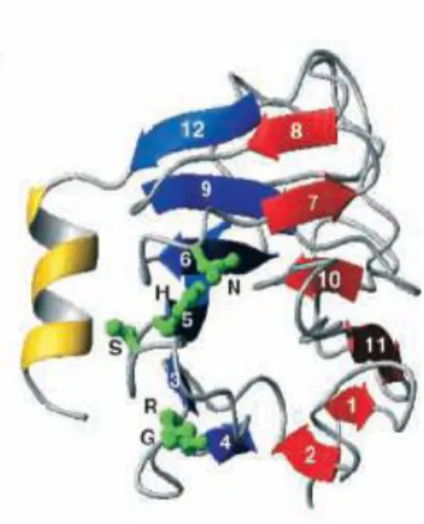
consists of a β-sandwich 37
Figure 12. Structure of Chk2K249R dimer 39
Figure 13. Two main pathway for Rad53 activation involving Rad9 and Mrc1 40 Figure 14. Rad53 phosphorylation sites upon 4-NQO and MMS 41 Figure 15. Mutation of possible phosphorylation sites at T811+S812+S821
of Rad53 destabilizes the Asf1-Rad53 interaction 71 Figure 16. Different mutants truncated at C-terminus of Rad53 72 Figure 17. Asf1-myc co-precipitated with TAP-tagged Rad53 C-terminal
truncation mutants 74
Figure 18. Rad53 interacts with Asf1 77
Figure 19. Effect of the asf1-T265A+270A mutant on HU and MMS sensitivities 79 Figure 20. The rad53-ALRR mutant re-enters the S-phase more rapidly than
W303 in the absence/presence of MMS 80
Figure 21. The rad53-ALRR mutant is competent at repressing late origins
firing in the presence of genotoxic stress 82
Figure 22. Shown is the cell cycle distribution of different mutations affecting the
Asf1-Rad53 interaction by flow cytometry analysis of DNA content 85 85
Figure 23. Microcolony viability analysis in rad24 versus rad53-ALRR rad24 strains 87 Figure 24. Microcolony analysis of adaptation to MMS-induced cell-cycle arrest
in wild type, rad53-ALRR, rad24 and rad53-ALRR rad24 strains 89 Figure 25. Individual cells were visualized in a microscope after ON treatment
with 0.05% MMS 89
Figure 26. Example of a yeast cell expressing a focus of Rad52-YFP
observed by fluorescence microscopy 90
List of Tables
Introduction
Model: Saccharomyces cerevisiae
The budding yeast Saccharomyces cerevisiae is a useful model for studying higher eukaryotic organisms. S.cerevisiae has many technical advantages such as rapid growth, dispersed cells, mutant isolation, a well-defined genetic system, numerous selective markers and easy gene manipulation. The genome of S.cerevisiae was completely sequenced in 1996 (Goffeau et al, 1996). It is composed of about 13,000,000 bp and 6,275 genes. It is estimated that 23% of yeast genes have homologs in the human genome, and 30% of known genes involved in human diseases have yeast orthologs. Additionally, the conservation of many cellular processes in eukaryotes, such as DNA replication, DNA damage checkpoints and cell cycle control also establishes the usefulness of yeast in the study of human disease (Johnson and O’Donnell, 2005; Lee and Nurse, 1987; Perego et al, 2000; Zhou and Elledge, 2000).
1.1 Genome stability
The maintenance of genomic stability is beneficial for the survival of an individual cell and crucial for cancer avoidance. Cells invest huge resources to maintain genomic stability, and cancer cells undergo an array of genetic changes to escape these barriers.
Defects in chromatin modulation and choromosomal aberrations, including altered chromatin structure in repetitive DNA, chromosome rearrangements and chromosome loss, are reflective of genomic instability and are a hallmark of cancer cells (Myung et al, 2003; Prado et al, 2004; Melo et al, 2007; Kops et al, 2005; Mitelman et al, 2007).
In addition, the DNA checkpoint pathway was originally identified in S.cerevisiae, because their loss of fuction resulted in defects in cell cycle progression in response to DNA damge. The absence of checkpoints can be lethal to cells. In cells mutated for checkpoint components, the spontaneous and induced chromosomal rearrangements are significantly increased (Hartwell et al, 1994; Myung et al. 2001; Kolodner et al. 2002; Myung and Kolodner, 2002). These results demonstrate that checkpoint proteins also play important roles in maintaining genomic stability.
1.2 Chromatin
1.2.1 Nucleosome and chromatin structure
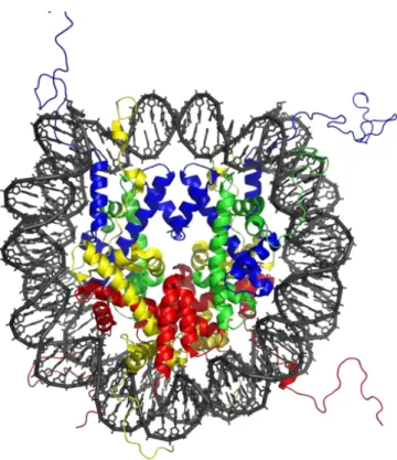
In eukaryotic cells, genomic DNA is packaged in chromatin as nucleosomes. The basic unit of chromatin is the nuclesome, consisting of 147bp of DNA wrapped around a histone octamer containing two copies each of histones H2A, H2B, H3 and H4 (Figure 1). The histone proteins are highly conserved across all eukaryotes. Histone proteins contain two functional domains: a core histone-fold domain involved in histone/histone and histone/DNA interactions, and a flexible N-terminal tail domain where a variety of covalent post-translational modifications sites have been studied (Luger et al, 1997). However, little is known about the conformation of the histone tails.
Two nuclesome core particles are separated by linker DNA varying in length from 10 to 80 bp. Histone H1 locks the linker DNA at the entry and exit points of the nucleosome. H1 participates in nucleosome positioning or spacing and formation of the higher-order chromatin structure (Ramakrishnan, 1997; Widom, 1998; Thomas, 1999; Maier et al. 2008). The primary chromatin structure consisting of nucleosomes assembled along DNA resembles ‘beads on a string’ as seen by electron microscopy (Woodcock et al, 2006). This chromatin fiber may then be further folded and compacted into higher-order chromatin structure that allows the packaging of the genomic DNA into the nucleus (Horn et al, 2002). The light-microscopy studies have revealed at least two types of chromatin: heterochromatin that stays condensed after cell division and euchromatin that decondenses during interphase (Grewal et al. 2002; Elgin et al. 2003; Maison et al. 2004). Euchromatin can either be actively transcribed or repressed whereas heterochromatin is commonly defined as transcriptionally repressed appearing in large units at the centromeres and telomeres (Grewal et al. 2007).
Figure 1. The structure of a nucleosome core particle defined by X-ray crystallography at 2.8 Å resolution. The core particle contains of two copies of H2A, H2B, H3 and H4 and
DNA. The view is from the top through the superhelical axis (Lunger et al, 1997). The histone globular domain consists of three α helices connected by two flexible loops and is referred to as the histone fold domain which allows histones to dimerize head to tail in a handshake manner (Arents and Moudrianakis, 1995).
1.2.2 Chromatin dynamics
Chromatin structure and packaging of the genome is important for regulating the cellular processes such as transcription, replication and repair (Kornberg et al, 1999). Therefore, factors that can alter chromatin structure are essential for and provide additional regulatory points in these cellular processes. The chromatin structure is highly dynamic to enable rapid unfolding, disassembly and refolding. Three main mechanisms that control the dynamics of chromatin structure have been identified: histone post-translational modification (PTMs), histone variants and ATP-dependent chromatin remodeling factors (remodelers).
1.2.2.1 Histone modifications
PTMs constitute reversible covalent modifications of amino acidic residues, such as serine and threonine phosphorylation, lysine acetylation, lysine and arginine methylation and lysine ubiquitination, among others. These PTMs modulate chromatin structure and/or recruit proteins to chromatin to mediate distinct cellular processes, including gene transcription, DNA replication and DNA repair (Strahl and Allis, 2000; Turner, 2000; Jenuwein and Allis, 2001; Vidanes et al., 2005). Each of these modifications is catalyzed by
a specific family of enzymes. For example, histone acetyltransferases, methyltransferases, kinases and ubiquitin ligases, as well as the enzymes that remove these modifications. The majority of histone PTMs were found in the N-terminal tails of histones that extend out from the globular core of the nucleosome, creating chromatin structures favourable either for activation or repression of genes through altering the degree of chromatin compaction (Luger et al, 1998). However, recent work has shown that modifications in the globular core play crucial roles in regulating the structure and function of chromatin and controlling biological function, such as H3K56 and H4K91 acetylation (Cosgrove et al, 2004; Masumoto et al, 2005; Xu et al, 2005; Hyland et al, 2005; Ye et al, 2005).
Some histone PTMs are induced in response to DNA damage (Figure 2). These histone PTMs may increase the plasticity of chromatin and facilitate the access of DNA repair and checkpoint proteins to sites of DNA lesions. After repair of lesions, PTMs are cleared for restoration of the chromatin structure and the shut down of checkpoint signaling. Several typical conserved PTMs are introduced here.
In mammals, phosphorylation of serine-139 in the C-terminal SQE motif of histone H2AX is rapidly induced at DSBs, and was named γ-H2AX (Rogakou et al, 1998). In yeast, this phosphorylation (γ-H2A) occurs at serine 129 in the most abundant form of histone H2A (Downs et al, 2000). This phosphorylation is catalysed by DNA damage checkpoint protein kinases: ATM, ATR and DNA-PK kinases in human cells (Burma et al, 2001; Ward et al, 2001; Stiff et al, 2004) or their homologues Mec1 and Tel1 in budding yeast (Downs et al, 2000; Redon et al, 2003; Nakamura et al, 2004) These kinases are recruited to DSBs through their association with partner proteins that recognise DNA lesions either directly or indirectly (Zou et al, 2003; Falck et al, 2005). The formation of γH2AX nuclear foci has been proven to be a DSB marker in mammalian cells. Although γH2AX is not essential for the initial recruitment of DSBresponse factors, it stabilizes the binding of the checkpoint factors, and it is required for effective repair of DSBs by both the NHEJ and HR pathways (Karagiannis et al, 2007; Celeste et al, 2002; 2003). Dephosphorylation of γH2AX by HTP-C (Histone phosphatase H2A complex) is necessary for the efficient recovery from DNA
damage after DNA repair. γH2AX recruits ubiquitin ligases that participate in the further recruitment of downstream players in the DNA damage response (Huen et al, 2007; Mailand et al, 2007; Kolas et al, 2007).
In yeast, H3K56 acetylation by Rtt109 is important for DNA damage response signaling and histone reassembly after DNA repair (Schneider et al, 2006). These acetylations regulate the binding of H3-H4 with histone chaperone CAF-1, but not Rtt106, to promote nucleosome assembly (Burgess et al, 2010). H3K56-Ac has been shown to require the histone chaperone Asf1 and occurs at the S phase in unstressed cells. Its role will be discussed in chapter 3.2.2.
The methyltransferase Dot1 mediates H3K79 methylation in both yeast and mammalian cells. In budding yeast, increased accessibility of H3K79me3 at DSBs is implicated in the recruitment of the Rad9 checkpoint adaptor protein (Bonilla et al, 2008; Huyen et al, 2004; Wysocki et al, 2005).
The recent studies have revealed that histone acetylation is important for promoting nucleosome assembly by enhancing histone binding with distinct histone chaperones. For example, the most highly-conserved mark of newly-synthesized histones is H4-K5, 8, 12, 16 acetylation which is conserved from yeast to humans. They are generally acetylated by a number of HATs such as NuA4, Gcn5 and in yeast. These acetylations may facilitate histone assembly into nucleosomes and facilitate DNA repair by non-homologous end joining (NHEJ) and homologous recombination (HR) (Sobel et al, 1995; Parthun et al, 1996; Bird et al, 2002; Murr et al, 2006; Murr et al, 2007). Following deposition onto the DNA, the newly synthesized H4 is rapidly deacetylated, which is required for proper chromatin maturation (Sobel et al, 1995; Loyola et al, 2006). H4-K16Ac inhibits the formation of compact 30-nanometer–like fibres and impedes the ability of chromatin to form cross-fibre interactions (Shogren-Knaak et al, 2006). In addition, methylation of histone H4-K20 is essential for recruiting the orthologous checkpoint proteins 53BP1 (mammals) and Crb2 (fission yeast) to sites of DSBs and subsequent activation of a DNA damage checkpoint.
Figure 2. Some histone PTMs are induced in response to DNA damage in yeast, such as γH2A/H2A.X, H3K56 Ac, H3K79 me3 and H4K5, 8, 12,16 Ac, which may increase the plasticity of chromatin, facilitate the access of DNA repair and checkpoint proteins to sites of DNA lesions.
1.2.2.2 Histone variants
Histone variants are distinct, non-allelic isoforms of the major histone types (Redon et al, 2002). The incorporation of the histone variants into nucleosomes may specify chromatin for particular biological roles (Malik and Henikoff, 2003; Talbert and Henikoff, 2010).
In mammals, there are three major classes of histone H3 variants: the replicative histones H3.1 and H3.2, the replacement histone H3.3, and the centromeric protein A (CENP-A) (Loyola et al, 2007). The replicative variants H3.1 and H3.2 represent the bulk of the histones and are expressed and deposited mostly in a replication-coupled manner during S phase. The replacement variant H3.3 is expressed constitutively at low levels throughout the cell cycle and incorporated in a replication-independent (RI) manner. S.cerevisiae has only a single H3 variant, most closely related to H3.3 (Tagami et al, 2004 ; Nakatani et al., 2004; Henikoff and Ahmad, 2005; Kamakaka and Biggins, 2005). CENP-A is a centromere specific form of the H3 and essential for centromere function in yeast and mammals (Black and Bassett, 2008).
Histone H2A has the largest number of variants, including H2A.X, H2A.Z, macroH2A and H2A.Bbd. H2A.Z (Htz1 in yeast) is essential in mammals but not in yeast (Faast et al, 2001), and its deletion increases the need for chromatin remodeling enzymes to promote
transcription (Santisteban et al, 2000). H2A.Z stabilizes the nucleosome and facilitates the formation of higher order structures (Park et al, 2004; Hoch et al, 2007; Fan et al, 2002), and is also required for the expression of genes that cluster near the sub-telomeric region where it has been proposed to act as a boundary element to stop the spread of heterochromatin (Meneghini et al. 2003; Raisner and Madhani, 2006). H2AX is similar to canonical H2A and is involved in the repair of DSBs as described above. In yeast there is no histone H2AX variant, but the major form of H2A is phosphorylated in a similar way and fulfils a similar role. (Shroff et al, 2004; Lydall and Whitehall, 2005). The functions of MacroH2A and H2A.Bbd are not fully understood. However, MacroH2A associates with repressive chromatin (Chakravarthy and Luger, 2006), whereas H2A.Bbd seems to be associated with transcriptionally active chromatin in mammals (Chadwich and Willard, 2001).
1.2.2.3 ATP-dependent chromatin remodeling enzymes
ATP-dependent chromatin remodeling complexes (consisting of between 4 and 17 subunits) render DNA more accessible by weakening DNA-histone contacts, sliding nucleosomes along DNA, or removing H2A-H2B dimers from the nucleosome (Becker and Horz, 2002; Flaus and Owen-Hughes, 2004; Saha et al, 2006). Chromatin remodeling enzymes utilize the energy of ATP hydrolysis to alter the contacts of histones with DNA (van Vugt et al, 2007). ATP-dependent chromatin remodeling enzymes possess a conserved Snf2 helicase domain that is capable of binding and hydrolyzing ATP (Eisen et al, 1995). They can be classified into four main families: Swi/Snf , Iswi, Ino80 and CHD.
SWI/SNF: In addition to the Snf2 helicase domain, The Swi/Snf (mating type switching/sucrose non-fermenting) family proteins possess a bromodomain that binds acetylated histone tails (Marfella et al, 2007; Wang et al, 2007). Drosophila brahma (BRM), mammalian BRG1 (Brahma related gene 1) and yeast SNF2 are examples of proteins that belong to this family. Swi/Snf complexes function in various cellular processes such as DNA replication, repair and transcription (Wang et al, 2007).
Iswi: the Iswi (imitation switch) family proteins possess a SANT (SWI3, ADA2, NCOR, TFIIIB) domain that binds histone tails (Marfella et al, 2007; Wang et al, 2007). The chromatin accessibility complex (CHRAC), nucleosome remodeling factor (NURF) complex and ATP-utilizing chromatin assembly and remodeling factor complex (ACF) are examples of the Iswi family (Tsukiyama(a) et al, 1995; Tsukiyama(b) et al, 1995; Varga-Weisz et al, 1997; Ito et al, 1997).
INO80: The Ino80 complex is reported to facilitate exonucleolytic resection, promote Ku70/80 recruitment and displace nucleosomes during a successful strand invasion event.
It also contributes to the cell-cycle checkpoint response. SRCAP (SNF2-related CREB-activator protein) and p400 are examples of Ino80 (inositol requiring 80) family proteins in mammals.
CHD: Like all ATP-dependent remodeling enzymes, CHD (Chromodomain helicase DNA binding) family proteins possess a conserved Snf2 helicase domain, and also a chromodomain that binds methylated lysines in the N-terminal tail of histone H3.
1.2.2.4 Histone chaperone
Histone chaperones deliver histones to the DNA during chromatin assembly, as well as remove histones from the DNA to facilitate chromatin disassembly, through binding to the positively charged histones and shielding their charge from the highly negatively charged DNA (Tyler et al, 2002). In budding yeast, several histone chaperones have been identified (Eitoku et al, 2008). Histone chaperones that bind preferentially to histones H3/H4 include Asf1, CAF-1 (chromatin assembly factor 1), HIR (histone regulator), Spt6 and Rtt106 (Emili et al, 2001; English et al, 2005; English et al, 2006; Green et al, 2005; Verreau et al, 1996; Tagami et al, 2004; Bortvin and Winston, 1996; Huang et al, 2005). Furthermore, FACT (facilitates chromatin transcription) (Orphanides et al, 1999), Nap1 and Vps75 can associate with all four core histones (Park et al, 2008; Andrew et al, 2008; Selth et al, 2009).
1.3 Asf1
CIA/Asf1 (cell cycle gene 1 (CCG1)-interacting factor A or antisilencing function 1) was first identified in a genetic screen based on its ability to disrupt transcriptional silencing in budding yeast when overexpressed (Le et al, 1997). Later, it was shown that Asf1 was a histone chaperone involved in both replication-coupled and replication-independent nucleosome assembly (Green et al, 2005; Tagami et al, 2004; Tyler et al, 1999). Asf1 is a highly conserved histone chaperone among eukaryotes that assembles and disassembles chromatin during transcription, replication and repair (Le et al, 1997; Tyler et al, 1999; Munakata et al, 2000; English et al, 2006). Although Asf1 is not required for viability in
S.cerevisiae, it is essential in S. pombe, Drosophila, chicken and humans (Umehara et al,
2002; Moshkin et al, 2002; Sanematsu et al, 2006; Groth et al, 2005). In mammals, there are two forms of Asf1, Asf1a and Asf1b, which appear to have common functions, as both proteins are present in the complexes co-purified with H3.1. However, they appear to have distinct functions in a replication-independent assembly pathway, since Asf1a interacts with HIRA, but Asf1b does not (Tagami et al, 2004).
Asf1 has a wide variety of functions in transcription, DNA replication and DNA repair (De Koning et al, 2007; Eitoku et al, 2008; Park and Luger, 2008; Sharp et al, 2001; Chimura et al, 2002; Adkins (a) et al, 2004; Korber et al, 2004; Schwabish et al, 2006; Williams et al, 2008; Le et al, 1997; Tyler et al, 1999; Tagami et al, 2004; Das et al, 2009; Emili et al, 2001; Hu et al, 2001). Yeast deleted for ASF1 exhibit spontaneous DNA damage, display increased frequencies of genome rearrangements (Myung et al, 2003; Prado et al, 2004), and are sensitive to a number of genotoxic agents that damage DNA during replication (Tyler et al, 1999; Linger et al, 2005). This is physiologically significant because chromosome rearrangements are an important source of tumourigenic mutations and often arise through replication-linked DNA damage.
1.3.1 Structure of Asf1
The N-terminal domain containing 155 residues is the highly-conserved core region of Asf1. In contrast, the C-terminal tail is variable, unstructured and flexible (Daganzo et al, 2003; Mousson et al, 2005). In S.cerevisiae and S.pombe, the Asf1 C-terminal sequence is extremely rich in asparatates and glutamates and this type of tail is common in histone chaperones. In vertebrates, the Asf1 C-terminal sequences are not as rich in acidic residues, but they are phosphorylated by Tousled-like kinases. The major Tlks (Tousled-like kinases) phosphorylation sites are located in the C-terminal part of Drosophila and human Asf1 within a (D/E)-N-S-(L/M) consensus motif, and both proteins cooperate in control of chromatin dynamics and cell cycle progression (Carrera et al, 2003; Sillje and Nigg, 2001; Mello et al, 2002). This phosphorylation by Tlk is inhibited by ATM/ATR/Chk1 kinases in response to DNA damage (Silje and Nigg, 2001; Groth et al, 2003). The loss of Tlk activity or mutation of phosphorylation sites for Asf1 results in degradation of Asf1 by both proteasome-dependent and independent pathways (Pilyugin et al, 2009).
The N-terminal domain of Asf1 consists of three helical linkers on top of a compact immunoglobulin-like β-sandwich fold. This domain is sufficient for all currently known functions of the full-length protein (Daganzo et al, 2003). Asf1 has a large electro-negative surface potential surrounding one side, and a highly conserved hydrophobic groove that interacts with histone proteins (Daganzo et al, 2003; Mousson et al, 2005).
The 3D structure of the functional N-terminal domain of budding yeast was determined by X-ray crystallography (Figure 3a)(Daganzo et al, 2003). Florence Mousson and Françoise Ochsenbein determined the structure of the human Asf1a N-terminal domain by nuclear magnetic resonance spectroscopy (NMR) (Mousson et al, 2005). The structures of yeast and human Asf1 N-terminal domains are quite similar. Recently, the YEATS domain of yeast Yaf9 was shown to have a highly similar structure to Asf1. Yaf9 is a subunit of both
the NuA4 histone H4 acetyltransferase complex and the SWR1 remodeling complex (Wang et al, 2009). The authors suggested that Yaf9 may have a similar histone-binding capacity as Asf1.
b
Figure 3. (a) Structure of the Asf1 N-terminal domain is well conserved. The superposition between hAsf1a (1-156) (purple) and S.cerevisiae Asf1(green) (Mousson et al, 2005; Daganzo et al, 2003).
(b) Ribbon diagram model of the Asf1p N-terminal domain bound to heterodimer histone H3 (cyano) and H4 (green) (English et al, 2006).
Structure of the Asf1-H3/H4 complex
This structure shows that Asf1 binds to a histone H3/H4 heterodimer (Fig 3b) (English et al, 2006; Natsume et al, 2007). The hydrophobic groove of Asf1 binds the histone H3-H4 heterodimer by enveloping the C-terminus helix of histone H3, thereby blocking the formation of a (H3-H4)2 heterotetramer. Furthermore, the C-terminus of histone H4, that
forms a mini-β sheet with histone H2A in the nucleosome, undergoes a major conformational change upon binding to Asf1 and adds a β strand to the Asf1 β sheet sandwich (English et al, 2006; Natsume et al, 2007). Additionally, the non-conserved acidic C-terminal tail of yeast Asf1 may strengthen the interaction between Asf1 and H3/H4 (English et al, 2006; Daganzo et al, 2003).
1.3.2 Asf1 at the crossroads of chromatin and DNA checkpoint pathways
Chromatin assembly occurs in a stepwise manner: a tetramer of histones H3/H4 is deposited first followed by two dimers of H2A/H2B on the outside of the tetramer (Verreault et al, 1996). Asf1 interacts with both CAF-1 and HIR and hands off histones to CAF-1 and HIRA facilitating replication-dependent and replication-independent chromatin assembly respectively (Krawitz et al, 2002; Nakatani et al, 2004; Green et al, 2005, Mousson et al, 2007).
1.3.2.1 Role for Asf1 in chromatin assembly/disassembly
Replication-dependent chromatin assembly/disassembly
The association of DNA with the histones in the nucleosome makes it difficult to access the DNA by protein molecules. The nucleosome is disassembled into two H2A-H2B dimers and a (H3-H4)2 tetramer ahead of the moving fork during DNA replication, transcription and
repair. Then the parental histones are relocated behind the replication fork and the full nucleosome density is completed by the deposition of newly synthesized histones. (Tagami et al, 2004; Falbo and Shen, 2006; Groth et al, 2007). In S.cerevisiae, passage through S phase in the absence of core histone synthesis results in a loss of viability that cannot be rescued by re-expression of histones in G2 (Kim et al, 1988).
The H2A-H2B chaperone FACT has been shown to be associated with the MCM (Minichromosome maintenance) helicase that unwinds DNA in front of the replication fork (Tan et al, 2006). Asf1 is also associated with the MCM helicase, suggesting that Asf1 plays a role in disrupting parental nucleosomes and potentially transfering them onto the nascent DNA behind the fork (Groth et al, 2007).
Asf1 acts in both chromatin assembly and disassembly (Adkins et al, 2004a,b; Adkins et al, 2007; Korber et al, 2006). Yeast Asf1 and both human isoforms Asf1a and Asf1b can interact with CAF-1 p60, promoting replication-dependent chromatin assembly synergistically with CAF-1 (Figure 5c) (Verreault, 2000; Sharp et al, 2001; Krawitz et al, 2002; Mello et al, 2002; Loyola and Almouzni, 2004). This pathway ensures that histones are promptly assembled onto newly replicated DNA to minimize the potential for DNA damage, as well as being important for inheritance of epigenetic information during DNA replication and repair (Ye et al, 2003; Groth et al, 2007; Henikoff et al, 2004). CAF-1 associates with the replication forks through an interaction with proliferating cell nuclear antigen (PCNA), a component essential for DNA replication and DNA repair (Shibahara and Stillman, 1999; Zhang et al, 2000; Moggs et al, 2000).
CAF-1 is essential in humans, as depletion of p60 CAF-1 triggers apoptosis in proliferating cells (Nabatiyan and Krude, 2004). In contrast to human cells, Asf1 and CAF-1 are not essential for cell viability in S.cerevisiae, probably because of the existence of other chaperones for histone H3-H4, such as Rtt106 (Huang et al, 2005). Besides Asf1, CAF-1 has been shown to mediate histone deposition onto DNA assisted by Rtt106 that binds to CAF-1 as well (Huang et al, 2005; Tyler et al, 2001). Although CAF-I is not essential in S. cerevisiae, its inactivation results in increased sensitivity to UV radiation and reduced silencing of genes adjacent to telomeric DNA (Kaufman et al, 1997).
CAF-1 is an evolutionarily conserved complex. In both yeast and human cells, CAF-1 consists of three subunits: Cac1, Cac2 and Cac3 in yeast ; p150, p60 and p48 in human cells; and p180, p105, p75 and p55 in Drosophila. The smallest subunit p55 binds the N-terminal part of histone H4 via a β-propeller structure (Smith and Stillman 1989; Kaufman et al, 1995; Kaufman et al, 1997; Song et al, 2008).
Asf1N binds a B domain motif found in both the p60 subunit of CAF-1 and HirA (Mello et al, 2002; Sanematsu et al, 2006; Tyler et al, 2001) (Figure 5b). CAF-I and HirA thus compete for binding to the same surface of Asf1N that is distinct from the surface of Asf1N that binds H3/H4 (Malay et al, 2008).
a b
Figure 4. A view of H3.1-H4 deposition by CAF-1. (a) The structure of Asf1-H3/H4 complex shows two binding sites for human ASFa on the histone dimer. Orange triangle indicates CAF-1-binding site. (b) Interaction of yeast Asf1 (SpAsf1) with a peptide from the p60 subunit of CAF-1, spCac2 (Malay et al, 2008). (c) Both Asf1a and Asf1b act as histone donors for CAF-1, promoting H3.1 deposition (De Koning et al, 2007).
Replication-independent chromatin assembly
Outside the S phase, histones can be deposited onto DNA by HIRA (HIR complex in S. cerevisiae) via a replication-independent pathway (Henikoff and Ahmad, 2005). The interaction between Asf1 and Hir was initially found in budding yeast and Asf1 can copurify with all four subunits of HIR (Hir1, Hir2, Hir3 and Hpc2). This interaction is necessary for telomeric silencing (Sharp et al, 2001; Daganzo et al, 2003; Green et al, 2005). Similarly, Asf1 forms complexes with histones H3 and H4 as well as HIRA in humans (Figure 4c). HIRA preferentially deposits the histone replacement variant H3.3 in nucleosomes (Loppin et al, 2005; Nakayama et al, 2007; Tagami et al, 2004; Van der Heijden et al, 2007). Since H3.3 is predominantly incorporated into actively transcribed genes (Mito et al, 2005; Schwartz et al, 2005; Wirbelauer et al, 2005), the HIRA/ASF1a complex is thought to mediate transcription-coupled deposition of histone H3.3 (Henikoff et al, 2008; Nourani et al, 2006; Prather et al, 2005; Ray-Gallet et al, 2002; Tagami et al, 2004). The overexpression of HIRA can also inhibit histone expression and lead to an S-phase arrest (Nelson et al, 2002).
The human Asf1 N-terminal domain has been shown to interact with the B-domain of HIRA (Daganzo et al, 2003; Zhang et al, 2005) in the form of an antiparallel β-hairpin (Tang et al, 2006) (Figure 4b). The evolutionarily conserved B-domain of HIRA (425-472) is located in the central portion of the protein. This surface is located on the opposite side of the H3 binding site of Asf1 (Tang et al, 2006). The ASF1 D37R+E39R double mutant disrupts the ASF1a-HIRA interaction, but does not affect the ASF1a-H3 complex (Daganzo et al, 2003; Tang et al, 2006; Mousson et al, 2005).
a b
c
Figure 5. A view of H3.3-H4 deposition by HIRA. (a) The structure of Asf1-H3/H4 complex shows two binding sites for human ASFa on the histone dimer. Orange triangle indicates HIRA-binding site. (b) hAsf1a-HIRA Ribbon diagram of a HIRA B domain peptide (green) bound to hAsf1a N-terminal domain (gray) (Tang et al, 2006). (c) Asf1a cooperate with HIRA to deposit H3.3.
1.3.2.2 Asf1 and DNA damage checkpoint pathway
Asf1 promotes genomic stability and protects against DNA damage and replication stress (Mousson et al, 2007). Asf1 participates in the regulation of the DNA damage checkpoint pathway by interacting with the central checkpoint kinase Rad53. Additionally, Asf1 is required for histone H3K56 acetylation during DNA repair.
Asf1 and Rad53
Asf1 is important for genomic stability, since cells lacking Asf1 are sensitive to agents that cause DNA damage or replication stress, such as methyl methane sulfonate (MMS), hydroxyurea (HU), camptothecin (CPT), bleomycin and cisplatin (Emili et al, 2001; Hu et al,
2001; Mousson et al, 2005; Ramey et al, 2004; Tamburini et al, 2005). HU treatment inhibits the ribonucleotide reductase (RNR) and leads to depletion of dNTPs and stalling of DNA replication (Elledge et al, 1992; Slater et al, 1973). MMS methylates DNA bases and can indirectly lead to the presence of abasic sites as well as single- and double-strand breaks in DNA. CPT is an interfacial inhibitor of DNA topoisomerase I that stabilizes the covalent complex formed by DNA topoisomerase I when it relaxes DNA by cleaving one DNA strand. DSBs are formed when the replication machinery collides with the CPT-stabilized complex of DNA topoisomerase I bound to the DNA (Pommier et al, 2003). Yeast Asf1 forms a complex with Rad53 in the absence of genotoxic stress. A part of the Rad53-Asf1 interaction is mediated by the FHA1 domain of Rad53 binding to a phsophorylated form of Asf1 (Schwartz et al, 2003). The Asf1-Rad53 complex was reportedly dissociated when yeast cells were subjected to genotoxic stress in a Mec1-dependent manner (Emilie et al, 2001; Hu et al, 2001). It was suggested that the phosphorylation sites of Rad53 induced by DNA damage could inhibit the Asf1-Rad53 interaction. These observations suggested that activation of Rad53 and liberation of Asf1 may be an important cellular response to DNA damage acting perhaps at the level of chromatin assembly (Emili et al, 2001; Hu et al, 2001). However, in mammalian cells, Asf1 does not interact with Chk2 (homologue of Rad53), but rather with Tlk kinases that are not conserved in yeast (Shillje and Nigg, 2001). The Tlk kinases phosphorylate C-terminal sequences of Asf1 during S phase, and this phosphorylation is inhibited in response to genotoxic stress. The functional significance of this phosphorylation is poorly understood, although there is some data suggesting that phosphorylation can affect the half-life of Asf1 isoforms.
Asf1 and H3K56Ac
Acetylation of H3K56 is an abundant modification of all newly synthesized H3 in budding yeast, fission yeasts and in Tetrahymena thermophilus (Masumoto et al, 2005; Garcia et al, 2007; Xhemalce et al, 2007), but is much less abundant in mammalian cells (Jasencakova et al, 2010). H3K56 is located at the DNA entry/exit point on the nucleosome core (Davey et al, 2002). Although H3K56 acetylation does not appear to greatly alter the overall structure of the nucleosome, acetylation at this residue increases the plasticity of nucleosomes, which may facilitate access of necessary protein factors to the DNA (Neumann et al, 2009; Watanabe et al, 2010).
H3K56Ac is involved in the response to DNA damage during replication (Collins et al, 2007; Driscoll et al, 2007; Han et al, 2007a,b,c; Masumoto et al, 2005; Tsubota et al, 2007). In yeast, H3K56ac peaks during S phase where it plays a role in the DNA damage response
and is largely deacetylated by the Sir2-related HDACs Hst3 and Hst4 during the G2/M phase of the cell cycle (Masumoto et al, 2005; Han et al, 2007a,b,c; Chen et al, 2008; Zhou et al, 2006; Celic et al, 2006; Maas et al, 2006).
H3K56 acetylation is catalyzed by Rtt109 (also known as KAT11) in S.cerevisiae and Schizosaccharomyces pombe (Xhemalce et al, 2007; Schneider et al, 2006; Tsubota et al, 2007; Han et al, 2007a,b,c; Driscoll et al, 2007; Allis et al, 2007), and by CBP/p300 and/or Gcn5 in mammalian cells (Das et al, 2009; Tjeertes et al, 2009). In addition to sensitivity to genotoxic stress, cells lacking the H3K56 acetyltransferase Rtt109 or cells expressing H3 with K56 mutated to arginine (H3K56R) exhibit an increased frequency of spontaneous chromosome breaks (Allis et al, 2007; Driscoll et al, 2007; Han et al, 2007a,b,c). Rtt109 harbors very low acetyltransferase activity on its own (Driscoll et al, 2007; Tsubota et al, 2007), but its activity is strongly stimulated by either Asf1 or Vps75 (Albaugh et al, 2010; Berndsen et al, 2008; Han et al, 2007c; Collins et al, 2007). Asf1 physically interacts with Rtt109 and is absolutely required for H3K56 acetylation. Vps75 also interacts with Rtt109 to promote H3K56ac. However, the accumulation of H3K56ac in vivo is dependent on Rtt109 and Asf1, but not Vps75. The asf1Δ or rtt109Δ mutants lack H3K56 acetylation, but no decrease was observed in cells lacking Vps75 (Tsubota et al, 2007; Schneider et al, 2006; Recht et al, 2006; Selth and Svejstrup, 2007).
H3K56 acetylation is required for S-phase chromatin assembly and was proposed to be a critical signal for turning off the DNA damage checkpoint following DNA repair followed (Chen et al, 2008), although this latter claim is controversial (Kim and Haber, 2009). H3K56 acetylation can increase the binding affinity between H3-H4 with CAF-1 and Rtt106 to promote efficient deposition of H3-H4 onto replicating DNA by these two histone chaperones (Li et al, 2008).
H3K56Ac is also involved in DSB repair. The mutations affecting H3K56Ac lead to increased sensitivity to agents that cause DSBs. The persistence of K56 acetylation when DSBs are present is due to the presence of DNA damage checkpoint proteins, and may be important for replication fork progression in the presence of DNA damage. (Chen et al, 2008).
Asf1 is thought to maintain the integrity of the replisome through H3K56 acetylation. Asf1 has been shown to directly interact with origins of replications and can also associate with components of the replisome (Groth et al, 2007; Han et al, 2007c). Indeed, in absence of Asf1 or H3K56 acetylation, components of the replisome are lost upon HU treatment (Franco et al, 2005; Han et al, 2007c). It appears that Asf1 and H3K56 acetylation promote the stability of stalled replications forks, contributing to cellular survival upon replication stress.
1.3.2.3 Transcriptional regulation by Asf1
In yeast, Asf1 facilitates chromatin disassembly at the PHO5 promoter to promote transcriptional activation, suggesting that it acts as a histone acceptor (Adkins and Tyler, 2004). Asf1 travels with the transcription machinery and/or rapidly fills in gaps left in nucleosome arrays following passage of RNA polymerase (Schwabish et al, 2006). The nature of Asf1 as an interactor with the TFIID subunit Bdf1 also suggests its participation in transcription control at various RNA polymerase II-dependent gene loci (Chimura et al, 2002; Zabaronick and Tyler, 2005). Asf1 is also involved in developmental gene expression control by mediating transcriptional repression of NOTCH target genes in Drosophila (Goodfellow et al, 2007).
In yeast, the loss of Asf1 results in impaired cell proliferation and minor defects of gene silencing at telomere and silent mating loci HMR and HML. These effects are greatly enhanced by inactivation of CAF-1, but not Hir (Tyler et al, 1999; Sharp et al, 2001; Krawitz et al, 2002). Asf1 and Hir participate together in a pathway for telomeric silencing that is independent of a pathway dependent on CAF-I (Daganzo et al, 2003).
In addition, Asf1 was found to mediate histone H3 eviction and deposition during transcriptional elongation. Furthermore, Asf1 has been implicated in transcription-dependent, replication-independent histone H3 exchange at promoters, another process which can deposit K56-acetylated H3.
1.3.2.4 Asf1 and histone modification
Asf1 can affect the PTM state of histones. In addition to its role in promoting H3-K56-acetylation described above, Asf1 also contributes to the H3-K56-acetylation of H3K9 and can promote trimethylation of H3K36 by Set2 in yeast (Adkins et al, 2007b; Lin et al, 2010). In human U2OS cells, histones bound to Asf1 showed two typical chromatin marks: H4K16Ac and H3K9Me3, giving rise to the hypothesis that Asf1 handles both new and parental histones during DNA replication (Groth et al, 2007).
1.4. DNA damage Checkpoints
Checkpoints were defined as molecular signaling cascades that trigger cell-cycle delay or arrest in response to DNA damage, providing sufficient time for repair from the damage (Hartwell et al. 1989). The DNA damage checkpoints control all the cell cycle phases in response to DNA damage. This damage results from the effect of exogenous mutagens, such as UV light, ionizing irradiation or chemical compounds, as well as spontaneous
damage that can arise from endogenous reactive oxygen species, or due to difficultes encountered during genomic DNA replication. If not repaired by continuously active repair pathways, DNA damage will lead to base mutations or single and double-strand chain breaks (Sancar et al, 2004). The DNA damage checkpoint cascades are evolutionarily conserved in eukaryotic organisms (Paulovich and Hartwell, 1995; Zhou and Elledge, 2000).
1.4.1 DNA damage checkpoint proteins and checkpoint pathway
Table 1. The proteins involved in the DNA-damage checkpoints and their orthologues (Harrison et al, 2006).
Checkpoint signaling consists of damage sensors, transducers and effectors (Ellege 1996). The sensors recognize the damaged DNA and initiate the signaling response. Transducers can be activated by the DNA damage signal passed from the sensors, then amplify the damage signal by phosphorylating downstream effectors. Finally, the effectors excute the regulation of different cellular processes.
DNA checkpoint pathways are conserved in eukaryotes and require a family of serine/threonine protein kinases which show strong similarity to the lipid kinase phosphatidyl-inositol-3-kinase (PI3K). Mec1 and Tel1 in budding yeast and their homologues Ataxia-Telangectasia Mutated (ATM), Ataxia-Telangectasia Related (ATR) and DNA-PK in humans are members of this family (Harrison and Haber, 2006). Other downstream kinases are also conserved and consist of Chk1 and Rad53 in budding yeast, Chk1 & Chk2 in vertebrates, Chk1 & Cds1 in fission yeast (Harrison, 2006).
of ssDNA at DSBs or at stalled replication forks is essential for activation of the DNA damage checkpoint. Single-strand DNA may be generated at stalled forks by the continued unwinding of DNA by MCM helicases ahead of the stalled replication fork (Sogo et al. 2002; Byun et al. 2005; Nedelcheva et al. 2005). The ssDNA is subsequently bound by RPA. The RPA-coated ssDNA, a structure commonly found after replicative stress or as a DNA repair intermediate, is critical for Mec1-Ddc2 recruitment. (Rouse and Jackson 2002; Zou and Elledge, 2003; Harrison and Harber, 2006; Branzei and Foiani, 2008).
Activation of PIKK family members also depends on other DNA damage sensors, such as the PCNA-like Ddc1-Mec3-Rad17 complex and the Rad24-Rfc2-5 alternative replication (RFC) complex. The budding yeast Ddc1-Mec3-Rad17 complex is a PCNA-like checkpoint clamp (orthologous to the human 9-1-1 complex) that was shown to be loaded onto the single- and double-stranded DNA junction of stalled replication forks by the clamp loader Rad24-RFC complex (Kondo et al, 2001; Melo et al, 2001; Majka et al. 2003). The Ddc1-Mec3-Rad17 complex stimulates the kinase activity of Mec1 (Majka et al, 2006), and Mec1 phosphorylates the Rad24 subunit of the clamp loader and the Ddc1 and Mec3 subunits of the clamp. Interestingly, it was shown that the co-localization of the Ddc1-Mec3-Rad17 complex and Mec1-Ddc2, is sufficient to activate Mec1 even without induced DNA damage (Bonilla et al, 2008).
Mec1 is an essential phosphatidylinositol 3-kinase-like kinase (PIKK) that associates with the DNA binding protein Ddc2 to form a checkpoint sensor complex (Paciotti et al, 2000). The phosphorylation of RPA may be required for later steps in the checkpoint cascade through interaction with other checkpoint proteins, or maybe required for the dissociation from DNA (Bartrand et al, 2004; Harrison and Haber, 2006). After binding to ssDNA, Mec1 functions in activating the checkpoint signal cascade via phosphorylation of the tranducer proteins Rad9 and Mrc1, and subsequent phosphorylation of the essential checkpoint effector kinase Rad53 (Branzei and Foiani, 2006). Rad53 is hyperphosphorylated and activated in response to DNA damage or DNA replication stress (Sun et al. 1996; Sanchez et al. 1996). Rad53 mutants are hypersensitive to genotoxic stress (Allen et al. 1994; Sun et al. 1996; Pellicioli et al. 1999). Activated Rad53 is critical for cellular processes through its downstream targets.
Another pathway involves the PIKK kinase Tel1 (ATM). Compared to Mec1 (ATR), Tel1 plays a minor role in response to DSBs in yeast. Deletion of TEL1 results in telomere shortening (Smogorzewska and de Lange, 2004), but does not show obvious checkpoint signalling defects or increased sensitivity towards DNA damage agents (Morrow et al, 1995). Instead of using Ddc2, Tel1 binds to DNA through its association with the MRX complex (Mre11-Rad50-Xrs2). In the presense of DSBs, Tel1 can activate the DNA
damage checkpoint pathway when Mec1 is absent (Nakada et al, 2003). Tel1 can respond to DSBs in a Mec1-dependent and –independent manner. In the Mec1 dependent manner, Tel1 is considered to contribute to DNA resection and produce ssDNA by activating an exonuclease that may correspond to MRX (Mantiero et al, 2007).
In contrast to budding yeast, both ATM and ATR have important functions in the checkpoint response in mammalian cells and are thought to be activated by different kinds of DNA damage. ATM is specially involved in the reponse to unprocessed DSBs, whereas ATR apprears to be activated by processed DSB ends, replicative stress and intermediates of DNA repair pathway (Jazayeri et al, 2006; Longhese et al, 2006).
Chk1 primarily contributes to the cell cycle arrest response to DNA damage in budding yeast, while Rad53 is more widely responsible response to DNA damage and replication stress. Chk1 has a major role in metazoan checkpoints, but a minor role in budding yeast.
Figure 6. DNA damage checkpoint pathways in S.cerevisiae (Harrison et al. 2006). Black arrows indicate protein kinase phosphorylations of several target proteins that activate downstream events, whereas a black line terminated in a bar indicates an inhibitory modifications. Grey arrows indicate protein interactions that facilitate checkpoint activation.
1.4.2 Downstream targets
The DNA checkpoint pathways are involved in cell cycle progression, DNA repair, DNA replication fork stabilization, DNA replication origin firing, chromatin remodeling, transcription of DNA damage response genes, and induced apoptosis and senescence (Zhou et al. 2000; Shiloh Y 2003; Abraham RT 2001; Bartek and Lukas, 2001; Santocanale
and Diffley 1998; Shirahige et al. 1998; Foiani et al. 2000; Nyberg et al. 2002; Tercero et al. 2003).
1.4.2.1 Cell cycle arrest
In response to DNA damage, checkpoints delay or arrest the cell cycle to provide sufficient time for repair. The phase of the cell cycle where the damage occurs determines the specific response. DNA damage checkpoints include G1/S checkpoints that delay or block cells before entry into S phase and S-phase and G2/M checkpoints that delay or block cells before the entry into mitosis (G2 in most organisms, but pre-anaphase in budding yeast). The G1/S checkpoint recognizes DNA damage during G1 phase and inhibits entry into S phase. The S-phase checkpoint/replication checkpoint, which is activated by DNA damage or impeded replication forks during S-phase, is crucial for stabilizing stalled replication forks and regulating late origin firing. The G2/M checkpoint prevents cells from initiating mitosis in the presence of damage to ensure that damaged chromosomes do not undergo chromosomal segregation during mitosis (Nyberg et al, 2002).
Pds1, a yeast securin, is required for normal cell cycle arrest in response to DNA damage (Cohen-Fix et al, 1997; Yamamoto et al, 1996). After DNA damage, Pds1 is hyperphosphorylated in a Mec-1, Rad9-, Chk1-dependent, but Rad53-independent manner (Cohen-Fix et al, 1997). At the entry into mitosis, this checkpoint-dependent phosphorylation prevents its degradation by ubiquitination by Cdc20/ APC (Anaphase Promoting Complex) (Agarwal et al, 2003; Sanchez et al, 1999). However, Rad53 inhibits mitotic exit. Rad53 is required to maintain CDK activity during the checkpoint arrest likely through inhibition of Cdc5 (Cheng et al, 1998; Sanchez et al, 1999). Cdc5 inhibits the Bub2/Bfa1 complex which in turn inhibits the mitotic exit network (MEN) (Geymonat et al, 2003; Hu et al, 2002; de Bettignies and Johnston, 2003). Rad53-dependent inhibition of Cdc5 could therefore inhibit progression through mitosis and help maintain the checkpoint arrest.
1.4.2.2 Transcriptional response and regulation of RNR
Dun1, a kinase downstream of Mec1/Rad53, was originally identified as a mutant deficient in the transcriptional induction of genes after DNA damage (Zhou and Elledge, 1993). Dun1 is recruited to activated Rad53 through the Dun1 FHA domain (Bashkirov et al, 2003), and then activated by Rad53 dependent phosphorylation of the Dun1 activation loop (Chen et al, 2007). Dun1 inhibits the transcriptional repressor Crt1 by phophorylation to prevent Crt1 binding to DNA, resulting in the up-regulation of several genes involved in DNA repair or
dNTP biosynthesis, including the RNR3 gene encoding a large subunit of the ribonucleotide reductase and the HUG1 gene encoding a small protein of unknown function (Elledge et al, 1992; Basrai et al,1999). DNA damage induced activation of Dun1 increases RNR activity by derepressing RNR gene transcription and by targeting by phosphorylation the RNR inhibitor Sml1 for proteolytic degradation (Zhao and Rothstein, 2002). Dun1 thus controls both the abundance and the activity of ribonucleotide reductase. The lethality of both MEC1 and RAD53 deletion can be suppressed either by deletion of SML1 or by overexpression of RNR large subunit genes (Zhao et al, 1998; Desany et al, 1998). These results indicate that Mec1 and Rad53 are important for increasing the synthesis of dNTPs. Additionally, Rad53 phosphorylates Swi6 that is a subunit of transcription factor SBF/MBF (Sidorova et al. 1997). This phosphorylation is thought to contribute to the delay in G1 after DNA damage.
1.4.2.3 Histone modification
As highlighted above, the rapid phosphorylation of serine 129 on histone H2A (γH2AX) is induced by DNA damage in a Mec1 and Tel1-dependent manner. γH2A is important for amplifying the checkpoint response in mammalian cells via recruitment of the checkpoint mediator Mdc1 (Su, 2006) and maintaining high checkpoint activity in yeast (Keogh et al, 2006). γH2A is also required for the recruitment of both the INO80 and SWR1 ATP-dependent chromatin remodeling complexes, as well as cohesins to DSBs. Mec1/Tel1 also phosphorylates Ino80.
When cells are exposed to genotoxic stress, Mec1-dependent downregulation of the Hst3 and Hst4 deacetylases lengthens the persistence of H3K56ac in chromatin (Maas et al, 2006, Thaminy et al, 2007, Masumoto et al, 2005). In addition, Rad9 binds H3K79-me3 in budding yeast. Removal of Rad9 from methylated histone leads to increased resection activity and partially bypasses the requirement for CDK activation of DSB processing (Lazzaro et al, 2008).
1.4.2.4 Activation of DNA repair
Different kinds of lesions require different repair pathways. The checkpoint pathway facilitates and induces DNA repair mechanisms (Harrison and Haber, 2006; Nyberg et al, 2002). So far, the data are not sufficient to define specific mechanisms. However, the activities of at least some DNA repair proteins are modified after DNA damage in S.cerevisiae. Several DNA damage checkpoint proteins contain tandem BRCT (BRCA1 carboxyl terminus) domains, a known phosphopeptide binding motif that is common among checkpoint and repair proteins (Glover et al, 2004). These proteins, such as BRCA1 and
BRCA2 in mammalian cells, are involved in DSB repair, and deficiencies in these proteins result in increased genomic instability (Hoeijmakers, 2001).
DSBs are potentially lethal lesions because segregation of chromosomes in the presence of un-repaired DSBs can result in the loss of large amounts of genetic information. DSBs can be induced by endogenous free oxygen radicals, collapsed replication forks, or by the physical force generated when dicentric or catenated chromosomes are pulled to opposite poles during mitosis (Acilan et al, 2007). DSBs are also produced exogenously when cells are exposed to DNA damaging agents, such as ionizing radiation (IR), chemical agents such as chemotherapeutics that poison topoisomerase I or topoisomerase II (Degrassi et al, 2004), or UV light that creates pyrimidine dimers and crosslinks (Limoli et al, 2002; Bosco et al, 2004).
DSB repair is carried out by two major pathways: non-homologous end joining (NHEJ) and homologous recombination (HR) (Haber et al, 2000). HR can be further subdivided into gene conversion and single strand annealing (SSA). HR, which occurs mainly during late S-G2 phase, takes advantage of sequence homology from an undamaged sister chromatid or homologous chromosome to repair the lesion with high fidelity. NHEJ, that involves processing and ligation of broken DNA ends, is the major pathway for repairing non-replication-associated breaks and occurs predominantly in the G1 phase of the cell cycle (Daley et al, 2005). The tightly packaged chromatin structure impedes DNA repair and the current DNA repair model on chromatin is: access-repair-restore (Smerdon et al, 1991). The phosphorylation of various repair factors is dependent on DNA damage checkpoints. In budding yeast, the checkpoint kinases have been shown to phosphorylate and regulate the recombination factors Srs2, Rad55 and Slx4 (Liberi et al, 2000; Bashkirov et al, 2000; Flott et al, 2007). Srs2, a DNA helicase/translocase, influences various steps of the recombination process by removing Rad51 from DNA. It is not known whether checkpoint-dependent Srs2 phosphorylation influences its role in recombination, however, it was proposed that Srs2 might be involved in removing checkpoint proteins from DBSs after repair to resume cell cycle progression (Vaze et al, 2002). The recombination protein Rad55, a Rad51 paralog, is phosphorylated by Mec1 upon DNA damage and this modification may play a role in activating recombinational repair (Bashkirov et al, 2000; Herzberg et al, 2006). Mec1/Tel1-dependent phosphorylation of Slx4 also controls the single-strand annealing (SSA) sub-pathway of DSB repair (Flott et al, 2007). Rtt107 is implicated in Mms2-dependent DNA repair during S phase, and its phosphorylation by Mec1 requires Slx4 (Rouse, 2004; Roberts et al, 2006). Nej1 phosphorylated by Dun1 has been shown to effect NHEJ (Ahnesorg and Jackson, 2007).
In S.cerevisiae, a mutation in Rad24 that affects activation of the Mec1-dependent pathway slowed down the kinetics of DSB resection and promoted ectopic recombination with short
homologous donor sequences (Aylon ad Kupiec, 2003). In fission yeast, the phosphorylation of Crb2 (a putative Rad9 ortholog) by CDK1 is important to mediate later steps of HR implicating the RecQ helicase Rqh1 and the Top3 topoisomerase (Caspari et al, 2002).
1.4.2.5 S-phase specific downsteam targets
During DNA replication, a DNA lesion caused by stresses such as UV damage, oxidative damage, genotoxic drugs or growth medium deprivation may slow down or even arrest progression of the replication fork. HU depletes the cellular dNTP pool and causes replication fork stalling. MMS also slows fork progression (Tercero and Diffley, 2001). MMS was found to inhibit replication fork progression independently of checkpoint kinases, but the inhibition of late origin firing is dependent on Mec1 and Rad53 activation.
Role of replication checkpoint in stabilizing stalled replication forks
The replication checkpoint is required to stabilize stalled replication forks to prevent their collapse and promote their restart (Longhese et al, 2003). Fork collapse can often result in DSBs, chromosomal rearragements and genomic instability (Branzei and Foiani, 2005). Cells lacking MEC1 or RAD53 are subject to irreversible collapse of stalled replication forks (Tercero et al. 2001). Many replication proteins dissociate from the stalled replication forks in the absence of the replication checkpoints (Blow et al. 2005; Cobb et al. 2003; Lucca et al. 2004). The replication checkpoints may stabilize the stalled replication fork by phosphorylating components of the replication fork, such as RPA, DNA polymerase α, Mcm2-7, Exo1 and Esc4/Rtt107 (Brush et al, 1996; Pellicioli et al, 1999; Cobb et al. 2003; Cobb et al, 2005; Cotta-Ramusino et al, 2005; Segurado and Diffley, 2008; Chin et al, 2006). These phosphorylations may prevent the collapse of arrested forks and the formation of abnormal replication intermediates, including DSBs. However, the targets of Rad53 and Mec1 that are important in preventing replication fork collapse have not yet been identified. MMS also slows fork progression as mentioned above. Cells lacking Mec1 are more sensitive to MMS than cells lacking Rad53, which suggests that Mec1 can stabilize stalled forks at least partly independently of Rad53 (Tercero et al. 2001).
Rad53 inhibits the exonuclease Exo1 by phosphorylation. Deletion of EXO1 can rescue the MMS, UV and IR, but not HU, sensitivity of rad53 mutants (Segurado et al, 2008). Thus, Exo1 is an important target of Rad53 in the cellular response to MMS, UV, and IR.
Additionally, Mrc1 is phosphorylated in a checkpoint-dependent manner. Mrc1 is associated with the replication fork and is required for normal replication fork progression, but also is