. . . .
. . . .
Transapical aortic valve implantation with a
self-expanding anatomically oriented valve
Volkmar Falk
1*
, Thomas Walther
2, Ehud Schwammenthal
3, Justus Strauch
4,
Diana Aicher
5, Thorsten Wahlers
4, Joachim Scha¨fers
5, Axel Linke
6, and
Friedrich W. Mohr
71
Division of Cardiovascular Surgery, University of Zu¨rich, Zu¨rich, Switzerland;2
Department of Cardiac Surgery, Kerckhoff Heart Center, Bad Nauheim, Germany;3
Sheba Medical Center, Tel Hashomer, Israel;4
Department of Cardiac and Thoracic Surgery, University of Cologne, Cologne, Germany;5
Division of Thoracic and Cardiovascular Surgery, Universita¨tskliniken des Saarlandes, Homburg, Germany;6
Department of Cardiology, Heart Center, Leipzig, Germany; and7
Department of Cardiac Surgery, Heart Center, Leipzig, Germany
Received 3 August 2010; revised 17 September 2010; accepted 21 October 2010; online publish-ahead-of-print 9 December 2010
This paper was guest edited by Professor Patrick W. Serruys, Erasmus Medical Center Rotterdam, The Netherlands
Aims The Medtronic EngagerTMaortic valve bioprosthesis is a self-expanding valve with support arms facilitating
anatomi-cally correct positioning and axial fixation. Valve leaflets, made of bovine pericardium, are mounted on a Nitinol frame. Here, we report the first in man study with this new implant (Trial Identifier NCT00677638).
Methods and results
Thirty patients (mean age 83.4 + 3.8 years; 83% female) with tricuspid aortic valve stenosis were included in the study. Mean logistic EuroSCORE was 23.4 + 11.9. Mean aortic annulus diameter was 21.8 + 1.4 mm. For this study, the Engager was available in only one size (23 mm), to fit aortic annuli of 19 – 23 mm. Standard transapical valve implantation was performed using predilation of the aortic valve and rapid ventricular pacing during ballon val-vuloplasty and most valve deployments. Accurate valve placement was achieved in 29/30 cases (97%). Post-implant peak-to-peak gradient was 13.3 + 9.3 mmHg. In 80% of the patients, no more than grade I paravalvular leakage was observed, in 13% grades I – II and in 3% grade II. Three patients (10%) required permanent pacemaker implantation for higher-degree or complete atrioventricular block. Four dissections (13%) occurred during positioning of the valve and were treated surgically in three cases. Thirty-day and in-hospital mortality were 20% and 23%, respectively, and 6-month survival was 56.7%. No structural failure occurred for up to 1 year.
Conclusion This series established the feasibility of implanting a novel self-expanding transapical aortic valve prosthesis predicta-bly into an anatomically correct position. Observed complications led to complete redesign of the delivery system for upcoming clinical studies with the goal of establishing safety and performance.
-Keywords Transapical aortic valve implantation
Introduction
Transcatheter aortic valve implantation is a novel technology, which is rapidly becoming a routine procedure in many centres
of expertise1,2 even before results from controlled randomized
studies are available. A major reason driving this development is that despite the excellent results of surgical aortic valve replace-ment, a substantial proportion of patients, mostly octogenarians with multiple comorbidities, are not referred for valve replacement
because they are not perceived to be suitable candidates.3,4In this
patient population, only one-third are expected to be alive by 1
year,5if left untreated.
In those undergoing transcatheter aortic valve implantation, a high incidence of paravalvular leaks has been observed, which is not always well tolerated by the hypertrophied stiff left ventricles of elderly patients with long-standing aortic stenosis. This, in addition to challenges in subcoronary device positioning and annular fixation with existing implants, has led to the development
*Corresponding author: Klinikdirektor Klinik fu¨r Herz- und Gefa¨sschirurgie, Universita¨tsspital Zu¨rich, Ra¨mistrasse 100, CH-8091 Zu¨rich. Tel:+41 44 255 3298, Fax: +41 44 2554446, Email:[email protected]
of different design concepts for transcatheter valves. Virtually, all previously reported experience with transapical transcatheter implantations relates to balloon-expandable prostheses.
The Medtronic Engager (originally Ventor Embracer) is a self-expanding valve with support arms to allow for anatomical posi-tioning and axial fixation. Here, we report the first-in-man study with this new transapical implant.
Methods
Approval by the competent authority as well as the local ethical com-mittees was obtained for a multi-centre feasibility study of 30 elderly patients (.75 years of age) with severe (mean gradient .40 mmHg, aortic valve area ,0.8 cm2) symptomatic aortic stenosis deemed to be at high risk (logistic EuroSCORE .11%) for surgical aortic valve replacement. The specific inclusion and exclusion criteria of the study are listed in Supplementary material online, Appendix. All patients gave written informed consent. The study was registered with the NIH at Clinicaltrials.gov (Trial identifier NCT00677638).
The valve
The EngagerTMAortic Valve Bioprosthesis (Medtronic, Inc.,
Minneapo-lis, MN, USA), shown in Figure1, is a flexible heart valve prosthesis composed of three leaflets, cut from tissue-fixated bovine pericardium, sewn to a polyester sleeve, and mounted on a compressible and self-expanding Nitinol frame (stent assembly). The stent assembly consists of a main frame and a support frame, which are coupled together so as to form the commissural posts of the valve. Two types of sewing materials are used: Polyester and expanded Polytetrafluoroethylene. The shaped valve prosthesis has a maximal inlet diameter of 28 mm, a waist diameter of 18 mm, and a diameter at the outlet of 23 mm. The fluid-dynamic shape is intended to minimize pressure losses at
the inlet and maximize pressure recovery at the outlet. The valve shape also creates an anatomical fit to facilitate periannular implan-tation and fixation at the target site with minimal risk of coronary obstruction (prosthesis confined at the level of the aortic cusps oppo-site the coronary ostia). Total length of implanted prosthesis is 24 mm upon deployment, with up to 7 mm seated subannularly (inlet).
For this study, the Engager was available in only one size (labelled as 23 mm according to its diameter at the commissural outlet), which was designed to fit an aortic annulus size of 19 – 23 mm.6The valve is ster-ilized and stored in a glutaraldehyde solution.
Implant procedure
The study was performed from June 2008 to October 2009 in Leipzig (26), Cologne (2), and Bad Homburg (2). Except for two implantations, all procedures were performed in a surgical hybrid suite. All pro-cedures were performed with the patient prepped and draped as for full sternotomy and cardiopulmonary bypass in stand-by. Prior to the procedure, the valve was crimped and mounted on the delivery system by pulling the valve through a cone-shaped converging tube using strings. The Engager is mounted onto the delivery catheter at the shaft’s top connector and the main frame is loaded into the deliv-ery tube while the support arms and the distal end of the Engager remain outside.
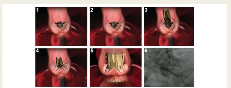
A standard transapical approach was performed.7The patient was placed in a supine position with the left chest slightly elevated. A 6F femoral arterial sheath was inserted into the right femoral artery and a pigtail catheter was placed into the aortic root for contrast aortogra-phy. A 6F introducer sheath was inserted into the right femoral vein and a guidewire placed in the right atrium to establish access for fast cannulation in case conversion to extracorporeal circulation became necessary (safety precaution). Low-dose heparin was given with a target activated clotting time of 150 s. An incision (5 – 7 cm) was per-formed in the fifth or sixth intercostal space for transapical access. An epicardial temporary pacemaker wire was sutured to the left ventricle and tested. The pericardium was incised and fixed with stay sutures. Two apical purse-string sutures with Teflon-felt pledgets were placed lateral and 2 cm above the true apex with an inner diameter of approximately 2 – 3 cm. Rotational angiography for 3D reconstruc-tion of the aortic root was performed in some cases as previously described.8 This technique facilitated orthogonal orientation of the C-arm and improved the ease of anatomically orientating the device. The left ventricular apex was punctured with an 18 G Seldinger-type needle. A 14F soft sheath was introduced and positioned across the aortic valve. A 0.0035′′ super-stiff guidewire was then positioned across the aortic arch. A balloon valvuloplasty catheter, selected according to the annulus diameter, was positioned under fluoroscopic and transoesophageal echocardiographic guidance and balloon valvulo-plasty was performed under a brief period of rapid ventricular pacing. The introducer sheath (30 Fr over-tube) containing the dilator was introduced over the guidewire and positioned across the aortic valve using fluoroscopic guidance. After removal of the dilator, the over-tube sheath was held in its position. At this point, the Engager delivery system with the mounted Engager valve was inserted into the over-tube via the cone-shaped entrance, the support arms were pressed against the shaft of the delivery catheter, and thus crimped into the over-tube. Commissural alignment was performed under fluoroscopic control with the valve still in the over-tube (Figures2and3). Once the delivery system exited the over-tube, the Engager support arms were exposed in the aortic root, expanded laterally, and were gently with-drawn under fluoroscopic guidance until they engaged against the floor of the aortic sinuses, thus providing tactile feedback to the oper-ator. Correct subcoronary positioning was verified by aortic root
Figure 1 Self-expanding Medtronic Engager (originally Ventor Embracer) first-generation aortic valve bioprosthesis used in this first-in-man series.
Figure 3 Alignment of prosthetic and native commissures using angiography and fluoroscopy. (A) The left anterior oblique (LAO) view can visualize the right (R) and left (L) coronary cusp and the commissure in between (a right anterior oblique view visualizes the commissure between right and non-coronary cusp); (B) the commissure between the right and left aortic cusp (CRL) is defined by the tip of the intercusp triangle spared by the contrast medium (arrow) and establishes the delivery axis; (C) a commissural post is aligned with the delivery axis when the operator can see en face through its window (circle); anterior position of the post is verified by confirming movement to the right of the image upon clockwise rotation of the delivery catheter under fluoroscopy (manoeuvre not shown); (D) when prosthetic and native commis-sures are aligned, the support arms are released directly above the sinuses and, when pulled back, engage against the sinuses; (E) aortic root angiography verifies correct subcoronary position of the support arms before deployment.
Figure 2 Principle of anatomically correct rotational positioning. 1 and 2, the commissural posts of the crimped prosthesis are rotated until aligned with the native commissures (see also Figure3); 3, when prosthetic and native commissures are aligned, the support arms are released directly above the sinuses; 4, the system is pulled back until the support arms engage against the sinuses; 5, unsheathing of the mainframe results in self-expanding release of the device and deployment is completed; 6, deployed device in situ (aortography).
angiography in left and right anterior oblique (RAO) projections (Figure3). If necessary, repositioning could be performed under fluoro-scopic/angiographic guidance by rotating the valve and re-aligning the commissural posts with the native commissures. The safety button of the delivery system was unlocked and rotation of the knob on the deliv-ery system in a clockwise direction allowed gradual self-expanding unco-vering of the prosthesis from its downstream to its upstream end. This manoeuvre was performed under a second period of rapid ventricular pacing (unless poorly tolerated by patient) and under continuous pull to maintain stable valve position during release. With the last turn of the knob, the device was released. Valve position and function were immediately assessed using angiographic and echocardiographic imaging as well as by simultaneous recording of the left ventricular and aortic pressure curves (Figure4). The transapical delivery system, including the guidewire, was removed and the apex was closed with the purse-string sutures. Intercostal blockade was performed using a local anaesthetic. The pericardium was partially closed over the apex and a left lateral chest tube inserted. The intercostal incision was closed in a standard fashion. Femoral sheaths were removed. Postopera-tive device-specific medical therapy consisted of aspirin 100 mg daily indefinitely and clopidogrel 75 mg daily for 3 months except in patients older than 85 years.
Statistical analysis
Standard methods for descriptive statistics have been used with data presented as the mean + standard deviation, or as the percentage of patient sample, as appropriate. Statistics were calculated using Micro-soft Excel version 2003.
Results
Patient characteristics
The baseline characteristics of the 30 enrolled are summarized in
Table1. Mean age was 83.4 + 3.8 years; 83% were female. The
majority of patients was in NYHA class III (77%) or IV (13%) and close to two-thirds had at least moderately impaired renal func-tion. Half the patient had coronary artery disease. Mean logistic EuroSCORE was 23.4 + 11.9. Mean aortic annulus diameter as assessed by transoesophageal echocardiography was 21.8 + 1.4 mm. Peak and mean aortic valve pressure gradients were 85.5 + 21.7 mmHg and 52.1 + 14.1 mm Hg, respectively. Fifty-seven per cent had aortic insufficiency grade 1 and 26% aortic insufficiency grade 2 or more.
Procedural details
Procedural details are summarized in Table2. Accurate valve
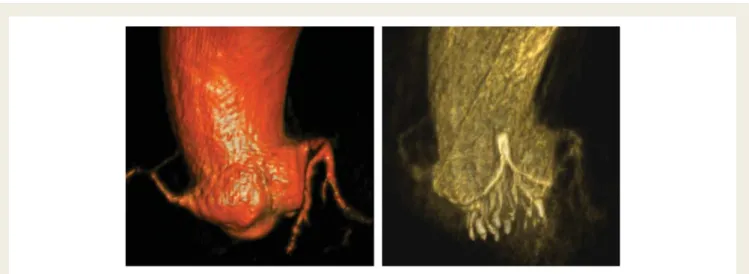
place-ment was achieved in 29/30 cases (97%) (Figure5). Mean fluoroscopy
time was 7.5 + 2.6 min; 129 + 58 mL of contrast medium was used per procedure. Skin-to-skin time was 74 + 26 min (excluding con-versions to sternotomy). The invasively measured peak-to-peak gra-dient after valve implantation was 13.3 + 9.3 mmHg; corresponding to a Doppler mean pressure gradient of 12.6 + 5.9 mmHg (peak instantaneous Doppler gradient 24.6 + 10.0 mmHg). Aortic regurgi-tation due to paravalvular leak grade 1 or less was present in 80%, grades I – II in 13%, and grade II in 3% of the patients. Aortic insuffi-ciency greater than grade II was not observed.
Procedure-related complications
Three patients (10%) required permanent pacemaker implantation for complete or higher-degree atrioventricular block, or bradyar-rhythmia with episodes of asystole. Five patients required temporary dialysis or continuous veno-venous haemo-filtration in the early postoperative period; of those, four had a diagnosis of pre-existing chronic renal failure. In four patients (13%), aortic dissection was diagnosed during or after implantation. One patient with localized dissection of the ascending aorta was treated conservatively. The
Figure 4 Simultaneous recordings of the pressure tracings in the aorta (pigtail catheter) and the left ventricle (using an introducer sheath) after implantation of a Medtronic Engager aortic valve in a patient with atrial fibrillation
patient is alive and well at 1-year follow-up with normal valve func-tion and mild paravalvular leakage. One patient with uneventful implantation showed a type A dissection on post-implant transoeso-phageal echocardiographic assessment and immediately underwent root replacement and complete arch replacement. In one patient, initial positioning of the support frame showed that one arm was deployed in the left ventricular outflow tract, i.e. below instead of
within the non-coronary sinus. The delivery catheter was advanced to reposition the non-coronary support arm above the valve. Intra-procedural angiography and transoesophageal echocardiography revealed a type A dissection posteriorly above the non-coronary sinus. The procedure was converted to replacement of the aortic root and the ascending aorta. The location of the dissection was confirmed to be located posteriorly above the non-coronary sinus. The fourth patient, after an uneventful implant procedure, experienced an episode of severe chest pain on the regular ward on day six. Computed tomography of the chest revealed a type A dissection starting at the sinotubular junction extending circumfer-entially for approximately 2708, with the left posterior aspect spared. The patient was re-operated a day later, and the ascending aorta and part of the aortic arch were replaced. Retrospective review of the intraoperative angiography after device positioning,
but before deployment (Figure6), revealed that a local dissection
above the non-coronary sinus had already been present. In all con-verted cases, the Engager valve, which was located in an anatomi-cally correct position, was explanted. Two of the three patients who underwent surgical repair for type A dissection were dis-charged from the hospital alive.
In one patient, post-dilatation was performed because of an increased gradient after deployment, resulting in an acceptable result (peak and mean gradients: 22/10 mmHg, grade II AR). Ten hours post procedure, the patient developed atrial fibrillation, pul-monary oedema and low cardiac output. Transoesophageal echo-cardiography suggested mitral regurgitation grade III and outflow tract turbulence compatible with moderate aortic regurgitation. Reoperation was performed for suspected dislodgement of the valve into the left ventricular outflow tract causing mitral valve interference. At surgical inspection, the valve was found stable in the proper position without detectable paravalvular gaps/dehis-cence, nor any structural damage or impairment of the mitral valve. The Engager valve was explanted and a 21 mm Mitroflow valve was implanted. Retrospective analysis of the transoesopha-geal echocardiographic study supports the view that the mitral regurgitation was secondary to systolic anterior motion of the mitral valve, also causing dynamic subaortic obstruction. The patient had an uneventful recovery.
. . . . . . . . . . . . . . . . . . . . . . . . . . . . . . . . . . . .
Table 1 Patient baseline characteristics
Characteristic No. of patients % Age (83.4 + 3.8) ≥90 years 2 7 80 – 89 years 25 83 Logistic EuroSCORE (23.4 + 11.9) Female sex 25 83 Body mass index (kg/m2; 27.2 + 5.0)
≥25 21 70
Body surface area (m2; 1.74 + 0.17)
≥1.8 11 37
New York Heart Association class
II 3 10 III 23 77 IV 4 13 Hypertension 28 93 Syncope/presyncope 10 33 Dizziness on exertion 9 30 Angina pectoris 12 40 Diabetes 14 47 Coronary artery disease 14 47 Chronic pulmonary disease 5 17 Estimated glomerular filtration rate ,60 mL/
min/1.73 m2
19 63 Previous stroke 3 10 Extracardiac arteriopathy 14 47 Previous cardiac surgery 1 3 Ejection fraction ,50% 9 30 Mitral regurgitation III 0 0 IV 0 0 Aortic regurgitation None 5 17 I 17 57 II 7 23 III 1 3
Pulmonary hypertension (systolic pressure .60 mmHg)
1 3
Atrial fibrillation 8 27 Pre-existing pacemaker 2 7
. . . .
Table 2 Procedure details
Characteristic
Aortic annulus diameter (mm) 21.8 + 1.4 Accurate device placement (patients) 29 (97%) Skin-to-skin time (min)a 74 + 16 Valve implantation time (min)b 6.0 + 2.4 Contrast medium volume (mL) 130 + 58 Fluoroscopy time (min) 7.5 + 2.6 Post-implantation dilatation (patients) 4 (13%)
a
Skin-to-skin time is exclusive of the four patients who were converted to open surgery.
b
Valve implantation time is defined as the time from over-tube insertion to completion of valve deployment.
Clinical outcome
Six patients (20%) died within 30 days post-implantation; a seventh patient (90 years old EuroSCORE 63%) died after a prolonged hos-pital stay after 61 days due to mesenteric ischaemia and sepsis (in-hospital mortality 23%). In one patient, one support arm was placed in the left ventricular outflow tract rather than the sinus resulting in aortic insufficiency grade II. Since the patient was haemodynamically stable, no re-intervention was performed. After initial extubation, the patient suffered from respiratory dys-function and was re-intubated. Reoperation was again considered but ultimately not performed since aortic insufficiency was only moderate and the overall condition was judged unfavourable for
open surgery. The patient developed low cardiac output and expired on the fifth postoperative day. In one patient, severe bleeding resulted from injury of the apex and required sternotomy and cardiopulmonary bypass support to repair the left ventricle. After a second revision, the patient required high-dose catechol-amine support, developed metabolic acidosis, and died on the same day. One patient was found unresponsive and pulseless on the regular ward on the sixth postoperative day after an initially uneventful recovery. Cardiac resuscitation was successful (echo-cardiography showed a normally functioning valve prosthesis and pericardial tamponade was ruled out), but the patient died of anoxic brain damage on the tenth postoperative day. One patient with initially uneventful recovery developed renal failure requiring re-intubation and subsequently died of multi-organ failure secondary to intestinal malperfusion on the third postopera-tive day. Another patient with a prolonged recovery acquired a norovirus infection and developed severe diarrhea. Despite ade-quate supportive therapy, the patient died on the 29th postopera-tive day of multi-organ failure. One patient developed intractable atrial fibrillation with rapid ventricular response in the context of a thyrotoxic crisis and expired due to refractory circulatory failure. In all but the first patient, proper valve position and function were confirmed by echocardiography. Mean and median hospital stay were 25.0 + 20.6 days and 18 days (interquartile range 13.75 – 27.25), respectively. Survival at 6 months was 56.7%.
Discussion
Transapical aortic valve implantation (TA-AVI) has evolved into a routine procedure for high-risk patients with aortic valve stenosis
in many centres of expertise.7,9,10Despite the lack of randomized
trials comparing TA-AVI with standard open surgical aortic valve replacement, it is generally assumed that the transcatheter approach may potentially provide benefit to higher risk patients. This assumption is based on the notion that minimally invasive transcatheter valve implantation—by avoiding cardiopulmonary
bypass, cardiac ischaemia, and sternotomy—may limit
procedure-related risks in elderly patient. While some excellent
Figure 5 Pre- and post-implant intraoperative rotational angiography (DynaCT, Siemens) demonstrates anatomical positioning of the valve.
Figure 6 Angiography before deployment (right anterior
oblique projection). A local dissection (extravasation) can be identified in direct continuation of the exposed prosthetic com-missural post of the crimped valve (arrow) above the non-coronary sinus (right posterior) during implantation of the Embracer valve.
results have been published, others report new complications associated with transcatheter valve implantation, including valve
dislocation,11,12 left main stem occlusion,13 impairment of mitral
valve function,14 rupture of the aortic annulus,15 ventricular
septal defect,16 and aortic dissection.17 Data from the SOURCE
registry have shown that vascular complications, the necessity for conversion to sternotomy, and residual aortic regurgitation greater than grade II all have a direct impact on procedure-related outcomes and are independent predictors of increased 30-day
mortality.2,18
Here, we report the results of a feasibility study with the Med-tronic Engager transapical self-expanding valve. The shaped valve is equipped with sinus support arms that allow for anatomically correct positioning and deployment in a self-guided procedure, and for axial fixation. This design prevents the native leaflets from being pushed against the coronary arteries as they are held against the main frame to provide additional sealing and mini-mize paravalvular leakage. Commissural posts and leaflet design create a mildly diverging outlet for optimal haemodynamic per-formance by avoiding flow separation and enhancing pressure recovery. The flared subvalvular inlet with barbs anchors the prosthesis at the upstream side of the aortic annulus to prevent device migration, provides a smooth entrance of flow into the prosthesis, and seals the outflow tract to limit paraprosthetic regurgitation.
While implantation of the valve was uneventful in the vast majority of cases and accurate placement of the valve with all support arms seated in the sinuses could be confirmed in all but one patient, a number of complications were encountered and need to be addressed. Of concern was the high rate of aortic dis-sections observed in this study. Careful review of the imaging data from procedures that resulted in dissections, revealed as the most likely common mechanism an interaction between an exposed prosthetic commissural post of the crimped bioprosthesis and the posterior aspect of the aorta, above the non-coronary cusp, as the delivery system was advanced into the ascending aorta
(Figure 6). The straight and rigid delivery system was unable to
conform to the angle between the left ventricle and the ascending aorta. Therefore, the vector of advancement into the ascending aorta had not only the intended superior component, but also an undesired posterior component (towards the aortic wall). This ventriculo-aortic angle can only be appreciated in the RAO, but not the left anterior oblique view, since the latter looks at
this angulation en face (Figure 7). Elevation of the left ventricle
by stay sutures to enhance exposure, as customary in TA-AVI, may further increase the ventriculo-aortic angle, and thus increase the risk of aortic injury, particularly in the presence of small aortic
roots. As shown in Figure8, the interaction between the straight
delivery system and the angulated anatomy was also the source of potential misplacement of a support arm below the non-coronary sinus that was observed in one case. It was therefore concluded that a straight and rigid delivery system should be aban-doned in favour of a delivery system with the following key
attri-butes: (i) flexible shaft for over-the-wire tracking, thus
conforming to the ventriculo-aortic angle and allowing co-axial alignment with the ascending aorta; (ii) protective cover over the prosthetic commissural posts until the valve’s final position prior to the deployment of the main frame is confirmed. Based on these learnings, such a system has been recently developed
(Figure 9) and is currently undergoing final testing. In addition,
several design modifications have been implemented in the valve prosthesis itself: (i) increase in the space between support arms and main frame to accommodate bulkier calcified leaflets and com-missures; (ii) increase in radial strength at the waist (native annulus) level to avoid the potential need for post-dilatation; (iii) reduction in the length of the support arms to improve prosthesis confor-mance to the native root anatomy.
Malpositioning (but without device embolization) was found in 3% (one case) which is in line with the data from other trials
using different implants.12,19Importantly, the procedure has now
been modified to include confirmation of support arm position in two planes, so that proper placement of all three support arms can be ascertained prior to main frame deployment. In this series, we did not observe coronary occlusion, which may
Figure 7 CT example showing that the ventriculo-aortic angle will be apparent in the RAO, but not the left anterior oblique equivalent of the corresponding angiographic projections.
Figure 8 Angiography before deployment (right anterior oblique view). Left: one support arm misplaced below the non-coronary sinus. Right: lowering the shaft of delivery catheter to attenuate the angle resulted in successful capture of the non-coronary sinus. All support arms are now above the valve and each is within an aortic sinus. The valve was successfully deployed.
Figure 9 Delivery system redesigned as a direct consequence of the FIM learnings. Left, single-piece apical introducer sheath containing flex-ible delivery catheter with valve loaded and covered; right, delivery catheter after advancement and exposure of the support arms, with the valve main frame and commissural posts still fully covered.
potentially occur due to ostial obstruction by a displaced native leaflet, inappropriately high positioning of a sealing cuff, the sup-porting stent posts of an implanted valve, thrombo-embolic
com-plications, or dissection of the aortic annulus.10
The 20% 30-day and 23% in-hospital mortality in this first-in-man series needs to be evaluated in the context of the early transapical valve experiences with other implants. In the US feasibility trial for the Sapien valve, a 17.5% 30-day and 22.5%
in-hospital mortality were reported.20In the Partner EU trial, a
multi-centre trial that included 69 patients receiving an Edwards Sapien valve, a similar mortality of 18.8% was reported. More recent large single-centre studies and registry data show mortality rates
in the range of 8 – 17%2,21 and similar or better results than in
risk-adjusted groups of patients undergoing conventional surgery.22
Besides major cardiac and non-cardiac complications, the fre-quent occurrence of paravalvular leakage and atrioventricular block are of concern in transcatheter valve implantations. Paravalv-ular leak resulting in aortic insufficiency of grade II or more is
reported at an incidence of up to 50%,11,12may lead to elevated
left ventricular filling pressures, left ventricular dysfunction, haemo-lysis, and promote endocarditis. Consequently, paravalvular leaks should not simply be viewed as acceptable unavoidable collateral
damage.23 The rate of paravalvular leakage with the Engager
valve was within the range of the more favourable reported implant series, with a low incidence of aortic insufficiency grade II. The His bundle and its left bundle branch pass adjacent to the non-coronary cusp of the aortic valve within the central fibrous body. Local compression of the His bundle by the expand-ing frame, especially in the settexpand-ing of a thickened non-coronary cusp, may therefore lead to atrioventricular block requiring
permanent pacemaker implantation and has a reported incidence of 10 – 30% in patients undergoing transcatheter aortic valve
implantation.24 For the Engager valve, we observed a 10%
inci-dence of atrioventricular blocks requiring pacemaker implantation, which is in line with previous reports.
While some of the reported complications are inherent to the procedure and will not be preventable, others may be reduced by careful patient selection and improved procedure planning. This would include a better understanding of the behaviour of the native valve during implantation and the interaction of the implant and the native leaflets. Even the normal aortic valve
complex has significant regional asymmetry and anisotropy.25
The presence of asymmetric calcification patterns, variations in aortic root geometry and in mechanical properties of the aorta, the valve and the aortic annulus all may further impact the final position of the valve and determine potential collateral damage such as leaks and atrioventricular block. The valve selection process may be further optimized based on individual anatomic cri-teria that may favour one implant over the other for different path-ologies. Models that have been developed so far allow to account
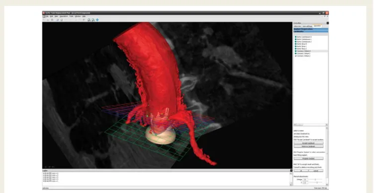
for native aortic root and device geometry (Figure10).26Given the
different behaviour of device materials (deformable Nitinol frames conforming to the local anatomy vs. stainless steel remodelling the
implantation site27,28), modelling of their mechanical properties (in
addition to that of the pathology) would also be important to achieve the important goal of knowledge-based valve selection. This is of increasing interest as a variety of valve designs and sizes is currently entering clinical trials. Enhanced imaging and mod-elling, as well as augmented reality testing will most likely help to select the best possible implant based on individual anatomic and
Figure 10 Preoperative planning using virtual implants in 3D reconstruction of the aortic root derived from intraoperative Dyna-CT data demonstrating perfect anatomical alignment.
morphologic data and facilitate the process of implantation in the
near future.29
Conclusion
This series has demonstrated the feasibility of implanting a novel self-expanding transapical aortic valve prosthesis predictably into an anatomically correct position within relatively short fluoroscopy times. However, important complications have been observed, which elicited a thorough review of procedure, delivery system, and prosthesis. As a result of this process, the delivery system has been completely redesigned, the valve prosthesis underwent minor modifications, and both will be tested in upcoming clinical studies with the goal of establishing safety and performance.
Supplementary material
Supplementary material is available at European Heart Journal online.
Acknowledgements
The authors would like to thank Mrs Sharon Sax for her help in collecting and preparing the data.
Conflict of interest: E.S. is a Co-Founder of Ventor Technol-ogies Ltd., in which he had an equity position until the company was acquired by Medtronic Inc. in February 2009. He is a paid consultant to Medtronic Ventor Ltd.
References
1. Ye J, Cheung A, Lichtenstein SV, Nietlispach F, Albugami S, Masson JB, Thompson CR, Munt B, Moss R, Carere RG, Jamieson WR, Webb JG. Transapical transcatheter aortic valve implantation: follow-up to 3 years. J Thorac Cardiovasc Surg 2010;139:1107 – 1113.
2. Wendler O, Walther T, Nataf P, Rubino P, Schroefel H, Thielmann M, Treede H, Thomas M. Trans-apical aortic valve implantation: univariate and multivariate ana-lyses of the early results from the SOURCE registry. Eur J Cardiothorac Surg 2010; 38:119 – 127.
3. Varadarajan P, Kapoor N, Bansal RC, Pai RG. Clinical profile and natural history of 453 nonsurgically managed patients with severe aortic stenosis. Ann Thorac Surg 2006;82:2111 – 2115.
4. Iung B, Baron G, Butchart EG, Delahaye F, Gohlke-Ba¨rwold C, Levang OW, Tornos P, Vanoverschelde JL, Vermeer F, Boersma E, Ravaud P, Vahanian A. A prospective survey of patients with valvular disease in Europe: the Euro Heart Survey on valvular disease. Eur Heart J 2003;24:1231 – 1243.
5. Lindroos M, Kupari M, Heikkila¨ J, Tilvis R. Prevalence of aortic valve abnormalities in the elderly: an echocardiographic study of a random population sample. J Am Coll Cardiol 1993;21:1220 – 1225.
6. Falk V, Schwammenthal E, Kempfert J, Linke A, Schuler G, Mohr FW, Walther T. New anatomically oriented transapical aortic valve implantation. Ann Thorac Surg 2009;87:925 – 926.
7. Walther T, Dewey T, Borger MA, Kempfert J, Linke A, Becht R, Falk V, Schuler G, Mohr FW, Mack M. Transapical aortic valve implantation: step by step. Ann Thorac Surg 2009;87:276 – 283.
8. Kempfert J, Falk V, Schuler G, Linke A, Merk D, Mohr FW, Walther T. Dyna-CT during minimally invasive off-pump transapical aortic valve implantation. Ann Thorac Surg 2009;88:2041.
9. Al-Attar N, Himbert D, Descoutures F, Iung B, Raffoul R, Messika-Zeitoun D, Brochet E, Francis F, Ibrahim H, Vahanian A, Nataf P. Transcatheter aortic valve implantation: selection strategy is crucial for outcome. Ann Thorac Surg 2009;87: 1757 – 1762;discussion 1762 – 1763.
10. Webb JG, Altwegg L, Boone RH, Cheung A, Ye J, Lichtenstein S, Lee M, Masson JB, Thompson C, Moss R, Carere R, Munt B, Nietlispach F,
Humphries K. Transcatheter aortic valve implantation: impact on clinical and valve-related outcomes. Circulation 2009;119:3009 – 3016.
11. Clavel MA, Webb JG, Pibarot P, Altwegg L, Dumont E, Thompson C, De Larochellie`re R, Doyle D, Masson JB, Bergeron S, Bertrand OF, Rode´s-Cabau J. Comparison of the hemodynamic performance of percutaneous and surgical bio-prostheses for the treatment of severe aortic stenosis. J Am Coll Cardiol 2009;53: 1883 – 1891.
12. Clavel MA, Dumont E, Pibarot P, Doyle D, De Larochellie`re R, Villeneuve J, Bergeron S, Couture C, Rode´s-Cabau J. Severe valvular regurgitation and late prosthesis embolization after percutaneous aortic valve implantation. Ann Thorac Surg 2009;87:618 – 621.
13. Kapadia SR, Svensson L, Tuzcu EM. Successful percutaneous management of left main trunk occlusion during percutaneous aortic valve replacement. Catheter Car-diovasc Interv 2009;73:966 – 972.
14. Wong DR, Boone RH, Thompson CR, Allard MF, Altwegg L, Carere RG, Cheung A, Ye J, Lichtenstein SV, Ling H, Webb JG. Mitral valve injury late after transcatheter aortic valve implantation. J Thorac Cardiovasc Surg 2009;137: 1547 – 1549.
15. Himbert D, Descoutures F, Al-Attar N, Iung B, Ducrocq G, De´taint D, Brochet E, Messika-Zeitoun D, Francis F, Ibrahim H, Nataf P, Vahanian A. Results of transfe-moral or transapical aortic valve implantation following a uniform assessment in high-risk patients with aortic stenosis. J Am Coll Cardiol 2009;54:303 – 311. 16. Al-Attar N, Ghodbane W, Himbert D, Rau C, Raffoul R, Messika-Zeitoun D,
Brochet E, Vahanian A, Nataf P. Unexpected complications of transapical aortic valve implantation. Ann Thorac Surg 2009;88:90 – 94.
17. Walther T, Falk V, Kempfert J, Borger MA, Fassl J, Chu MW, Schuler G, Mohr FW. Transapical minimally invasive aortic valve implantation; the initial 50patients. Eur J Cardiothorac Surg 2008;33:983 – 988.
18. Thomas M, Schymik G, Walther T, Himbert D, Lefe`vre T, Cartier J, Treede H, Eggebrecht H, Rubino P, Michev L, Lange R, Wendler O. 30 Day Results of the SOURCE Registry: A European Registry of Transcatheter Aortic Valve Implantation using the Edwards SAPIENTM Valve Circ 2010;122:62 – 69.
19. Al Ali AM, Altwegg L, Horlick EM, Feindel C, Thompson CR, Cheung A, Carere RG, Humphries K, Ye J, Masson JB, Webb JG. Prevention and management of transcatheter balloon-expandable aortic valve malposition. Catheter Cardiovasc Interv 2008;72:573 – 578.
20. Svensson LG, Dewey T, Kapadia S, Roselli EE, Stewart A, Williams M, Anderson WN, Brown D, Leon M, Lytle B, Moses J, Mack M, Tuzcu M, Smith C. United States feasibility study of transcatheter insertion of a stented aortic valve by the left ventricular apex. Ann Thorac Surg 2008;86:46 – 54. 21. Eltchaninoff H, on behalf of the FRANCE registry investigators. FRench Aortic
National Corevalve and Edwards registry. American Heart Association 2009 Scientific Sessions (http://www.theheart.org/article/1021541.do).
22. Walther T, Schuler G, Borger MA, Kempfert J, Seeburger J, Ru¨ckert Y, Ender J, Linke A, Scholz M, Falk V, Mohr FW. Transapical aortic valve implantation in 100 consecutive patients: comparison to propensity-matched conventional aortic valve replacement. Eur Heart J 2010;31:1398 – 1403.
23. Walther T, Falk V. Hemodynamic evaluation of heart valve prostheses paradigm shift for transcatheter valves? J Am Coll Cardiol 2009;53:1892 – 1893.
24. Jilaihawi H, Chin D, Vasa-Nicotera M, Jeilan M, Spyt T, Ng GA, Bence J, Logtens E, Kovac J. Predictors for permanent pacemaker requirement after transcatheter aortic valve implantation with the CoreValve bioprosthesis. Am Heart J 2009; 157:860 – 866.
25. Gundiah N, Kam K, Matthews PB, Guccione J, Dwyer HA, Saloner D, Chuter TA, Guy TS, Ratcliffe MB, Tseng EE. Asymmetric mechanical properties of porcine aortic sinuses. Ann Thorac Surg 2008;85:1631 – 1638.
26. Karar ME, Chalopin C, Merk DR, Jacobs S, Walther T, Burgert O, Falk V. Local-ization and tracking of aortic valve prosthesis in 2D fluoroscopic image sequences. Proc SPIE MI 2009;7261:72911Q.
27. Schultz CJ, Weustink A, Piazza N, Otten A, Mollet N, Krestin G, van Geuns RJ, de Feyte P, Serruys PW, de Jaegere P. Geometry and degree of apposition of the CoreValve ReValving system with multislice computed tomography after implan-tation in patients with aortic stenosis. J Am Coll Cardiol 2009;54:911 – 918. 28. Ng CT, Delgado V, van der Kley F, Shanks M, van de Veire NRL, Bertini M,
Nucifora G, van Bommel RJ, Tops LF, de Weger A, Tavilla G, de Roos A, Kroft LJ, Leung DY, Schujf J, Schalij MJ, Bax JJ. Comparison of aortic root dimen-sions and geometries before and after transcatheter aortic valve implantation by 2- and 3-dimensional transesophageal echocardiography and multislice computed tomography. Circ Cardiovasc Imaging 2010;3:94 – 102.
29. Gessat M, Karar ME, Walther T, Falk V, Burgert O. Computer assisted transapical aortic valve implantation. In Do¨ssel O, Schlegel WC, eds. Proceedings - World Con-gress on Medical Physics and Biomedical Engineering. Vol. 25. Springer; Mu¨nchen: 2009. pp. 303 – 306.