Christian R. Baumann Martin Hersberger Claudio L. Bassetti
Hypocretin-1 (orexin A)
levels are normal
in Huntington’s disease
Received: 24 August 2005
Received in revised form: 9 January 2006 Accepted: 24 January 2006
Published online: 5 April 2006
Sirs: Sleep-wake disturbances are common in Huntington’s disease (HD), and include disturbances of the sleep-wake cycle, insomnia/ disturbed night sleep with noc-turnal awakenings, and more rarely excessive daytime sleepi-ness, and disturbed REM sleep [1–
3]. Furthermore, alteration of nutrition status and energy bal-ance (including weight loss and altered plasma concentrations of ghrelin and leptin) are common in HD [4,5].
At the present time, there are no biochemical markers for the diagnosis of HD.
Hypocretins (orexins) are hypothalamic neuropeptides which are involved in the regula-tion of arousal, feeding behavior and energy expenditure (i.a. via
interactions with ghrelin and leptin). In the sleep-wake disor-der narcolepsy-cataplexy, which is associated with altered energy expenditure, cerebrospinal fluid (CSF) hypocretin-1 levels are de-creased due to a loss of hypotha-lamic hypocretin neurons [6,7]. Petersen et al. recently reported a significant loss of hypocretin neurons and dramatically
decreased CSF hypocretin-1 levels in the best studied rodent model of HD [8]. Furthermore, the au-thors observed a decrease of hyp-ocretin-immunopositive neurons in the hypothalamus of five human HD patients. In our preliminary study, therefore, we intended to test whether CSF hypocretin-1 levels are decreased in human HD patients.
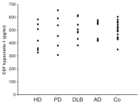
In seven genetically confirmed HD patients (mean age 51 years, range 35–74 five men), we as-sessed duration of disease (de-fined as time from onset of first symptom to lumbar puncture), body mass indices and sleep-wake disturbances by standardized sleep questionnaires (including Epworth Sleepiness Scale=ESS; available in six patients). CSF hypocretin-1 levels were deter-mined in all patients in a single radioimmunoassay as previously described [9]. Levels were com-pared with those of controls without neurological or other sleep-wake disorders (n=20), and with patients with other neuro-degenerative disorders (Parkin-son’s disease, n=6, mean age 71 years; dementia with Lewy bodies, n=9, mean age 73 years; Alzhei-mer’s disease, n=7, mean age 72 years).
In the HD patients, mean duration of disease was 4.1 years (range 1–10). Mean BMI was 26.3 (range 22–33). Four patients re-ported unintentional weight loss. Sleep-wake disorders were
com-mon complaints, and included insomnia (n=4), fragmented night sleep without daytime tiredness (n=1), and REM sleep behavior disorder (n=1). Exces-sive daytime sleepiness (defined as ESS score>10) was not re-ported (mean ESS score 6.3, range 4–8).
CSF hypocretin-1 levels were normal in all HD patients (mean 441 pg/ml, range 326–583) (Fig.
1). Levels did not differ signifi-cantly from those of patients with Parkinson’s disease (487 pg/ml, range 307–654 pg/ml), dementia with Lewy bodies (504 pg/ml, range 382–667), and Alzheimer’s disease (474 pg/ml, range 333– 564). There were no associations between the presence of sleep-wake disorders, Epworth Sleepi-ness Scale, duration of the dis-ease, body mass index,
unintentional weight loss, and CSF hypocretin-1 levels.
We could not find a hypocretin neurotransmission deficiency – as assessed by determination of CSF hypocretin-1 levels – in human HD. However, Petersen et al. re-ported an approximate 27 % loss of hypocretin neurons in human HD [8]. We think that this discrepancy may be due to quantitative effects: Gerashchenko and colleagues [10] found that an average loss of 14 % of rodent hypocretin neurons was not followed by a decrease of CSF hypocretin levels. An average loss of 73 %, however, was associated with a significant 50 % decline in CSF hypocretin levels. The authors assumed that surviving hypocretin neurons might compensate for hypocretin neuronal loss by in-creased hypocretin ligand produc-tion. Thus, interpreting Petersen’s finding together with the observa-tions of Gerashchenko, it is con-ceivable that CSF hypocretin-1 levels are normal in HD patients.
C.R. Baumann (&) Æ C.L. Bassetti Department of Neurology University Hospital Frauenklinikstrasse 26 8091 Zurich, Switzerland Tel.: +41/1 255-55 11 Fax: +41/1 255-43 80 E-Mail: christian.baumann@usz.ch M. Hersberger
Institute of Clinical Chemistry University Hospital
Zurich, Switzerland
LETTER TO THE EDITORS
J Neurol (2006) 253: 1232–1233 DOI 10.1007/s00415-006-0146-7
JON
In conclusion, we found normal CSF hypocretin-1 levels in patients with Huntington’s disease, inde-pendent of the presence of sleep-wake disorders, and independent of nutritional status. CSF hypo-cretin-1 determination may not be helpful in the diagnosis of HD. Further studies to evaluate hypo-cretin neuronal loss in HD are required.
References
1. Starr A (1967) A disorder of rapid eye movements in Huntington’s chorea. Brain 90:545–564
2. Brotini S, Gigli GL (2004) Epidemiol-ogy and clinical features of sleep dis-orders in extrapyramidal disease. Sleep Med 5:169–79
3. Wiegand M, Moller AA, Lauer CJ, Stolz S, Schreiber W, Dose M, Krieg JC (1991) Nocturnal sleep in Huntington’s disease. J Neurol 238:203–208 4. Trejo A, Tarrats RM, Alonso ME, Boll
MC, Ochoa A, Velasquez L (2004) Assessment of the nutrition status of patients with Huntington’s disease. Nutrition 20:192–6
5. Popovic V, Svetel M, Djurovic M, Petrovic S, Doknic M, Pekic S, Miljic D, Milic N, Glodic J, Dieguez C, Casanueva FF, Kostic V (2004) Circulating and cerebrospinal fluid ghrelin and leptin: potential role in altered body weight in Huntington’s disease. Eur J Endocrinol 151:451–5
6. Nishino S, Ripley B, Overeem S, Lam-mers GJ, Mignot E (2000) Hypocretin (orexin) deficiency in human narco-lepsy. Lancet 355:39–40
7. Peyron C, Faraco J, Rogers W, Ripley B, Overeem S, Charnay Y, Nevsimalova S, Aldrich M, Reynolds D, Albin R, Li R, Hungs M, Pedrazzoli M, Padigaru M, Kucherlapati M, Fan J, Maki R, Lam-mers GJ, Bouras C, Kucherlapati R, Nishino S, Mignot E (2000) A mutation in a case of early onset narcolepsy and a generalized absence of hypocretin peptides in human narcoleptic brains. Nat Med 6:991–7
8. Petersen A, Gil J, Maat-Schieman ML, Bjorkqvist M, Tanila H, Araujo IM, Smith R, Popovic N, Wierup N, Norlen P, Li JY, Roos RA, Sundler F, Mulder H, Brundin P (2005) Orexin loss in Hun-tington’s disease. Hum Mol Genet 14:39–47
9. Baumann CR, Dauvilliers Y, Mignot E, Bassetti CL (2004) Normal CSF hypo-cretin-1 (orexin A) levels in dementia with Lewy bodies associated with excessive daytime sleepiness. Eur Neurol 52:73–6
10. Gerashchenko D, Murillo-Rodriguez E, Lin L, Xu M, Hallett L, Nishino S, Mignot E, Shiromani PJ (2003) Rela-tionship between CSF hypocretin levels and hypocretin neuronal loss. Exp Neurol 184:1010–6 0 100 200 300 400 500 600 700 C S F hypoc re ti n-1 ( pg/ m l) HD PD DLB AD Co
Fig. 1 CSF hypocretin-1 levels (in pg/ml) in Huntington’s disease (HD, n=7), Parkinson’s disease (PD, n=6), dementia with Lewy bodies (DLB, n=9), Alzheimer’s disease (AD, n=7), and healthy controls (Co, n=20)