ELSEVIER
FEMS Microbiology Letters I35 (1996) 85-92Osa protein encoded by plasmid pSa is located at the inner
membrane but does not inhibit membrane association of VirB and
VirD virulence proteins in Agrobacterium tumefaciens
Chao-Ying Chen ‘, Clarence I. Kado
*LInl,is Crobvn Gall Group, Department of Plant Pathdog!, Uni~~ersity of Cal(fornia. lIaris, CA 95616. USA Received 14 September 1995; revised 7 October 1995; accepted 17 October 1995
Abstract
The osu gene of IncW plasmid pSa encodes a 21-kDa protein that completely abolishes the oncogenic activity encoded by virulence genes in Agrohacterium tumefaciens. osa is the last gene of a four-gene operon in pSa, the expression of which appears to be highly regulated since the Osa protein is absent when either pSa or the osu operon is present in the
Apdcrctrriurn cell. When the osa gene alone or together with upstream genes within the operon are expressed under the control of a constitutive promoter, Osa protein is produced, enabling us to determine its subcellular location. Immunoblot analyses located Osa protein at the inner membrane of both A. tumefuciens and Ercherichiu co/i. Because Osa inhibits oncogenicity of A. tumqfucien.s, and because alterations of the products of the c,irB and l,irD genes affect oncagenicity. studies were conducted to determine if there are changes in their specific association with the membranes in the presence Osa. Immunoblot analyses of VirB2, VirB3, VirB4, VirB9, and VirD4 in the presence and absence of Osa revealed no differences between the two treatments in these Vir protein associations with the membranes. These results indicate that both
rirB and GrD gene products are produced in the presence of Osa; that they appear unaffected in their association with the membranes: and that Osa is associated with the inner membrane. where VirB2, VirB4, and VirD4 proteins are also located. Kewod: Agrohtrcteriurn furnyfaciem: Oncogenicity; Crown gall tumors: IncW plasmid; pSa plasmid: Ti plasmid: RPI plasmid: Vir proteins: Fertility inhibition
1. Introduction
The oncogenicity of Agrobacterium tumefaciens
is dependent on the presence of the Ti plasmid, which contains virulence (cir> genes necessary for the delivery of a specific 25kb sector (the T-DNA containing oncogenes) of this plasmid into plant cells, culminating in the incorporation of this DNA
’ Corresponding author.
’ Present address: Department of Plant Pathology and Entomol- ogy. National Taiwan University. Taipei, Taiwan 106.
into the plant genome. Mutations of rir gerles such as CirB and virD genes required for the processing and transfer of the T-DNA results in the loss of oncogenicity. Mutations of chromosomal genes such as chl, genes involved in virulence also appreciably affect oncogenicity. An ancillary phenomenon of oncogenic suppression has been observed by the transfer of the IncW plasmid pSa into A. tum~fa- ciens [l]. The oncogenic inhibition by pSa is abso- lute, indicating that some step in the T-DNA process- ing or transfer is completely blocked in the presence of pSa. The genetic element on pSa conferring this 037% I097/96/$I2.00 0 I996 Federation of European Microbiological Societies. All rights reserved
activity rests in a single gene, nsa (for oncogenic suppressive activity) [2]. This gene is part of the osa operon containing at least four open reading frames (orfs) [3]. The functions of ~~~1 and orJ3 are un- known, while OI$? encodes a nuclease [4], and oyf4 encodes the 21-kDa Osa protein [2]. Amino acid sequence analysis of Osa has revealed an interesting homology to proteins involved in fertility inhibition of pSa by 1ncP plasmid RPl [3], suggesting that Osa might also play a role in fertility inhibition such as affecting delivery of the T-DNA by a conjugative mechanism [3]. Credence of this hypothesis comes from the fact that the r!irB genes are directly in- volved in T-DNA transfer, and are homologs of genes involved in the synthesis and assembly of a conjugative pilus [5,6]. Nonetheless, the mechanism by which Osa blocks T-DNA delivery is not pre- cisely understood; its activity is not associated with either the inhibition of transcription of 13ir genes, or preventing the production of T-DNA intermediates [7]. Useful information can be obtained from amino acid sequence data; however, examination of the amino acid sequence of Osa has not revealed any homologies to known proteins. The sequence showed no potential signal sequences, but a region rich in hydrophobic amino acid residues near its carboxyl terminus is present, suggesting that Osa might poten- tially associate with the bacterial membrane [3]. Be- cause a number of possibilities as to the function of Osa have been ruled out, the subcellular location of Osa would provide an important clue leading to its function.
In this communication, we show that Osa is asso- ciated with the inner membrane of A. tumgfuciens. Since many of the VirB proteins are associated with the inner membrane [9- 141, one notion has been that there may be competitive inhibition between VirB and Osa proteins. However, such competition ap- pears not to occur.
2. Materials and methods
2.1. Bacterial strains, growth conditions, und plus- mids
To express Osa protein, Escherichiu coli BL21(DE3) containing a chromosomal T7 RNA
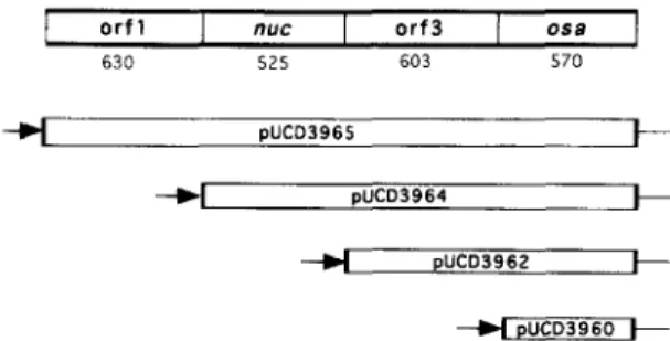
orf1 ““CT orf3 ma
630 525 603 570
-4 pUtD3962e
+ pUCD3960 + Fig. I. Organization of the OSCI operon and its deletion derivatives. Each deletion derivative contains the indicated orfs or genes, of which the upstream most gene is fused to the constitutive kanamycin/spectinomycin resistance operon promoter (arrow). The length in base pairs is indicated below each orf.
polymerase gene under the control of a laclJV5 promoter [ 151 was used as a host for plasmid PET-3a [ 161 containing the osu gene under the control of a T7 promoter. The cells were grown at 37°C in medium 2YT [ 171 containing 100 pug ml ’ ampi- cillin. E. coli DH5a containing pUCD3960 [3] was grown in a similar fashion and used as a host for Osa production. A. tumefuciens LBA4301 (pTiC58Tra”) was grown in AB induction medium [ 181 at 28°C. LBA4301 cells were exposed to 200 FM acetosyrin- gone (= 3’,5’-dimethoxy-4’-hydroxy-acetophen- one) at mid-exponential phase and were allowed to grow for an additional 14 h before they were col- lected by centrifugation (5000 X g, 4”C, 10 min).
The osa gene was cloned into PET-3a that had been cleaved by double digestion with restriction enzymes BumHI and NdeI. The osa gene with flanking BumHI and NdeI cleavage sites generated by polymerase chain reaction amplification was in- serted in the respective cloning site of PET-3a. The resulting plasmid designated pUCD3985 was trans- ferred to E. coli BL21(DE3) by electroporation. In addition, as shown in Fig. 1, progressive deletion derivatives of each og of the osu operon were cloned in the KpnI restriction site within pUCD105 described previously [3], and resulted in recombinant plasmids pUCD3965, pUCD3964, pUCD3962, and pUCD3960, each under the control of a promoter of the kanamycin/spectinomycin operon described pre- viously [8]. Each clone was transformed into E. coli DH5a. We also used pUCD1311, which contained a 3. I-kb DNA sector of pSa containing the entire I>.YU operon [2]. pUCD3940-30 is a derivative of
C.-Y. Chen, Cl. Kado / FEMS Microbinln~~ Letters 135 ( I9961 X5-92 x7
pUCDl3 1 I containing a TnCAT insertion 2025 bp upstream of the S-end of osa [3].
2.2. Protein pur$cation. antibody production, and immurtobkotting
Osa protein was produced in E. coli BL21(DE3) cells containing the osa gene cloned in PET-3a and designated as pUCDl3 18. At mid-exponential phase, the cells were exposed to 0.4 mM IPTG (= isopropyl-P-D-thiogalactopyranoside) and incubated for 2 h. They were then lysed by sonication at maximum setting with five 20-s pulses using an ultrasonicator (Heat Systems-Ultrasonics, Inc., Form- ingdale, NY). The proteins were fractionated by SDS-PAGE [ 191, transferred to nitrocellulose mem- brane (0.45 mm, BA8.5, Schleicher and Schuell, Inc.). and visualized by staining with Ponceau S. The banded protein was excised, sliced into small pieces and ground into a powder in liquid nitrogen with a mortar and pestle. The powder was resuspended in normal saline and used as the immunogen in the presence of complete adjuvant. Rabbit antiserum was raised by Antibodies, Inc., Davis, CA. Non-specific cross-reacting antibodies were removed by immuno- cross-absorption with clairfied whole-cell lysates of E. co/i Bl2l(DE3) containing PET-3a, and with that of A. tum~fuciens LBA4301 (pTiC58Tra’) contain- ing cloning vector pUCD105 [20].
Proteins from total cell lysates were fractioned into subcellular components by SDS-PAGE and transferred by electroblotting to nitrocellulose mem- branes. Proteins on the membrane were reacted with specific Osa antibodies followed by goat anti-rabbit horse raddish peroxidase-conjugated secondary anti- bodies commercially supplied in the ECL system (Amersham, Arlington Heights, IL). Polyclonal rab- bit antibodies against A. tumgfaciens Vir proteins, VirB2, VirB3. VirB4, VirB9, and VirD4, were raised at Antibodies, Inc., Davis. CA.
2.3. Frrrctionutiov:
of
.s~hcellular componentsPeriplasmic, cytoplasmic, inner and outer mem- brane components were fractionated by differential centrifugation [21]. All steps were performed at 4°C. A. tumqfaciens with or without acetosyringone was suspended in 5 ml lysis buffer (25 mM HEPES, pH
7.6, 20% sucrose, 2 mM EDTA, 0.2 mg ml _ ’ lysozyme) for 30-60 min. The lysate was clarified by centrifugation (7000 X g, 15 min). The resulting pellet was resuspended in 5 ml HEPES buffer (25 mM, pH 7.6) and sonicated three times for 201 s each at full output. Cellular debris was removed by cen- trifugation (7000 X g, 15 min) and KC1 was added to the supematant to a final concentration of O.2M. The suspension was centrifuged at I 10000 X g for 1 h to yield the cytoplasmic fraction. The inner and outer membranes were obtained from cells collected by centrifugation of 250 ml of culture at mid-exponen- tial phase. The cells were resuspended in IO ml of a solution containing 25 mM HEPES. pH 7.6. 20% sucrose, 0.2M KCl, 0.2 mM dithiothreitol, 0.2 mg ml-’ desoxyribonuclease, and 0.2 mg ml ’ ribo- nuclease A, and passed through a French pressure cell (Amicon. Beverly. MA) three times at 16000 psi. Lysozyme was then added to the cracked cell suspension to a final concentration of 1 rng nilF ‘. The mixture was incubated for 30-60 min and then centrifuged (7000 X g for 10 min) to remove,cellular debris. The supernatant containing the cell envelopes was centrifuged at 110000 X g for 1 h and the resulting pellet was resuspended in 1 ml solution containing 5 mM EDTA, pH 7.5. 0.2 mM dithio- threitol and 20% sucrose. This suspension \hias lay- ered with a discontinuous density gradient consisting of I ml of 70% sucrose on top of which 2.9 ml of 53% sucrose was layered, both in 5 mM EDTA. pH 7.5, The gradient was centrifuged at IO0000 X ,q for 24 h to separate the inner and outer membranes. Fractions (0.5 ml) were collected from the bottom of the gradient and each fraction was assayed ‘for pro- tein concentration using a protein assay reagent and analysed by the Softmax program (BioRad, Her- cules, CA). NADH-oxidase activity. naturally associ- ated with the inner membrane and serving as a marker for this subcellular fraction. was analysed as described previously [ 1 I].
3. Results
3. I. Isolation and pur$cution
of Osu
Osa protein was produced in E. coli DH5a con- taining pUCD3960 and the whole cell lyoate was
88 C.-Y. Clam, C.I. Ktrdo / FEMS Mic~rohiolo,q~ Lrtterc 1.35 ClYYhi KS-Y2
analysed by SDS-PAGE. Densitometry of the frac- tionated proteins showed that Osa represented ap- proximately 0.2% of the total cellular protein in exponential phase cells, which therefore provided a minimal source of the protein (Fig. 2). On the other hand, Osa produced in E. coli BL21(DE3) cells containing pUCD3985 represented approximately 5% of the total cellular protein. Excessive production of Osa stimulated by subjecting the cells to IPTG was lethal to the cells. Thus, even in the absence of induction with IPTG, there was sufficient expression of the osa gene that resulted in Osa production, which was amenable for purification. Cell lysates of the latter host were therefore routinely used to obtain Osa which conveniently fractionated into the insolu- ble fraction (Fig. 2). Proteins in this fraction were fractionated electrophoretically by SDS-PAGE and the separated proteins were transferred by electro- blotting to a nitrocellulose membrane. The mem- brane portion, containing the Osa protein, visualized by staining with Ponceau S, was used to raise poly- clonal antibody purified further by cross absorption as described in Materials and methods. The speci- ficity of the antibody was demonstrated by Western blotting whole-cell lysates prepared from E. coli BL21(DE3) with and without the osa gene cloned in its vector plasmid (Fig. 2). Only those cells bearing osa displayed a protein band reactive to the antibody. 3.2. Osa protein production in A. tumqfaciens is regulated
Whole-cell lysates prepared from A. tumqfaciens LBA430 l(pTiC58Tra’) harboring either pSa or pUCD13 11 were analysed by SDS-PAGE and by immunoblotting with Osa antibody. The Osa protein was barely detectable by staining with Coomassie brilliant blue and by immunoblotting, suggesting that the expression of osa is regulated in this organism (Fig. 2). As expected, the interuption of transcription of the osa operon by the insertion of TnCAT up- stream of osa failed to produce the Osa protein.
Since osa is part of an operon containing three additional genes upstream of osa [3], clones bearing a systematic deletion of each of these genes with a constitutive promoter driving the remaining genes downstream of the deletion (Fig. 1) were analysed in the above A. tumefuciens test strain. Cells contain-
(A)
I -1. __II 97.4 66.2 42.7 21.5(B)
1 2 3 4 5m
12 345 31.0 - -1. 21.5 - J1 14.4 -Fig. 2. SDS-PAGE analyses of soluble and insoluble proteins prepared from E. co/i DH5 a cells containing one of the follow- ing plasmids: (A) soluble fraction: pUCDIO5 (lane I); pUCD3960 (lane 2): pUCD3985 (lane 3); pUCDl318 (lane 4): and PET-3a (lane 5). The protein band (arrow) in lane 3 is a fusion protein between Osa and the first I2 amino terminal amino acid residues of T7qlO gene of PET-3a. resulting in a protein larger than the expected size of Osa (lane 4). (B) Insoluble fraction: pUCDIO5 (lane I): pUCD3960 (lane 2): pUCD3985 (lanes 3 and 5); pUCDl3 18 (lane 4). The same fusion protein described above is indicated by the arrow in lanes 3 and 5. The protein bands resolved in 15% acrylamide gels were stained with Coomassie brilliant blue. (C) Western blot of Osa as the fusion protein (described in A) shown in lanes 2 and 3, and Osa protein (lane 4). Lanes I and 5 contain proteins derived from cells with the respective vectors pUCDIO5 and PET-3a. Specific Osa antibody was used and detected by goat anti-rabbit antibody conjugated with horse radish peroxidaae.
C.-Y. Chen, C.I. Kado / FEMS Microbiology Letters 135 (IYY6J 85-92 89 1 2 3 4 5 6 97.4 - 66.2 - (, I 42.7 - .j j 31.0 - 21.5 -
(B)
1 2 3 4 5 31.0 - mm9 21.5 -Fig. 3. Western blot analysis of A. tumefariens LBA4301tpTiC58Tra‘ ) containing the following plasmids: (A) whole cell lysate: pUCDlO5 (lane 1); pUCD3960 (lane 3): pUCD3962 (lane 4), pUCD3964 (lane 5); pUCD3965 (lane 6). Purified Osa fusion protein (lane 2) as in lane 3 of Fig. 2. (B) Subcellular fractions: outer membrane (lane I); inner membrane (lane 2); cytoplasm (lane 3); periplasm (lane 4). Lane 5: same as lane 2 of (A). The amount of protein in each lane was adjusted to represent that recoverable from a single A. fumefnciens cell.
Numhers on the left are in kDa based on protein standards.
ing osa and either the penultimate locus orf3, or n~lc and orf3, in clones pUCD3962 and pUCD3964, re- spectively, produced highly detectable Osa protein (Fig. 2). Each Osa-producing test strain containing pTiC58 was avirulent on Jimson weed (Daturu stru- monium) as anticipated (not shown). These results suggest that the osa operon is regulated in A. tume- ,fuciens. This hypothesis is supported by the lack of
expression of a promoter-less chloramphenicol acetyltransferase gene fused to osu of the osu operon in A. tumqfuciens [3].
3.3. Osu is located at the inner-membrane
A. tumefuciens LBA430l(pTiC58Tra’) cells con- taining pUCD3960 were separated into cytoplasmic, periplasmic. and inner- and outer-membrane frac- tions as described in Materials and methods. Proteins from each fraction were separated by SDS-PAGE and analysed by immunoblotting. As shown in Fig. 3, the Osa protein was found solely in the inner membrane fraction. The homogeneity of the inner
and outer membrane preparations was verified by the presence of NADH oxidase activity (64 nmol
min-
’
(mg protein)-’ ) only in the inner membrane fraction. The amount and subcellular location of this protein was not altered when A. tumefuciens cells were induced with acetosyringone, indicating that the reg- ulation of osu is independent of acetosyringone in- duction. Western blot analysis of the subcellular fractions of E. coli DHSa containing pUCD3960 also showed that Osa is located at the inner mem- brane (not shown). These results indicate that Osa associates mainly with the inner membrane, irrespec- tive of acetosyringone induction and bacterial chro- mosomal background.
3.4. Osu does not qfect the production and s#bcellu-
lur locution qf VirB2, VirB3, VirB4. VirB9. and
VirD4 proteins
Proteins encoded by cirB and r,irD get7es are essential for virulence of A. tumefuciens. Because Osa inhibits A. tumefuciens virulence completely, its mode of action may be directed at c%ir gene products. As we show here, the transcription and translation of the l>ir genes are not blocked, but neither protein-to-protein, nor competitive protein- to-membrane target interactions have been com- pletely ruled out. Comparative analyses were there- fore made of LBA4301(pTiC58Tra’) cells containing either the osu-bearing plasmid pUCD3960, or the vector plasmid pUCDl05 in the absence of osu. In either case, VirB2. VirB3, VirB4, VirB9 and VirD4 proteins were detected in the total membrane fraction (not shown). Upon fractionation of the membranes, Western blots revealed that VirB2 and Vi@9 pro- teins were associated with both the inner and outer membranes as would normally be the case (Fig. 4). confirming previous VirB protein localization studies [ 111. VirB4 and VirD4 were found in the inner membrane (not shown), as found previously, in other studies [lo, 13,221. These results show that in the presence of osu, the l:irB and r,irD operons ure fully expressed.
In each instance, no differences were observed between the association of these Vir proteins with their respective membranes. Shifts in protein-to- membrane patterns can easily be detected as shown previously with the observed loss of membrane asso-
C.-Y. Chnz, C.I. Kudo / FEM.7 Microbiolo,q~ Le:wr.c- 135 f iYY61 X-Y2 90 (A) 123456 78 6.5 - 3.4 -
9)
1 234 5 6 7 8Fig. 4. Western blot analyses for the effect of Osa on the subcellular locations of VirB proteins in A. fumefaciens. Proteins were resolved by SDS-PAGE. (A) Proteins in each subcellular fraction were probed with VirB2 antibody: lanes 1 and 5 contain cytoplasmic proteins; lanes 2 and 6 contain the periplasmic frac- tion; lanes 3 and 7 contain the outer membrane proteins; and lanes 4 and 8 contain the inner membrane proteins, (B) The same sets of protein fractions were probed with VirB9 antibody. Proteins loaded in each lane were pre-adjusted to be equivalent on a per cell basis. The numbers on the left are positions of molecular mass standards in kDa.
ciation of VirB3 when VirB4 is mutated 1231. Hence, it appears that VirB2, VirB3, VirB9 and VirD4 remain unaffected by Osa in associating with their respective membranes.
4. Discussion
Osa protein produced in E. coli was isolated and purified, leading to the preparation of specific poly- clonal antibody. This antibody was used to detect Osa in various subcellular fractions derived from A. tumefaciens cells containing pSa or a clone bearing the osa operon. These studies curiously showed the absence of the Osa protein in A. tumefaciens cells containing pSa or the entire osa operon, suggesting that the expression of osa may be tightly regulated and may require some type of external inducer. The nature of such an inducer is presently being explored in another study (L.-Y. Lee and C.I. Kado, unpub- lished). To circumvent the lack of osa expression in A. tumefaciens, transcription of the osa gene was placed under the control of a constitutive promoter
derived from a kanamycin/spectinomycin operon [3,24]. Immunoblot analysis of the subcellular com- ponents of A. tumefaciens cells, containing the con- stitutively expressed osa gene, clearly showed the presence of Osa protein that specifically localizes to the inner membrane fraction. Examination of its amino acid sequence reveals a stretch of hydropho- bic residues near the carboxyl terminus of Osa, which might serve to localize at least this part of the polypeptide to the membrane. The manner by which Osa spans the inner membrane awaits further studies. The fact that Osa completely inhibits A. tumcfa- dens oncogenicity, and that the protein associates with the inner membrane as shown in this study, leads us to hypothesize that Osa might be blocking the proper generation of the postulated T-DNA trans- port apparatus which spans the membrane. Based on recent comparative amino acid sequence homology studies [5,11,22], and on the identification of a pro- cessed propilin-like VirB2 protein ([ 111; A.L. Jones, E.-M. Lai and C.I. Kado, submitted), as well as electron microscopic observations ([lo]; 0. Ches- nokova, J. Coutinho, I. Kahn, and C.I. Kado, submit- ted), the assembly of a conjugative pilin-like struc- ture may be inhibited by Osa at its membrane loca- tion.
To assess this possibility, immunoblot studies us- ing antibodies specific to various VirB proteins, including a VirD4 protein, were used to analyse A. tumefaciens cells harboring a Ti plasmid plus either an osa clone or the wild-type pSa plasmid. The results of these studies showed no obvious difference in the location or the amount of each VirB or VirD protein analysed in subcellular fractions of cells with and without osa. Thus, the Osa protein may either not be competing for specific sites on the membrane where VirB2, VirB3, VirB4, VirB9 and VirD4 pro- teins dock, or the interaction may be too subtle to detect at the sensitivity level of the immunoblot analyses, or the activity of Osa is not associated with these proteins. Although not all of the Vir proteins were analysed, the possibility that we have over- looked one that does react with Osa seems remote because recent cross-linking studies revealed no pro- tein-to-protein interactions [26]. Additional studies underway using in situ electron microscopic analysis and immunogold-labelled antibodies should verify these results.
C.-Y. Chm, Cl. Kado/ FEMS Microbiology Letters 135 llYY6) k-Y2 91
Previously, by comparative amino acid sequence homology studies, we found that there are relatively close homologies between the amino acid sequence of Osa and that of an IncP plasmid RPI protein involved in fertility inhibition of IncW plasmids such as pSa [3]. Conjugative transfer requires the close association of specific proteins and plasmid DNA to form a complex at the membrane [24,25]. Fertility inhibition may result either through the alteration of such complexes by a protein identified to inhibit plasmid conjugative transfer, or by plugging the postulated pore through which the donor DNA must pass into the recipient cell. Thus, one of these mech- anisms is that used by Osa. Our recent studies using agroinfection and intron-P-glucuronidase (GUS) as- says to measure the transfer of the T-DNA from A. tumqfuciens to plants indicate that T-DNA transfer is indeed inhibited by Osa [26]. This further supports the notion that Osa may be interfering with the T-DNA transfer mechanism.
Acknowledgements
We thank Louise Jones and Ken Shirasu for technical assistance and helpful discussions. Special thanks goes to Shih-Tung Liu for generously provid- ing laboratory space and equipment at Chang-Gung Medical College, Taoyan, Taiwan during a summer stay by the senior author. This work was supported by US Public Health Service grant GM45550 from National Institutes of Health.
References
[I] Loper, J.E. and Kado, C.1. (1979) Host range conferred by the virulence-specifying plasmid of Agrobactrrium tumqfa- cirrrs. J. Bacterial. 139, 591-596.
121 Close, S.M. and Kado, C.I. (1991) The osa gene of pSa encodes a 2 I. 1-kilodalton protein that suppresses Agrobac- trrium tumefaciens oncogenicity. J. Bacterial. 173, 5449- 5456.
[3] Chen, C.-Y. and Kado. C.I. (1994) Inhibition of Agrobuc- terium tumefuciens oncogenicity by the osa gene of pSa. J. Bacterial. 176, 5697-5703.
[4] Close, S.M. and Kado, C.I. (1992) A gene near the plasmid pSa origin of replication encodes a nuclease. Mol. Microbial. 6. 52 I-527. 151 161 [71 [81 [91 1101 [Ill [I21 [I31 [141 [I51 [I61 1171
Kado, C.I. (1994) Promiscuous DNA transfer system of Agrobacterium tumefaciens: role of the rirB operon in sex pilus assembly and synthesis. Mol. Microbial. 12. 17-22. Kado. C.I. (1994) T-DNA transfer to plants is mediated by pilus-like apparatus encoded by the Ti plasmid t,irB operon. In: Advances in Plant Biotechnology (Ryu. D.D.Y. and Furusaki, S. Eds.), pp. 23-36. Elsevier, Amsterdam. Close. S.M. (1991) Ph.D. thesis. University of California, Davis, CA.
Tait, R.C.. Rempel. H., Rodriguez, R.L. and ICado, C.I. (I 985) The aminoglycoside-resistance operon of the plasmid pSa: nucleotide sequence of the streptomycin+pectinomycin resistance gene. Gene 36, 97- 104.
Ward. J.E. Jr.. Dale, E.M., Nester. E.W. and Binns. A.N. (1990) Identification of a VirBlO protein aggregate in the inner membrane of A,qrobtrctrrium tumefiuk.\. J. Bacterial. 172. 5200-52 IO.
Berger. B.R. and Christie. P.J. (1993) The A~rtrbuctrr-ium twtwfticien.~ rrrB4 gene product is an essential virulence protein requiring an intact nucleo\ide triphosphate-binding domain. J. Bacterial. 175, 1723-1734.
Shirasu. K. and Kado, C.I. (1993) Membrane location of the Ti plasmid VirB proteins involved in the biosynthesis of a pilin-like conjugative structure of A~robtrc~terirrm tumyfir- cirns. FEMS Microbial. Lett. 1 I I. 287-293.
Beijersbergen, A.. Smith. S.J. and Hooykaas. P.J.J. (1994) Localization and topology of VirB protein\ of A,qrobtrc- trrium tum@cicns. Plasmid 32. 2 12-2 18.
Thorstenson, Y.R., Kuldau, G.A. and Zambryski, P.C. (1993) Suhcellular localization of seven VirB proteins of Ap4wc- tvrium rumyfiuks: implication5 for the formation of a T- DNA transport structure. J. Bacterial. 175. 5233-5241. Thorstenson. Y.R. and Zambry\ki, P.C. (1994) The essential virulence protein VirB8 localizes to the inner membrane of ARrobnctrrium twm$cien.s. J. Bacterial. 176. I7 I I - I7 17. Studier, F.W. and Moffatt, B.A. (1986) Use of bacteriophage T7 RNA polymerase to direct selection of high level expres- sion of cloned genes. J. Mol. Biol. 189. I 13-130.
Rosenberg. A.G.. Lade, B.N.. Chiu. D.. Dunn, J.J. and Studier. W. (1987) Vectors for selective expression ot cloned DNAa by T7 RNA polymerase. Gene 56. I25- 135. Sambrook. J., Fritsch. E.F. and Maniatis, T. (1989) Molecu- lar Cloning: A Laboratory Manual. 7 edn. Cold Spring Harbor Laboratory, Cold Spring Harbor. NY.
[I81 Cangelosi. G.A.. Ankenbauer. R.G. and Nester, E.W. (1990) Sugars induce the A,qrobactrrrum virulence genes through a periplaamic binding protein and a transmembrane signal pro- tein. Proc. Natl. Acad. Sci. USA 87. 6708-6712.
[19] Laemmli, U.K. (1970) Cleavage of structural proteins during the assembly of the head of bacteriophage T3. Nature 227, 6X0-685.
[20] Valentine, C.R.I. and &ado. C.I. (1989) Molecular genetics of IncW plasmids. In: Promiscuous Plasmids of Gram-nega- tive Bacteria (Thomas, CM.. Ed.). pp. 125- 163. Academic Press, London.
92 C.-Y. Chen, C.I. Kodo/ FEMS Microhiolo~,v Letter.5 135 (19%) X5-92
Rhizobium leguminosarum cells into outer membrane, cyto-
plasmic membrane, periplasmic and cytoplasmic compo- nents. J. Bacterial. 167, 1083-1085.
[22] Shirasu, K., Koukolikova-Nicola, Z., Hohn, B. and Kado, C.I. (1994) An inner-membrane-associated virulence protein essential for T-DNA transfer from Agrobacterium tumefu-
ciens to plants exhibits ATPase activity and similarities to
conjugative transfer genes. Mol. Microbial. 11, 58 I-588. [23] Jones, A.L., Shirdsu, K. and Kado, C.I. (1994) The product
of the r,irBI gene of Agrobacterium tumefaciens promotes accumulation of VirB3 protein, J. Bacterial. 176, 5255-5261. [24] Frost, L.S., Ippen-Ihler, K. and Skurray, R.A. (1994) Analy-
sis of the sequence and gene products of the transfer region of the F sex factor. Microbial. Rev. 58, 162-210.
[25] Willetts, N. and Skurray, R. (1987) Structure and function of the F factor and mechanism of conjugation. In: Escheric~hicr
co/i and Scr/ntonellr/ typhimurium, Cellular and Molecular
Biology (Neidhardt, F.C., Ingraham, J.L., Low. K.B.. Maga- sanik, B., Schaechter, M., Umbarger, H.E., Eds.). pp. I I IO- 1133. American Society for Microbiology, Washington. D.C. [26] Lee, L.-Y. and Ado, C.I. (1995) The oncogenic suppressive
gene osn is part of an operon in plasmid pSa and encodes a 21 kDa protein which blocks T-DNA conjugative transfer from A,yrohncterium to plants. Proc. Int. Conf. Agmbtrc-