HAL Id: inserm-00435658
https://www.hal.inserm.fr/inserm-00435658
Submitted on 28 Dec 2010
HAL is a multi-disciplinary open access
archive for the deposit and dissemination of
sci-entific research documents, whether they are
pub-lished or not. The documents may come from
teaching and research institutions in France or
abroad, or from public or private research centers.
L’archive ouverte pluridisciplinaire HAL, est
destinée au dépôt et à la diffusion de documents
scientifiques de niveau recherche, publiés ou non,
émanant des établissements d’enseignement et de
recherche français ou étrangers, des laboratoires
publics ou privés.
Impact of genotype on survival of children with T-cell
acute lymphoblastic leukemia treated according to the
French protocol FRALLE-93: the effect of
TLX3/HOX11L2 gene expression on outcome.
Paola Ballerini, Judith Landman-Parker, Jean Michel Cayuela, Vahid Asnafi,
Myriam Labopin, Virginie Gandemer, Yves Perel, Gérard Michel, Thierry
Leblanc, Claudine Schmitt, et al.
To cite this version:
Paola Ballerini, Judith Landman-Parker, Jean Michel Cayuela, Vahid Asnafi, Myriam Labopin, et
al.. Impact of genotype on survival of children with T-cell acute lymphoblastic leukemia treated
according to the French protocol FRALLE-93: the effect of TLX3/HOX11L2 gene expression on
outcome.. Haematologica, Ferrata Storti Foundation, 2008, 93 (11), pp.1658-65.
�10.3324/haema-tol.13291�. �inserm-00435658�
Impact of genotype on survival of children with T-cell
acute lymphoblastic leukemia treated according to the
French protocol FRALLE-93: the effect of TLX3/HOX11L2
gene expression on outcome
Paola Ballerini,
1Judith Landman-Parker,
2* Jean Michel Cayuela,
3Vahid Asnafi,
4Myriam Labopin,
5* Virginie Gandemer,
6Yves Perel,
7Gérard Michel,
8Thierry Leblanc,
9Claudine Schmitt,
10Sylvie Fasola,
2Anne Hagemejier,
11François Sigaux,
3Marie Françoise Auclerc,
9Luc Douay,
1* Guy Leverger,
2* and André Baruchel
91Laboratory of Hematology, Hôpital d’Enfants Armand Trousseau APHP:2Department of Pediatric Hematology
Oncology and *Unité INSERM 832, Université Pierre et Marie Curie-Paris 6, Paris; 3Laboratory of Hematology,
Hopital Saint Louis, APHP, Paris; 4Laboratory of Hematology, Hopital Necker Enfants Malades APHP, Paris;
5CEREST-TC, Hôpital Saint Antoine, Université Pierre et Marie Curie-Paris 6; 6Department of Pediatrics,
Onco-hema-tology Unit, CHRU, Hôpital Sud, Rennes; 7Department of Pediatric Hematology and Oncology, Groupe Hospitalier
Pellerin, Bordeaux; 8Department of Hematology, Hôpital La Timone, Marseille; 9Department of Hematology, Hôpital
Saint Louis, APHP, Paris; 10Department of Oncology and Hematology, Hôpital d’Enfants, Vandoeuvres Les Nancy,
France; 11Department of Human Genetics, University of Leuven, Leuven, Belgium
Acknowledgements: we especially thank the cytopathologists Mircea Adam and Marie Thérèse Daniel for their expert morphological evaluation of leukemia samples. We are indebt-ed to Hélène Morsy and Corinne Bronstein for their excellent technical assistance. We thank E.A. Macintyre and F. Pflumio for their critical read-ing of this manuscript.
Funding: supported by Chugai France (Dr. Thierry Guillot) and SFCE (Société Française des Cancers de l’Enfant).
Manuscript received April 29, 2008. Revised version arrived on July 1, 2008. Manuscript accepted July 10, 2008.
Correspondence: Paola Ballerini,
Laboratoire d’Hématologie,
Hôpital A. Trousseau, INSERM E0210. E-mail: paola.ballerini@trs.aphp.fr The online version of this article con-tains a supplemental appendix.
ABSTRACT
Background
The prognostic value of the ectopic activation of TLX3 gene expression, a major oncogenetic event associated with pediatric T-cell acute lymphoblastic leukemia, is controversial. Likewise, the frequency and the prognostic significance in pediatric T-cell acute lymphoblastic leukemia of the newly characterized NUP214-ABL1 fusion transcript is not yet clear.
Design and Methods
Two hundred children with T-cell acute lymphoblastic leukemia were treated in the French FRALLE-93 study from 1993 to 1999. The expression of TLX3, TLX1 and SILTAL1 genes was ana-lyzed in samples from 92 patients by real-time quantitative reverse transcriptase polymerase chain reaction. Most of these samples were further studied for NUP214-ABL1 and CALM-AF10 fusion transcripts.
Results
The median follow-up was 7.9 years. At 5 years the overall survival (± standard deviation, %) was 62 (±3%) and leukemia-free survival was 58 (±3%). Patients with T-cell acute lymphoblas-tic leukemia positive for TLX3 had a poorer survival compared to those with T-ALL negative for
TLX3 (overall survival: 45±11% vs. 57±5%, p=0.049). In multivariate analysis, TLX3 expression
was an independent adverse risk factor predicting relapse with a hazard ratio of 2.44 (p=0.017) and an overall survival with a hazard ratio of 3.7 (p=0.001). NUP214-ABL1 was expressed in 16.6% of patients with TLX3-positive T-ALL (3 of 18); all of the patients with this association died before completion of the treatment. SILTAL expression did not significantly affect the prognosis of patients with T-cell acute lymphoblastic leukemia. Only three of 92 patients expressed the TLX1 gene and all three are alive.
Conclusions
TLX3 gene expression is an independent risk factor predicting poor survival in childhood T-cell
acute lymphoblastic leukemia. When co-expressed with TLX3, NUP214-ABL1 transcripts may increase the risk of poor survival.
Key words: childhood T-cell acute lymphoblastic leukemia, TLX3/HOX11L2, NUP214-ABL1,
SIL-TAL1, outcome.
Citation: Ballerini P, Landman-Parker J, Cayuela J, Asnafi V, Labopin M, Gandemer V, Perel Y, Michel G, Leblanc T, Schmitt C, Fasola S, Hagemejier A, Sigaux F, Auclerc MF, Douay L, Leverger G and Baruchel A. Impact of genotype on survival of children with T-cell acute lymphoblastic leukemia treated according to the French protocol FRALLE-93: the effect of TLX3/HOX11L2 gene expression predicts on outcome. Haematologica 2008; 93:1658-1665. doi: 10.3324/haematol.13291
Introduction
T-cell acute lymphoblastic leukemia (T-ALL) accounts for 10 to 15% of cases of childhood acute lymphocytic leukemia and is often associated with unfavorable features. The prognosis of this disease, although improved over the last years because of the use of more intensive treatments and risk-adapted therapy, remains unsatisfactory.1-5 Thus, the
identifi-cation of prognostically relevant molecular markers at diagnosis will be fundamental to guide risk-adapted strategies and contribute to the optimization of T-ALL treatment.
T-ALL are characterized by recurrent translocations affecting the T-cell receptor loci (TCR). Consequently, enhancer elements from the TCRA/D locus (14q11) and TCRB locus (7q34) come close to target genes and activate their ectopic expression. This mechanism implies a number of transcription factors such as the helix-loop-helix genes (TAL1/SCL, TAL2, LYL1),6-10the
LIM-domain-only (LMO1, LMO2) genes, the home-obox genes of class-I (HOXA10, HOXA11)11,12or
class-II (HOX11/TLX1, HOX11L2/TLX3)13,14 and the
NOTCH1 gene.15The same genes are frequently
tar-geted during the malignant transformation of T cells by others mechanisms, such as interstitial deletion on chromosome 1 for TAL1/SCL or point mutations for
NOTCH1.16,17
Other translocations, not involving TCR loci, have also been described: the t(10;11)(p13;q14) transloca-tion,18,19resulting in the CALM-AF10 fusion gene; the
cryptic t(5;14) (q34;q32) translocation,14 resulting in
the ectopic transcription of TLX3/HOX11L2 and the episomal 9q34 recombination between NUP214 and
ABL1 genes.20These two latter events are highly
spe-cific to T-ALL, occurring in, respectively, 20-24% and 5-6% of cases. For an unexplained reason,
NUP214-ABL1 fusion appears closely associated with TLX1 and TLX3 deregulation. Until recently there have been no
reports of this episomal recombination in others types of leukemia and very little is known about its frequen-cy in pediatric T-ALL. Furthemore, it is still unclear how these genetic lesions affect the prognosis of patients with T-ALL. Few cohorts of patients have been analyzed so far and for more recently character-ized lesions such as the NUP214-ABL1 fusion gene, no studies in childhood T-ALL are available. Preliminary analyses of the prognostic value of frequent lesions such as ectopic TLX3 activation have led to contrast-ing conclusions.21-14
Here, we analyzed the prognostic value of TLX3 expression on the clinical outcome of 92 patients with T-ALL enrolled in the FRALLE-93 study between 1993-1999. Moreover, to gain insights into the rela-tionship between TLX3 and other oncogenetic events we extended our analysis to SILTAL and TLX1 gene expression, as well as to molecular lesions such as
NUP214-ABL1 and CALM-AF10 gene fusions which
have not yet been extensively evaluated in pediatric T-ALL.
Design and Methods
Patients and the FRALLE-93 trial design
Between 01.1993 and 31.12.1999, 1395 children (778 boys and 617 girls) with ALL were included in the FRALLE-93 protocol. The leukemia was classified as B-cell lineage in 1195 cases (86%) and T-B-cell lineage in 200 cases (14%). All the patients with T-ALL were included in this study and uniformly assigned to the high-risk group. Informed consent was obtained from the patients’ parents in all cases. The treatment design is summarized in Online Supplementary Figure S1. High-risk patients were assigned to three different treatment sub-groups according to their response to prednisone at day 8 and the blast count in bone marrow evaluated at day 21. Prednisone response was defined as good when the number of blasts circulating in the blood was < 1000/µL at day 8 or poor when the circulating blast count was >1000/µL at day 8. The persistence of blasts in the bone marrow at day 21 was categorized as M1 when the blast count was <5%, M2 when the count was between 5% and 25% and M3 when it was >25%. Group C1, patients, who had a good response to prednisone and M1 or M2 bone marrow involvement, received two delayed intensification blocks; group C2, patients con-sidered at a very high risk because of poor response to prednisone and/or M3 bone marrow involvement, received six intensification blocks, followed by autolo-gous stem cell transplantation or, group C3, by allogene-ic stem cell transplantation if an HLA-matched sibling was available. Central nervous system prophylaxis con-sisted in triple intrathecal injection with methylpred-nisolone, methotrexate, cytarabine and 18 Gy radio-therapy. The details of the treatment for group C2 have been published elsewhere25The main characteristics of
the patients studied are presented in Table 1. T-cell lin-eage was assessed on the basis of expression of CD3, CD2, CD5 and CD7 antigens and exclusion of B-cell-associated antigens according to the European EGIL rec-ommendations.26 Karyotypes and fluorescence in situ
hybridization (FISH) analysis of t(5;14) translocation of cases analyzed for TLX3 expression have already been reported.27Molecular studies for SILTAL, TLX3 and TLX1
were possible in 92 out 200 samples (46%). This was mainly related to the fact that systematic cryopreserva-tion of T-ALL blasts was not performed in all the partic-ipating centers. Additional analysis of CALM-AF10 and
NUP214-ABL1 was carried out in 84 and 52 patients,
respectively.
Molecular studies
RNA was extracted and reverse transcribed according to previously described procedures.21Expression of
SIL-TAL, TLX3 and TLX1 was analyzed by real time
quanti-tative polymerase chain reaction (RQ-PCR) on a Taqman 7700 or 7500 real time PCR device (Applied Biosystems, Les Ulis, France) and their levels of expres-sion were quantified relative to the expresexpres-sion of the endogenous control gene, ABL. Primers and probes for
ABL1 and SILTAL as well as the RQ-PCR procedures are
described in the Europe-Against-Cancer Program.28,29
TLX1 and TLX3 gene expression was detected as
report-ed elsewhere.21CALM-AF10 fusion transcripts, both 5’
and 3’ fusions, and the more frequent NUP214-ABL1 fusion transcripts were screened for as reported previ-ously.20, 30
Statistical analysis
Two different descriptive analyses were performed, the first on the overall population of patients, the sec-ond on the sub-population of patients for whom molec-ular data were available. Variables considered were patients’, age and gender, and disease and treatment characteristics. Leukemia-free survival was defined as survival without evidence of relapse or progression. All patients still alive in first continuous remission were censored at their last follow-up. Probabilities of overall survival and leukemia free survival were calculated using the Kaplan-Meier estimate. Further analyses were performed only on patients with available molecular data to evaluate the clinical significance of the oncoge-netic events. Patient-, disease-, and treatment-related variables of the groups divided according to oncogenet-ic events were compared using the χ2statistic for
cate-gorical variables and the Mann-Whitney test for contin-uous variables. The log-rank test and Cox proportional hazards model were used to determine the univariate and independent prognostic importance of several vari-ables. All variables with a p value less than 0.2 in the univariate analysis or differing in distribution were entered into the model. The type I error rate was fixed at 0.05 for the determination of factors associated with time to event outcomes. Statistical analyses were per-formed with SPSS (Inc., Chicago, USA) software pack-ages.
Results
Patients and FRALLE-93 trial design
The biological and clinical characteristics of the entire cohort of FRALLE-93 T-ALL patients are summarized in Table 1. One hundred and sixty-eight patients out of 200 reached complete remission. The follow-up period ranged from 4.3 to 11.8 years (median, 7.9 years). The overall and leukemia-free survival rates of the FRALLE-93 T-ALL population were 62% (±3%) and 58% (±3%), respectively, at 5 years and 60% (±4%) and 56% (±4%) at 10 years. As shown in Table 1 the subgroup of 92 patients who underwent genetic analysis is representa-tive of the total population, as there were no differences in clinical presentation, initial response to chemothera-py, or subsequent treatments between the subgroup and the total population.
Frequency and distribution of the genetic lesions
among the study population
High and specific over-expression of TLX3 and TLX1 was found in 21.7% (20/92) and 3.2% (3/92) of the cases, respectively. SILTAL and CALM-AF10 5’- or 3’-fusion transcripts were detected in an additional 21.7% (20/92) and 4.7% (4/84) of samples tested.
NUP214-ABL1 fusion transcripts were found in three (5.8%) of
52 T-ALL samples: two had a fusion between NUP214 exon 29 and ABL1 exon 2 and one between NUP214 exon 30 and ABL1 exon 2. All the three cases of T-ALL positive for a NUP214-ABL1 fusion were also positive for TLX3 activation.
Clinical and biological characteristics at diagnosis
according to the oncogenetic lesions
Five distinct molecular subgroups could be identified within the study population SILTAL, HOX11L2/TLX3,
HOX11/TLX1, CALM-AF10 and NUP214-ABL1. Except
for the NUP214-ABL1 gene fusion, none of the other lesions studied was found to be co-expressed in the same leukemic sample. As shown in Table 2 there were no differences between these molecular subgroups in sex ratio, age or mediastinal involvement, although ini-tial leukocytosis did differ. The white blood cell count was significantly higher in the SILTAL subgroup than in the TLX3, TLX1 and NUP214-ABL1 subgroups. Marked hyperleukocytosis and mediastinal involvement were also noted in the group of patients with CALM-AF10 T-ALL. Conventional cytogenetic analysis was conducted in 169 patients and was normal in half of these cases
Table 1. Clinical characteristic of the overall pediatric T-ALL cohort and of the study population.
FRALLE-93 T-ALL cohort Study population
Total n=200 n=92
Median age, years, at diagnosis 9.15 (1.1-19.5) 9.7 (1.7-18.5) (range)
Median year of diagnosis 1996 (93-99) 1997 (93-99) Patients’ gender: male/female 139/61 63/29
69% - 31% 68% - 32% Median WBC at diagnosis, 98,85 (0,6-1247) 149.5 (0.6-736) ×109/L (range)
WBC >50 ×109/L 126 (63%) 68 (74%) Mediastinal involvement 141 (70%) 66 (71%) Response to pre- phase treatment
PPR 109 (57%) 45 (52%) GPR 82 (43%) 41 (39%) M status M1 135 (69%) 68 (73%) M2 21 (11%) 11 (12%) M3 40 (20%) 11 (12%) NE 4 2
Risk group of the protocol
C1 100 (59%) 46 (55%)
C2 44 (26%) 23 (28%)
C3 24 (14%) 14 (17%)
Stem cell transplant (SCT)
allogeneic 32 (16%) 16 (17%)
autologous 39 (20%) 18 (20%)
no SCT 129 (65%) 58 (63%)
Follow-up, years (range) 7.9 (4.3-11.8) 7.4 (4.2-11.8) 5-year leukemia-free survival 58±3% 64±5% 5-year overall survival 62 ± 3% 56 ± 5%
WBC, white blood cell count; GPR: good prednisone response; PPR: poor pred-nisone response; M1-2-3 status: percentage of blasts in the bone marrow at day 21.
(87/169). Within the TLX3 subgroup, only four cases displayed abnormal karyotypes involving chromosome 5 (addition of 5q, monosomy 5, and deletion of 5q). FISH analysis confirmed the presence of translocation of the TLX3 locus on 5q35 in all the cases analyzed (8/16). In the SILTAL subgroup, six cases displayed karyotype abnormalities, mostly involving chromosome 6 (dele-tion of 6p, n=1, or 6q, n=3). No 10q24 chromosome rearrangement was detected in the TLX1 subgroup. In the CALM-AF10 subgroup, the karyotypes were normal in three of four cases. Episomal distribution of
NUP214-ABL1 was confirmed by FISH analysis in the only case
studied in this subgroup. Overall, the frequency of cyto-genetic alterations was similar in the different oncoge-netic subgroups.
Analysis of the prognostic impact of oncogenetic
events
TLX3 and SILTAL. The clinical response to treatment
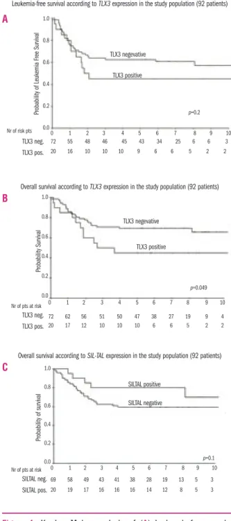
and the survival of patients in each oncogenetic sub-group are detailed in Table 3. Kaplan-Meier estimates of overall survival for patients with TLX3 and SILTAL lesions are represented in Figure 1. T-ALL positive for
TLX3 gene expression did not differ significantly from TLX3-negative ones with respect to prednisone-poor
response (p=0.11), bone marrow blast count (M status) at day 21 (p=0.81), complete remission rate (p=0.54) and treatment subgroups: C1 vs. C2 vs. C3 (p=0.44). Patients with SILTAL-positive T-ALL had hyperleukocytosis and many had a poor response to prednisone at day 8 (58%), but they had an excellent response to induction chemotherapy (95% M1 for SILTAL-positive patients vs. TLX3/HOX11L2 expression predicts adverse outcome in childhood T-ALL
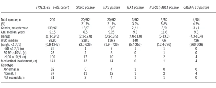
Table 2.Clinical and biological characteristics at diagnosis of the patients divided according to the presence of oncogenetic lesions.
FRALLE-93 T-ALL cohort SILTAL positive TLX3 positive TLX1 positive NUP214-ABL1 positive CALM-AF10 positive
Total number, n 200 20/92 20/92 3/92 3/52 4/84
(%) 21.7% 21.7% 3.2% 5.8% 4.7%
Gender, male/female 139/61 13/7 13/7 2 / 1 3/0 3 /1
Age, median, years 9.15 6.5 9.25 9.8 11.6 9.8
(range) (1.1-19.5) (2.3-17.8) (3.2-18.5) (4.8-11.8) (5-13.5) (4.3-16.4) WBC, median 98.85 238.5 116.7 140 66 426 (range,×109/L) (0.6-1247) (3.5-636) (1.9 - 736) (5.4-256) (12.4-736) (260-606) <50 ×109/L (n) 75 1 7 1 1 0 50-99 ×109/L (n) 25 2 3 0 1 0 ≥100 ×109/L (n) 100 17 10 2 2 4 Mediastinal involvement, (n) 141 13 14 0 1 4 Karyotype Abnormal, n 82 6 4 1 0 0 Normal, n 87 11 12 1 2 4 Not evaluable, n 31 3 4 1 1 0
Table 3. Clinical response to treatment, distribution and type of events, and survival status in subgroups divided according to the pres-ence of oncogenetic lesions.
FRALLE-93 T-ALL all TLX3 neg. TLX3 pos SILTAL pos TLX1 pos NUP214-ABL1 pos CALM-AF10 pos
Total number, (n) (200) (72) (20) (20) (3) (3) (4)
Response to prephase, n
died before evaluation 4 2 0 0 0 0 0
GPR <1000 blasts/µL 109 32 13 8 3 0 4
PPR >1000 blasts/µL 82 35 6 11 0 3 0
not evaluated 5 3 1 1 0 0 0
M status at day 21, n
died before evaluation 4 2 0 0 0 0 0
M1 135 54 14 19 2 1 3 M2 21 8 3 1 1 0 1 M3 40 8 3 0 0 2 0 Type of event, n No eventa 110 42 9 12 3 0 3 Failure 20 5 1 0 0 0 0 early deaths 4 1 0 0 0 0 0 relapses 66b 24 10a 8d 0 3 1 toxic deaths 3 0 0 0 0 0 0
Leukemia-free survival % ± SE 58±3 60±5 45±11 70±10 not evaluable not evaluable not evaluable Overall survival % ± SE 62±3 57±5 45±11 80±9 not evaluable not evaluable not evaluable
Alive, n 122 64 9 15 3 0 1
a
Continuous complete remission; b
Relapses occurred in bone marrow (BM) (n=42), central nervous system (CNS) (n=6), BM+CNS (n=5), testis (n=3) and medi-astinum (n= 10); CRelapses occurred in BM(n= 9) and testis (n=1) ; dRelapses occurred in BM (n=3), CNS (n=3), BM+CNS (n=1) and mediastinum (n=1).
65% MI for SILTAL-negative patients, p=0.008) (Table 3). In univariate analysis only two factors were significant-ly correlated with a lower 5-year overall survival rate: a poor response to prednisone (p=0.001) and TLX3 status (p=0.049) (Table 4). A shorter leukemia-free survival was significantly correlated only to a poor response to pred-nisone (p=0.002). It is noteworthy that M3 status did not significantly predict patients’ outcome (leukemia-free survival p=0.11; overall survival p=0.09). Patients with
SILTAL-positive T-ALL appeared to have a better
long-term outcome than those without this genetic lesion,
although the difference was not statistically significant (p=0.1) (Figure 1). In a multivariable analysis, a poor response to prednisone at day 8 and over-expression of the TLX3 gene independently predicted adverse out-come. The hazard ratios for 5-year leukemia-free sur-vival and overall sursur-vival for patients with a poor response to prednisone were 3.6 (p=0.01) and 4.8 (p<0.0001), respectively. The corresponding hazard ratios for patients with TLX3-positive T-ALL were 2.44 (p 0.017) and 3.7 (p=0.001), respectively (Table 5).
Outcome of T-ALL in other oncogenetic groups
A detailed description of the other subgroups is given in Table 3. The low number of positive samples in each
Figure 1. Kaplan Meier analysis of (A) leukemia-free survival according to TLX3 expression and (B) overall survival according to
TLX3 expression and (C) overall survival according to SILTAL expression.
Table 4.Univariate analysis of factors associated with 5-year over-all survival (OS) and leukemia-free survival (LFS) in the study pop-ulation. OS p LFS p Age Below median 67±7% 63±7% Median or above 61±7% 54±7% 0.47 0.4 Year of diagnosis Before median 61±7% 54±7% Median or after 67±7% 63±7% 0.56 0.44
White blood cell count
< median (149.5 ×10⁹/L) 65±7% 59±7%
≥ median 63±7% 59±7%
0.93 0.89
Response to steroids at day 8
Good 80±6% 73±6% Poor 46±8% 41±8% 0.001 0.002 M status M1+ M2 68±5% 63±5% M3 45±15% 36±14% 0.09 0.11 TLX3 negative 69±5% 63±6% positive 45±11% 45±11% 0.049 0.2 SILTAL negative 59±6% 55±6% positive 80±9% 70±10% 0.1 0.26
M1-2-3 status: percentage of blasts in the bone marrow at day 21.
Table 5.Multivariate analysis of factors associated with leukemia-free survival and overall survival at 5 years in the FRALLE-93 T-ALL study population.
Leukemia-free survivala Hazard ratio 95% confidence interval p value
Poor response to prednisone 3.57 7.14-1.72 0.001
TLX3 positive vs. negative 2.43 5.26-1.17 0.017
Overall survivala,b
Poor response to prednisone 4.76 11.11-2.08 <0.0001
TLX3 positive vs. negative 3.7 8.33-1.69 0.001
aCox regression model with all covariates associated with a p value less than 0.2
by univariate analysis: bsame as for leukemia-free survival but without SILTAL
status which had a p value of 0.26.
Leukemia-free survival according to TLX3 expression in the study population (92 patients)
Overall survival according to TLX3 expression in the study population (92 patients)
Overall survival according to SIL-TAL expression in the study population (92 patients)
1.0 0.8 0.6 0.4 0.2 0.0 1.0 0.8 0.6 0.4 0.2 0.0 1.0 0.8 0.6 0.4 0.2 0.0 0 1 2 3 4 5 6 7 8 9 10 0 1 2 3 4 5 6 7 8 9 10 0 1 2 3 4 5 6 7 8 9 10 72 62 56 51 50 47 38 27 19 9 4 69 58 49 43 41 38 28 19 13 5 3 20 19 17 16 16 16 14 12 8 5 3 20 17 12 10 10 10 6 6 5 2 2 72 55 48 46 45 43 34 25 6 6 3 20 16 10 10 10 9 6 6 5 2 2 Probability of Leukemia Free Survival Probability Survival Probability of survival TLX3 neg. TLX3 negevative TLX3 negevative TLX3 positive TLX3 positive SILTAL positive SILTAL negative TLX3 pos. TLX3 neg. TLX3 pos. Nr of risk pts Nr of pts at risk SILTAL neg. SILTAL pos. Nr of pts at risk p=0.2 p=0.049 p=0.1 A B C
group precluded a reliable statistical evaluation of their clinical impact, nevertheless all three patients with
TLX1 expression had an excellent outcome whereas all
three patients with the association of NUP214-ABL1 and TLX3 expression died before completion of their treatment.
Discussion
In the present study, we analyzed the prognostic value of five different genetic abnormalities, SILTAL,
TLX3, TLX1, CALM-AF10 and NUP214-ABL1 in a large
panel of cases of pediatric T-ALL treated in the FRALLE-93 trial. We were able to classify at the molecular level up to 50% (47/92) of the T-ALL cases studied and demonstrate that TLX3 overexpression is an independ-ent risk factor in pediatric T-ALL, predicting an inferior outcome. That risk may increase when the
NUP214-ABL1 fusion is co-expressed.
Very little is known about the role of the TLX3 gene in leukemic hematopoiesis. Recently, unsupervised hier-archical analysis of T-ALL gene expression profiles demonstrated a major cluster sharing a common gene expression signature, which included TLX1, TLX3-expressing cases and HOXA-translocated cases.12
Nervertheless, the contributions of TLX3 and TLX1 to the leukemic process could be quite different since actu-al data on clinicactu-al outcome seem to be opposing for these two lesions in adult T-ALL22,31,32as well in pediatric
T-ALL.24Moreover, the high frequency of ectopic TLX3
activation in pediatric T-ALL suggests that this gene plays a crucial role. Conflicting results have been
report-ed21-24 on the prognostic value of TLX3 activation in
pediatric T-ALL. Confounding elements, such as hetero-geneity of patient cohorts and clinical trials and differ-ences in therapeutic strategies, might explain such dis-crepancies.
The survival of patients with T-ALL has improved over the past 10 years, especially in paediatric co-opera-tive studies, because of more intensive post-induction chemotherapy.2,3Nevertheless, a subgroup usually
iden-tified as being at very high risk, generally including prednisone-poor responders and/or slow responders to early chemotherapy (M3), still have a high mortality rate. Our analysis confirmed the pejorative impact of a poor response to prednisone on both leukemia-free and overall survival in the FRALLE-93 T-ALL cohort and showed that TLX3 activation is of significant value in predicting worse outcome in terms of overall and leukemia-free survival independently of the prednisone response at day 8. Interestingly, we observed that TLX3 expression was more significantly associated with infe-rior survival than was M3 status (TLX3 positivity,
p≥0.049 vs. M3 status, p=0.09). Our data confirm the
findings of Ferrando22 in a cohort of 59 children and
young adults treated at St. Jude hospital,33and the more
recent results of van Grotel et al. in two independent cohorts of 72 and 53 children treated according to the DCOG ALL7/8 and COALL-97 protocols, respectively.24
In the latter study, with a median follow up of 3 years,
TLX3 expression was significantly predictive of a poor
outcome in both univariate (log-rank p=0.014) and mul-tivariate analysis (log-rank p=0.039). As in our study, the patients had a high median white blood cell count (135×109/L) and similarly, the most marked
hyperleuko-cytosis was observed in the SILTAL and CALM-AF10 subgroups.
Our data reinforce results from adult T-ALL
stud-ies.31,34An analysis of potential risk factors among the 92
T-ALL patients treated in the LALA94 trial31clearly
indi-cated an association between TLX3 expression and infe-rior overall survival (13% vs. 47%; p=0.009). Similar data emerged from the analysis of 100 patients with T-ALL treated in the GMT-ALL 05/93 and 06/99 trials between 1993 and 2000, reported by Baldus et al.34
Our results do, however, differ from those reported by Cavé et al.23These authors did not observe any
pejo-rative effect associated with TLX3 expression in a cohort of 138 T-ALL cases treated with EORTC 58881 (n=75) and 58951(n=78) protocols between 1989 and 2002. As in the case of the FRALLE-93 protocol, the gen-eral design of the 58881 trial had many similarities with that of the ALL-BFM90 trial and treatment results of T-ALL patients included in the very high risk and standard risk groups were also similar, with global leukemia-free survival rates of 61.7%±3.6 and 63.8%±6.7, respective-ly, at 8 years. The overall survival of T-ALL patients reported by Cavé et al. was 78%, thus higher than in the 58881 study. This difference could result from a bias in sample recruitment over the period, reflecting the better outcome of patients treated in the more recent 58951 study. In any case, the low number of events in that cohort of patients does not allow the detection of signif-icant differences among subgroups.
Some features of the patient population at diagnosis in the Cavé’s study differed strikingly from our and from other studies. Firstly, the median white blood cell count in the EORTC cohort was lower than in the FRALLE cohort (49×109/L vs. 98.85×10⁹/L); this was also
the case for the subgroups expressing TLX3 (34×109/L
vs. 120×109/L), SILTAL (124×109/L vs. 238.5×10⁹/L) and
TLX1 (29×109/L vs. 140×109/L). Secondly, the median
age of patients positive for TLX3, and TLX1 was lower than in the FRALLE cohort (6 and 5 vs. 9.2 and 9.8 years, respectively) whereas the median age of SILTAL- posi-tive patients was higher (11 vs. 6 years). These differ-ences are difficult to explain but they could be revelant to the interpretation of the results.
Besides the differences in patient populations and treatment efficacy, another explanation of the difference in the prognostic relevance of TLX3 may be the pres-ence of associated oncogenetic events. The recent find-ing of NUP214-ABL1 episomal fusions within T-ALL patients positive for TLX3 or TLX1 activation could sup-port this hypothesis. We observed a global frequency of
NUP214-ABL1 of 5.8%, which is in agreement with a
previous report,20with the frequency increasing to 16%
(3 of 18) among the TLX3-positive subgroup. Our data show that this association may effectively worsen clin-ical outcome, as suggested by Graux et al., and a search for NUP214-ABL1 should be systematically included in the investigation of TLX3-positive cases of T-ALL.
In keeping with the studies mentioned above, patients TLX3/HOX11L2 expression predicts adverse outcome in childhood T-ALL
with SILTAL-positive T-ALL showed very high white blood cell counts at diagnosis and were more frequently poor responders to prednisone at day 8 of treatment. Nevertheless, SILTAL expression did not affect signifi-cantly either leukemia-free survival or overall survival. One possible explanation is that TAL1 expression and/or reactivation is a frequent oncogenetic event associated with T-ALL, even in the absence of structural alterations of its locus.12,21The levels of TAL1 expression are highly
variable and not directly linked to the underlying mech-anism of activation. Thus, it would be interesting, as in the case of TLX1 expression,35to identify what level of
expression has a direct impact on the prognosis. We observed a favorable outcome in patients with
TLX1-positive T-ALL, which appears consistent with
observations in adults suggesting a positive association between TLX1 expression and good responder patient.32
Finally, we were unable to confirm previous data sug-gesting a pejorative role of CALM-AF10 fusion tran-scripts.30,36 but with only four positive cases and one
event in this subgroup, statistical analysis of the clinical impact of this lesion would not be reliable.
In conclusion, our study highlights the importance of
TLX3 activation as a new and powerful risk predictor in
pediatric patients with T-ALL. TLX3 expression and the presence of NUP214-ABL1 should be taken into account when evaluating patients with high-risk disease in order to improve outcome in pediatric T-ALL.
Authorship and Disclosures
PB, JL-P, GL, AB: conception and design of the study; GL, J-LP, LD, FS, AB: administrative support; PB, J-LP, VA, JMC, SF, VG, YP, MB, TL, CS, LD, AH, AB: provi-sion of study materials or patients; PB, JL-P, VA, JMC, MFA: collection and assembly of the data; PB, ML, JL-P, AB: data analysis and interpretation; PB, JL-P: manu-script writing. The authors reported no potential con-flicts of interest.
References
1. Nachman JB, Sather HN, Sensel MG, Gaynon PS, Arthur DC, Lange BJ, et al. Augmented post-induction thera-py for children with high-risk acute lymphoblastic leukemia and a slow response to initial therapy. N Engl J Med 1998;338:1663-71.
2. Amylon MD, Shuster J, Pullen J, Berard C, Link MP, Wharam M, et al. Intensive high-dose asparaginase consolidation improves survival for pediatric patients with T cell acute lymphoblastic leukemia and advanced stage lymphoblastic lym-phoma: a Pediatric Oncology Group study. Leukemia 1999;13:335-42. 3. Pui CH, Sandlund JT, Pei D,
Campana D, Rivera GK, Ribeiro RC, et al. Improved outcome for children with acute lymphoblastic leukemia: results of Total Therapy Study XIIIB at St Jude Children’s Research Hospital. Blood 2004;104:2690-6. 4. Schrauder A, Reiter A, Gadner H,
Niethammer D, Klingebiel T, Kremens B, et al. Superiority of allo-geneic hematopoietic stem-cell transplantation compared with che-motherapy alone in high-risk child-hood T-cell acute lymphoblastic leukemia: results from ALL-BFM 90 and 95. J Clin Oncol 2006;24:5742-9. 5. Pullen J, Shuster JJ, Link M,
Borowitz M, Amylon M, Carroll AJ, et al. Significance of commonly used prognostic factors differs for children with T cell acute lymphocytic leukemia (ALL), as compared to those with B-precursor ALL. A Pediatric Oncology Group (POG) study. Leukemia 1999;13:1696-707. 6. Carroll AJ, Crist WM, Link MP,
Amylon MD, Pullen DJ, Ragab AH, et al. The t(1;14)(p34;q11) is nonran-dom and restricted to T-cell acute lymphoblastic leukemia: a Pediatric Oncology Group study. Blood 1990; 76:1220-4.
7. McGuire EA, Hockett RD, Pollock KM, Bartholdi MF, O'Brien SJ, Korsmeyer SJ. The t(11;14)(p15;q11) in a T-cell acute lymphoblastic leu-kemia cell line activates multiple transcripts, including Ttg-1, a gene encoding a potential zinc finger pro-tein. Mol Cell Biol 1989;9:2124-32. 8. Royer-Pokora B, Loos U, Ludwig
WD. TTG-2, a new gene encoding a cysteine-rich protein with the LIM motif, is overexpressed in acute T-cell leukaemia with the t(11;14) (p13;q11). Oncogene 1991; 6:1887-93.
9. Xia Y, Brown L, Yang CY, Tsan JT, Siciliano MJ, Espinosa R III, et al. TAL2, a helix-loop-helix gene acti-vated by the (7;9)(q34;q32) transloca-tion in human T-cell leukemia. Proc Natl Acad Sci USA 1991;88:11416-20.
10. Mellentin JD, Smith SD, Cleary ML. lyl-1, a novel gene altered by chro-mosomal translocation in T cell leukemia, codes for a protein with a helix-loop-helix DNA binding motif. Cell 1989;58:77-83.
11. Speleman F, Cauwelier B, Dastugue N, Cools J, Verhasselt B, Poppe B, et al. A new recurrent inversion, inv(7)(p15q34), leads to transcrip-tional activation of HOXA10 and HOXA11 in a subset of T-cell acute lymphoblastic leukemias. Leukemia 2005;19:358-66.
12. Soulier J, Clappier E, Cayuela JM, Regnault A, García-Peydró M, Dombret H, et al. HOXA genes are included in genetic and biologic net-works defining human acute T-cell leukemia (T-ALL). Blood 2005;106: 274-86.
13. Hatano M, Roberts CW, Minden M, Crist WM, Korsmeyer SJ. Deregul-ation of a homeobox gene, HOX11, by the t(10;14) in T cell leukemia. Science 1991;253:79-82.
14. Bernard OA, Busson-LeConiat M, Ballerini P, Mauchauffé M, Della Valle V, Monni R, et al. A new recur-rent and specific cryptic
transloca-tion, t(5;14)(q35;q32), is associated with expression of the Hox11L2 gene in T acute lymphoblastic leu-kemia. Leukemia 2001;15:1495-504. 15. Ellisen LW, Bird J, West DC, Soreng AL, Reynolds TC, Smith SD, et al. TAN-1, the human homolog of the Drosophila notch gene, is broken by chromosomal translocations in T lymphoblastic neoplasms. Cell 1991; 66:649-61.
16. Brown L, Cheng JT, Chen Q, Siciliano MJ, Crist W, Buchanan G, et al. Site-specific recombination of the tal-1 gene is a common occurrence in human T cell leukemia. EMBO J 1990;9:3343-51.
17. Weng AP, Ferrando AA, Lee W, Morris JP 4th, Silverman LB, Sanchez-Irizarry C, et al. Activating mutations of NOTCH1 in human T cell acute lymphoblastic leukemia. Science 2004;306:269-71.
18. Breit S, Stanulla M, Flohr T, Schrappe M, Ludwig WD, Tolle G, et al. Activating NOTCH1 mutations predict favorable early treatment response and long-term outcome in childhood precursor T-cell lym-phoblastic leukemia. Blood 2006; 108:1151-7.
19. No authors listed. t(10;11)(p13-14; q14-21): a new recurrent transloca-tion in T-cell acute lymphoblastic leukemias. Groupe Francais de Cyto-genetique Hematologique (GFCH). Genes Chromosomes Cancer 1991; 3:411-5.
20. Dreyling MH, Martinez-Climent JA, Zheng M, Mao J, Rowley JD, Bohlander SK, et al. The t(10;11) (p13;q14) in the U937 cell line results in the fusion of the AF10 gene and CALM, encoding a new member of the AP-3 clathrin assembly protein family. Proc Natl Acad Sci USA 1996; 93:4804-9.
21. Berger R, Dastugue N, Busson M, Van Den Akker J, Pérot C, Ballerini P, et al. t(5;14)/HOX11L2-positive T-cell acute lymphoblastic leukemia. A collaborative study of the Groupe
Francais de Cytogenetique Hémato-logique (GFCH). Groupe Français de Cytogénétique Hématologique (GFCH). Leukemia 2003;17:1851-7. 22. Graux C, Cools J, Melotte C, Quentmeier H, Ferrando A, Levine R, et al. Fusion of NUP214 to ABL1 on amplified episomes in T-cell acute lymphoblastic leukemia. Nat Genet 2004;36:1084-9.
23. Ballerini P, Blaise A, Busson-Le Coniat M, et al. HOX11L2 expres-sion defines a clinical subtype of pediatric T-ALL associated with poor prognosis. Blood 2002;100: 991-7.
24. Ferrando AA, Neuberg DS, Staunton J, Loh ML, Huard C, Raimondi SC, et al. Gene expression signatures define novel oncogenic pathways in T cell acute lymphoblastic leukemia. Cancer Cell 2002;1:75-87.
25. Cavé H, Suciu S, Preudhomme C, Poppe B, Robert A, Uyttebroeck A, et al. Clinical significance of HOX11L2 expression linked to t(5;14)(q35;q32), of HOX11 expres-sion, and of SILTAL fusion in child-hood T-cell malignancies: results of EORTC studies 58881 and 58951. EORTC-CLG. Blood 2004;103:442-50.
26. van Grotel M, Meijerink JP, Beverloo HB, Langerak AW, Buys-Gladdines JG, Schneider P, et al. The outcome of molecular-cytogenetic subgroups in pediatric T-cell acute lymphoblas-tic leukemia: a retrospective study of patients treated according to DCOG or COALL protocols. Hae-matologica 2006;91:1212-21. 27. Gaynon PS, Desai AA, Bostrom BC,
Hutchinson RJ, Lange BJ, Nachman JB, et al. Early response to therapy and outcome in childhood acute lymphoblastic leukemia: a review. Cancer 1997;80:1717-26.
28. Michel G, Landman-Parker J, Auclerc MF, Mathey C, Leblanc T, Legall E, et al. Use of recombinant human granulocyte colony-stimulat-ing factor to increase chemotherapy dose-intensity: a randomized trial in very high-risk childhood acute lym-phoblastic leukemia. J Clin Oncol 2000;18:1517-24.
29. Bene MC, Castoldi G, Knapp W, Ludwig WD, Matutes E, Orfao A, et al. Proposals for the immunological classification of acute leukemias. European Group for the Immunolo-gical Characterization of Leukemias (EGIL). Leukemia 1995; 9:1783-6. 30. Gabert J, Beillard E, van der Velden
VH, Bi W, Grimwade D, Pallisgaard
N, et al. Standardization and quality control studies of ‘real-time’ quanti-tative reverse transcriptase poly-merase chain reaction of fusion gene transcripts for residual disease detection in leukemia - a Europe Against Cancer program. Leukemia 2003;17:2318-57.
31. Beillard E, Pallisgaard N, van der Velden VH, Bi W, Dee R, van der Schoot E, et al. Evaluation of candi-date control genes for diagnosis and residual disease detection in leukemic patients using ‘real-time’ quantitative reverse-transcriptase polymerase chain reaction (RQ-PCR) - a Europe Against Cancer pro-gram. Leukemia 2003;17:2474-86. 32. Asnafi V, Radford-Weiss I, Dastugue
N, Bayle C, Leboeuf D, Charrin C, et al. CALM-AF10 is a common fusion transcript in T-ALL and is specific to the TCRγδ lineage. Blood 2003;102: 1000-6.
33. Su XY, Busson M, Della Valle V, Ballerini P, Dastugue N, Talmant P, et al. Various types of rearrangements target TLX3 locus in T-cell acute lymphoblastic leukemia. Genes Chromosomes Cancer 2004;41:243-9.
34. Baldus CD, Burmeister T, Martus P, Schwartz S, Gökbuget N, Bloomfield CD, et al. High expres-sion of the ETS transcription factor ERG predicts adverse outcome in acute T-lymphoblastic leukemia in adults. J Clin Oncol 2006;24:4714-20.
35. Asnafi V, Buzyn A, Thomas X, Huguet F, Vey N, Boiron JM, et al. Impact of TCR status and genotype on outcome in adult T-cell acute lymphoblastic leukemia: a LALA-94 study. Blood 2005;105:3072-8. 36. Logan C, Wingate RJ, McKay IJ,
Lumsden A. Tlx-1 and Tlx-3 homeo-box gene expression in cranial sen-sory ganglia and hindbrain of the chick embryo: markers of patterned connectivity. J Neurosci 1998;18: 5389-402.
37. Watt PM, Kumar R, Kees UR. Pro-moter demethylation accompanies reactivation of the HOX11 proto-oncogene in leukemia. Genes Chro-mosomes Cancer 2000;29:371-7. 38. Hansen-Hagge TE, Schäfer M, Kiyoi
H, Morris SW, Whitlock JA, Koch P, et al. Disruption of the RanBP17/ Hox11L2 region by recombination with the TCRδ locus in acute lym-phoblastic leukemias with t(5;14) (q34;q11). Leukemia 2002;16:2205-12.
39. Hawley RG, Fong AZ, Reis MD, Zhang N, Lu M, Hawley TS. Trans-forming function of the HOX11/ TCL3 homeobox gene. Cancer Res 1997;57:337-45.
40. Kawabe T, Muslin AJ, Korsmeyer SJ. HOX11 interacts with protein phos-phatases PP2A and PP1 and disrupts a G2/M cell-cycle checkpoint. Nature 1997;385:454-8.
41. Ferrando AA, Neuberg DS, Dodge RK, Paietta E, Larson RA, Wiernik PH, et al. Prognostic importance of TLX1 (HOX11) oncogene expres-sion in adults with T-cell acute lym-phoblastic leukaemia. Lancet 2004; 363:535-6.
42. Asnafi V, Beldjord K, Libura M, Villarese P, Millien C, Ballerini P, et al. Age-related phenotypic and oncogenic differences in T-cell acute lymphoblastic leukemias may reflect thymic atrophy. Blood 2004; 104:4173-80.
43. Schrappe M, Reiter A, Zimmermann M, Harbott J, Ludwig WD, Henze G, et al. Long-term results of four consecutive trials in childhood ALL performed by the ALL-BFM study group from 1981 to 1995. Berlin-Frankfurt-Munster. Leukemia 2000; 14:2205-22.
44. Pui CH, Boyett JM, Rivera GK, Hancock ML, Sandlund JT, Ribeiro RC, et al. Long-term results of total therapy studies 11, 12 and 13A for childhood acute lymphoblastic leukemia at St Jude Children’s Research Hospital. Leukemia 2000; 14:2286-94.
45. De Keersmaecker K, Marynen P, Cools J. Genetic insights in the pathogenesis of T-cell acute lym-phoblastic leukemia. Haematologica 2005;90:1116-27.
46. Bergeron J, Clappier E, Radford I, Buzyn A, Millien C, Soler G, et al. Prognostic and oncogenic relevance of TLX1/HOX11 expression level in T-ALLs. Blood 2007;110:2324-30. 47. Dreyling MH, Schrader K, Fonatsch
C, Schlegelberger B, Haase D, Schoch C, et al. MLL and CALM are fused to AF10 in morphologically distinct subsets of acute leukemia with translocation t(10;11): both rearrangements are associated with a poor prognosis. Blood 1998;91: 4662-7.