HAL Id: hal-02081581
https://hal.sorbonne-universite.fr/hal-02081581
Submitted on 27 Mar 2019
HAL is a multi-disciplinary open access
archive for the deposit and dissemination of
sci-entific research documents, whether they are
pub-lished or not. The documents may come from
teaching and research institutions in France or
abroad, or from public or private research centers.
L’archive ouverte pluridisciplinaire HAL, est
destinée au dépôt et à la diffusion de documents
scientifiques de niveau recherche, publiés ou non,
émanant des établissements d’enseignement et de
recherche français ou étrangers, des laboratoires
publics ou privés.
Narrative review: clinical assessment of peripheral tissue
perfusion in septic shock
Geoffroy Hariri, Jérémie Joffre, Guillaume Leblanc, Michael Bonsey,
Jean-Rémi Lavillegrand, Tomas Urbina, Bertrand Guidet, Eric Maury, Jan
Bakker, Hafid Ait-Oufella
To cite this version:
Geoffroy Hariri, Jérémie Joffre, Guillaume Leblanc, Michael Bonsey, Jean-Rémi Lavillegrand, et al..
Narrative review: clinical assessment of peripheral tissue perfusion in septic shock. Annals of Intensive
Care, SpringerOpen, 2019, 9, pp.37. �10.1186/s13613-019-0511-1�. �hal-02081581�
REVIEW
Narrative review: clinical assessment
of peripheral tissue perfusion in septic shock
Geoffroy Hariri
1,2, Jérémie Joffre
1,2, Guillaume Leblanc
3,4, Michael Bonsey
1, Jean‑Remi Lavillegrand
1,2,
Tomas Urbina
1, Bertrand Guidet
1,2,5, Eric Maury
1,2,5, Jan Bakker
6,7,8,9and Hafid Ait‑Oufella
1,2,10*Abstract
Sepsis is one of the main reasons for intensive care unit admission and is responsible for high morbidity and mortality. The usual hemodynamic targets for resuscitation of patients with septic shock use macro‑hemodynamic parameters (hearth rate, mean arterial pressure, central venous pressure). However, persistent alterations of microcirculatory blood flow despite restoration of macro‑hemodynamic parameters can lead to organ failure. This dissociation between macro‑ and microcirculatory compartments brings a need to assess end organs tissue perfusion in patients with septic shock. Traditional markers of tissue perfusion may not be readily available (lactate) or may take time to assess (urine output). The skin, an easily accessible organ, allows clinicians to quickly evaluate the peripheral tissue perfusion with noninvasive bedside parameters such as the skin temperatures gradient, the capillary refill time, the extent of mottling and the peripheral perfusion index.
Keywords: Septic shock, Microcirculation, Capillary refill time, Temperatures gradient, Peripheral perfusion index,
Mottling, Skin
© The Author(s) 2019. This article is distributed under the terms of the Creative Commons Attribution 4.0 International License
(http://creat iveco mmons .org/licen ses/by/4.0/), which permits unrestricted use, distribution, and reproduction in any medium,
provided you give appropriate credit to the original author(s) and the source, provide a link to the Creative Commons license, and indicate if changes were made.
Background
International guidelines emphasized that fast identifica-tion, assessment and treatment combining early anti-biotic therapy, fluid administration and vasopressor infusion are crucial steps in the management of septic shock. However, despite early management, mortality of
patients with septic shock remains high [1]. A possible
explanation may be the persistent tissue hypoperfusion despite restoration of macro-hemodynamic parameters.
The usual hemodynamic targets for resuscitation of patients with septic shock use macro-hemodynamic parameters (heart rate, mean arterial pressure, cen-tral venous pressure). However, persistent alterations of microcirculatory blood flow despite restoration of macro-hemodynamic parameters can lead to organ fail-ure. In a meta-analysis of 252 patients, De Backer et al.
[2] showed that microcirculatory perfusion alterations
predict mortality during serious infections, whereas mean arterial pressure or cardiac output did not. In critically ill patients, cardiac output optimization using increasing doses of dobutamine did not improve
micro-vascular blood flow in the sublingual area [3, 4]. In
another study, modulating mean arterial pressure by increasing norepinephrine dose had variable unpredict-able effects on microcirculatory flow, which occasionally
worsened [5, 6]. This dissociation between macro- and
microcirculatory compartments, defined by Ince as «a
loss of hemodynamic coherence» [7], brings a need to
assess end organs tissue perfusion in patients with septic shock and to develop tools to analyze microcirculatory
blood flow [8]. The direct identification of severe
micro-circulatory alterations remains difficult at bedside. Tra-ditional markers of tissue perfusion may not be readily available (lactate) or may take time to assess (urine out-put). The skin, an easily accessible organ, allows clinicians to quickly evaluate the peripheral tissue perfusion with noninvasive bedside parameters such as the skin tem-peratures gradient, the capillary refill time, the extent of mottling and the peripheral perfusion index.
Open Access
*Correspondence: hafid.aitoufella@aphp.fr
1 Service de réanimation médicale, Assistance Publique‑Hôpitaux de
Paris (AP‑HP), Hôpital Saint‑Antoine, 184 rue du Faubourg Saint‑Antoine, 75571 Paris Cedex 12, France
Page 2 of 9 Hariri et al. Ann. Intensive Care (2019) 9:37
The aim of this review is to evaluate whether peripheral tissue perfusion assessment in septic patients could be helpful in evaluating organ failure severity and to screen patients at high risk of mortality. Finally, we analyze avail-able data regarding implementation of peripheral perfu-sion evaluation in sepsis management.
Skin as a tool for the evaluation of the microcirculation and tissue perfusion
The skin provides important information in patients with septic shock. As a visible and easily accessible organ, the skin allows simple observation of local microcirculatory perfusion through skin temperature alterations (skin temperature gradient), perfusion (capillary refill time) and color (mottling). The pathophysiology of these clini-cal disorders has not been investigated in depth, but sev-eral authors assume that the main driven mechanism of reduced blood flow is local vasoconstriction mediated by
sympathetic neuroactivation [8]. Additional mechanisms
could participate to impair microvascular blood flow
(Fig. 1) [9, 10] such as local endothelial dysfunction [11,
12] (Fig. 2), leukocyte adhesion, platelet activation and
fibrin deposition [13]. These clinical, noninvasive,
easy-to-use, parameters are attractive tools to follow micro-circulatory perfusion in patients with acute micro-circulatory
failure [14, 15]. In 2014, several European experts
recom-mended to integrate abnormal skin perfusion parameters
in the definition and treatment of shock [16].
Subjective assessment of peripheral skin temperature may be a valuable tool in the evaluation of patients with
septic shock. Eighty years ago, Ebert et al. [17] described
the skin of septic shock patients as being «pale, often
sweaty». Altemeier et al. [18] then noticed that a moist
and cold skin was a factor of worse prognosis in patients with septic shock. Cold hands and feet, and abnor-mal skin color are the first clinical signs that developed
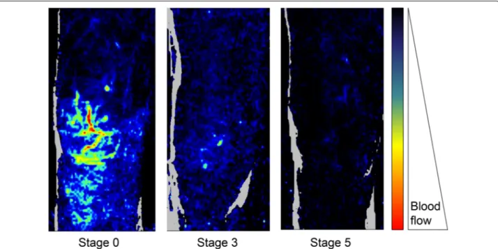
Fig. 1 Examples of skin microvascular perfusion evaluation using laser Doppler imaging in the knee area according to the mottling score. Skin
perfusion decreases when mottling score worsens. Adapted from [9]
Knee area (Perfusion Units)
0 20 40 60 2 4 6 8 10 12 minutes 80 100 Sepsis
Septic shock survivor Septic shock non-survivor
Fig. 2 Examples of skin microcirculatory endothelial reactivity in
the knee area in a patients with sepsis, in a patient with septic shock that was alive at day 14 and in a patient with septic shock that was ultimately dead at day 14. Skin microcirculatory blood flow was measured at baseline and after acetylcholine iontophoresis. Adapted from [11]
in meningococcal disease in children [19]. In a cohort of 264 surgical ICU patients, patients with cold skin on extremities and knees had significantly lower central venous saturation and higher lactate level as compared to patients with normal skin temperature (4.7 ± 1.5 vs
2.2 ± 1.6 mmol/L, p < 0.05) [20]. In a prospective cohort
study of 50 critically ill patients with circulatory dys-function, including 26 patients with septic shock, Lima
et al. [21] observed that patients with cold skin on the
extremities had a higher rate of organ failure at 48 h after resuscitation as compared to patients with normal skin temperature.
However, skin temperature gradients may be more accurate in the evaluation of patients with septic shock. Several studies investigated quantitative temperature gradients in critically ill patients, particularly between
peripheral and ambient temperatures [22], central and
peripheral body temperatures [23] and finger and
fore-arm skin temperatures [24]. Temperature gradients do
not correlate with cardiac output [22, 25, 26] but are
predictive of both organ failure severity and worse
out-come. Joly et al. [22] measured toe-to-ambient
tempera-ture gradients 3 h after admission in a mixed population of critically ill patients, and non-survivors had a mean toe-ambient temperature gradient of 0.9 °C, whereas survivors had a gradient of 3.4 °C. Normalization of central-peripheral temperature gradients (< 7 °C) within the 6 first hours of resuscitation predicted correction of
hyperlactatemia in septic shock patients [27]. In a recent
study including 103 septic patients, Bourcier et al. [28]
reported higher central-to-toe temperature gradients and lower toe-to-ambient temperature gradients in patients with septic shock, compared to patients with sepsis. Moreover, a rise in the toe-to-ambient temperature gra-dient was independently associated with decreased ICU mortality (OR 0.7 [0.5, 0.9] per °C, p < 0.001).
Finger-to-forearm skin and toe-to-ambient tempera-ture gradients are more accurate tools that could be used in every patient without previous hypothermia, including patients with dark skin, providing quantitative
informa-tion with good reproducibility (Table 1, Fig. 3).
Capillary refill time
The capillary refill time (CRT) measures the amount of time necessary for the skin to return to baseline color after applying a pressure on a soft tissue (generally finger tip). The CRT gives important information on skin per-fusion and microcirculatory status but does not reflect
cardiac output [25, 29]. Visual measurement of CRT
associated with other clinical signs (tachycardia, mucosal dryness, etc.) helps to diagnose dehydration in children
[30]. In acute pathologies, such as gastro-intestinal
infec-tions or malaria [31], CRT represents an attractive and
easy-to-use tool for clinicians in the initial screening of
severely ill patients [32]. Inter-rater variability of CRT
was weak in non-trained physicians [33], but is better in
centers expert in tissue perfusion evaluation [34],
espe-cially in the knee area [35]. Standardization of finger-tip
pressure (i.e., How long? How strong the applied pres-sure?) might improve CRT reproducibility. Ait-Oufella
et al. [35] obtained good inter-rater concordance by
“applying a firm pressure for 15 s. The pressure applied was just enough to remove the blood at the finger tip of the physician’s nail illustrated by appearance of a thin white distal crescent (blanching) under the nail.”
Capillary refill time measurement correlates with the pulsatility index, a surrogate ultrasound-derived param-eter that reflects vascular tone of visceral organs in septic
shock patients [36]. CRT is an interesting tool to assess
the severity of an acute illness. In the intensive care
unit, Lima et al. [21] reported an association between a
prolonged CRT (> 4.5 s on the index finger) and hyper-lactatemia and a higher SOFA score. In septic shock patients, a prolonged CRT 6 h after resuscitation has been shown to be predictive of 14-day mortality, with an Area Under Curve (AUC) of 84% for a measure on the index finger, and 90% for a measure on the knee. A 2.4-second threshold value on the index finger predicted mortality with an 82% sensitivity (95% CI [60–95]) and a 73% specificity (95% CI [56–86]). On the knee, a thresh-old value of 4.9 s predicted 14-day mortality with an 82% sensitivity (95% CI [60–95]) and an 84% specificity (95%
CI [68–94]) [35].
Overall, when used as a qualitative variable (prolonged or not), CRT is a reliable triage tool to identify critically ill patients at risk of negative outcome. Quantitative measurement of CRT should be mainly used by trained
physicians in patients with non-dark skin (Table 1, Fig. 3).
Mottling
Mottling, a characteristic discoloration of the skin
fol-lowing reduced skin blood flow [9], is taught as a marker
of shock, but its clinical relevance has been poorly inves-tigated until recent years. A significant relationship between mottling extension and visceral organ vascular tone has been reported suggesting that mottling could
reflect gut, liver spleen and kidney hypoperfusion [36].
To assess the predictive value of mottling in criti-cally ill patients with severe infections, a semi-quanti-tative clinical score for mottling (ranging from 0 to 5), based on the extension of these purple patches from the patella toward the periphery, has been developed and validated with an excellent inter-observer
reproduc-ibility [37] (Kappa 0.87% (CI 95% [0.72–0.97]) (Fig. 4).
Mottling score reliably reflects organ failure sever-ity in patients with sepsis or septic shock and helps to
Page 4 of 9 Hariri et al. Ann. Intensive Care (2019) 9:37
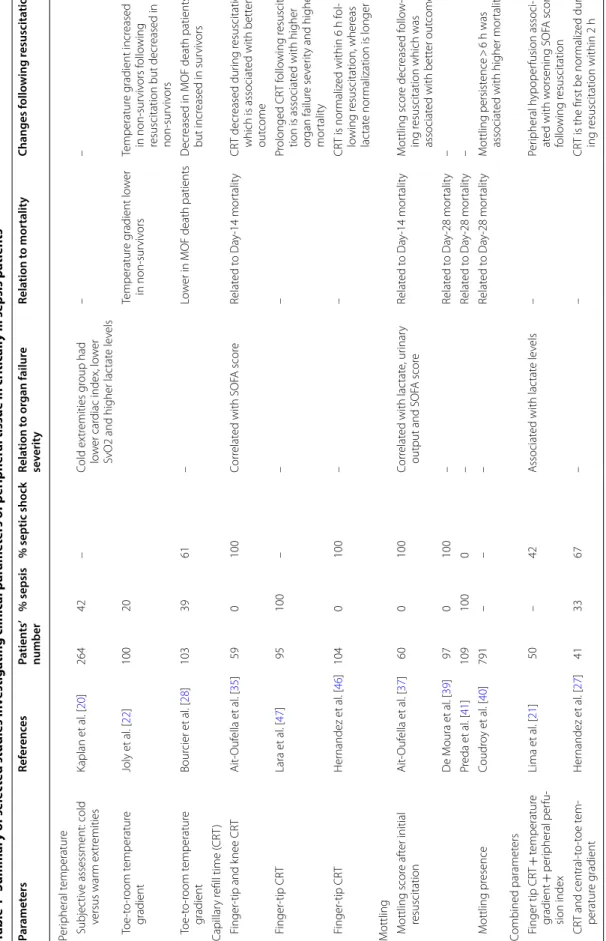
Table 1 Summar y of selec ted studies in vestiga ting clinic al par amet ers of p eripher al tissue in critic
ally ill sepsis pa
tien ts CR T capillar y r efill time , MO F multior gan failur e, SO FA sequen tial or gan failur e assessmen t Par amet ers Ref er enc es Pa tien ts ’ number % sepsis % septic shock Rela tion t o or gan failur e se verit y Rela tion t o mor talit y Changes f ollo wing r esuscita tion Per ipheral t emperatur e Subjec tiv e assessment: cold versus war m ex tr emities Kaplan et al . [ 20 ] 264 42 – Cold ex tr emities g roup had lo w er car diac index, lo w er
SvO2 and higher lac
tat e le vels – – T oe ‑t o‑ room temperatur e gradient Joly et al . [ 22 ] 100 20 Temperatur e g radient lo w er in non ‑sur viv ors Temperatur e g radient incr eased in non ‑sur viv ors f ollo wing
resuscitation but decr
eased in non ‑sur viv ors T oe ‑t o‑ room temperatur e gradient Bour cier et al . [ 28 ] 103 39 61 – Lo w
er in MOF death patients
D
ecr
eased in MOF death patients
but incr eased in sur viv ors Capillar y r efill time ( CR T) F inger ‑tip and k nee CR T Ait ‑Ouf ella et al . [ 35 ] 59 0 100 Cor relat ed with SOF A scor e Relat ed t o Da y‑ 14 mor talit y CR T decr eased dur ing r esuscitation which is associat ed with bett er out come F inger ‑tip CR T Lara et al . [ 47 ] 95 100 – – – Pr olonged CR T f ollo wing r esuscita ‑ tion is associat ed with higher or gan failur e se ver
ity and higher
mor talit y F inger ‑tip CR T Her nandez et al . [ 46 ] 104 0 100 – – CR T is nor maliz ed within 6 h f ol ‑ lo wing r esuscitation, wher eas lac tat e nor malization is longer M
ottling Mottling scor
e af ter initial resuscitation Ait ‑Ouf ella et al . [ 37 ] 60 0 100 Cor relat ed with lac tat e, ur inar y
output and SOF
A scor e Relat ed t o Da y‑ 14 mor talit y M ottling scor e decr eased f ollo w ‑ ing r
esuscitation which was
associat ed with bett er out come D e M oura et al . [ 39 ] 97 0 100 – Relat ed t o Da y‑ 28 mor talit y – Pr eda et al . [ 41 ] 109 100 0 – Relat ed t o Da y‑ 28 mor talit y – M ottling pr esence Coudr oy et al . [ 40 ] 791 – – – Relat ed t o Da y‑ 28 mor talit y M ottling persist ence > 6 h was associat
ed with higher mor
talit y Combined paramet ers F inger tip CR T + temperatur e gradient + per ipheral per fu ‑ sion index Lima et al . [ 21 ] 50 – 42 A ssociat ed with lac tat e le vels – Per ipheral h ypoper fusion associ ‑ at ed with w orsening SOF A scor e follo wing r esuscitation CR T and central ‑t o‑ toe t em ‑ peratur e g radient Her nandez et al . [ 27 ] 41 33 67 – – CR
T is the first be nor
maliz ed dur ‑ ing r esuscitation within 2 h
identify critically ill patients with worse outcome. In a study including septic shock patients, the mottling score at 6 h after resuscitation was predictive of death at day 14 (odds radio [OR] 16, CI 95% 4–81, for stages 2–3; vs 74, CI 95% 11–1568, for stages 4–5). Mortality occurred within 12–24 h for stages 4–5, within 24–72 h for stages 2–3 and later than 72 h for the rare deaths for stages 0–1 (Kaplan–Meier charts, p < 0.0001). In the same study, cardiac output and blood pressure were not associated with mortality at day 14, confirming the disparity between microcirculatory and
macrocircula-tory parameters [37]. These results were confirmed in
cirrhotic patients with septic shock [38]. In addition,
in mottling groups ≤ 3, knee CRT improved patient discrimination according to their outcome, with non-survivors presenting a significantly higher knee CRT
[35]. Another South American study confirmed these
results in septic shock patients. Mortality rate at day 28 was 100% when the mottling score was higher or equal to stage 4, 77% for stages 2 and 3, and 45% for
stages 1 or lower [39]. Prognostic value of mottling
was also reported in unselected ICU patients: Persis-tent (> 6 h) mottling extending over the knee (> stage 2) was an independent risk factor for mortality (OR 3.29,
95% CI 2.08–5.19; p < 0.0001) [40]. Finally, Preda et al.
[41] found the good predictive value of the mottling
score for mortality at day 28 in patients with sepsis not receiving vasopressors.
In summary, mottling score is a reliable semi-quan-titative tool that reflects organ failure severity in non-selected septic patients with or without vasopressors and is helpful to identify critically ill patients with pejorative outcome and also to monitor changes during resuscita-tion. In patients with mottling score ranging from 0 to 3, knee CRT measurement could be associated with
improving risk stratification (Table 1, Fig. 3).
Peripheral perfusion index
Peripheral perfusion index is defined as the difference between the pulsatile and non-pulsatile portion of pulse wave, measured by plethysmography. Peripheral perfu-sion index (PPI) gives information on peripheral vascular tonus by the pulsatility, decreasing in vasoconstriction
and raising in vasodilation [42]. Peripheral perfusion
index is an early predictor of central hypovolemia [43].
In a prospective observational study in an emergency department, PPI was not significantly different between patients admitted to the hospital and patients discharged from the emergency department suggesting that it could
not be used as a triage tool [44]. However, in critically
Dark skin ?
No
Yes
Temperature gradient -Finger-to-Forearm -Toe-to-Room Mottling score Stage >3 ? Yes No Finger or knee CRT Hypothermia ? No Yes P Perfusion Index Trained physician ? Yes Quantitative CRT Qualitative CRTNo Finger threshold 3 s Knee threshold 5 sPage 6 of 9 Hariri et al. Ann. Intensive Care (2019) 9:37
ill patients, PPI is significantly lower in patients with a peripheral perfusion alteration (0.7 vs 2.3, p < 0.01)
[21]. He et al. [45] showed that the PPI is altered in
sep-tic shock patients, as compared to control subjects in postoperative scheduled surgery. Moreover, in the same study, the PPI was significantly lower in non-survivors. With a 0.20 cutoff value, PPI was predictive of ICU mor-tality with an AUC of 84% (69–96), a sensitivity of 65% and a specificity of 92%.
Discussion
Abnormal skin perfusion evaluation and resuscitation
Despite some differences between micro and macrovas-cular compartments, it would be over-simplifying and possibly wrong to completely separate these two
vascu-lar compartments. In the study by Ait-Oufella et al. [37]
focusing on mottling, global hemodynamic improvement
within the first hours following resuscitation, based on blood volume optimization and catecholamine use, was associated with mottling improvement. Patients whose mottling score improved through the first 6-hour resus-citation had a good prognosis, whereas those whose score was stable or even worsened had a poor progno-sis (14-day mortality: 23% vs 88%, p < 0.001). Finger-tip CRT is also quickly normalized in septic shock patients within 2–6 h after resuscitation, whereas hyperlactatemia
requires longer time to recover [27, 46]. Interestingly,
patients in whom CRT did not recover after fluid
infu-sion had pejorative outcome [47]. Altogether, these
stud-ies suggest that peripheral tissue perfusion could be used as triage tool at the early steps of sepsis manage-ment at admission and after fluid infusion. The ongo-ing ANDROMEDA-SHOCK trial aims to compare two resuscitation strategies during the first hours of sepsis 1 2 3 4 5 STAGE 4
a
b
Fig. 4 a The mottling score, ranging from 0 to 5, is based on skin mottling area extension on legs. Score 0 represents no mottling, score 1
represents small mottling area (coin size) localized to the center of the knee, score 2 represents mottling area not exceeding the superior edge of the knee cap, score 3 represents mottling area not exceeding the middle thigh, score 4 represents mottling area not exceeding the fold of the groin and score 5 otherwise. b Example of mottling score 5. Adapted from [37]
treatment on 28-day mortality, one based on CRT
meas-urement and the other on arterial lactate clearance [48].
During ICU stay, evaluation of peripheral perfusion could also be helpful. A «proof-of-concept» study has been done comparing a volume expansion strategy based on peripheral perfusion, clinical parameter assessment, to a classical strategy based on mean arterial pressure, central venous pressure and cardiac index. Peripheral perfusion was assessed through CRT, index-forearm tem-perature gradient, peripheral perfusion index, and StO2. The resuscitation strategy based on clinical tissue perfu-sion assessment led to a reduction in fluid therapy vol-ume in the first 72 h (7565 ± 982 mL vs. 10,028 ± 941 mL, p = 0.08) and to a reduction in hospital length of stay (16
[5–28] vs. 43 [8–45] days, p < 0.05) [49]. A task force of
six international experts with extensive bedside experi-ence recently proposed to integrate peripheral tissue perfusion tools in risk stratification and management of septic patients in resource-limited intensive care units, especially CRT, mottling score and temperature gradients
[50].
As bedside evaluation of tissue perfusion using the skin improves risk stratification in patients with sepsis, there is a possibility that it could be used as a tool to guide
resuscitation. Lavillegrand et al. [51] reported that a mild
arterial hypotension (MAP between 55 and 65 mmHg) could be safely tolerated in patients without any sign of hypoperfusion. Such «personalized» management requires close monitoring (in an ICU) but may decrease the use of invasive devices and vasopressors, both having potential side effects. Conversely, patients with markers of tissue hypoperfusion require rapid ICU transfer, and also, we hypothesized that they should be good candidate for therapeutic approaches targeting microcirculation for resuscitation in the future. For example, nitroglyc-erin infusion had no beneficial effect in unselected
sep-sis patients [52] but improved peripheral perfusion in
selected patients with prolonged CRT and/or increased
finger-tip-to-forearm skin gradient temperatures [53].
Ilomedin has been also recently proposed as a rescue therapy in sepsis shock with refractory tissue
hypoper-fusion [54] and will be tested soon in a prospective
ran-domized multicenter trial (I-MICRO NCT03788837). In the future, it is important to evaluate whether drugs targeting the microcirculation could improve outcome of selected patients with persistent peripheral
hypoperfu-sion despite initial resuscitation [55]. The first results of
ANDROMEDA-SHOCK, an international multicenter trial recently completed, support that a tissue
perfusion-guided resuscitation is beneficial [48, 56]. Indeed,
Her-nandez et al. [56] showed in septic shock adults that an
early peripheral perfusion-targeted resuscitation, aim-ing at normalizaim-ing capillary refill time, was associated
with less organ dysfunction at day 3 and a trend toward reduced 28-day mortality when compared to a lactate-level-targeted therapeutic strategy.
Limitations
In this review, almost all data were obtained in small-sized monocenter observational studies and were performed by experts in tissue perfusion evaluation, suggesting potential biases. In addition, no published multicenter randomized trial is available showing that the implementation of bedside tissue perfusion assess-ment improves septic patients manageassess-ment and in fine outcome. This narrative review did not provide strong recommendation regarding the use of tissue perfusion parameters in septic patients according to GRADE meth-odology but only proposed how and when to implement them.
Conclusion
In patients with septic shock, tissue microvascular hypoperfusion can be evaluated at bedside using indica-tors of skin perfusion. After initial resuscitation, these parameters are helpful in identifying patients with severe organ failure and at high risk of mortality. However, there is a need in the future to investigate these bedside tissue microvascular perfusion parameters as management tar-gets for resuscitation in septic shock patients.
Abbreviations
CI: confidence interval; CRT : capillary refill time; ICU: intensive care unit; MAP: mean arterial pressure; NIRS: near‑infrared spectroscopy; NO: nitric oxide; OR: odds ratio; PPI: peripheral perfusion index; SOFA: sequential organ failure assessment; SAPS II: Simplified Acute Physiologic Score II; ROC: receiver operat‑ ing characteristics.
Authors’ contributions
Drafting and critical revision of manuscript was done by all authors. All authors read and approved the final manuscript.
Author details
1 Service de réanimation médicale, Assistance Publique‑Hôpitaux de Paris
(AP‑HP), Hôpital Saint‑Antoine, 184 rue du Faubourg Saint‑Antoine, 75571 Paris Cedex 12, France. 2 Sorbonne Université, Université Pierre‑et‑Marie Curie‑Paris
6, Paris, France. 3 Division of Critical Care Medicine, Department of Anesthesi‑
ology and Critical Care Medicine, Université Laval, Québec City, QC, Canada.
4 Population Health and Optimal Health Practices Research Unit (Trauma –
Emergency – Critical Care Medicine), Centre de recherche du CHU de Québec – Université Laval, Université Laval, Québec City, QC, Canada. 5 Inserm U1136,
Paris 75012, France. 6 Department Intensive Care Adults, Erasmus MC Univer‑
sity Medical Center, Rotterdam, The Netherlands. 7 Department of Pulmonol‑
ogy and Critical Care, Columbia University Medical Center, New York, USA.
8 Department of Pulmonology and Critical Care, New York University Medical
Center – Bellevue Hospital, New York, USA. 9 Department of Intensive Care,
Pontificia Universidad Católica de Chile, Santiago, Chile. 10 Inserm U970, Centre
de Recherche Cardiovasculaire de Paris (PARCC), Paris, France. Acknowledgements
Page 8 of 9 Hariri et al. Ann. Intensive Care (2019) 9:37
Competing interests None.
Availability of data and materials Not applicable.
Consent for publication Not applicable.
Ethics approval and consent to participate Not applicable.
Source of funding None.
Publisher’s Note
Springer Nature remains neutral with regard to jurisdictional claims in pub‑ lished maps and institutional affiliations.
Received: 7 January 2019 Accepted: 1 March 2019
References
1. Rhee C, Dantes R, Epstein L, Murphy DJ, Seymour CW, Iwashyna TJ, et al. Incidence and trends of sepsis in US hospitals using clinical vs claims data, 2009–2014. JAMA. 2017;318(13):1241–9.
2. De Backer D, Donadello K, Sakr Y, Ospina‑Tascon G, Salgado D, Scolletta S, et al. Microcirculatory alterations in patients with severe sepsis: impact of time of assessment and relationship with outcome. Crit Care Med. 2013;41(3):791–9.
3. De Backer D, Creteur J, Dubois MJ, Sakr Y, Koch M, Verdant C, et al. The effects of dobutamine on microcirculatory alterations in patients with septic shock are independent of its systemic effects. Crit Care Med. 2006;34(2):403–8.
4. Hernandez G, Bruhn A, Luengo C, Regueira T, Kattan E, Fuentealba A, et al. Effects of dobutamine on systemic, regional and microcirculatory perfusion parameters in septic shock: a randomized, placebo‑controlled, double‑blind, crossover study. Intensive Care Med. 2013;39(8):1435–43. 5. Dubin A, Pozo MO, Casabella CA, Palizas F Jr, Murias G, Moseinco MC,
et al. Increasing arterial blood pressure with norepinephrine does not improve microcirculatory blood flow: a prospective study. Crit Care. 2009;13(3):R92.
6. Thooft A, Favory R, Salgado DR, Taccone FS, Donadello K, De Backer D, et al. Effects of changes in arterial pressure on organ perfusion during septic shock. Crit Care. 2011;15(5):R222.
7. Ince C. Hemodynamic coherence and the rationale for monitoring the microcirculation. Crit Care. 2015;19(Suppl 3):S8.
8. Lima A, Bakker J. Noninvasive monitoring of peripheral perfusion. Inten‑ sive Care Med. 2005;31(10):1316–26.
9. Ait‑Oufella H, Bourcier S, Alves M, Galbois A, Baudel JL, Margetis D, et al. Alteration of skin perfusion in mottling area during septic shock. Ann Intensive Care. 2013;3(1):31.
10. Ait‑Oufella H, Bourcier S, Lehoux S, Guidet B. Microcirculatory disorders during septic shock. Curr Opin Crit Care. 2015;21(4):271–5.
11. Bourcier S, Joffre J, Dubee V, Preda G, Baudel JL, Bige N, et al. Marked regional endothelial dysfunction in mottled skin area in patients with severe infections. Crit Care. 2017;21(1):155.
12. Becker L, Prado K, Foppa M, Martinelli N, Aguiar C, Furian T, et al. Endothe‑ lial dysfunction assessed by brachial artery ultrasound in severe sepsis and septic shock. J Crit Care. 2012;27(3):316e9–14.
13. Ait‑Oufella H, Maury E, Lehoux S, Guidet B, Offenstadt G. The endothe‑ lium: physiological functions and role in microcirculatory failure during severe sepsis. Intensive Care Med. 2010;36(8):1286–98.
14. Ait‑Oufella H, Bakker J. Understanding clinical signs of poor tissue perfu‑ sion during septic shock. Intensive Care Med. 2016;42(12):2070–2. 15. Lima A, Bakker J. Clinical assessment of peripheral circulation. Curr Opin
Crit Care. 2015;21(3):226–31.
16. Cecconi M, De Backer D, Antonelli M, Beale R, Bakker J, Hofer C, et al. Con‑ sensus on circulatory shock and hemodynamic monitoring. Task force of the European Society of Intensive Care Medicine. Intensive Care Med. 2014;40(12):1795–815.
17. Ebert RV, Stead EA. Circulatory failure in acute infections. J Clin Invest. 1941;20(6):671–9.
18. Altemeier WA, Cole W. Septic shock. Ann Surg. 1956;143(5):600–7. 19. Thompson MJ, Ninis N, Perera R, Mayon‑White R, Phillips C, Bailey L, et al.
Clinical recognition of meningococcal disease in children and adoles‑ cents. Lancet. 2006;367(9508):397–403.
20. Kaplan LJ, McPartland K, Santora TA, Trooskin SZ. Start with a subjective assessment of skin temperature to identify hypoperfusion in intensive care unit patients. J Trauma. 2001;50(4):620–7 (discussion 7–8). 21. Lima A, Jansen TC, van Bommel J, Ince C, Bakker J. The prognostic value of
the subjective assessment of peripheral perfusion in critically ill patients. Crit Care Med. 2009;37(3):934–8.
22. Joly HR, Weil MH. Temperature of the great toe as an indication of the severity of shock. Circulation. 1969;39(1):131–8.
23. Ibsen B. Treatment of shock with vasodilators measuring skin tem‑ perature on the big toe. Ten years’ experience in 150 cases. Dis Chest. 1967;52(4):425–9.
24. House JR, Tipton MJ. Using skin temperature gradients or skin heat flux measurements to determine thresholds of vasoconstriction and vasodila‑ tation. Eur J Appl Physiol. 2002;88(1–2):141–5.
25. Bailey JM, Levy JH, Kopel MA, Tobia V, Grabenkort WR. Relationship between clinical evaluation of peripheral perfusion and global hemody‑ namics in adults after cardiac surgery. Crit Care Med. 1990;18(12):1353–6. 26. Vincent JL, Moraine JJ, van der Linden P. Toe temperature versus trans‑
cutaneous oxygen tension monitoring during acute circulatory failure. Intensive Care Med. 1988;14(1):64–8.
27. Hernandez G, Pedreros C, Veas E, Bruhn A, Romero C, Rovegno M, et al. Evolution of peripheral vs metabolic perfusion parameters dur‑ ing septic shock resuscitation.A clinical‑physiologic study. J Crit Care. 2012;27(3):283–8.
28. Bourcier S, Pichereau C, Boelle PY, Nemlaghi S, Dubee V, Lejour G, et al. Toe‑to‑room temperature gradient correlates with tissue perfusion and predicts outcome in selected critically ill patients with severe infections. Ann Intensive Care. 2016;6(1):63.
29. Tibby SM, Hatherill M, Murdoch IA. Capillary refill and core‑peripheral temperature gap as indicators of haemodynamic status in paediatric intensive care patients. Arch Dis Child. 1999;80(2):163–6.
30. Gorelick MH, Shaw KN, Murphy KO. Validity and reliability of clinical signs in the diagnosis of dehydration in children. Pediatrics. 1997;99(5):E6. 31. Evans JA, May J, Ansong D, Antwi S, Asafo‑Adjei E, Nguah SB, et al. Capil‑
lary refill time as an independent prognostic indicator in severe and complicated malaria. J Pediatr. 2006;149(5):676–81.
32. Gove S, Tamburlini G, Molyneux E, Whitesell P, Campbell H. Develop‑ ment and technical basis of simplified guidelines for emergency triage assessment and treatment in developing countries. WHO Integrated Management of Childhood Illness (IMCI) Referral Care Project. Arch Dis Child. 1999;81(6):473–7.
33. Alsma J, van Saase J, Nanayakkara PWB, Schouten W, Baten A, Bauer MP, et al. The power of flash mob research: conducting a nationwide observational clinical study on capillary refill time in a single day. Chest. 2017;151(5):1106–13.
34. van Genderen ME, Paauwe J, de Jonge J, van der Valk RJ, Lima A, Bakker J, et al. Clinical assessment of peripheral perfusion to predict postopera‑ tive complications after major abdominal surgery early: a prospective observational study in adults. Crit Care. 2014;18(3):R114.
35. Ait‑Oufella H, Bige N, Boelle PY, Pichereau C, Alves M, Bertinchamp R, et al. Capillary refill time exploration during septic shock. Intensive Care Med. 2014;40(7):958–64.
36. Brunauer A, Kokofer A, Bataar O, Gradwohl‑Matis I, Dankl D, Bakker J, et al. Changes in peripheral perfusion relate to visceral organ perfusion in early septic shock: a pilot study. J Crit Care. 2016;35:105–9.
37. Ait‑Oufella H, Lemoinne S, Boelle PY, Galbois A, Baudel JL, Lemant J, et al. Mottling score predicts survival in septic shock. Intensive Care Med. 2011;37(5):801–7.
38. Galbois A, Bige N, Pichereau C, Boelle PY, Baudel JL, Bourcier S, et al. Exploration of skin perfusion in cirrhotic patients with septic shock. J Hepatol. 2015;62(3):549–55.
39. de Moura EB, Amorim FF, da Cruz Santana AN, Kanhouche G, de Souza Godoy LG, de Jesus Almeida L, et al. Skin mottling score as a predictor of 28‑day mortality in patients with septic shock. Intensive Care Med. 2016;42(3):479–80.
40. Coudroy R, Jamet A, Frat JP, Veinstein A, Chatellier D, Goudet V, et al. Incidence and impact of skin mottling over the knee and its duration on outcome in critically ill patients. Intensive Care Med. 2015;41(3):452–9. 41. Preda G, Bourcier S, Joffre J, Boelle PY, Dubee V, Baudel JL, et al. Mottling
score is associated with 28‑day mortality in critically ill patients with sepsis. Minerva Anestesiol. 2017;83(6):664–6.
42. Hales JR, Stephens FR, Fawcett AA, Daniel K, Sheahan J, Westerman RA, et al. Observations on a new non‑invasive monitor of skin blood flow. Clin Exp Pharmacol Physiol. 1989;16(5):403–15.
43. van Genderen ME, Bartels SA, Lima A, Bezemer R, Ince C, Bakker J, et al. Peripheral perfusion index as an early predictor for central hypovolemia in awake healthy volunteers. Anesth Analg. 2013;116(2):351–6. 44. Oskay A, Eray O, Dinc SE, Aydin AG, Eken C. Prognosis of Critically ill
patients in the ED and value of perfusion index measurement: a cross‑ sectional study. Am J Emerg Med. 2015;33(8):1042–4.
45. He HW, Liu DW, Long Y, Wang XT. The peripheral perfusion index and transcutaneous oxygen challenge test are predictive of mortality in septic patients after resuscitation. Crit Care. 2013;17(3):R116. 46. Hernandez G, Luengo C, Bruhn A, Kattan E, Friedman G, Ospina‑Tascon
GA, et al. When to stop septic shock resuscitation: clues from a dynamic perfusion monitoring. Ann Intensive Care. 2014;4:30.
47. Lara B, Enberg L, Ortega M, Leon P, Kripper C, Aguilera P, et al. Capillary refill time during fluid resuscitation in patients with sepsis‑related hyper‑ lactatemia at the emergency department is related to mortality. PLoS ONE. 2017;12(11):e0188548.
48. Hernandez G, Cavalcanti AB, Ospina‑Tascon G, Zampieri FG, Dubin A, Hurtado FJ, et al. Early goal‑directed therapy using a physiological holistic
view: the ANDROMEDA‑SHOCK—a randomized controlled trial. Ann Intensive Care. 2018;8(1):52.
49. van Genderen ME, Engels N, van der Valk RJ, Lima A, Klijn E, Bakker J, et al. Early peripheral perfusion‑guided fluid therapy in patients with septic shock. Am J Respir Crit Care Med. 2015;191(4):477–80.
50. Misango D, Pattnaik R, Baker T, Dunser MW, Dondorp AM, Schultz MJ, et al. Haemodynamic assessment and support in sepsis and septic shock in resource‑limited settings. Trans R Soc Trop Med Hyg. 2017;111(11):483–9. 51. Lavillegrand JR, Dumas G, Bige N, Zafimahazo D, Guidet B, Maury E, et al. Should we treat mild hypotension in septic patients in the absence of peripheral tissue hypoperfusion? Intensive Care Med. 2018;44(9):1593–4. 52. Boerma EC, Koopmans M, Konijn A, Kaiferova K, Bakker AJ, van Roon EN, et al. Effects of nitroglycerin on sublingual microcirculatory blood flow in patients with severe sepsis/septic shock after a strict resuscitation protocol: a double‑blind randomized placebo controlled trial. Crit Care Med. 2010;38(1):93–100.
53. Lima A, van Genderen ME, van Bommel J, Klijn E, Jansem T, Bakker J. Nitroglycerin reverts clinical manifestations of poor peripheral perfusion in patients with circulatory shock. Crit Care. 2014;18(3):R126.
54. Depret F, Sitbon A, Soussi S, De Tymowski C, Blet A, Fratani A, et al. Intra‑ venous iloprost to recruit the microcirculation in septic shock patients? Intensive Care Med. 2018;44(1):121–2.
55. Legrand M, Ait‑Oufella H, Ince C. Could resuscitation be based on micro‑ circulation data? Yes. Intensive Care Med. 2018;44(6):944–6.
56. Hernandez G, Ospina‑Tascon GA, Damiani LP, Estenssoro E, Dubin A, Hurtado J, et al. Effect of a resuscitation strategy targeting peripheral perfusion status vs serum lactate levels on 28‑day mortality among patients with septic shock: the ANDROMEDA‑SHOCK randomized clinical trial. JAMA. 2019;321(7):654–64.