HAL Id: hal-03192930
https://hal.sorbonne-universite.fr/hal-03192930
Submitted on 8 Apr 2021
HAL is a multi-disciplinary open access
archive for the deposit and dissemination of
sci-entific research documents, whether they are
pub-lished or not. The documents may come from
teaching and research institutions in France or
abroad, or from public or private research centers.
L’archive ouverte pluridisciplinaire HAL, est
destinée au dépôt et à la diffusion de documents
scientifiques de niveau recherche, publiés ou non,
émanant des établissements d’enseignement et de
recherche français ou étrangers, des laboratoires
publics ou privés.
protein-integral immune evasion domain
Einat Seidel, Liat Dassa, Shira Kahlon, Boaz Tirosh, Anne Halenius, Tal
Seidel Malkinson, Ofer Mandelboim
To cite this version:
Einat Seidel, Liat Dassa, Shira Kahlon, Boaz Tirosh, Anne Halenius, et al.. A slowly cleaved viral
signal peptide acts as a protein-integral immune evasion domain. Nature Communications, Nature
Publishing Group, 2021, 12 (1), pp.2061. �10.1038/s41467-021-21983-x�. �hal-03192930�
A slowly cleaved viral signal peptide acts as
a protein-integral immune evasion domain
Einat Seidel
1, Liat Dassa
1, Shira Kahlon
1, Boaz Tirosh
2, Anne Halenius
3,4, Tal Seidel Malkinson
5&
Ofer Mandelboim
1✉
Stress can induce cell surface expression of MHC-like ligands, including MICA, that activate NK cells. Human cytomegalovirus (HCMV) glycoprotein US9 downregulates the activating immune ligand MICA*008 to avoid NK cell activation, but the underlying mechanism remains unclear. Here, we show that the N-terminal signal peptide is the major US9 functional domain targeting MICA*008 to proteasomal degradation. The US9 signal peptide is cleaved with unusually slow kinetics and this transiently retained signal peptide arrests MICA*008 maturation in the endoplasmic reticulum (ER), and indirectly induces its degradation via the ER quality control system and the SEL1L-HRD1 complex. We further identify an accessory, signal peptide-independent US9 mechanism that directly binds MICA*008 and SEL1L. Col-lectively, we describe a dual-targeting immunoevasin, demonstrating that signal peptides can function as protein-integral effector domains.
https://doi.org/10.1038/s41467-021-21983-x OPEN
1The Lautenberg Center for General and Tumor Immunology, The Faculty of Medicine, The Hebrew University Medical School, IMRIC, Jerusalem, Israel.2The Institute for Drug Research, Hebrew University Faculty of Medicine, Hebrew University of Jerusalem, Jerusalem, Israel.3Institute of Virology, Medical Center University of Freiburg, Freiburg, Germany.4Faculty of Medicine, University of Freiburg, Freiburg, Germany.5Paris Brain Institute, Sorbonne Université, Inserm UMRS 1127, CNRS UMR 7225, Paris, France. ✉email:oferm@ekmd.huji.ac.il
123456789
N
-terminal cleavable signal peptides (SP) direct nascent soluble and type-I transmembrane (TM) polypeptides into the endoplasmic reticulum (ER) and the secretory pathway1–3. SP sequences vary in composition and length, but all have polar regionsflanking a hydrophobic core which inserts into the ER membrane3–5. The cleavage site is determined by small uncharged residues in positions−3 and −16. Cleavage is medi-ated by signal peptide peptidase and generally occurs co-trans-lationally, though a few exceptions were reported3–5.The ER provides an environment suitable for proper protein folding and maturation. In it, proteins undergo N-linked glyco-sylation, disulfide bond formation, and attachment of glycosyl-phosphatidylinositol (GPI) membrane anchors7,8. When a protein in the ER fails to fold correctly, it is recognized by the ER quality control (ERQC) machinery. N-linked glycosylations play an essential role in glycoprotein quality control: glycan compo-sition signals whether a protein is misfolded and should be retained in the ER for additional folding attempts. Furthermore, glycans serve as timers which limit such folding attempts. Spe-cifically, ER mannosidases progressively trim N-linked glycosy-lations until they become too short to support further folding attempts9. Lectins then deliver the terminally misfolded protein to an E3-ligase complex that retrotranslocates the misfolded protein from the ER to the cytosol for proteasomal degradation, a process called ER-associated degradation (ERAD)10.
Human cytomegalovirus (HCMV) is a herpesvirus which persistently infects most of the human population. It is the leading infectious cause of birth defects in the developed world, and poses significant risks to immunosuppressed patients11. Its complex dsDNA genome encodes hundreds of proteins and several classes of non-coding RNAs12,13. A significant portion of its genome is devoted to evading immune responses, ensuring viral persistence for the host’s lifetime14. A hallmark of HCMV immune evasion is manipulation of the ERAD and secretory pathways to prevent cell surface expression of activating immune ligands and subsequent recognition by immune cells15.
Natural killer (NK) cells constitute a major HCMV immune evasion target14. Best known for their ability to kill cancer cells and virally infected cells16, NK cells are crucial in controlling HCMV infections17. NK cells are also important sources of cytokines and chemokines16. NK cells express a wide array of activating and inhibitory receptors, and are triggered when signal summation tips toward activation18. To counter NK cell activa-tion, HCMV upregulates the expression of cellular and viral inhibitory NK ligands, while simultaneously suppressing the expression of activating NK ligands14,19.
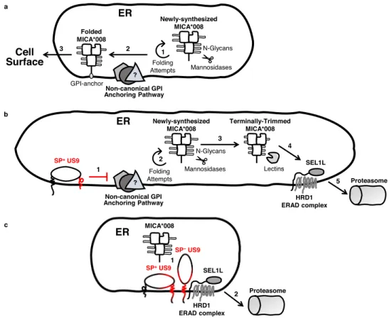
A key activating ligand targeted by HCMV is major histo-compatibility complex (MHC) class I polypeptide-related sequence A (MICA). MICA is part of a family of eight stress-induced MHC-like ligands, along with MICB and UL16-binding proteins 1–6 (ULBP1-6)20. These ligands are upregulated in response to stress such as DNA damage and viral infection. The activating NK receptor NKG2D binds these ligands and facil-itates the elimination of dangerous cells20. MICA is the most polymorphic member of the family, with about 100 known alleles21. Of these alleles, MICA*008 is one of the most prevalent21, and has unique biological properties. Due to a fra-meshift mutation in its transmembrane domain, MICA*008 is first synthesized as a truncated soluble protein. It then remains in the ER until it becomes GPI-anchored, permitting ER egress and surface expression. The slow process that directs MICA*008 to GPI anchoring despite its lack of a canonical GPI-anchoring signal is poorly characterized, and the factors involved remain unknown22. In contrast, non-truncated MICA alleles are trans-membrane proteins and follow a different and faster maturation pathway22.
Several HCMV immune evasion proteins inhibit MICA expression. US18 and US20 act together to send MICA to lyso-somal degradation23, as does UL148A, which acts in tandem with an unknown viral binding partner24. Finally, UL142 retains MICA in the cis-Golgi apparatus25–27. The truncated allele MICA*008 is, however, resistant to all of these mechanisms24,25. Consequently, until recently MICA*008 was considered an example of human adaptation to viral selective pressure14. In our previous work, we found that MICA*008 is in fact susceptible to HCMV28. A viral glycoprotein called US9 selectively targets MICA*008 for proteasomal degradation, hampering NKG2D-mediated NK killing of HCMV-infected cells. Interestingly, US9 function seems to depend on MICA*008’s non-standard maturation pathway. US9 degrades MICA*008 with slow kinet-ics, after a prolonged lag in the ER but prior to the GPI-anchoring step. Moreover, US9 function is only possible when MICA*008 undergoes non-canonical GPI-anchoring, since substituting the frameshifted MICA*008 TM domain with a canonical GPI-anchoring signal confers resistance to US928.
Here, we explore US9 structure-function relationship and underlying mechanisms of action. We show that the US9 SP is cleaved with unusually slow kinetics. We establish that the US9 SP is the major US9 domain targeting MICA*008 to degradation, thanks to this slow cleavage. We further show that US9 contains a second, SP-independent mechanism targeting MICA*008 that directly interacts with MICA*008 and the SEL1L-HRD1 ERAD complex. Last, we show that the US9 SP arrests MICA*008 maturation and indirectly induces ERQC-mediated degradation, also via the SEL1L-HRD1 ERAD complex.
Results
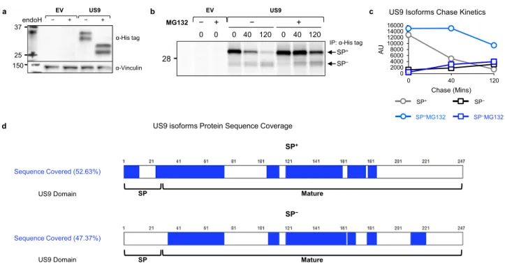
US9 SP is cleaved with unusually slow kinetics. US9 is a type-I transmembrane ER-resident glycoprotein of approximately 30 kDa, expressed early in HCMV infection13,29–31. In gel electro-phoresis, US9 migrates as two distinct protein bands, previously ascribed to differential glycosylation31. Hence, we expected that endoglycosidase H (endoH), an enzyme that digests the high mannose N-linked glycans of ER-resident glycoproteins, would cause US9 to run as a single band. We used previously described28 RKO cells which endogenously express very low levels of the full-length allele MICA*007:01, and overexpress MICA*008 fused to an N-terminal HA tag. We transduced the cells with an empty vector (EV) or US9. However, when we performed endoH diges-tion followed by immunoblotting, we observed that deglycosylated US9 still migrated as a doublet (Fig.1a), suggesting that a differ-ence in the polypeptide backbone itself is responsible for these two forms. The difference could not be at the C-terminus since the US9 construct we used bears a C-terminal 6XHis tag and cleavage of that end would have prevented recognition by the tag anti-body, so we suspected that N-terminal processing caused the observed size difference. The approximately 3 kDa size difference between the two US9 forms matched the predicted size of the N-terminal SP of 27 amino acids (AA) (see annotated US9 sequence in Fig. S1a). We therefore hypothesized that the two US9 bands represent a SP+precursor form and a SP−processed form. Of note, a similar two-band appearance caused by delayed N-terminal SP cleavage has been described for US1132.
To determine the nature of the two US9 forms we conducted a pulse-chase experiment (Fig.1b and S1b, quantified in Fig.1c). The larger SP+form was thefirst to appear, while the smaller SP− form accumulated very slowly, suggesting a precursor-product relationship. The SP+form was almost completely gone by 120 min of chase, but its quantity decreased more rapidly than the SP− form accumulated, suggesting rapid turnover of the SP+ form. Accordingly, treatment with the proteasome inhibitor
MG132 stabilized the SP+form but did not significantly impact the accumulation of the more stable SP− form. We further validated these results with a cycloheximide (CHX) chase assay (Fig. S1c). Under treatment with the protein translation inhibitor CHX, the SP+ form was initially present, but slowly vanished until it was no longer detectable at 4 h of chase, even in the presence of the proteasome inhibitor epoxomicin (EPX).
To directly assess SP presence, we immunoprecipitated US9, excised each of the two US9 isoforms from an electrophoresis gel, and detected peptides derived from each band using mass spectrometry (Fig. 1d). A sequence coverage rate of about 50% was obtained for both forms, and notably, only the SP+forms contained peptides derived from the SP, including peptides spanning the predicted cleavage area (Supplementary Data 1–2). We next addressed the question of the SP cleavage site. Specifically, there are two such predicted sites in the literature: S2433, and S2734 (Fig. S1d, residues highlighted in red). We mutated each of these residues to arginine to prevent SP peptidase cleavage35, creating mutants named S24R and S27R. We then overexpressed the mutants in RKO MICA*008-HA cells and immunoblotted cell lysates. Importantly, S24R remained a doublet, while S27R migrated as a single band the same size as the SP+form (Fig. S1d). Both mutants were endoH-sensitive (Fig. S1e), ruling out defective ER insertion. These findings confirm S27 is the US9 SP cleavage site. Taken together, ourfindings show that US9 has two isoforms: the precursor SP+ US9, which is slowly cleaved into the SP−US9 product.
Generation of US9 mutants. We previously showed that US9 specifically targets MICA*008 for proteasomal degradation28. We next wanted to identify the domains involved in US9 function. Based on Uniprot34 and Phobius36 predictions, US9 contains
several putative domains (diagram in Fig.2a, annotated sequence in Fig. S1a): the N-terminal SP (27 AA), an ER-luminal domain (166 AA), a transmembrane domain (30 AA), and a short cytosolic tail (24 AA). The ER-luminal domain of US9 contains a composi-tionally biased serine-rich area near the N-terminus (59 AA), and an immunoglobulin (Ig)-like fold (97 AA). Two N-linked glyco-sylation sites are predicted within the Ig-like fold (N97, N158).
US9 resembles its neighboring genes: US2 and US1137, which target the MHC class I heavy chain (MHC I) for proteasomal degradation to evade cytotoxic T lymphocytes38,39. Structurally, US2 and US11’s ER-luminal domains bind MHC I37,40–43, while their transmembrane (TM) or cytosolic domains recruit specific ERAD complexes41,43–48. We therefore hypothesized that US9 might function similarly. We aligned US9 and US11 protein sequences (Fig. S2a) that share 35.7% similarity and found that US9 contains a conserved glutamine (Q214, highlighted in red) in its TM domain. The equivalent glutamine in US11 (Q192) is responsible for recruiting the ERAD complex constituent Derlin-141,44,45.
We decided to systematically mutate US9 and perform a structure-function analysis (Fig. 2b). We created three cytosolic and TM domains mutants:ΔCyto+TM—removal of the cytosolic and TM domains; Q214A—conserved glutamine mutated to alanine; and 8TM—the TM domain was swapped with that of US8, a related protein which does not affect MICA*008, recently shown to target TLR3 and TLR4 to degradation28,49. We created three luminal deletion mutants: Δlum—deletion of the C-terminal part of the luminal domain up to the Ig-like fold;ΔIg— deletion of the Ig-like fold; andΔSer—deletion of the serine-rich area up to the 13 N-terminal AA. Last, we created two mutants for the SP and adjacent luminal area:ΔN-Ser—deletion of the 13 N-terminal AA (KESLRLSWSSDES) of the serine-rich area, up to the SP cleavage site; and 8SP—the US9 SP was swapped with the
a EV US9 b − + − + 37 25 150 endoH α-Vinculin α-His tag
IP: α-His tag
MG132 − 0 0 40 120 0 40 120 0 + − + 28 SP− SP+ US9 EV
d US9 isoforms Protein Sequence Coverage
Sequence Covered (52.63%)
US9 Domain SP Mature
SP+ Mature SP− SP Sequence Covered (47.37%) US9 Domain 0 2000 4000 6000 8000 10000 12000 14000 16000 0 40 120 AU Chase (Mins) US9 Isoforms Chase Kinetics
SP−
SP−MG132
SP+
SP+MG132
c
Fig. 1 US9 contains a slowly cleaved SP. a RKO MICA*008-HA cells co-expressing an empty vector (EV) or US9 attached to a C-terminal 6XHis tag were lysed. Lysates were mock-treated (−) or digested with endoglycosidase H (endoH; +) and then blotted. Anti-His tag was used to visualize protein expression, with anti-vinculin as loading control. Representative of at least three independent experiments.b RKO MICA*008-HA cells expressing EV or US9 were radioactively labeled for 20 min (pulse) and then chased in the absence (−) or presence (+) of 10 µM MG132. Digitonin-lysates were prepared at the indicated chase times (in minutes). An anti-His tag immunoprecipitation was performed. All samples were digested with endoH. Representative of three independent experiments. Ladder was visualized at a higher contrast, shown in Fig. S1b.c Quantification of the US9 isoforms shown in (b). AU arbitrary units.d Graphic representation of US9 protein sequence coverage detected by mass spectrometry analysis of the two US9 isoforms: SP+and SP−. Sequence coverage % and US9 domains are indicated. See also Supplementary Data 1–2, figure S1 and the Source Data file for experimental data.
US8 SP to maintain proper ER insertion33. All the constructs were fused to a C-terminal 6XHis tag.
US9 mutants are properly expressed and ER-localized. The US9 mutants were transduced into RKO MICA*008-HA cells. First, we verified the expression of the US9 mutants by immunoblot (Fig. S2b). All the mutants were expressed, but their abundance varied. We validated ER localization by endoH sensitivity assay (Fig.2c). Almost all mutants were endoH-sensitive, indicating ER localization. The endoH assay is irrelevant for the ΔIg mutant that lacks N-linked glycosylations. Therefore, we verified by immunofluorescence that like wild type (WT) US9, ΔIg co-localizes with the ER marker PDI (Fig. S2c).
Delayed cleavage depends on SP sequence and the N-Ser domain. Intriguingly, the two-band appearance indicating the presence of both SP+and SP−forms (named SP±in Fig.2c) was
lost in certain mutants: ΔIg, ΔSer, ΔN-Ser, and 8SP (Fig. 2c, right). This was to be expected for 8SP, since US8 migrates as a single band in immunoblot and pulse-chase experiments, indi-cating rapid SP cleavage28,31,49,50. However, the single band appearance in other mutants suggested that US9 SP cleavage was affected by additional domains. To study the SP status of these mutants, we compared the predicted and observed molecular weights of each deglycosylated mutant. For ΔIg and ΔSer, the observed sizes were compatible with unprocessed forms con-stitutively retaining their SP (named SP+in Fig.2c). In contrast, ΔN-Ser and 8SP sizes were compatible with the predicted pro-cessed size (named SP−in Fig.2c), indicating rapid cleavage.
We further validated the SP status of theΔIg, ΔSer, and ΔN-Ser mutants by mass spectrometry, which identified SP-derived sequences in ΔIg and ΔSer, but not in ΔN-Ser (Fig. S2d and Supplementary Data 3–5). Finally, we conducted a pulse-chase experiment comparing US9 andΔN-Ser maturation kinetics, which showed no evidence of a SP-containing precursor form in the ΔN-Ser mutant (Fig. S2e). This suggested rapid, co-translational SP cleavage occurring before His tag synthesis. Notably,ΔN-Ser was also considerably more stable than SP+US9.
These results suggest that large luminal domain deletions can prevent US9 SP cleavage, regardless of the deleted domain. In contrast, deletion of the N-Ser area or swapping US9 SP with a rapidly cleaved SP sequence restored rapid SP cleavage. Of note, SP retention inversely correlated with US9 protein abundance: SP+ mutants were less abundant than WT US9, while SP−mutants were more abundant than WT US9.
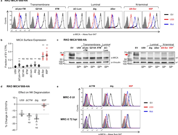
US9 N-terminal mutants are impaired in inducing MICA*008 degradation. We next assessed US9 mutant functionality. We began by measuring the surface expression of MICA*008 in the presence of each mutant using flow cytometry (Fig. 3a and S2f, quantified in
Fig.3b, summarized in Table1). Remarkably, all mutants retained some MICA*008 downregulating capacity. Mutating Q214 caused no significant difference compared to WT US9, indicating that this residue is redundant for MICA*008 targeting. The ΔCyto+TM and ΔIg mutations had a statistically significant but relatively small effect on US9 functional capacity; both mutants continued to cause a greater than tenfold reduction in MICA*008 surface levels. Unex-pectedly, the two mutants which showed the greatest functional impairment were the N-terminal mutants with rapid SP cleavage,
N-terminal SP+ SP− SP− SP± α-Vinculin α-His tag ΔSer ΔN-Ser 8SP US9 − + − + − + − + endoH ΔIg − + SP+ Luminal 150 37 25 20
EV US9 ΔCyto+TM Q214A 8TM ΔC-Lum
− + − + − + − + − + − + Transmembrane Luminal α-His tag α-Vinculin SP± SP± SP± SP± SP± 37 25 20 150 endoH a b c Ig-like 97 AA Ser-rich 59 AA HIS Luminal 166 AA TM 30 AA Cyto 24 AA SP 27 AA US9 247 AA ΔCyto+TM 193 AA HIS −54 AA Q214A 247 AA Q214A HIS 8TM 237 AA 20 AA HIS ΔC-Lum 222 AA HIS −25 AA ΔIg 158 AA HIS −89 AA ΔSer 208 AA HIS −39 AA ΔN-Ser 234 AA HIS −13 AA 8SP 241 AA 21 AA HIS
Fig. 2 Delayed US9 SP cleavage requires the N-Ser area. a Diagram of US9 domains: signal peptide (SP), serine-rich compositionally biased area (Ser), Ig-like fold (Ig), transmembrane (TM), cytosolic (Cyto), 6XHis tag (HIS). Total protein length without tags and the length of each domain in amino acids (AA) are indicated.b Diagrams of US9 deletion and substitution mutants arranged according to the mutated domain. The total length of each construct without tags and of each modified domain are indicated. Dashed red lines show deleted domains, solid red lines show substitutions with a homologous US8 sequence, arrow shows a single mutated residue.c The US9 mutants described in (a, b) were expressed in RKO MICA*008-HA cells. Lysates prepared from the indicated transfectants were untreated (−) or digested with endoH (+) and then immunoblotted. Mutants are grouped by mutated region (indicated). Anti-His tag was used to visualize protein expression, with anti-vinculin as loading control. Annotation beneath the panel indicates deglycosylated US9 forms with, without, or both with and without the SP (SP+/−/±). Different panels depict separate gels. Different segments of the same gel are shown together for clarity in the left panel. Representative of two independent repeats. See also Fig. S2.
ΔN-Ser, and 8SP (Fig.3a, b, highlighted in red). MICA*008 surface
levels were increased by about tenfold in cells expressing these mutants compared to WT US9-expressing cells.
To determine if the mutants still induced MICA*008 degradation, we assessed their effect on total MICA*008 protein quantity by immunoblot. We have previously shown that US9 does not reduce the quantity of ER-resident, non-GPI-anchored MICA*008, while post-ER, GPI-anchored MICA*008 vanishes28. In RKO cells, the ER-resident and post-ER forms of MICA*008 are easily distinguishable by size since the post-ER form of MICA*008 migrates at ~70 kDa due to Golgi-acquired glycosylation modifications, compared to the ~60 kDa ER-resident form28. Most of the US9 mutants maintained this pattern (Fig. 3c), substantially reducing the quantity of post-ER
MICA*008 (red arrows) compared to empty-vector (EV) control cells. Only the two N-terminal SP−mutations partially restored post-ER MICA*008 quantities (Fig.3c, red font), demonstrating impaired MICA*008 degradation. In summary, we concluded that US9 operates differently than US2/11 and its main functional domain is localized to its N-terminus.
US9 SP is required for evasion of NK cell-mediated killing. We next wondered about the functional significance of the partial MICA*008 restoration in the N-terminal US9 mutants. To address this question, we conducted a CD107a-degranulation assay. CD107a is a cytotoxic granule marker transiently expressed on NK cell surface following degranulation51. NK cells were co-incubated for 2 h with target cells expressing EV, US9, ΔCyto+TM, ΔIg, or a
b c
α-MICA – Alexa fluor 647
ΔCyto+TM Q214A 8TM
Transmembrane
ΔC-Lum ΔIg ΔSer
Luminal ΔN-Ser 8SP EV US9 Mut N-terminal 100 101 102 103 104 0 25 50 75 100 4 Count s RKO MICA*008-HA MRC-5 UI MRC-5 72 hpi ΔCTM ΔIg 8SP EV US9 Mut Count s
α-MICA – Alexa fluor 647
100 101 102 103 104 0 13 25 38 50 4 100 101 102 103 104 0 25 50 75 100 4 d e US9 ΔCyto+TM Q2 1 4 A 8TM ΔC-l um ΔIg ΔSer ΔN-Ser 8SP 0.0 0.2 0.4 0.6 Frac ti o n o f E V CT R L ** NS NS ** ** NS NS **
MICA Surface Expression
RKO MICA*008-HA US9 ΔCTM ΔIg 8SP -40 -30 -20 -10 0 10 % C h ange in C D 107a NS NS Effect on NK Degranulation * RKO MICA*008-HA ΔCyto +TM Q214A 8TM ΔC-Lum 150 75 50 α-Vinculin α-MICA EV US9 Transmembrane Luminal SP± SP± SP± SP± SP± 100
ΔIg ΔSer ΔN-Ser 8SP
EV US9 150 N-terminal Luminal α-Vinculin α-MICA SP+ SP+ SP− SP− SP± 75 50 100
Fig. 3 US9 N-terminus is required for effective MICA*008 downregulation and immune evasion. a Flow cytometry of MICA surface expression in RKO MICA*008-HA cells expressing an EV (black), US9 (red), or the indicated US9 mutants (blue), grouped by mutated region (indicated). Red font highlights MICA*008 downregulation impairment. Gray-filled histograms represent secondary antibody staining of EV cells. Figure combines representative results from separate experiments.b Quantification of MICA mean flourescence intensity (MFI) shown in (a), normalized to the EV control. Data show mean ± SEM for at least four independent experiments per mutant. A one-way ANOVA was performed with a significant effect at the p < 0.05 level for all conditions [F(8,75)= 70.48, p = 9.2 × 10⁻³²]. MICA MFI for US9 (dashed line) was compared to each mutant’s MFI using a post hoc Dunnett’s test. **p < 0.01. NS = non-significant. Red circles highlight MICA*008 downregulation impairment. c Lysates from the indicated cells described in (a) were blotted using anti-MICA, anti-His tag, and anti-vinculin as loading control. Panels depict separate gels. Annotation beneath the panels indicates SP status of the US9 constructs (SP+/−/±). Red arrows indicate post-ER MICA*008. Red font highlights MICA*008 degradation impairment. Representative of three independent experiments.d NK cell degranulation following incubation with the indicated target cells was measured by CD107a staining. Relative degranulation reduction was calculated for each US9 mutant. Shown are mean ± SEM from three independent repeats. A one-way ANOVA was performed with a significant effect at the p < 0.05 level for all conditions [F(3,8) = 8.52, p = 0.0071]. US9 degranulation reduction (dashed line) was compared to each mutant using a post hoc Dunnett’s test. *p < 0.05. NS = non-significant. Red circles highlight degranulation reduction impairment. e The indicated MRC-5fibroblasts were uninfected (UI; top panels) or infected with ΔUS9 HCMV and stained for MICA expression at 72 h post infection (hpi; bottom panels). Gray-filled histograms represent isotype-control staining of EV cells. Red font highlights MICA*008 downregulation impairment. Representative of three independent experiments. See also Table1, Fig. S2, and the Source datafile for experimental data and full statistics.
8SP. The percentage of CD107a+NK cells was measured byflow cytometry, and the reduction in NK degranulation induced by US9 and the mutants was calculated (Fig. 3d). US9,ΔCyto+TM, and ΔIg, reduced NK cell degranulation by 15–20% compared to EV control and there was no significant difference between them. In contrast, 8SP was significantly less efficient than US9 and failed to reduce NK cell degranulation, indicating that SP removal hampered US9 NK cell evasion capacity.
US9 SP is required for MICA*008 targeting during HCMV infection. We wondered what the impact of the US9 SP deletion during HCMV infection would be, since its effect could poten-tially be masked by other, as-yet unidentified MICA*008-target-ing viral genes28. To test this, we expressed EV, US9,ΔCyto+TM, ΔIg, or 8SP in HCMV-permissive MRC-5 fibroblasts, which we genotyped and determined to be MICA*008 homozygous. We then infected the transduced MRC-5 cells with an HCMV mutant lacking US9 (ΔUS9) we previously generated28. We assessed MICA*008 surface expression in uninfected (UI) and infected cells at 72 h post infection (hpi; Fig.3e). In uninfected cells, only 8SP partially restored MICA*008 levels. Following infection, ΔCyto+TM behaved similarly to WT US9, ΔIg showed partial impairment in MICA*008 downregulation, and 8SP was greatly impaired in MICA*008 downregulation, to the extent that MICA*008 levels in 8SP-expressing cells approached those in EV control cells. We therefore concluded that the US9 SP was the most significant MICA*008-targeting domain of US9, even in the context of HCMV infection.
US9 SP is sufficient for inducing MICA*008 degradation. Substitution of the SP and deletion of the N-Ser domain both impaired US9 function to a similar extent. There are two possible explanations to this finding: that both sequences were required for N-terminal domain function; or that the SP is an independent functional domain, and the N-Ser area has a supporting role in delaying its cleavage. To address this and discover whether the SP was sufficient for MICA*008 downregulation, we generated reciprocal chimeras based on US8 (Fig. 4a). First, we replaced US8’s endogenous SP with that of US9 alone (9SP) or together with the adjacent N-Ser domain (9SP+N-Ser). In addition, to discover if the N-Ser domain is independently functional, we
wanted to insert it alone into US8, but a SP was needed to ensure ER insertion. Therefore, we first had to show that the US9 N-terminal domain could downregulate MICA*008 even when inserted after a different SP. We inserted the US9 SP and the N-Ser area (a total of 40 AA) after the US8 endogenous SP as a control (40ins). We inserted an extended sequence including the last seven AA of the SP and the N-Ser area (a total of 20 AA) after US8’s SP (20ins), to increase the chances that the inserted sequence would be functional.
We overexpressed the US8 chimeras in RKO MICA*008-HA cells and verified by immunoblot all were expressed (Fig. 4b). Intriguingly, we observed that the 9SP chimera was larger than WT US8. Since the only sequence difference between the two is the SP itself, this showed that the US9 SP was constitutively retained on this construct (SP+). The 9SP+N-Ser chimera regained the typical doublet appearance of US9, indicating that the addition of the N-Ser domain restored slow SP cleavage (SP±). The 40ins chimera looked identical to 9SP+N-Ser in forms and size, indicating that the US8 SP was rapidly cleaved, and then the US9 SP was slowly cleaved (SP±). In contrast, the 20ins chimera had only one form which was larger than the SP−form of 40ins/ 9SP+ N-Ser, indicating the SP cleavage site in the extended N-Ser sequence we inserted was not functional and that the entire inserted sequence was retained (SP−). Since US8 is known to partially localize to the Trans-Golgi network and lysosomes, rendering it endoH resistant31,49,50, we used Peptide:N-glycosi-dase F (PNGaseF), an enzyme that digests all N-linked glycans, to validate the US9 SP status of the chimeras (Fig. S3a, annotated beneath the panel).
We assessed the cellular localization of US8 and the chimeras by an endoH assay (Fig. S3b) and found that SP+forms seemed to be entirely endoH-sensitive, indicating they were being retained in the ER. In comparison, 20ins gained partial endoH resistance similarly to WT US8.
We then measured the chimeras’ effect on MICA*008 surface expression by flow cytometry (Fig. 4c, quantified in Fig. 4d). While parental US8 and the 20ins chimera had no effect on MICA*008 surface expression, 9SP, 9SP+N-Ser, and 40ins all robustly downregulated MICA*008 by ~10-fold. In agreement with the surface expression data, immunoblot analysis (Fig. 4e) showed that 9SP, 9SP+N-Ser, and 40ins greatly reduced the quantity of post-ER MICA*008 (red arrow), showing they, too, Table 1 Characteristics and function of US9 mutants.
Construct name Mutated domain US9 SP status Effect on MICA*008a Relatedfigure
US9 − Slowly cleaved ++ Fig.3
ΔCyto+TM Cytosolic and transmembrane Slowly cleaved ++
Q214A Transmembrane Slowly cleaved ++
8TM Transmembrane Slowly cleaved ++
ΔC-Lum Luminal Slowly cleaved ++
ΔIg Luminal Uncleaved ++
ΔSer Luminal Uncleaved ++
ΔN-Ser N-terminal Rapidly cleaved +
8SP N-terminal Swapped +
8SPΔCyto N-terminal, Cytosolic Swapped + Fig.5
8SPΔCyto+TM N-terminal, Cytosolic and transmembrane Swapped −
8SPQ214A N-terminal, transmembrane Swapped +
8SP8TM N-terminal, transmembrane Swapped +
8SPΔC-Lum N-terminal, luminal Swapped +
8SPΔIg N-terminal, luminal Swapped −
8SPΔSer N-terminal, luminal Swapped +
8SPΔN-Ser N-terminal Swapped +
induced its degradation. ER-resident MICA*008 forms were spared, recapturing the US9 phenotype. These results show that US9 SP is sufficient for targeting MICA*008, and that the N-Ser domain does not independently target MICA*008.
US9 SP is sufficient for MICA*008 targeting during HCMV infection. Since we observed that the US9 SP was sufficient for targeting MICA*008, we wondered whether the same would be true during HCMV infection. We overexpressed EV, US9, US8, and 9SP in MRC-5fibroblasts, infected them with ΔUS9 HCMV, and measured MICA*008 surface expression at 72 hpi compared to uninfected (UI) cells (Fig.4f). Both in UI and inΔUS9-infected cells, the 9SP construct reduced MICA*008 levels to the same extent as US9 itself.
US9 SP is functional when affixed to the unrelated protein EYFP. To address the possibility that some structural feature conserved in US8 and US9 was required for US9 SP function, we
decided to attach it to an unrelated protein—enhanced yellow fluorescent protein (EYFP) (Fig. S4a). N-terminal tags were removed from parental FLAG-HA-tagged EYFP protein (named FLAG-HA-EYFP), and the following chimeras were generated: 8SP EYFP, 9SP EYFP, and 9SP+N-Ser EYFP.
We transfected the EYFP constructs into RKO MICA*008-HA cells and checked their expression by immunoblot (Fig. S4b). EYFP harbors no glycosylations so we did not conduct an endoH assay. We observed a doublet appearance of 9SP EYFP, indicating that the US9 SP sequence was sufficient for inducing slow cleavage in the context of the EYFP sequence. Importantly, the relative abundance of SP+EYFP increased with addition of the N-Ser area, in support of this sequence’s role in slowing SP cleavage.
We then assessed the effect on MICA*008 surface expression by flow cytometry (Fig. S4c, quantified in Fig. S4d). While parental EYFP and 8SP EYFP did not downregulate MICA*008, 9SP EYFP moderately downregulated MICA*008 surface levels, and 9SP+N-Ser EYFP (both highlighted in red) strongly
c d f e a 20ins 247 AA US8 227 AA SP 21 AA HIS Mature 206 AA +13 AA +13 AA 27 AA 9SP 233 AA HIS 27 AA 9SP+N-Ser 246 AA HIS +13 AA +27 AA HIS 40ins 267 AA +7 AA HIS 100 101 102 103 104 0 25 50 75 100 US8 9SP EV US9 US8 Mut
α-MICA – Alexa fluor 647
100 101 102 103 104 0 13 25 38 50 US8 9SP Count s MRC-5 UI MRC-5 72 hpi
α-MICA – Alexa Fluor 647
Count s US8 9SP 9SP+N-Ser 100 101 102 103 104 0 25 50 75 100 4 40ins 20ins EV US9 US8 Mut RKO MICA*008 b 100 37 25 US8 9SP 9SP+ N-Ser 20ins 40ins EV US9 α-Vinculin α-His tag SP− SP± SP+ SP± SP− SP± US9 US8 9SP 9SP+N-Se r 40in s 20ins 0.0 0.2 0.4 0.6 0.8 1.0 1.2 1.4 Frac ti o n o f E V CT R L ** ** ** NS MICA Surface Expression
150 75 50 US8 9SP 9SP+ N-Ser 20ins 40ins EV US9 α-Vinculin α-MICA SP− SP± SP+ SP± SP− SP± RKO MICA*008 100 4 4
Fig. 4 The US9 SP is sufficient for targeting MICA*008 to degradation. a Schematic representation of US8 and the indicated US8 mutants. US9 domains of the indicated length (solid red lines) substituted US8’s endogenous SP or were inserted after it. The total length of each construct without tags is indicated.b Lysates from RKO MICA*008-HA cells transduced with the constructs described in (a) were immunoblotted. Anti-His tag was used to visualize protein expression, with anti-vinculin as loading control. Annotation beneath the panel indicates the US9 SP status of the constructs (SP+/−/±). Representative of three independent experiments.c Flow cytometry of MICA surface expression in cells transduced with an EV (black histograms) US9 (red histograms) or the indicated US8 constructs (blue histograms). Red font highlights gain-of-function constructs. Gray-filled histograms represent secondary antibody staining of EV cells.d Quantification of MICA MFIs shown in (c), normalized to the EV control. Data show mean ± SEM for at least four independent repeats per mutant. A one-way ANOVA was performed with a significant effect at the p < 0.05 level for all conditions [F(4,31) = 68.528, p = 6 × 10⁻¹⁵]. A post hoc Dunnett’s test was used to compare the MFI for 8SP (dashed line) to that of each mutant. **p < 0.01, NS non-significant. Red circles highlight gain-of-function.e Lysates obtained from the indicated mutants described in (a) were blotted using MICA, His tag, and anti-vinculin as loading control. Red font highlights gain-of-function, and the red arrow indicates post-ER MICA*008. Representative of three independent experiments.f MRC-5fibroblasts were transduced with an EV (black histograms), WT US9 (red histograms) or the indicated US8 constructs (blue histograms), and then left uninfected (UI; left panels) or infected withΔUS9 HCMV and stained for MICA expression at 72 h post infection (hpi; right panels), as part of the same experiment shown in Fig.3e. Gray-filled histograms represent isotype-control staining of EV cells. Red font highlights gain-of-function. Representative of three independent experiments. See also Figs. S3–4and the Source datafile for experimental data and full statistics.
downregulated MICA*008 surface levels to about 25% of the FLAG-HA-EYFP control levels. No other stress-induced ligand or MHC I were affected by the US9 SP, confirming that this effect was MICA-specific (Fig. S4e). We then tested total MICA*008 protein levels by immunoblot (Fig. S4f), to determine if the EYFP chimeras merely downregulated surface MICA*008 or caused its degradation. Expression of 9SP+N-Ser EYFP and to a lesser extent of 9SP EYFP (both highlighted in red), induced MICA*008 degradation since the post-ER form of MICA*008 (shown by a red arrow) was reduced. Importantly, the magnitude of the effect on MICA*008 correlated with SP+EYFP abundance (Fig. S4d).
These results demonstrate that US9 SP is sufficient for MICA*008 degradation when attached to EYFP.
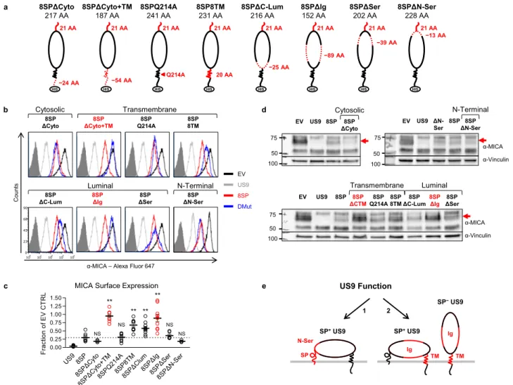
SP-independent US9 function is mediated by the Ig-like and TM domains. The SP is the major functional domain of US9 in effect size and functional significance. However, SP− US9 still
reduces MICA*008 levels by about 70% (Fig. 3b). To char-acterize the SP-independent functional domains, we constructed a panel of US9 double mutants. The 8SP mutation was com-bined with each of eight other mutations (Fig.5a): deletion of the cytoplasmic domain (8SPΔCyto), or of the cytoplasmic and TM domains (8SPΔCyto+TM); mutation of a conserved TM glutamine to alanine (8SPQ214A); substitution with US8’s TM domain (8SP8TM); deletion of the C-terminal part of the luminal domain up to the Ig-like fold (8SPΔC-lum); deletion of the Ig-like fold (8SPΔIg); deletion of the serine-rich area
a c b d −24 AA 8SPΔCyto+TM 187 AA HIS −54 AA 8SPQ214A 241 AA Q214A HIS 8SP8TM 231 AA 20 AA HIS 8SPΔC-Lum 216 AA HIS −25 AA 8SPΔIg 152 AA HIS −89 AA 8SPΔSer 202 AA HIS −39 AA 8SPΔN-Ser 228 AA HIS −13 AA 8SPΔCyto 217 AA 21 AA HIS 21 AA 21 AA 21 AA 21 AA 21 AA 21 AA 21 AA e SP SP+US9 TM US9 Function 1 2 SP−US9 Ig SP+US9 TM Ig N-Ser US9 8SP 8SPΔ Cyto 8SPΔCy to+T M 8SPQ 214 A 8SP8T M 8SPΔ Clum 8SP ΔIg 8SPΔ Ser 8SP ΔN-S er 0.00 0.25 0.50 0.75 1.00 1.25 1.50 Frac ti o n o f E V CT R L
MICA Surface Expression ** NS NS ** ** ** NS NS Transmembrane Luminal 8SP ΔCTM 8SP Q214A 8SP 8TM 8SP ΔIg 8SP ΔC-Lum 8SP 8SP ΔSer 100 50 75 α-Vinculin α-MICA EV US9 N-Terminal 8SP ΔN-Ser 50 75 α-Vinculin α-MICA 8SP 100 EV US9 ΔN-Ser 8SP ΔCyto 8SP EV US9 50 75 100 Cytosolic US9 8SP DMut EV
α-MICA – Alexa Fluor 647
8SP ΔCyto+TM 8SP Q214A 8SP 8TM 8SP ΔC-Lum 8SP ΔSer Count s 8SP ΔCyto 8SP ΔN-Ser 100 101 102 103 104 0 23 45 68 90 Transmembrane Luminal N-Terminal Cytosolic 8SP ΔIg
Fig. 5 SP-independent MICA*008 downregulation is mediated by the US9 Ig-like and TM domains. a Schematic representation of US9 double mutants, in which the SP was swapped with the US8 SP and combined with each of the indicated mutations in the following domains: serine-rich area (Ser), Ig-like fold (Ig), other luminal (Lum), transmembrane (TM), and cytosolic (Cyto). Dashed red lines show deleted domains, solid red lines show domains substituted with a homologous US8 sequence, arrow shows a single mutated residue. Mutants were 6XHis tagged (HIS). Mutated sequence length and the total length of each construct without tags are indicated.b Flow cytometry of MICA surface expression in RKO MICA*008-HA cells transduced with an EV (black histograms), US9 (gray histograms), 8SP (red histograms), or the indicated US9 double mutants (blue histograms). Gray-filled histograms represent secondary antibody staining of EV cells. Mutants are grouped by mutated region (indicated). Red font highlights complete loss-of-function. Figure combines representative results from separate experiments.c Quantification of MICA MFIs shown in (b), normalized to the EV control. Data show mean ± SEM for at least four independent experiments per mutant. A one-way ANOVA was performed with a significant effect at the p < 0.05 level for all conditions [F(8,69)= 20.073, p = 2.8 × 10⁻¹⁵]. A post hoc Dunnett’s test was used to compare the MFI for 8SP (dashed line) to that of each mutant. **p < 0.01, NS non-significant. d Lysates obtained from the indicated cells were blotted using anti-MICA, anti-His tag, and anti-vinculin as loading control. Different panels show different gels. Mutants are grouped by mutated region (indicated). Red font highlights complete loss-of-function, and the red arrow indicates post-ER MICA*008. Representative of three independent experiments.e Diagram of the two distinct mechanisms of US9-mediated MICA*008 degradation: (1) US9 SP-dependent mechanism, present only in the SP+isoform, requires the SP and the cleavage-delaying N-Ser domain; and (2) US9 SP-independent mechanism, present in both SP+and SP−isoforms, requires the Ig-like and TM domains. See also Fig. S5, Table1, and the Source datafile for experimental data and full statistics.
(8SPΔSer); and last, deletion of the 13 N-terminal AA of the serine-rich area (8SPΔN-Ser).
The double mutants were then transduced into RKO MICA*008-HA cells, and their expression was confirmed by immunoblot (Fig. S5a). ER localization was confirmed by endoH digestion or by confocal microscopy co-localization for 8SPΔIg, which lacks N-linked glycosylations (Fig. S5b-c). As expected, following deglycosylation, all double mutants migrated as a single band consistent with rapid SP processing, and their sizes matched the expected sizes for SP−forms.
We then measured MICA*008 surface expression by flow cytometry (Fig.5b, quantified in Fig.5c, summarized in Table1). Strikingly, only two double mutants completely lost their capacity to downregulate MICA*008 surface levels: 8SPΔCyto+TM and 8SPΔIg, indicating that the TM and Ig two domains are required for SP-independent US9 function. All other double mutants retained their function either fully (8SPΔCyto, 8SPQ214A, 8SPΔSer, and 8SPΔN-Sser) or partially (8SP8TM and 8SPΔC-Lum). It is notable that the two N-terminal mutations, 8SP and ΔN-Ser were not additive, further evidence that they disrupt the same mechanism. Immunoblot analysis of total MICA*008 protein quantity in the presence of the US9 double mutants (Fig. 5d) confirmed that the 8SPΔCyto+TM and 8SPΔIg mutants do not reduce post-ER MICA*008 protein quantity (shown by a red arrow), while the other mutants remained capable of inducing its degradation, to a degree which correlated with MICA*008 surface expression. We therefore concluded that the US9 TM and Ig-like domains mediate SP-independent MICA*008 degradation, resem-bling US2/11.
In summary, we found that US9 contains two independent MICA*008-targeting mechanisms (Fig. 5e, functional domains highlighted in red): thefirst and most significant (1) is SP-mediated, present only in the SP+isoform. This mechanism depends on the presence of the N-Ser domain which slows SP cleavage. The second, SP-independent mechanism (2) is mediated by the TM and Ig-like domains and is present in SP+and SP−isoforms.
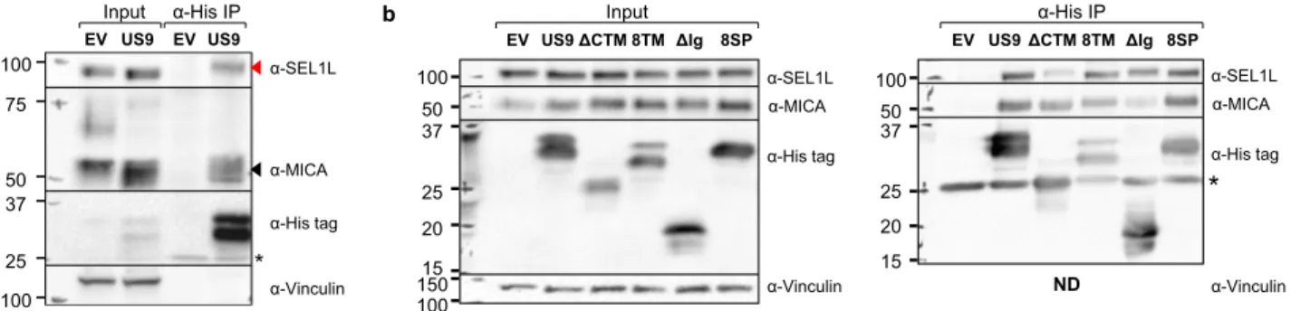
US9 co-precipitates with the ERAD component SEL1L and with MICA*008. Having identified US9’s functional domains, we wanted to study their underlying mechanisms of action. We began by performing an anti-His tag co-immunoprecipitation assay in EV controls and in US9-His-expressing cells to identify potential US9 interacting proteins. We used RKO MICA*008-HA cells and HeLa cells which endogenously express MICA*00828. Immunoprecipitated proteins were subsequently analyzed by
LC-MS/MS. We searched the results for proteins which specifically co-precipitated with US9 in both cell types. These analyses identified Protein Sel-1 Homolog 1 (SEL1L), an essential com-ponent of the HRD1 ERAD complex52, as a US9 interacting protein (mass spectrometry data available in Supplementary Data 6). Hence, we chose SEL1L for further analysis.
First, we performed an anti-His tag IP and immunoblot to validate the interaction between US9 and SEL1L in RKO MICA*008-HA and in Hela cells (Figs.6a and S6a, respectively). US9 specifically co-precipitated with SEL1L (shown by a red arrow). We also found that US9 co-precipitated with ER-resident MICA*008 (shown by a black arrow). To determine if the SEL1L-US9 interaction was MICA*008-dependent, we repeated the anti-His tag immunoprecipitation assay in RKO cells expressing MICA*004-HA, a full-length MICA allele which is not targeted by US928 (Fig. S6b). As expected, MICA*004 did not co-precipitate with US9, but SEL1L still co-co-precipitated with US9 (red arrow), demonstrating that its interaction with US9 does not require MICA*008.
The US9 TM and Ig-like domains bind SEL1L and MICA*008, respectively. Having established that US9 binds MICA*008 and SEL1L, we asked which US9 domains were responsible for these interactions. We repeated the co-immunoprecipitation described in Fig. 6a to test which US9 mutants would lose their ability to co-precipitate with ER-resident MICA*008 or with SEL1L. We tested the 8SP,ΔCyto+TM, 8TM, and ΔIg mutants (Fig.6b). Strikingly, despite being functionally impaired, the 8SP mutant was still able to co-precipitate with both MICA*008 and SEL1L. The ΔIg mutant showed diminished MICA*008 binding but continued to interact with SEL1L. Conversely, the ΔCyto+TM mutant showed dimin-ished SEL1L binding but continued to interact with MICA*008. It is intriguing that the 8TM mutant, in which the US9 TM domain was swapped with the equivalent US8 sequence, continued to bind SEL1L, suggesting that US8 itself might also bind this factor. These results are consistent with the requirement for the Ig-like and TM domains for MICA*008 targeting by SP−US9 (Fig. 5). We
con-cluded that the Ig-like domain binds MICA*008 and the TM domain binds SEL1L, while the SP does not bind either protein. US9-mediated degradation of MICA*008 requires SEL1L. Based on these IP results, we hypothesized that only SP-independent US9 function requires SEL1L. To investigate the functional sig-nificance of the US9-SEL1L interaction, we knocked down SEL1L. RKO MICA*008-HA cells were transduced with a scrambled
a 75 50 37 25 100 100 α-Vinculin α-His tag α-MICA α-SEL1L * EV US9 EV US9 α-His IP Input b α-Vinculin 150 37 25 20 15 50 100 100 α-His tag α-MICA α-SEL1L 8SP EV US9ΔCTM 8TMΔIg Input α-Vinculin α-His tag α-MICA α-SEL1L
*
8SP EV US9ΔCTM 8TM ΔIg α-His IP 37 25 20 15 50 100 NDFig. 6 The US9 Ig-like and transmembrane domains, but not the US9 SP, bind MICA*008, and the ERAD scaffold protein SEL1L. a Lysates prepared from RKO MICA*008-HA cells co-transduced with EV or US9 were immunoprecipitated using anti-His tag antibody. Immunoblotting was performed using anti-His tag, anti-MICA, and anti-SEL1L to visualize protein co-precipitation, with anti-vinculin as input loading control. Arrows indicate bands which specifically co-precipitated with US9 (red—SEL1L; black—MICA*008). * antibody light chain. Representative of three independent experiments. b Lysates prepared from the indicated RKO MICA*008-HA transfectants were immunoprecipitated using anti-His tag (A—input; B—IP) and immunoblotted using anti-His tag, anti-MICA, and anti-SEL1L. Anti-vinculin served as input loading control. * antibody light chain. Representative of two independent experiments. ND not detected. See also Fig. S6 and Supplementary Data 6.
shRNA control (referred to as shSCR) or with two different shRNAs directed at SEL1L: shRNA#1 and shRNA#2 (referred to as sh#1 and sh#2, respectively). Once stably expressing the shRNAs, cells were co-transduced with an EV control or with US9. Knockdown (KD) efficiency was validated by immunoblot (Fig. S7a).
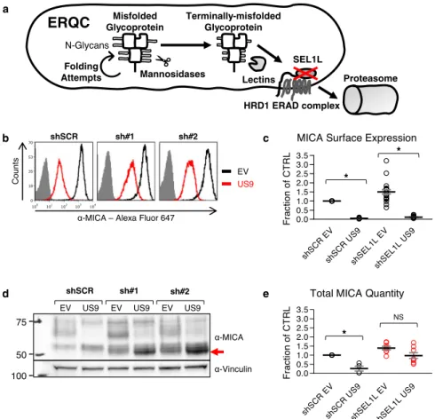
The SEL1L-HRD1 ERAD complex degrades misfolded luminal and transmembrane glycoproteins, including MHC I. Terminal trimming of N-linked glycans on the misfolded substrates causes dissociation from folding factors and recognition by lectins, which deliver the misfolded glycoprotein to the SEL1L-HRD1 ERAD complex for retrotranslocation and subsequent proteaso-mal degradation9,52–55. In addition to its role in quality control, the SEL1L-HRD1 complex regulates the quantity of folding-competent proteins, and is essential for many physiological and homeostatic functions, including energy metabolism, water balance, and cellular development56–62. Therefore, SEL1L knock-down is expected to induce accumulation of terminally trimmed misfolded glycoproteins (Fig.7a).
We stained the shRNA-expressing cells for surface MICA*008 expression (Fig.7b, quantified in Fig.7c). SEL1L KD somewhat increased MICA surface levels in EV and US9-expressing cells, but US9 still significantly reduced MICA*008 surface levels in the SEL1LKD cells by about tenfold (Fig. 7c). Next, we immuno-blotted lysates from the shRNA-expressing cells (Fig. 7d, quantified in Fig. 7e). In the SEL1L KD EV cells, surface and total MICA*008 levels increased by about 40–50% compared to shSCR EV cells, implying a role for SEL1L in intrinsic MICA*008 degradation. Importantly, even though SEL1L KD did not restore surface MICA*008, total MICA*008 protein levels were greatly increased in the KD cells compared to shSCR US9 cells, due to accumulation of a rapidly migrating MICA*008 form (shown by a red arrow). Moreover, quantitation revealed no significant differences in total MICA*008 protein levels when comparing EV to US9-expressing SEL1L KD cells (Fig.7e). This implied that in the KD cells, MICA*008 was retained intracellularly and was no longer degraded by US9.
d b e c Misfolded Glycoprotein Terminally-misfolded Glycoprotein Lectins Proteasome Mannosidases N-Glycans Folding Attempts
ERQC
HRD1 ERAD complex SEL1L a 100 75 50 α-MICA α-Vinculin shSCR sh#1 EV US9 EV US9 sh#2 EV US9 US9 EVα-MICA – Alexa Fluor 647
shSCR sh#1 Count s 100 101 102 103 104 0 18 35 53 70 sh#2 shSC RE V shSC RU S9 shS EL1L EV shS EL1L US9 0.0 0.5 1.0 1.5 2.0 2.5 3.0 3.5 F ra c tio n o f C TRL * *
MICA Surface Expression
shSC REV shSC RU S9 shSE L1L EV shS EL1 LUS 9 0.0 0.5 1.0 1.5 2.0 2.5 3.0 3.5 Fr ac tio n o f C T RL NS *
Total MICA Quantity
Fig. 7 SEL1L is required for US9-mediated MICA*008 degradation. a Schematic representation of the effect of SEL1L knockdown (red X) on the ER quality control (ERQC) pathway. Misfolded glycoproteins undergo folding attempts while mannosidases progressively trim their N-glycans, until lectins deliver terminally trimmed glycoproteins to the SEL1L-HRD1 ER-associated degradation (ERAD) complex, which retrotranslocates them to the proteasome. Therefore, SEL1L knockdown should induce the accumulation of terminally trimmed misfolded glycoproteins.b RKO MICA*008-HA cells transduced with the indicated shRNAs (shScrambled, shSEL1L#1 and shSEL1L#2, annotated as shSCR, sh#1 and sh#2), were co-transduced with an EV or with US9 and stained for MICA surface expression. Gray-filled histograms represent secondary antibody staining of EV cells. c Quantification of MICA MFIs shown in (b). MFIs were normalized to the MFI of shSCR EV cells. Data show mean ± SEM for eleven independent experiments. Two-tailed unequal variance Student’s t-tests were used to compare EV vs. US9 surface MICA levels in shSCR cells and in pooled shSEL1L cells (shSCR p = 1.92 × 10⁻¹⁷, shSEL1L p = 9.63 × 10⁻¹¹). Sidak’s correction was used to determine a corrected alpha level, *p < 0.0252. d Lysates obtained from the indicated cells were blotted using anti-MICA and anti-vinculin loading control. The red arrow indicates glycosylation-trimmed MICA*008.e Total MICA*008 protein levels shown in (d) were quantified relative to the shSCR EV control and normalized to the loading control. Data show mean ± SEM for four independent experiments. Two-tailed unequal variance Student’s t-tests were used to compare EV vs. US9 total MICA levels in shSCR cells and in pooled shSEL1L cells (shSCR p = 0.012, shSEL1Lp = 0.043). Sidak’s correction was used to determine a corrected alpha level, *p < 0.0252. NS = not significant. See also Figs. S7–8and the Source datafile for experimental data.
Intriguingly, the rapidly migrating MICA*008 form which accumulated in SEL1L KD US9 cells was smaller in size than MICA*008 in shSCR US9 cells (Fig.7d, shown by a red arrow). This size difference vanished upon endoH treatment (Fig. S7b), revealing that the retained MICA*008 form was identical to the ER-resident, non-GPI-anchored, 37 kDa form found in shSCR US9 cells. Reduced glycosylation size reflected glycosylation trimming of the ER-retained MICA*008.
To directly assess the kinetics of MICA*008 maturation and degradation in US9-expressing shSCR and shSEL1L cells, we conducted a CHX chase assay. Cells were treated with translation inhibitor CHX and were lysed at different time points during a 5-h chase. Lysates were then immunoblotted to assess MICA*008 levels (Fig. S7c, ER-resident MICA*008 quantified in Fig. S7d). In US9 shSCR control cells, ER-resident MICA*008 quantity (red arrow) slowly declined but did not become mature MICA*008, indicating MICA*008 is lost primarily through degradation. Importantly, in the SEL1L KD cells, ER-resident MICA*008 quantity changed very little throughout the chase, showing that in US9-expressing SEL1L-deficient cells, MICA*008 is retained in the ER instead of being degraded. Using immunofluorescence, we visualized MICA*008 accumulation in SEL1L-deficient US9-expres-sing cells, and verified that retained MICA*008 co-localized with the ER marker PDI (Fig. S7e). In summary, in the absence of SEL1L, US9 failed to induce MICA*008 degradation. Instead, non-GPI-anchored, glycan-trimmed MICA*008 accumulated in the ER.
Since US9 acts as an ER-retention factor in the absence of SEL1L, we postulated that SEL1L KD would also affect MICA*008-US9 complex stability. To examine this, we per-formed an anti-HA tag co-immunoprecipitation in the RKO MICA*008-HA shSCR and shSEL1L cells (Fig. S7f). While US9 failed to co-precipitate with MICA*008 in the shSCR cells, it was recovered from the SEL1L KD cells (red arrows). We concluded that SEL1L KD led to the stabilization of the MICA*008-US9 complex.
In conclusion, we found that US9 lost its ability to degrade MICA*008 in SEL1L-depleted cells, even though only SP-independent functional domains directly bound SEL1L. This suggests that both SP-dependent and SP-independent US9 mechanisms require SEL1L for MICA*008 degradation. US9 SP arrests MICA*008 maturation despite lack of direct binding. To directly assess the reliance of each US9 functional domain on SEL1L and establish which US9 domains mediate MICA*008 maturation arrest, we tested the impact of SEL1L KD on different US9 mutations. RKO MICA*008-HA expressing shSCR or shSEL1L#2 were co-transduced with EV, US9 or the following mutants: ΔIg which lacks MICA*008 binding; 8SP which lacks the US9 SP; and 9SP, in which only the US9 SP is present. We stained the cells for surface MICA*008 expression and quantified the results (Fig.8a). SEL1L KD slightly increased MICA*008 surface levels across all cell types but did not restore MICA*008 surface levels in any of the US9 mutants. We then blotted lysates obtained from the various shSCR and shSEL1L#2 transfectants to visualize ER accumulation of MICA*008 (Fig. 8b). US9 and all mutants caused at least some degree of MICA*008 ER retention in the SEL1L KD cells, as shown by accumulation of the glycan-trimmed form (red arrow). This accumulation confirms that MICA*008 degradation was impaired in all US9 mutants in SEL1L’s absence.
MICA*008 accumulation was very prominent in SEL1L KD cells expressing 9SP and ΔIg, two mutants where the US9 SP is the only active domain. 8SP, where the Ig-like domain is the only active one (since SEL1L is depleted so the TM domain cannot bind it), also retained MICA*008, but less robustly. We
concluded that despite the lack of direct interaction between it and MICA*008, the US9 SP is the main mediator of MICA*008 maturation arrest in SEL1L-depleted cells, with a secondary contribution from the Ig-like domain.
MICA*008 accumulates in a soluble form in SEL1L-depleted cells. In addition to its role in ERQC as an indispensable part of the HRD1 E3-ligase complex, SEL1L also has HRD1-independent functions. In particular, SEL1L inhibits the aggregation of lipo-protein lipase (LPL) in the ER, enabling its maturation and ER egress. In SEL1L’s absence, ER-retained detergent-insoluble LPL aggregates are degraded primarily by autophagy56. We therefore wondered whether SEL1L’s effect is mediated by the HRD1 ubiquitin-proteasome pathway, or by another, independent mechanism.
First, we wanted to further characterize MICA*008 which accumulates in US9-expressing SEL1L-depleted cells. In immu-nofluorescence, ER-retained MICA*008 appeared diffusely dis-tributed within the ER (Fig. S7e), rather than forming puncta that would suggest aggregation56,58. We next separated shSCR and SEL1LKD RKO MICA*008-HA cells expressing either EV or US9 into detergent-soluble and detergent-insoluble fractions (Fig. S7g). Mature MICA*008 is GPI-anchored and localizes to detergent-resistant membranes22. Accordingly, this form, present only in the EV-expressing cells, partitioned into detergent-insoluble pellets. In contrast, the ER-resident form of MICA*008 (red arrow) was detergent-soluble in EV and US9-expressing control cells. Importantly, in SEL1LKD cells, the ER-resident form remained largely soluble, even in the presence of US9, and only a minor quantity partitioned to the detergent-insoluble pellet. These results indicate that ER-retained MICA*008 does not require SEL1L to remain soluble. These results are supported by our previous finding that proteasomal but not lysosomal inhibitors impede US9-mediated degradation of MICA*00828, ruling out autophagy as a mechanism of MICA*008 degradation. US9-mediated degradation of MICA*008 requires HRD1. To directly address HRD1’s function in MICA*008 degradation, we knocked down HRD1. We hypothesized that HRD1 KD would phenocopy SEL1L KD, and induce the accumulation of glycosylation-trimmed MICA*008 (Fig. S8a). RKO MICA*008-HA cells were transduced with a shSCR control or with two different shRNAs directed at HRD1: shRNA#3 and shRNA#4 (referred to as sh#3 and sh#4, respectively). Knockdown efficacy was verified by immunoblot (Fig. S8b). We then assayed MICA*008 surface expression by flow cytometry (Fig. S8c, quantified in Fig. S8d). Similar to SEL1L KD, HRD1 depletion mildly increased MICA*008 surface levels, but US9 still robustly reduced surface MICA*008 in the KD cells. Next, we assayed whole-cell MICA*008 levels by immunoblot (Fig. S8e, quantified in Fig. S8f). Again, as seen in SEL1L KD, depletion of HRD1 mildly increased total MICA*008 levels in the absence of US9, suggesting the SEL1L-HRD1 complex intrinsically degrades MICA*008. Crucially, HRD1 depletion resulted in the substantial accumulation of a rapidly migrating MICA*008 form (Fig. S8e, shown by a red arrow), and abolished the differences in total MICA*008 protein levels when comparing EV to US9-expressing HRD1 KD cells (Fig. S8f). This demonstrates that HRD1 KD impairs US9-mediated MICA*008 degradation, resembling SEL1L KD (Fig.7d, e).
Last, to directly demonstrate that MICA*008 is degraded by the E3-ligase activity of HRD1, we immunoprecipitated MICA*008 from US9-expressing shSCR and shHRD1#4 RKO MICA*008-HA cells, using an anti-HA-tag antibody. Cells were treated with 4 µM of the proteasomal inhibitor EPX for 5 h prior
to precipitation to induce the accumulation of polyubiquitiny-lated proteins. We immunoblotted the eluates using anti-MICA and anti-ubiquitin antibodies (Fig. S8g, relative quantification in Fig. S8h). While MICA*008 quantities increased in the HRD1 KD US9-expressing cells, ubiquitin-conjugated MICA*008 quantity decreased, so that the ratio of polyubiquitinylated MICA*008 to MICA*008 decreased by about 70%. Overall, these results show that MICA*008 is a bona fide substrate of the SEL1L-HRD1 ERAD complex.
US9 SP harnesses physiological ERQC to induce MICA*008 degradation. We found that the US9 SP induced MICA*008 degradation via the SEL1L-HRD1 complex without directly binding either, and that in SEL1L’s absence, the SP retained MICA*008 in a terminally trimmed form. We therefore specu-lated that US9 SP might be blocking MICA*008 maturation, leading to progressive glycosylation trimming and eventual degradation by the endogenous ER quality control machinery.
To test this hypothesis, we used kifunensine (KIF), a specific inhibitor of α1,2 mannosidases which inhibits N-linked glycan trimming and is a potent ERAD inhibitor63. Treatment with KIF causes the accumulation of untrimmed misfolded glycoproteins (Fig.9a). KIF also prevents Golgi-based glycosylation trimming and modification, so even post-ER glycoproteins remain endoH-sensitive64.
We incubated RKO MICA*008-HA cells expressing EV or US9 with 200 µM KIF for 24 h and lysed the cells. To allow comparison of all different MICA*008 forms, lysates were either mock-treated or digested with endoH or PNGaseF, and immunoblotted (Fig. 9b; different MICA*008 forms are
anno-tated). As expected, KIF treatment in the EV cells caused most of MICA*008 to lose its complex glycosylation modifications and consequently its size decreased (compare untreated and KIF-treated undigested EV samples). These non-modified glycosylations remained endoH-sensitive (compare KIF-treated EV samples digested with endoH to EV samples digested with PNGaseF). PNGaseF treatment revealed a band of ~42 kDa, which represents the deglycosylated native full-length
MICA*007:01 allele expressed by RKO cells at low levels, and this form (marked by *) also became endoH-sensitive following KIF treatment. In contrast, in US9-expressing cells, the size of glycosylated ER-resident MICA*008 increased, as expected due to the inhibition of ER glycosylation trimming (compare undigested US9 samples with and without KIF treatment).
Importantly, despite these differences in glycosylation size, KIF did not affect the GPI-anchoring status of MICA*008. In the EV cells, most MICA*008 remained in the GPI-anchored 34 kDa form upon deglycosylation. In the US9-expressing cells, the endoH-sensitive, non-GPI-anchored 37 kDa form is the only one present, and KIF treatment did not change this.
To compare the effect of KIF treatment on total MICA*008 quantity, PNGaseF-digested MICA*008, marked by red squares, was quantitated and the fold change in MICA*008 levels following KIF treatment was calculated (Fig. 9c). KIF treatment increased MICA*008 levels in EV cells by ~30%, but in US9-expressing cells, MICA*008 levels showed a 2.5-fold increase, indicating KIF treatment increased MICA*008 to significantly higher levels in the presence of US9.
Next, we wanted to test the impact of KIF treatment on US9 mutants which have or lack the SP. RKO MICA*008-HA cells expressing EV, US9, 8SP (that lacks the US9 SP), US8, and 9SP (that has only the US9 SP) were mock-treated or treated with KIF, lysed, and blotted as before. All lysates were digested with PNGaseF to facilitate quantitation (Fig.9d), and the fold change in MICA*008 levels following KIF treatment was calculated (Fig. 9e). Strikingly, Only US9 and 9SP showed a significant
increase of at least twofold in MICA*008 levels. EV, 8SP, and US8 all showed a mild increase of MICA*008 levels and were not statistically different from each other. These results show that only SP-dependent degradation of MICA*008 requires glycosyla-tion trimming, pointing to an indirect mechanism that harnesses physiological ER quality control.
Discussion
The mechanistic understanding of the ways by which HCMV manipulates host cells has been instrumental in elucidating basic
a
b
MICA Surface Expression
EVUS9 ΔIg8SP9SP EV US9ΔIg8SP9SP 0.0 0.5 1.0 1.5 2.0 F racti on o f C T R L shSCR shSEL1L ** ** ** ** shSCR EV US9 ΔIg 8SP 9SP α-Vinculin α-MICA sh#2 EV US9 ΔIg 8SP 9SP α-Vinculin α-MICA 100 75 50 100 75 50
Fig. 8 US9 SP arrests MICA*008 maturation in SEL1L’s absence. RKO MICA*008-HA cells expressing shScrambled (shSCR) or shSEL1L#2 (sh#2) were co-transduced the indicated constructs.a Quantification of MICA*008 surface expression on the indicated cells measured by flow cytometry. MFIs were normalized to the MFI of shSCR EV transfectants. Data show mean ± SEM for at least three independent experiments per construct. A two-way ANOVA (knockdown, US9 construct expression) was conducted to compare the normalized MFIs of the various constructs in shSCR vs. shSEL1L cells. The main effect of knockdown was significant at the p < 0.05 level [F(1,56) = 41.46, p = 2.01 × 10⁻⁸]. The main effect of US9 construct expression was also significant [F(4,56) = 276.73, p = 3.59 × 10−36]. There was a significant interaction between knockdown and US9 construct expression [F(4,56) = 20.16, p = 2.5 × 10⁻¹⁰]. A post hoc Tukey test was used to compare EV MFI (dashed line) to that of each construct in the shSEL1L-expressing cells. **p < 0.01. b Lysates were obtained from the indicated cells and blotted using anti-MICA and anti-vinculin loading control. The red arrow indicates glycosylation-trimmed MICA*008. Representative of two independent repeats. See also Figs. S7–8and the Source Datafile for experimental data and full statistics.
cellular processes. One such example is mammalian ERAD, where many of the key principles and components were discovered in a series of landmark studies on the US2/11-mediated degradation of MHC I38,39,41,44,45,65,66. We initially assumed that US9, which is structurally related to US2/US11, operates similarly. Instead, we found that US9 targets MICA*008 to degradation by two distinct mechanisms: the dominant mechanism is SP-dependent, and only the second, accessory mechanism resembles US2/US11 function.
We found that like the US11 SP32, the US9 SP is slowly cleaved, giving rise to two isoforms: SP+and SP−. The SP sequence itself and the 13 AAflanking the cleavage site (N-Ser area) were both required for delayed SP cleavage in US9. However, cleavage kinetics were sensitive to additional sequence features. Large luminal deletions abrogated SP cleavage, possibly due to steric or conformational effects. Interestingly, when attached to other proteins, the SP alone could induce slow cleavage (EYFP) or entirely prevent cleavage (US8), indicating that the N-Ser area is dispensable in certain sequence contexts. Nonetheless, addition of N-Ser area always resulted in delayed SP cleavage.
Delayed cleavage permitted the SP to actfirst as an ER insertion signal, then as a transient transmembrane immune evasion domain. Rapid cleavage abolished SP immune evasion capacity, suggesting the SP cannot act as a free peptide. The reason for this requirement is not yet known. It may be the SP is degraded following its cleavage, or that cleavage otherwise inactivates it. Either way, when attached to any carrier protein, the SP alone was sufficient for MICA*008 degrada-tion provided it was uncleaved or slowly cleaved. This mode of SP function differs from previously described SP roles.
SPs are known to regulate ER insertion efficacy, folding steps within the ER, and even post-ER trafficking5. For instance, the SP of the HIV protein gp160 operates as an intramolecular chaperone: its cleavage is delayed until after proper protein folding, ensuring that only functional gp160 exits the ER66–69. Following their cleavage, SPs are generally degraded, but there are exceptions to this rule3,5,7. The best known example is that of the HCMV protein UL40, whose SP contains a conserved sequence also found in MHC I heavy chain SPs70–73. MHC I SP fragments are loaded onto the non-classical HLA-E molecule, an inhibitory NK ligand which serves as a reporter of proper MHC I expression.
EV US9 0 1 2 3 4 KI F/ M oc k d b e c EV US9 8SP US8 9SP 0 1 2 3 4 KI F /Mo c k ** NS NS ** a * Misfolded Glycoprotein Terminally-misfolded Glycoprotein Lectins Proteasome Mannosidases N-Glycans Folding Attempts ERQC HRD1 ERAD complex SEL1L Kifunensine (KIF) α-MICA * GPI−-Gly− GPI+-Gly− EV Mock EV PNGaseF US9 Mock US9 endoH EV endoH US9 PNGaseF − + − + − + − + − + − + 37 150 75 50 α-Vinculin Gly+ KIF
MICA*008 Fold Change
GPI−-Gly− GPI+-Gly− α-MICA α-Vinculin EV − + US9 − + 8SP − + KIF US8 − + 9SP − + 37 150 100
Fig. 9 US9 SP arrests MICA*008 maturation, indirectly leading to ERQC-mediated degradation. a Schematic representation of the effect of kifunensine (KIF) treatment (red line) on the ER quality control (ERQC) pathway. KIF inhibits mannosidases, preventing glycosylation trimming of misfolded glycoproteins and subsequent ER-associated degradation (ERAD). Therefore, KIF treatment should cause the accumulation of untrimmed misfolded glycoproteins.b RKO MICA*008-HA cells expressing EV or US9 were untreated (−) or treated (+) with 200 µM KIF and lysed. Lysates were mock-treated (Mock), or digested with endoH or with PNGaseF, and blotted using anti-MICA, with anti-vinculin as loading control. Annotated are the glycosylated forms of MICA*008 (Gly+), and deglycosylated MICA*008 forms with or without the GPI anchor (GPI+-Gly−and GPI−-Gly−, respectively). * Endogenous MICA*007:01 (42 kDa). Red squares highlight PNGaseF-digested forms used for protein quantitation.c Deglycosylated MICA*008 levels were normalized to the loading control and quantified relative to the mock EV control. The fold change in levels following KIF treatment was calculated. Data show mean ± SEM for four independent experiments. A two-tailed unequal variance Student’s t-test was used to compare the fold change in EV vs. US9-expressing cells (p = 0.011). *p < 0.05. d The indicated cells were untreated (−) or treated (+) with KIF as described. Lysates were obtained, digested with PNGaseF, and blotted using anti-MICA, with anti-vinculin as loading control. Annotated are GPI-anchored (GPI+-Gly−) and unanchored (GPI−-Gly−) deglycosylated MICA*008 forms.e MICA*008 levels shown in (d) were normalized to the loading control and quantified relative to the untreated EV control. The fold change in total MICA*008 protein levels following KIF treatment was calculated. Data show mean ± SEM for four independent experiments. A one-way ANOVA was performed with a significant effect at the p < 0.05 level for all conditions [F(4,15) = 15.36, p = 3.4 × 10⁻⁵]. A post hoc Dunnett’s test was used to compare the fold change for EV (dashed line) to that of US9 and the mutants. Red columns show significant KIF-induced increase in total MICA*008 levels. **p < 0.01, NS non-significant. See also the Source data file for experimental data and full statistics.