Annals of Oncology 23: 531–536, 2012 doi:10.1093/annonc/mdr126 Published online 28 April 2011
First-line temozolomide combined with bevacizumab in
metastatic melanoma: a multicentre phase II trial (SAKK
50/07)
R. von Moos
1*, B. Seifert
2, M. Simcock
3, S. M. Goldinger
4, S. Gillessen
5, A. Ochsenbein
6,
O. Michielin
7, R. Cathomas
1, M. Schla¨ppi
5, H. Moch
4, P. H. Schraml
4, D. Mjhic-Probst
4,
C. Mamot
8, N. Scho¨newolf
4& R. Dummer
4on behalf of the Swiss Group for Clinical Cancer
Research (SAKK)
1
Kantonal Hospital Graubuenden, Chur;2
University Hospital Basel, Basel;3
SAKK Coordinating Centre, Bern;4
University Hospital Zurich, Zurich;5
Kantonal Hospital St Gallen, St Gallen;6
Inselspital, Bern;7
CHUV, Lausanne;8
Kantonal Hospital Aarau, Aarau, Switzerland
Received 22 October 2010; revised 1 February 2011; accepted 3 March 2011
Background:
Oral temozolomide has shown similar efficacy to dacarbazine in phase III trials with median progression-free survival (PFS) of 2.1 months. Bevacizumab has an inhibitory effect on the proliferation of melanoma and sprouting endothelial cells. We evaluated the addition of bevacizumab to temozolomide to improve efficacy in stage IV melanoma.Patients and methods:
Previously untreated metastatic melanoma patients with Eastern Cooperative Oncology Group performance status of two or more were treated with temozolomide 150 mg/m2days 1–7 orally and bevacizumab 10 mg/kg body weight i.v. day 1 every 2 weeks until disease progression or unacceptable toxicity. The primary end point was disease stabilisation rate [complete response (CR), partial response (PR) or stable disease (SD)] at week 12 (DSR12); secondary end points were best overall response, PFS, overall survival (OS) and adverse events.Results:
Sixty-two patients (median age 59 years) enrolled at nine Swiss centres. DSR12 was 52% (PR: 10 patients and SD: 22 patients). Confirmed overall response rate was 16.1% (CR: 1 patient and PR: 9 patients). Median PFS and OS were 4.2 and 9.6 months. OS (12.0 versus 9.2 months; P= 0.014) was higher in BRAF V600E wild-type patients.Conclusions:
The primary end point was surpassed showing promising activity of this bevacizumab/temozolomide combination with a favourable toxicity profile. Response and OS were significantly higher in BRAF wild-type patients.Key words:
antiangiogenic therapy, bevacizumab, first-line treatment, metastatic melanoma, temozolomideintroduction
Cutaneous melanoma today is considered a genetically
heterogeneous disease characterised by a wide variation of
genetic alterations, including the most frequent mutation in the
BRAF gene [1]. This has resulted in molecular definition of
melanoma subtypes [2]. Chemotherapy for metastatic
melanoma remains disappointing. The median survival time is
6–9 months, depending on the bulk and location of disease at
the time of tumour recurrence, and has not improved
significantly in the last few decades with the currently available
chemotherapy regimens. Dacarbazine, the generally accepted
standard, has response rates in phase III trials of 9.8%–12% [3,
4]. Temozolomide is at least as effective in conventional dosing
as it can cross the blood–brain barrier and can be given orally
[3]. Two phase II trials that combined extended dose
temozolomide with thalidomide or with pegylated interferon
a2b showed response rates of approximately 30% [5, 6]. In
analogy to the European Organisation for Research and
Treatment of Cancer 18032 study (phase III: dacarbazine versus
temozolomide) [4], we decided to use the extended dosing
temozolomide of 150 mg/m
2days 1–7 every 14 days.
Bevacizumab is a monoclonal antibody against vascular
endothelial cell growth factor (VEGF) that has shown
significant survival advantage when combined with
chemotherapy in advanced colorectal and non-small-cell lung
cancer [7, 8]. A significant advantage in progression-free
survival (PFS) was attained in advanced breast cancer [9] and
non-small-cell lung cancer [10] when administering
bevacizumab in combination with chemotherapy and in renal
cell cancer when combined with interferon [11, 12]. Beside this,
bevacizumab has the orphan drug status for glioblastoma based
on a randomised phase II trial [13]. Bevacizumab recognises all
isoforms of VEGF but does not recognise other peptide growth
factors tested (fibroblast growth factor, epidermal growth
*Correspondence to: Dr R. von Moos, Department of Medicine Oncology, KantonsspitalGraubu¨nden, Loe¨strasse 170, 7000 Chur, Switzerland. Tel:+41-81-256-6111; Fax:+41-81-256-6640; E-mail: roger.vonmoos@ksgr.ch
factor, hepatocyte growth factor, platelet-derived growth factor
and nerve growth factor). It may exert a direct antiangiogenic
effect by binding and clearing VEGF from the tumour
environment. Additional antitumour activity may be obtained
via the effects of bevacizumab on tumour vasculature,
interstitial pressure and blood vessel permeability that can lead
to enhanced chemotherapy delivery to tumour cells.
VEGF receptors were found in melanocytes as well as in
malignant melanoma cells and the surrounding stromal cells
[14]. It could be shown in several melanoma cell lines that
treatment with dacarbazine can cause higher secretion of
interleukin 8 (IL8) and VEGF. Metastatic cell lines secreting
high levels of IL8 and VEGF were more resistant to dacarbazine
treatment [15]. Exogenously added VEGF (10 ng/ml) was able
to stimulate up to 40% increased proliferation of A375
melanoma cells following a 48-h period of quiescence,
suggesting that VEGF indeed plays a role in autocrine as well as
paracrine stimulation of melanoma growth [16]. It was shown
in human melanoma xenografts that anti-VEGF therapy
inhibits melanoma growth [17]. However, tumour cells appear
to express endothelial markers that do not respond to normal
angiogenic control. In a recently published study, it was shown
that vascular endothelial growth factor-A-driven autocrine loop
promotes human melanoma cell ability to invade the
extracellular matrix, which strongly supports the hypothesis
that activation of vascular endothelial growth factor receptor-2
plays a primary role in this process [18]. Immunohistochemical
studies have found that expression of VEGF in melanoma
metastases is higher than in primary tumours and increased
serum concentrations of VEGF have been found to correlate
with tumour progression and survival [19]. A case series [20] as
well as a few phase II studies of bevacizumab in combination
with different nonstandard chemotherapy (nab-paclitaxel,
paclitaxel as single agent or in combination with carboplatin)
have been published so far [21–24]. One trial has combined
bevacizumab with interferon a2b, and another study tested the
combination with everolimus [25, 26]. The results of these
trials are encouraging and warrant further evaluation of
combination therapy of bevacizumab with other agents.
Considering all the points mentioned and in view of the
fact that the combination of VEGF antibodies and standard
chemotherapy can improve time to progression and overall
survival (OS) in different tumour entities, it appeared to us
a logical step to investigate the combination of
chemotherapy and bevacizumab in melanoma patients as
well. Because temozolomide is a standard chemotherapy
regimen in metastatic melanoma, has a favourable side-effect
profile and nonoverlapping toxicity with bevacizumab, we
decided to test this combination in our trial. Especially, we
were interested to evaluate the efficacy of this therapeutic
approach in patients with BRAF-positive compared with
BRAF-negative metastatic melanoma.
patients and methods
Adult patients who had histologically confirmed stage IV metastatic melanoma; had measurable disease according to RECIST; had Eastern Cooperative Oncology Group (ECOG) performance status of two or less; had haemoglobin ‡90 g/l (may be transfused to maintain or exceed this
level), neutrophils ‡1.5 · 109/l, platelets ‡100 · 109/l, bilirubin £1.5 · upper
limit of normal (ULN), alanine transaminase and alkaline phosphatase £2.5 · ULN (£5 · ULN acceptable in patients with liver metastases) and serum creatinine <177 lmol/l and had not received prior systemic chemotherapy were included. Prior cytokine or vaccine adjuvant therapy was allowed if completed more than 4 weeks before trial registration. Prior vaccine therapy for stage IV as well as therapy of locoregional disease with perfusional therapy (limb and liver) were allowed as well. The main exclusion criteria were patients with ocular melanoma, brain metastases [magnetic resonance imaging (MRI) mandatory], uncontrolled hypertension, use of full-dose oral or parenteral anticoagulants, thrombolytic agents or use of aspirin (>325 mg/day) or clopidogrel (>75 mg/day). Major surgery within 30 days or minor surgery within 24 h before registration, serious nonhealing wound, active peptic ulcer, nonhealing bone fracture or bleeding skin metastases were also considered as exclusion criteria. Patients with history of abdominal disease, such as fistula, gastrointestinal perforation or
intraabdominal abscess, not able to swallow tablets, receiving a treatment in a clinical trial within 30 days before registration, receiving concurrent treatment with other experimental drugs or other anticancer therapy, who had previous therapy with bevacizumab or other angiogenic inhibitors were not allowed to enter the trial. Pregnant or lactating women were excluded. The trial was approved by the local ethics review boards as well as by Swissmedic and was registered at the National Institute of Health (www.clinicaltrial.gov; identifier number: NCT00568048). All patients gave informed consent before any trial procedure.
BRAF mutation status—PCR-based method
In order to determine the mutation status for the amino acid exchange at position V600E of exon 15 of the BRAF gene, quantitative real-time PCR was carried out by means of Taqman 7900HT Fast Real-Time PCR Systems (Applied Biosystems, Foster City, CA) after DNA extraction from paraffin-embedded melanoma tissue [27]. The exact method has been described elsewhere and has been used in a variety of tumour types [28].
treatment
Participating patients received a two-drug regimen containing temozolomide 150 mg/m2on days 1–7 every 14 days orally and bevacizumab 10 mg/kg body weight i.v. over 90 min for the first infusion, 60 min for the second and 30 min for the third and subsequent infusions every 14 days. In case of grade 3 haematologic toxicity, a two-dose reduction for temozolomide was requested (dose 1: 112.5 mg/m2and dose
2: 75 mg/m2). Therapy was given until progression, unacceptable toxicity or intolerability of either of the drugs. Prophylactic antiemetic treatment with a 5-HT3-antagonist was administered before temozolomide on day 1. From day 2, prophylaxis was replaced by metoclopramide 10 mg or domperidone 10 mg. Because continued administration of temozolomide has been associated with severe lymphocytopenia with increased risk for opportunistic infections, in particular pneumocystis jiorvecii pneumonia, prophylaxis with trimethoprim–sulfamethoxazole, was recommended.
clinical assessment
Screening assessments included full physical examination and medical history, MRI of the brain, computed tomography as indicated for tumour assessment, haematology, chemistry testing and urinalysis. Physical examination, updates of the medical history, haematology (haemoglobin, neutrophils, platelets) and urine analysis were carried out before each cycle. Tumour response was assessed by investigators according to the RECIST criteria 1.0 at the end of every three cycles (i.e. every 6 weeks). Investigators were required to document all sites of disease at baseline. The longest diameter measurement of all lesions large enough to be reliably detected at baseline was summed at each tumour evaluation [i.e. sum of the longest
diameters (SLD)]. The first two tumour assessments after trial registration were reviewed by an independent blinded radiologist. The first documented partial response (PR) and complete response (CR) were confirmed by the next assessment 6 weeks later. Adverse events (AEs) were graded according to National Cancer Institute’s Common Terminology Criteria of Adverse Events, version 3.0.
statistical design and analysis
This single-arm open-label multicentre phase II trial used the Simon’s optimal two-stage design with the primary end point being disease stabilisation rate at 12 weeks (DSR12) after trial registration. The trial therapy would be considered promising if the proportion of patients with disease stabilisation [CR, PR or stable disease (SD)] was 35% or more and uninteresting if 20% or less. Allowing for one interim analysis with 80% power and a 5% significance level, the total sample size comprised 62 patients. This translates into the trial therapy being considered promising if, during the final analysis, 18 or more patients experience disease stabilisation at week 12.
AEs were summarised by event type and grade over the total number of patients (worst recorded AE grade per patient).
For secondary end points, three time-to-event analyses were carried out: PFS, duration of response stabilisation (RD) and OS. PFS was defined as the time from trial registration until either a disease progression or death with patients censored at the time of starting a second-line therapy or the last time they were known to be alive without progression. RD included only patients with disease stabilisation (CR, PR or SD) and was read as the time from disease stabilisation until disease progression or death; patients were censored at the last time they were known to be alive and without disease progression if no event was observed. All time-to-event analyses were carried out using the intention-to-treat principle and estimated with the Kaplan–Meier method.
Other secondary end points included best overall response and confirmed response as well as the effect the BRAF mutation had on the primary and secondary end points. Between-group comparisons were carried out using the chi-squared test for categorical/binary end points and the log-rank test for time-to-event end points.
The data were analysed in SAS version 9.2 (SAS Institute Inc., Cary, NC).
results
Between January 2008 and April 2009, 62 patients (40 male and
22 female) were enrolled. None of the patients were found
ineligible or withdrew participation before the start of treatment.
The median age at enrolment was 59 (range: 29–82) years and
the median follow-up time was 20.1 (range: 1.7–32.0) months.
All patients underwent at least one cycle of therapy. Further
characteristics of these patients are presented in Table 1.
disease stabilisation rate at 12 weeks
The unreviewed clinical DSR12 according to the investigators
was 58% including 1 patient with a CR, 11 patients with a PR
and 24 patients with SD. The independently reviewed DSR12
was 52% (10 PR and 22 SD).
overall best response
The independently reviewed and confirmed objective response rate
was 16.1% with one patient with CR and nine patients with PR.
Patients with elevated lactate dehydrogenase (LDH) (28 patients)
did not have a higher response rate than patients with LDH within
normal range (33 patients) (6.6% versus 9.8%; P value: 0.4899). No
correlation between experiencing hypertension and response rate
could be seen. At the end of the observation period, 1 of 32 patients
with reviewed disease stabilisation at 12 weeks had not experienced
progression and was censored. Median RD was 6.1 months
[95% confidence interval (CI): 5.3–8.1]. Maximum percentage
change in the sum longest diameter (SLD) from trial registration is
depicted by a waterfall plot (Figure 1).
PFS and OS
Median PFS was 4.2 months (95% CI: 2.7–5.4; Figure 2) and
median RD was 4.4 months (95% CI: 4.1–5.7). At 6 months, 33%
(95% CI: 21.7–44.8) of the patients survived without experiencing
disease progression. The 6 months survival probability was 77.4%
(95% CI: 64.9–86.0) and the median OS was 9.6 months (95%
CI: 8.0–11.9; Figure 2). Nonstatistically significant differences in
median OS was observed when stratified by LDH level: normal
11.5 months (95% CI: 8.3–13.6) versus elevated 8.8 months (95%
CI: 6.5–11.8; P = 0.1746; Figure 3).
toxicity
The toxicity analysis was based on all treated patients (n = 62).
The majority of observed AEs were mild to moderate (i.e.
grades 1 or 2) in severity. Thirty-two percent of all patients
experienced a serious AE during the trial. The most common
haematologic grade 3 and 4 AEs were thrombocytopenia (six
patients, 9.7%) and neutropenia (four patients, 6.5%).
Nonhaematological grade 3 AEs were hypertension (seven
patients, 11.3%), fatigue (five patients, 8.1%), haemorrhage
(three patients, 4.8%), nausea (three patients, 4.8%) and
vomiting (two patients, 3.2%). Other toxic effects were rare
(Table 2). Of the 54 patients who died since study entry, 51
deaths were attributed to disease progression, 1 was the patient
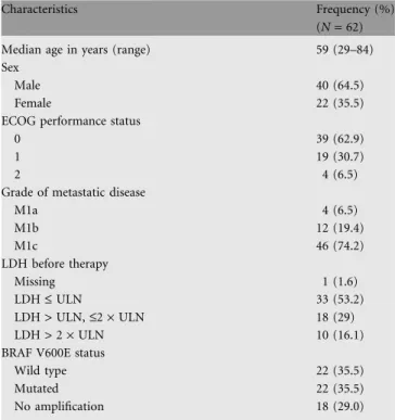
Table 1. Patient baseline characteristics
Characteristics Frequency (%)
(N = 62)
Median age in years (range) 59 (29–84)
Sex
Male 40 (64.5)
Female 22 (35.5)
ECOG performance status
0 39 (62.9)
1 19 (30.7)
2 4 (6.5)
Grade of metastatic disease
M1a 4 (6.5) M1b 12 (19.4) M1c 46 (74.2) LDH before therapy Missing 1 (1.6) LDH £ ULN 33 (53.2) LDH > ULN, £2 · ULN 18 (29) LDH > 2 · ULN 10 (16.1)
BRAF V600E status
Wild type 22 (35.5)
Mutated 22 (35.5)
No amplification 18 (29.0)
ECOG, Eastern Cooperative Oncology Group; LDH, lactate dehydrogenase; ULN, upper limit of normal.
decision, 2 are unknown and 2 deaths were attributed to the trial
drugs (of these latter 2 patients one died with extensive
abdominal tumour burden and bowel perforation and the other
died with pulmonary infection without leuco- or lymphopenia).
BRAF analysis
Of the 44 BRAF status results available for analysis, 22 patients
had the BRAF V600E mutation and 22 were BRAF wild type.
Based on the independent response review, a statistically
significant association was found between BRAF status and the
unconfirmed 12 weeks response favouring a response in the
wild-type group (P = 0.0088). Median PFS time in the mutated
group was 4.0 (95% CI: 1.7–5.4) versus 5.4 (95% CI: 2.6–8.2)
months in the wild-type group (P = 0.0556). Median duration of
disease stabilisation was 4.2 (95% CI: 3.0–5.5) versus 4.1 (95%
CI: 2.7–6.9) months between the mutated (n = 15) and wild-type
(n = 17) groups, respectively (P = 0.7292). A statistically
significant difference was found between the groups regarding
OS; median OS being 9.2 months (95% CI: 6.5–11.9) in the
mutated group compared with 12.0 months (95% CI: 7.4–16.4)
in the wild-type group (P = 0.0137; Figure 4). Other prognostic
biomarkers for metastatic melanoma, such as LDH, performance
status and age, did not differ significantly between the BRAF
mutated versus BRAF wild-type groups. Due to low numbers, no
multivariable analyses are carried out regarding BRAF status.
discussion
No significant advances in the treatment of metastatic
melanoma have been achieved in the last three decades. Despite
several promising results in phase II trials, all phase III trials
have reported negative results.
This multicentre single-arm phase II trial surpassed its
primary end point of 18 or more patients with a DSR12 with an
impressive 32 of 62 patients having confirmed, reviewed disease
stabilisation as defined in our trial. Our response rate of 16.1%
is in the same range as reported for single-agent temozolomide
conventional [3] (13.5%) and extended dosing schedule [4]
(14.5%), despite having studied a population at higher risk by
including patients with LDH > 2 ULN (16%) and/or ECOG
performance status of two (7%). The median PFS of 4.2
months compares favourably with the PFS seen in
single-agent temozolomide trials (1.9–2.3 months). The median OS of
temozolomide + bevacizumab was in the range of single-agent
temozolomide (9.6 versus 7.7–9.1 months) [3, 4]. The extent of
response rate as well as PFS is comparable to other trials that
have added bevacizumab to conventional chemotherapy in
melanoma patients [23, 24]. In the only randomised phase II
trial of its kind, carboplatin and paclitaxel (CPP) showed a PFS
of 4.2 versus 5.6 months (P = 0.14) when adding bevacizumab
(CPB) [23]. In another single-arm phase II trial with CPB
dosing paclitaxel weekly, the PFS was in the same range (6.5
months) [24]. Even though the absolute numbers of PFS are
slightly higher in the combination chemotherapy backbones
with CPB, the extent of improvement with bevacizumab when
combined with temozolomide seems to be higher compared
with other chemotherapy backbone. In addition, the
side-Figure 1. Waterfall plot of maximal percentage change in sum of longest diameter of target tumour lesion(s) size from baseline.
Figure 2. Kaplan–Meier plot of progression free survival (PFS) and overall survival (OS).
Figure 3. Kaplan–Meier plot of overall survival stratified by normal lactate dehydrogenase (LDH) and elevated LDH.
effects of single-agent oral temozolomide are lower and seem to
be better tolerable for the patients. In contrast to most
melanoma trials, our patients with elevated LDH showed
higher response rates and a statistically equal OS to the group
with normal LDH value. This raises the hypothesis whether
patients at high risk with rapidly progressive disease may
particularly profit more from the addition of bevacizumab,
which is in line with the results of the only randomised phase II
trial of its kind (BEAM trial): CPP showed a PFS of 4.2 versus
5.6 months (P = 0.14) when adding bevacizumab (CPB) [23].
Patients with a BRAF wild type had better PFS (borderline
significance), OS and response rate than patients with BRAF
mutation. It remains to be determined whether this mutation is
only a prognostic or also a predictive biomarker. Since there are
new promising targeted therapies for BRAF mutant patients
such as PLX4032 in phase III trials, it appears appealing to
re-evaluate these findings in BRAF wild-type patients.
The treatment was generally well tolerated. The incidence of
serious AEs in our trial was 32%, which is comparable to the
30% observed in single-agent temozolomide [4].
Bevacizumab-specific side-effects were in the range known from other trials;
no new or unexpected toxic effects occurred. The good
tolerability is of utmost importance in a disease where survival
is mostly short and the quality of life of the remaining weeks is
crucial for the patients.
In summary, the results of our trial suggest that the
combination of an alkylating agent such as temozolomide with
an agent that specifically targets VEGF might be a valid and
interesting therapeutic strategy for patients with metastatic
melanoma. A phase III trial stratifying for LDH level as well as
for BRAF status is urgently warranted in a disease where no
satisfactory first-line treatment exists.
acknowledgements
We would like to thank Martina Storz, Marie-Therese Abdou
and Nikita Kobert for their work with regards to the BRAF
amplification. Prior presentation: These data were previously
presented at the joint 15th Congress of the European CanCer
Organisation and 34th Congress of the European Society for
Medical Oncology, Berlin, Germany, 21 September 2009, and
the 2010 Annual Meeting of the American Society of Clinical
Oncology, Chicago, IL, 7 June 2010.
funding
This work was supported by Roche Pharma (Schweiz) AG,
Shering Plough and the Swiss State Secretariat for Education
and Research.
disclosures
R. von Moos had consultancy of advisory role at Roche and
Essex AG and speaking honoraria at Roche. R. Dummer had
consultancy of advisory role and research funding at
Astra-Table 2. AEs (worst grade observed per patient)
AE CTCAE grade, n (%)
Grade 3 Grade 4 Grade 5
ALT – 1 (2) – AST 1 (2) – – Alkaline phosphatase 1 (2) – – Anorexia 1 (2) – – Bilirubin 1 (2) – – Confusion – 1 (2) – Constipation 2 (3) – – Diabetes 1 (2) – – Diarrhoea 1 (2) – – Extensive abdominal tumour burden and bowel perforation – – 1 (2) Fatigue 4 (7) 1 (2) – Fracture 1 (2) – – GGT 1 (2) – – Hand–foot syndrome 1 (2) – – Haemorrhage 3 (5) – – Hypertension 7 (11) – –
Infection with unknown ANC–NOS 1 (2) – – Leucocytes 2 (3) – – Lymphopenia 1 (2) 1 (2) – Nausea 3 (5) – – Neutrophils 3 (5) 1 (2) – Pain 7 (11) 1 (2) – Platelets 4 (7) 2 (3) –
Pulmonary infection without leuco- or lymphopenia – – 1 (2) Thrombosis/thrombus/ embolism – 1 (2) – Trismus 1 (2) – – Vomiting 2 (3) – –
Only grade 3 and 4 AEs are shown.
AE, adverse events; ALT, alanine transaminase; ANC, absolute neutrophil count; AST, aspartate transaminase; CTCAE, Common Terminology Criteria for Adverse Events (version 3.0); GGT, gamma glutamyl transpeptidase; NOS, disorder not otherwise specified.
Figure 4. Kaplan–Meier plot of overall survival stratified by BRAF wild type and BRAF mutation.
Zeneca and Novartis. H. Moch is currently conducting research
sponsored by Roche and Novartis. All other authors declare no
conflict of interest.
references
1. Lin WM, Baker AC, Beroukhim R et al. Modeling genomic diversity and tumor dependency in malignant melanoma. Cancer Res 2008; 68: 664–673. 2. Curtin JA, Fridlyand J, Kageshita T et al. Distinct sets of genetic alterations in
melanoma. N Engl J Med 2005; 353: 2135–2147.
3. Middleton MR, Grob JJ, Aaronson N et al. Randomized phase III study of temozolomide versus dacarbazine in the treatment of patients with advanced metastatic malignant melanoma. J Clin Oncol 2000; 18: 158–166. 4. Patel MP, Suciu S, Mortier L et al. Extended schedule escalated dose
temozolomide versus dacarbazine in stage IV malignant melanoma: final results of the randomized phase III study (EORTC 18032). Ann Oncol 2008 LBA 8. 5. Hwu WJ, Krown SE, Menell JH et al. Phase II study of temozolomide plus
thalidomide for the treatment of metastatic melanoma. J Clin Oncol 2003; 21: 3351–3356.
6. Hwu WJ, Panageas KS, Menell JH et al. Phase II study of temozolomide plus pegylated interferon-alpha-2b for metastatic melanoma. Cancer 2006; 106: 2445–2451.
7. Hurwitz H, Fehrenbacher L, Novotny W et al. Bevacizumab plus irinotecan, fluorouracil, and leucovorin for metastatic colorectal cancer. N Engl J Med 2004; 350: 2335–2342.
8. Sandler A, Gray R, Perry MC et al. Paclitaxel-carboplatin alone or with bevacizumab for non-small-cell lung cancer. N Engl J Med 2006; 355: 2542–2550. 9. Miller KD. E2100: a phase III trial of paclitaxel versus paclitaxel/bevacizumab for
metastatic breast cancer. Clin Breast Cancer 2003; 3: 421–422.
10. Reck M, von Pawel J, Zatloukal P et al. Phase III trial of cisplatin plus gemcitabine with either placebo or bevacizumab as first-line therapy for nonsquamous non-small-cell lung cancer: AVAil. J Clin Oncol 2009; 27: 1227–1234.
11. Escudier B, Pluzanska A, Koralewski P et al. Bevacizumab plus interferon alfa-2a for treatment of metastatic renal cell carcinoma: a randomised, double-blind phase III trial. Lancet 2007; 370: 2103–2111.
12. Rini BI, Halabi S, Rosenberg JE et al. Bevacizumab plus interferon alfa compared with interferon alfa monotherapy in patients with metastatic renal cell carcinoma: CALGB 90206. J Clin Oncol 2008; 26: 5422–5428.
13. Friedman HS, Prados MD, Wen PY et al. Bevacizumab alone and in combination with irinotecan in recurrent glioblastoma. J Clin Oncol 2009; 27(28): 4733–4740. 14. Brychtova S, Bezdekova M, Brychta T et al. The role of vascular endothelial
growth factors and their receptors in malignant melanomas. Neoplasma 2008; 55: 273–279.
15. Lev DC, Onn A, Melinkova VO et al. Exposure of melanoma cells to dacarbazine results in enhanced tumor growth and metastasis in vivo. J Clin Oncol 2004; 22: 2092–2100.
16. Liu B, Earl HM, Baban D et al. Melanoma cell lines express VEGF receptor KDR and respond to exogenously added VEGF. Biochem Biophys Res Commun 1995; 217: 721–727.
17. Danielsen T, Rofstad EK. VEGF, bFGF and EGF in the angiogenesis of human melanoma xenografts. Int J Cancer 1998; 76: 836–841.
18. Lacal PM, Ruffini F, Pagani E et al. An autocrine loop directed by the vascular endothelial growth factor promotes invasiveness of human melanoma cells. Int J Oncol 2005; 27: 1625–1632.
19. Ugurel S, Rappl G, Tilgen W et al. Increased serum concentration of angiogenic factors in malignant melanoma patients correlates with tumor progression and survival. J Clin Oncol 2001; 19: 577–583.
20. Terheyden P, Hofmann MA, Weininger M et al. Anti-vascular endothelial growth factor antibody bevacizumab in conjunction with chemotherapy in metastasising melanoma. J Cancer Res Clin Oncol 2007; 133: 897–901. 21. Boasberg P, Cruickshank S, Hamid O et al. Nab-paclitaxel and bevacizumab as first-line therapy in patients with unresectable stage III and IV melanoma. J Clin Oncol 2009; 27 (Suppl): 15s (Abstr 9061).
22. Gonzalez-Cao M, Viteri S, Diaz-Lagares A et al. Preliminary results of the combination of bevacizumab and weekly paclitaxel in advanced melanoma. Oncology 2008; 74: 12–16.
23. O’Day SJ, Kim KB, Sosman JA et al. BEAM: a randomized phase II study evaluating the activity of bevacizumab in combination with carboplatin plus paclitaxel in patients with previously untreated advanced melanoma. Eur J Cancer 2009; 7(3): 13.
24. Perez DG, Suman VJ, Fitch TR et al. Phase 2 trial of carboplatin, weekly paclitaxel, and biweekly bevacizumab in patients with unresectable stage IV melanoma: a North Central Cancer Treatment Group study, N047A. Cancer 2009; 115: 119–127.
25. Varker KA, Biber JE, Kefauver C et al. A randomized phase 2 trial of bevacizumab with or without daily low-dose interferon alfa-2b in metastatic malignant melanoma. Ann Surg Oncol 2007; 14: 2367–2376.
26. Peyton JD, Spigel DR, Burriset HA et al. Phase II trial of bevacizumab and everolimus in the treatment of patients with metastatic melanoma: preliminary results. J Clin Oncol 2009; 27 (Suppl): 15s (Abstr 9027).
27. Newton CR, Graham A, Heptinstall LE et al. Analysis of any point mutation in DNA. The amplification refractory mutation system (ARMS). Nucleic Acids Res 1989; 17: 2503–2516.
28. Benlloch S, Paya A, Alenda C et al. Detection of BRAF V600E mutation in colorectal cancer: comparison of automatic sequencing and real-time chemistry methodology. J Mol Diagn 2006; 8: 540–543.