**Cestoda from Rattus assimilis (Gould, 1858) from
Australia
By *DOROTHEA F. SANDARS, M . S C , Ph.D.
From Institut de Zoologie, Universite de Neuch&tel and Department of Parasitology, London School of Hygiene and Tropical Medicine
Amongst material forwarded to me by Dr. M. J. Mackerras of the Queensland Institute of Medical Research, were cestodes collected from several specimens of Rattus assimilis (Gould, 1858), from Mt. Glorious, South Queensland and Innisfail, North Queens-land. These cestodes have been identified as (i) Hymenolepis diminuta Rudolphi, 1819; (ii) Raillietina (Raillietina) celebensis (Janicki, 1902) (see Baer and Sandars, 1956) ; (iii) Choanotaenia ratticola n.sp ; (iv) Hymenolepis australiensis n.sp. ; (v) one misshapen cysticercus of
Taenia taeniaeformis (Batsch, 1786). Of these (i), (ii) and (v) appear
to be new host records for Australia while (iii) and (iv) are new species.
Whole mounts of all these cestodes were prepared by staining in hydrochloric-alcohol-carmine and some were stained in weak haemalum. They were all mounted in Canada balsam. Scolices were also mounted in berlase medium, some of which were squashed to facilitate the detailed study of the rostellar hooks. Sections were cut at 9 \L and stained in haemalum and eosin. Measurements given were made from mounted specimens.
* On study leave from the University of Queensland and the Queensland Institute of Medical Research, Brisbane.
** Part of a thesis approved by the University of London for the award of the Ph.D. degree.
66 Cestodes from Rattus assimilis (2)
(i) HYAIENOLEPIS DIMINUTA Rudolphi, 1819
These were collected from Rattus assimilis from Innisfail, North Queensland. Each host had an infestation of numerous specimens. The male reproductory organs in the mature segments demonstrated a great variability in the number of testes present; they varied between none and four, even in segments adjacent to one another. This is typical for Hymenohpis diminuta.
(ii) RAILUETINA (RAILUETINA) CELEBENSIS (Janicki, 1902)
Several specimens were recovered from the duodenum of Rattus
assimilis from Mt. Glorious, South Queensland. The length is
35-175 mm. and the maximum width is 1.4-1.75 mm. The scolex is 274-411 jx longX480-803 \i diameter; the four suckers are 114-183 (x in diameter, each having numerous small spines on the inner edges. The rostellum is 105-123 fx in diameter and is armed with 160 hooks each 18-23 fi long, and arranged in two circlets. In the mature segments there are 28-30 testes (7-9 poral; 21 aporal), 37-46 (x in diameter. The cirrus pouch is 114-151 fx long x 50-73 jx in diameter. The genital pore always lies within the anterior quarter of the proglottid length. There are 100-140 egg capsules in each gravid proglottid and within each capsule 1-4 eggs, 27 (x in diameter.
Baer and Sandars (1956) have studied this species in detail.
(iii) CHOANOTAENIA KATTICOLA n.sp. (Figures 1-9)
A number of specimens identified as Choanotaenia were recovered from Rattus assimilis from Mt. Glorious, South Queensland. These cestodes were always in the duodenum and upper ileum of the host. They appear to belong to a new species for which the name
Choanotaenia ratticola is proposed.
Description : The mature strobila is 80-120 mm. long. It
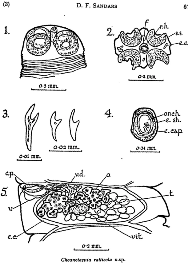
gradually increases in width from behind the scolex towards the posterior end. The maximum width, 0.75-1.5 mm. is across the gravid proglottides. The maximum diameter of the scolex, 434-471 fx is across the suckers and its maximum length is 297-457 jx. The length of the rostellum and its sac, when contracted, is 136-229 fx ; it bears 26 thorn-shaped hooks, each 16-20 fx long and arranged in two irregular circlets (Figure 1).
:k
o-smm.3.
O-02 mm. mm. mm. o-2 mm. Choanotaenia ratticola n.sp.Fig. I.—Scolex. Fig. 2.—^Transverse section of scolex showing suckers and rostellum. Fig. 3.—Rostellar hooks. Fig. 4.—Egg capsule. Fig. 5.—Mature proglottid.
ABBREVIATION'S
c.p.—cirrus pouch ; e.c.—excretory canal; e.cap.—egg capsule ; e.sh.— egg shell; o.—ovary ; onch.—onchosphere; r.—rostellum ; r.h.—rostellar hooks ; s.s.—section of sucker; t.—testes ; v.—vagina ; v.d.—vas deferens ; vit—vitellarium.
68 Cestodes from Rattus assimilis (4)
The neck region of the strobila is extremely short, being 479-640 p. wide. It is followed by between 10-12 immature segments of gradually increasing size, 0.02-1.10 mm. long x 0.53-0.87 mm. wide. There are 10-13 mature segments 0.09-0.27 mm. in length x 0.64-1.03 mm. width; the gravid segments, of which there are approximately
13-16, are 0.23-0.69 mm. x 0.69-1.37 mm. wide.
Q.
o-\
mm.
Choaitolaenia ratticola n.sp.
Fig. 6.—Cirrus pouch with cirrus invaginatcd. Fig. 7.—Cirrus pouch with cirrus evaginated.
The longitudinal musculature is very well developed. It has single large isolated fibres arranged in two rows, the inner row being of less numerous and more irregular fibres than the outer row. There are also scattered smaller longitudinal fibres in the cortex. Transverse and dorso-ventral muscle fibres are not evident.
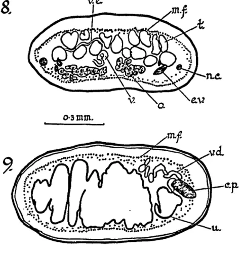
The longitudinal nerve cords lie outside both the dorsal and ventral longitudinal excretory canals on both sides of the strobila. They measure 18-23 (xx 23-25 p. (Figure 8).
TUL Z.XZ
o-3 mm.
i t .
Choanotaenia ratticola n.sp.
Fig. 8.—Transverse section of mature proglottid. Fig. 9.—Transverse section of gravid proglottid.
ABBREVIATIONS used in Figs. 6-9
a.p.—anterior proglottid margin : c.—cirrus ; c.p.—cirrus pouch ; e.v.— excretory vessel; g.a.—genital atrium ; m.f.—muscle fibres ; n.c—nerve cord ; r.a.—retractor muscle ; r.c.—receptaculum seminis ; s.c.—spines of cirrus ; t.—testis ; u.—uterus ; v.—vagina ; v.d.—vas deferens.
The excretory system conforms to the typical cestode pattern. The paired dorsal and ventral longitudinal ducts are all narrow, the dorsal vessel always overlying the ventral vessel. In each proglottid these longitudinal vessels markedly curve inwards to where they join the transverse excretory vessels. Sections show the excretory vessels to have thick cellular walls. The genital ducts pass dorsally to both dorsal and ventral longitudinal excretory vessels.
70 Cestodes from Raltus assimilis (6) In the male reproductory system there are 20-35 testes which are slightly lobed ; they measure 23-46 \i x 37-64 \i. The testes are arranged in two to three dorso-ventral layers (Figure 8), extend-ing right across the mature proglottides but lyextend-ing within the region delimited by the longitudinal excretory canals. The vasa efferentia join the vas deferens which arises in about the middle of each proglottid. The vas deferens passes forward between the lobes of the ovary and becomes much coiled anteriorly in the proglottid, before entering the cirrus pouch within which the duct forms only a few simple coils. The cirrus pouch does not extend beyond the longitudinal excretory vessels ; in mature proglottides it is 114-165 fx long X 37-55 {i in diameter. The cirrus pouch persists in gravid proglottides and is 101-183 fx long X 37-46 jx in diameter. The cirrus is heavily armed with small spines and appears to be 137-151 \i long when evaginated (Figures 6 and 7). It opens into a small inconspicuous genital atrium. There are no spines at the base of the genital atrium where the cirrus opens into it. The genital atria are irregularly alternate and are at the anterior of each proglottid.
The vagina, which appears to be unarmed, opens from the genital atrium posteriorly to the cirrus pouch. In several segments self fertilization was observed. The vagina passes inwards and slightly anteriorly and then passing posteriorly it expands to form a large receptaculum seminis 55-160 (xx 46-69 fx. This is most evident in the older mature segments and in the gravid segments. The ovary is deeply lobed, 205-434 [xx 69-137 (x. The vitellarium is only slightly lobed and is 46-55 ;x X 64-69 jx; it is posterior to the ovary (Figure 5). The uterus arises from the posterior region of each proglottid and as it develops passes anteriorly. It forms a lobed sac extending laterally beyond the excretory canals (Figure 9). The uterus eventually forms egg capsules, each capsule containing only one egg (Figure 4). The egg-capsules measure 27-40 fxX36-45 jx; the eggs are 26-34 fx x 28 fx ; the onchospheres are 22-25 fxxl7 fx. The six hooks within the oncho-sphere are 11-13 |x long.
Specific Diagnosis : CJwanotaenia ratticola n.sp. Body length
8-120 mm. Rostellum with 26 thorn-shaped hooks 16-20 jx long arranged in two irregular circlets. There are 20-35 slightly lobed testes in several layers. The cirrus pouch is 101-183 [i long x 37-55 jx in diameter. The cirrus is armed. Spines are absent from the base of the genital atrium near the cirrus opening. Genital atria irregularly alternate. Egg capsules present, each containing one egg.
Host: Rattus assimilis Gould.
Habitat in host : Duodenum and upper ileum.
Geographical distribution : Mt. Glorious, South Queensland.
Discussion : The genus Choanotaenia is typically a parasite
of birds, although three species have been described from insectivores from Europe and four species from North American rodents. The present record is the first one made from a rat.
The species from insectivore hosts are :—Choanotaenia
fila-mentosa (Goeze, 1782) from Talpa europaea (L) ; C. scutigerum
(Dujardin, 1845) from Sorex araneus tetragonnrus Herm., and
Crocidura russula russula Herm. : Choanotaenia hepaticum (Baer,
1932) from Sorex araneus Linnaeus.
Among the species of Choanotaenia parasitic in rodents, Hansen (1950) described C. nebraskensis from Microtus ochrogaster (Wagner) and Sciurus rufiventer Geoff. He placed Prochoanotaenia spermophili McLeod, 1933 from Citellus tridecemlineatus (Mitchill) and C.
richard-sonii (Sabine) within the genus Choanotaenia, so that it is therefore
a synonym of Choanotaenia spermophili (McLeod, 1933) Hansen, 1950. He also put Prochoanotaenia peromysci Erickson, 1938, which is parasitic in Peromyscus maniculatus gracilis (Le Comte), within the genus Choanotaenia. It is therefore synonymous with
Choanotaenia peromysci (Erickson, 1938) Hansen, 1950. Hansen
(1950) referred also to Choanotaenia sciuricola Harwood and Cooke, 1949, from Sciurus niger Linnaeus.
Rausch and Tiner (1949), recorded a single specimen of
Choano-taenia sp. from the intestine of Microtus ochrogaster from Illinois,
U.S.A., which they considered as possibly representing "an accidental infection". If this specimen was a Choanotaenia, which is impossible to say without the gravid segments, it is probably C. nebraskensis Hansen, 1950 (see Table 1).
Choanotaenia ratticola differs from all these species in the size and
shape of the rostellar hooks, in the size of the cirrus pouch and egg capsules and in the absence of small spines in the genital atrium where the cirrus opens (see Table 1). Hansen gives a "Key to the Species of Choanotaenia from Mammals" in which he makes no mention of C. sciuricola Harwood and Cooke, 1949, although referring to it in the text. Hansen lists C. crassiscolex, which is a synonym of
TAUL E I Compariso n o f specie s o f Choanotaenia fro m Insectivore s an d Rodents . 4 to Specie s C. filamtntosa (Goeze , 1782 ) C. hepaticum (Baer , 1932 ) *C . sculigerum (Dujardin , 1845 ) C. nebmskensis Hansen , 105 0 C. peromysci (Erickson , 1938 ) C. ratticola n.sp . C. sciuricola Hanvoo d an d Cooke , 194 9 C. spermophili (McLcod , 1933 ) C . sp . recorde d b y Rauscl i an d Tiner , 194 9 Rostella r Lengt h Cirru s Pouc h lioo k rostella r lengt h diamete r Egg s numbe r hook s ( x \i y. | i Hos t 2 4 3 2 4 6 3 8 16-1 8 52-5 5 24-2 5 28-3 1 30-3 5 190-7 0 57X4 9 Talpa europaea (capsule ) immatur e immatur e immatur e Sorex araneiis 15-2 0 31-4 3 130-14 0 15 0 Sorex araneiis tetragonunts X (capsule ) Crocidura russula russula 2 3 07-11 7 65-7 4 Microtttsochrogasler X X Sciurus rufivenler 20-3 3 56-6 5
9
cn o o> 8 2 6 2 2 23-2 5 2 6 3 2 16-2 0 3 8 34-3 7 3 0 <2 5 (21* ) 20-3 5 27-4 3 mea n 31 ) <2 5 (20« ) 3 1 84x3 5 101-18 3 X 37-5 5 150-13 0 X 31-3 8 200x3 3 80-11 5 27-4 0 X 36-4 5 (capsules ) 59x68. 5 Peromyscus maniculatus gracili s Rallies assimilis Sciurus niger Citellus tridecemlineatus C. richardsonii Microtus ochrogaster to issimins * Measurement s take n fro m diagram . G Oin the description given by Baer (1928), 16-18 hooks are stated to be present.
Choanotaenia ratticola is the first species of Choanotaenia described
from a mammal from Australia. The previous records from Australian hosts are all from birds viz : C. infundibulnm (Bloch, 1779) from Gallus gallus ; C. fieldingi (Maplestone and Southwell; 1923) from Craticus torqiiains, Lath., C. meliphagidarum T. H. Johnston, 1910, from Meliornis novae-hollandiae, Meliornis sericea,
Ptilotis chrysotis and P. leucotis ; Choanotaenia taylori T. H. Johnston,
1912, from Malurus cyanochlamys. Of these, Choanotaenia
infundi-bulnm (Bloch, 1779) is doubtlessly an introduced species, so that
there have therefore been only three species previously described, which may be considered as being endemic in Australia.
Choanotaenia zoniferae T. H. Johnston, 1912, described from Zonifer tricolor is now Paricterotaenia zoniferae (Johnston, 1912),
since there are no egg capsules present.
(iv) HYMENOLEPIS AUSTRAUENSIS n.sp. (Figures 10-12)
A number of cestodes recovered from Rattus assimilis from Innisfail, North Queensland, have been identified as a new species of Hymenolepis, to which the name H. australiensis is assigned.
Description : The total body length is 225-1,100 mms., the
strobila being of regular shape with a maximum width of 731-777 [L usually across the mature and gravid proglottides.
The scolex is 160-229 |x long with a maximum diameter of 183-229 (x across the suckers which are 68-78 fx in diameter. The rostellum and sac are 159-137 fx long x 69-102 fx diameter (Figure 10). The rostellum is armed with 31 hooks arranged in a single circlet; each hook is 18-21 (x long. The blade of each hook is large, the measurement from the end of the base of the hook to the tip of the blade being 21-21 fx (Figure 11).
The neck region is long with a width of 182-205 jx. The mature proglottides are 137-183 jx longX594-777 [x wide; the gravid proglottides are 183-237 jx long X 274-617 JX wide.
The musculature, longitudinal nerve cords and excretory system are of the usual hymenolepid pattern.
74 Cestodes from Raltus assitnilis (10)
There are three testes, which are usually spherical, 69 jx in diameter, but if subspherical measure 64-69 fxx 73-91 {x. The testes are usually arranged in a transverse row and with one testes poral in position and the other two aporal. However, the testes are often differently arranged in the proglottides of even one strobila. Hence they may all be aporal, or two may be poral and one aporal. These variations may be observed even in successive proglottides (Figure 12). The external seminal vesicle is 137-297 {x long x 69-91 fx wide and extends beyond both longitudinal excretory canals. The cirrus pouch is 137-169 fx long with a maximum dia-meter of 27-41 {x ; it does not reach either longitudinal excretory canal. The unilateral genital atria lie in the middle of the length of each proglottid.
The vagina opens into the genital atrium ventrally to the cirrus pouch. It dilates slightly before it reaches the female genital complex. The ovary is lobed (Figure 12) ; it has a maximum width of 69-114 (x and a maximum length of 46-69 (x. The vitellarium lies posterior to the ovary, frequently between the ovarian lobes. It is sometimes oval in shape and may be slightly lobed; it is then 23-46 (x in maximum width and 23-37 (x in length. Sometimes the vitellarium is spherical having a diameter of 46 fx. The female genital complex always lies slightly closer to the aporal side in each proglottid.
The eggs are 20-24 \L diameter; the onchospheres are 17-19 jx diameter with hooks 11-13 fx long.
Specific Diagnosis : Hymenolepis australiensis n.sp. The
rostellum is armed with 31 hooks 18-21 [x long and arranged in a single circlet. The cirrus pouch which is 137-169 fx long with a maximum diameter of 27-41 jx does not' reach the longitudinal excretory canals. The cirrus is unarmed. The three testes are arranged in a transverse row. The ovary is lobed; the vitellarium may be oval and slightly lobed or spherical. Eggs 20-24 (x diameter, onchosphere 17-19 fx diameter with hooks 11-13 fx long.
Host: Rattits assimilis (Gould, 1858). Habitat in host : intestine.
Geographical distribution : Eastern Australia.
Discussion : Mahon (1954), lists all the species of armed Hymenolepis which have been described from rodents. She also
JO.
12.
o-2 mm.
Hymenolepis australiensis n.sp.
Fig. 10.—Scolex. Fig. 11.—Rostellar hooks. Fig. 12.—Mature proglottides.
ABBREVIATIONS
cp.—cirrus pouch ; e.v.s.—external vesicula seminalis; g.s.—genital atrium ;
76 Cestodes from Rattus assimilis (12) Number of hooks 18-27 10 12-14 20-24 12 31 Size of hook 10-15 (JL 20-22 jx 18-24 (x 14-16 |x 34-37 {x 18-21 tx gives the following list for Hymenolepis spp. all of which have rostellar hooks of a similar shape. The species now described is now included in the list:—
Species
H. microstoma (Dujardin, 1845) Blanchard, 1891 H. evaginata Baker and Andrews, 1915
H. globirostris Baer, 1925
H. straminea (Goeze, 1782) Kowalewsky, 1904 H. uncinispinosa Joyeux and Baer, 1930 H. australiensis n.sp.
Altogether there have been only four other species of Hymenolepis [excluding H. terraereginae (see below)] described from the nature fauna of Australia. Only one of these is from an endemic mammal viz : H. peramelidarum Nybelin, 1917, from the bandicoot, Thylacis
obesuhis (syn. Perameles macrura). Hymenolepis peramelidarum has
also been recovered from Thylacis obesulus and Perameles nasuta recorded by Sandars (manuscript in course of preparation). The other three species recorded are all parasites of birds. They are :—
Hymenolepis collaris (Batsch, 1786), from Anas superciliosa and Nettion castaneum ; Hymenolepis ibidis T. H. Johnston, 1913, from Platibis fiavipes; Hymenolepis megalops (Creplin, 1829), from Anas superciliosa, Anseranas semipalmata, Nettion castaneum and
the "black duck" ; Hymenolepis robertsi Baylis, 1934, from
Quer-quedula gibberifrons and Anderanas semipalmata. Hymenolepis terraereginae is probably a synonym of H. collaris (Batsch, 1786).
It was described from "numerous scolexless tapeworms" and Johnston himself stated that its anatomy "closely resembles that of
H. collaris".
Hymenolepis australiensis n.sp. is the first record of a Hymenolepis
from a presumably ubiquitous Australian rodent.
(v) TAENIA TAENIAFORMJS (Batsch, 1786)
A mis-shapen cysticercus' of Taenia taeniaeformis was recorded from the liver of a Rattus assimilis from Mt. Glorious, South Queens-land.
SUMMARY
Four species of adult cestodes and one larval form were recovered from Rattus assimilis (Gould, 1858) from Queensland. Hymenolepis
diminuta Rudolphi, 1819 ; and the cysticercus of Taenia taentae-for mis (Batsch, 1786) are new host records taentae-for Australia; the same
infestations with Raillietina (Raillietina) celebensis (Janicki, 1902). have also been recorded by Baer and Sandars (1956). Two new species, viz :—Choanotaenia ratticola and Hymenolepis australiensis are described.
The type specimens are lodged in the Queensland Museum, Brisbane. Cotype and other specimens studied are also lodged in the Queensland Museum and in" the Institut de Zoologie, University de Neuchatel, Switzerland; the Department of Parasitology, London School of Hygiene and Tropical Medicine and in the British Museum (Natural History).
ACKNOWLEDGMENTS
I am indebted to Dr. M. J. Mackerras of the Queensland Institute of Medical Research, Brisbane, for forwarding the specimens to me for examination and description. I wish to express also my appreci-ation to Professor J. G. Baer of the Institut de Zoologie, Universite de Neuchatel for the hospitality in his laboratory and his keen interest in this research ; and also to Professor J. J. C. Buckley of the London School of Hygiene and Tropical Medicine where this
work was completed.
The research was done while holding both an Ohio State Grant 1955-56 awarded by the International Federation of University Women, and a grant for 1955-56 made available through the Trustees of the Science and Industry Endowment Fund of Australia.
REFERENCES
BAER, J. G., 1928.—"Contribution a la Faune helminthologique de Suisse."
Rev. Suisse ZooL. 35, 27-41 (W.L.19288).
1932.—"Contribution k la Faune helminthologique de Suisse (Deuxieme Partie)." Ibidem, 39, 1-56.
BAER, J. G. and SANDARS, D. F., 1956.—"The first record of Raillietina
(Raillietina) celebensis (Janicki, 1902) (Cestoda) in man from Australia, with a critical survey of previous cases." / . Helminth., 30, 173-182. (W.L. 11224c).
BAYLIS, H. A., 1934.—"Some parasitic worms from Australia." Parasitology,
78 Cestodes from Rattits assimilis (14)
ERIKSON, A. B., 1938.—"Parasites of some Minnesota Cricetidae and Zapodidae,
and a host catalogue of helminth parasites of native American Mice."
Amer. Midi. Nat., 20, 575-589.
HANSEN, M. F., 1950.—"A new dilepidid tapeworm and notes on other tapeworms
of Rodents." Ibidem, 43, 471-479.
HARWOOD, P. D. and COOKE, V., 1949.—"The helminths from a heavily
parasitized fox squirrel, Sciurus niger." Ohio J. Set., 49, 146-148. (W.L. 15627).
JOHNSTON, T. H., 1910.—"On Australian avian entozoa." J. Roy. Soc. N.S.W.,
44, 84-122. (W.L. 11492).
1911.—"New species of avian cestodes." Proc. Linn. Soc. N.S.W., 36,58-80. (W.L. 16791a).
1912.—"International parasites recorded from Australian birds."
Emu. 12, 105-112.
1912a.—"New species of cestodes from Australian birds." Mem.
Qd. Mits., 1, 211-215. (W.L. 13418).
-1912b.—"On a re-examination of the types of Krefft's species of cestoda in the Australian Museum, Sidney." Rec. Aust. Mus.. 9, 1-35. (W.L. 17736).
1913.—"Notes on some entozoa." Proc. Roy. Soc. Qd.. 24, 63-91. (W.L. 16903).
1913a.—"Cestoda and Acanthocephala." Rep. Aust. Inst. Trop. Med., 1911,75-96. (W.L. 17930e).
MAHON, J., 1954.—"Tapeworms from the Belgian Congo." Ann. Mus. Congo
Beige 4 to., 1, fasc. 2. 149-261. (W.L. 881).
MAPLESTONE, P. A. and SOUTHWELL, R., 1923.—"Notes on Australian Cestodes."
No. VII. Ann. Trop. Med. ParasiL, 17, 317-331. (W.L. 1063).
MCLEOD, J. A., 1933.—"A parasitological survey of the genus Citellus in
Manitoba." Canad. J. Res., 9, 108-127. (W.L. 5898c).
RAUSCH, R. and TINER, J. D.. 1949.—"Studies on the parasitic helminths of
the North Central States. II. Helminths of voles (Microtus spp.) preliminary report." Amer. Midi. Nat., 41, 665-694. (W.L.640)