HAL Id: hal-02018153
https://hal.archives-ouvertes.fr/hal-02018153
Submitted on 13 Feb 2019
HAL is a multi-disciplinary open access
archive for the deposit and dissemination of
sci-entific research documents, whether they are
pub-lished or not. The documents may come from
teaching and research institutions in France or
abroad, or from public or private research centers.
L’archive ouverte pluridisciplinaire HAL, est
destinée au dépôt et à la diffusion de documents
scientifiques de niveau recherche, publiés ou non,
émanant des établissements d’enseignement et de
recherche français ou étrangers, des laboratoires
publics ou privés.
Disulfide Oxidoreductase
Edwige Garcin, Olivier Bornet, Latifa Elantak, Nicolas Vita, Laetitia Pieulle,
Francoise Guerlesquin, Corinne Sebban-Kreuzer
To cite this version:
Edwige Garcin, Olivier Bornet, Latifa Elantak, Nicolas Vita, Laetitia Pieulle, et al.. Structural
and Mechanistic Insights into Unusual Thiol Disulfide Oxidoreductase. Journal of Biological
Chem-istry, American Society for Biochemistry and Molecular Biology, 2012, 287 (3), pp.1688-1697.
�10.1074/jbc.m111.288316�. �hal-02018153�
Structural and Mechanistic Insights into Unusual Thiol
Disulfide Oxidoreductase
□SReceived for publication, August 1, 2011, and in revised form, November 20, 2011Published, JBC Papers in Press, November 28, 2011, DOI 10.1074/jbc.M111.288316 Edwige B. Garcin, Olivier Bornet, Latifa Elantak, Nicolas Vita, Laetitia Pieulle, Franc¸oise Guerlesquin,
and Corinne Sebban-Kreuzer1
From the IMR, IFR88, CNRS, Aix-Marseille Universite´, Marseille 13402, France
Background:TDOR are ubiquitous and catalyze important cell redox reactions.
Results:Dtrx presents atypical physicochemical properties and a positive surface around its active site, suggesting a specificity for it(s) substrate(s).
Conclusion:Active site histidine plays an important role in the molecular mechanism of Dtrx catalysis.
Significance: Structural and functional studies of such atypical systems will give new insights into the TDOR catalytic mechanism.
Cytoplasmic desulfothioredoxin (Dtrx) from the anaerobe
Desulfovibrio vulgaris Hildenborough has been identified as a
new member of the thiol disulfide oxidoreductase family. The active site of Dtrx contains a particular consensus sequence, CPHC, never seen in the cytoplasmic thioredoxins and generally found in periplasmic oxidases. Unlike canonical thioredoxins (Trx), Dtrx does not present any disulfide reductase activity, but it presents instead an unusual disulfide isomerase activity. We have used NMR spectroscopy to gain insights into the structure and the catalytic mechanism of this unusual Dtrx. The redox potential of Dtrx (ⴚ181 mV) is significantly less reducing than that of canonical Trx. A pH dependence study allowed the deter-mination of the pKaof all protonable residues, including the cysteine and histidine residues. Thus, the pKavalues for the
thiol group of Cys31and Cys34are 4.8 and 11.3, respectively. The His33 pK
avalue, experimentally determined for the first
time, differs notably as a function of the redox states, 7.2 for the reduced state and 4.6 for the oxidized state. These data suggest an important role for His33in the molecular mechanism of Dtrx
catalysis that is confirmed by the properties of mutant DtrxH33G protein. The NMR structure of Dtrx shows a differ-ent charge repartition compared with canonical Trx. The results presented are likely indicative of the involvement of this protein in the catalysis of substrates specific of the anaerobe cytoplasm of DvH. The study of Dtrx is an important step toward revealing the molecular details of the thiol-disulfide oxidoreductase cata-lytic mechanism.
Thiol/disulfide oxidoreductases (TDOR)2are ubiquitous in
prokaryotes and eukaryotes and catalyze important redox
reac-tions in the cell (1, 2). All of the members of this family share the thioredoxin fold consisting of a central-sheet surrounded by four␣-helices and an active site with two conserved cysteine residues that specify the biological activity of the protein (3). Despite these similarities, thiol/disulfide oxidoreductases can be subdivided according to their cellular location. Although the cytoplasmic members of this family such as thioredoxin (Trx) and glutaredoxin catalyze the reduction of disulfide bonds, the members of oxidizing cellular compartments, such as protein-disulfide isomerase (PDI) from the endoplasmic reticulum and disulfide bond proteins DsbA and DsbC from the bacterial periplasm, are catalysts of disulfide bond formation during folding of secreted proteins.
The CXXC active site of these proteins is essential for TDOR activity. The sequence of the XX dipeptide located between the cysteines in the active site motif is very important in controlling the redox properties of the protein (5–7). Within each sub-group the active site contains a conserved consensus sequence (Trx, CGPC; glutaredoxin, CPYC; PDI, CGHC; DsbA, CPHC; DsbC, CGYC).
In all thioredoxin-like proteins, the reactivity is influenced by the pKavalue of the first active site cysteine residue and by the
redox potential (E0⬘) of the disulfide bond. There is a
remarka-ble correlation between the standard redox potential of these enzymes and their physiological role; indeed the members with the lowest redox potentials catalyze reducing processes in vivo (Trx,⫺270 mV (4); and glutaredoxin, ⫺233 to ⫺198 mV (5)), whereas the protein folding catalysts are strong oxidizing agents (PDI,⫺175 to ⫺147 mV (6, 7); and DsbA, ⫺163 to ⫺80 mV (8)). Exceptions are the thioredoxin-like proteins anchored to the inner bacterial membrane. As an example, TlpA (thiore-doxin-like protein A) from Bradyrhizobium japonicum exhibits a low redox potential (⫺259 mV) despite its periplasmic orien-tation and is required for cytochrome aa3maturation (9). How-ever, no high redox potential has ever been reported for cyto-plasmic TDOR.
Thioredoxins are a group of small (12 kDa) proteins. They have been characterized from various prokaryotic and eukary-otic organisms (10). The thioredoxin system comprises a thi-oredoxin with the active site consensus sequence WCGPC
□S This article containssupplemental Table S1 and Figs. S1–S6.
The atomic coordinates and structure factors (codes 2L6D and 2L6C) have been deposited in the Protein Data Bank, Research Collaboratory for Structural Bioinformatics, Rutgers University, New Brunswick, NJ (http://www.rcsb.org/).
1To whom correspondence should be addressed. Tel.: 33-4-91-16-44-53; Fax:
33-4-91-16-45-40; E-mail: [email protected].
2The abbreviations used are: TDOR, thiol-disulfide oxidoreductases; Dsb,
disulfide bond protein; Dtrx, desulfothioredoxin; PDI, protein-disulfide isomerase; Trx, thioredoxin; HSQC, heteronuclear single quantum coherence.
THE JOURNAL OF BIOLOGICAL CHEMISTRY VOL. 287, NO. 3, pp. 1688 –1697, January 13, 2012
© 2012 by The American Society for Biochemistry and Molecular Biology, Inc. Published in the U.S.A.
at CNRS on December 12, 2018 http://www.jbc.org/ Downloaded from at CNRS on December 12, 2018 http://www.jbc.org/ Downloaded from at CNRS on December 12, 2018 http://www.jbc.org/ Downloaded from at CNRS on December 12, 2018 http://www.jbc.org/ Downloaded from at CNRS on December 12, 2018 http://www.jbc.org/ Downloaded from at CNRS on December 12, 2018 http://www.jbc.org/ Downloaded from at CNRS on December 12, 2018 http://www.jbc.org/ Downloaded from at CNRS on December 12, 2018 http://www.jbc.org/ Downloaded from at CNRS on December 12, 2018 http://www.jbc.org/ Downloaded from at CNRS on December 12, 2018 http://www.jbc.org/ Downloaded from
(Trx), a thioredoxin reductase, and NADPH. The molecular catalytic mechanism actually proposed for the reduction of the oxidized substrate proteins by the canonical Trx involves a bimolecular nucleophilic substitution reaction. The reaction starts with a nucleophilic attack of the N-terminal thiol of the WCXXC motif on the disulfide bond of the target protein, releasing a free thiol and forming a mixed disulfide between Trx and its substrate. In the second step, the C-terminal thiol must be activated as a thiolate to allow the dissociation of the com-plex. For deprotonation of the C-terminal thiol, one hypothesis involves a conserved aspartate residue (11). Recently, it was suggested that when the N-terminal thiolate of Trx attacks its substrate disulfide to form a mixed disulfide complex, the leav-ing thiol group deprotonates the thiol of the C-terminal active site of Trx (12). Finally another hypothesis proposes that the cysteine is activated for its nucleophilic attack by hydrogen bonds between this residue and the backbone amide of the active site tryptophan (13).
In Desulfovibrio vulgaris Hildenborough, a Gram-negative sulfate-reducing bacteria, two cytoplasmic thioredoxin systems have been identified (14). The Trx1/TR1 system contains the ubiquitous thioredoxin with the active site consensus sequence WCGPC. The second system contains an atypical thioredoxin with a CPHC sequence at the active site identical to the DsbA motif and an unconventional thioredoxin reductase that uses preferentially NADH (14). The presence of these atypical pro-teins being restricted to Desulfovibrio organisms, they have been named desulfothioredoxin (Dtrx) and desulfothioredoxin reductase (15).
In this work, we have determined the disulfide isomerase and reductase activities and redox properties for the cytoplasmic CPHC active site. We have solved the structure of Dtrx and investigated the catalytic mechanism of this atypical enzyme. We identified important structural differences between Dtrx and the canonical bacterial Trx1 in the areas surrounding the catalytic sites.
EXPERIMENTAL PROCEDURES
Protein Production—The encoding sequence (DVU378) of desulfothioredoxin from D. vulgaris Hildenborough was cloned into expression vector pJF119-EH for production of a His-tagged protein at its C terminus.
pJF119-EH Dtrx plasmid with the desulfothioredoxin wild-type gene was used as DNA template in two separate PCRs to introduce the H33G mutation with two pair of primers (an internal mutagenic forward primer (H1: 5 ⬘-CCTGTGC-CCGGGCTGCAAGAAC-3⬘) and an external reverse primer (C2: 5⬘-GCTTCTGCGTTCTGATTTAATCTG-3⬘); and an internal mutagenic reverse primer (H2: 5 ⬘-GTTCTTGCAGC-CCGGGCACAGG-3⬘) and an external forward primer (C1: 5⬘-GCAGAAACGTGGCTGGCCTGG-3⬘)). The two PCR-pu-rified fragments were mixed, denatured, and extended. The product was then amplified in a third PCR using C1 and C2 primers. The resulting fragments were digested with BamHI and EcoRI and cloned into the pJF119-EH vector.
Transformed Escherichia coli TG1 cells were grown in a M9 minimal medium containing15NH
4Cl (1 g/liter) and [
13
C]glu-cose (2 g/liter) as the sole nitrogen and carbon sources. Protein
purification was achieved using nickel-nitrilotriacetic acid affinity chromatography and imidazole gradient (20 –500 mM) in 500 mMNaCl and 100 mMphosphate buffer at pH 8. The purity of the protein sample was checked by SDS-PAGE.
NMR Spectroscopy—NMR spectra were recorded on Bruker Avance III 600 MHz spectrometer equipped with a TCI cryo-probe or a Bruker Avance III 500 MHz spectrometer equipped with a TXI probe. All of the experiments were carried out at 298 K. The spectra were processed using Topspin (Bruker).
Activity Assay—The ability of Trx1 and Dtrx, wild-type and mutant, to catalyze insulin reduction in the presence of DTT was determined as previously described (16). The reaction mix-tures were prepared in cuvettes containing 130Minsulin, 5M protein catalyst in different 0.1Mbuffers (pH 4.22, 6.3, 7.06, 7.52, 8.11, and 8.98), and 2.5 mMEDTA. For Dtrx, we also used a 20 Mconcentration. The reactions were started with the addition of 1 mMDTT. The rate of precipitation was monitored by recording the increased turbidity of the reaction mixture, which was measured at 650 nm every 30 s at 33 °C and using an Uvikon spectrophotometer. The noncatalyzed reduction of insulin by DTT was monitored in a control reaction.
An in vitro assay involving refolding of scrambled RNase A was used to monitor the oxidase activity of Trx1, Dtrx, DtrxH33G mutant, and DsbC (ATGen, Seongnam-SI, South Korea) (17). Disulfide-scrambled RNase A was produced by incubating 30 mg of native RNase A (Sigma) overnight at room temperature in 50 mMTris-HCl, pH 8.5, in the presence of 6M guanidinium chloride and 130 mMDTT. After acidifying the solution (addition of 1l of 11Macetic acid), the DTT was removed by passing the sample through a desalting column. The RNase A concentration was then determined at 280 nm. The fully reduced sample was diluted to 0.5 mg䡠ml⫺1in water at pH 6.7. Reshuffling of scrambled RNase A (15M) was carried out by incubation 15 min in 5 mMpotassium phosphate buffer, pH 7.0, with 60M protein sample. The RNase activity was assayed by analysis of RNA hydrolysis after 30 min of reaction by NMR. For the determination of a percentage of the RNase A activity, the mean intensity of several isolated peaks in one-dimensional NMR spectrum of RNA was used relative to the RNA spectrum in the presence of native RNase A. The RNA spectrum in the presence of ScRNase A is used as blank.
An in vivo assay involving refolding of PalB (lipase B from Pseudozyma antarctica) was used to monitor the isomerase activity of Trx1 and Dtrx. DNA fragments containing the lead-erless palB gene were PCR-amplified using PrimeSTAR HS DNA polymerase (TAKARA), the primer pair (P1/P3 or P2/P3), and P. antarctica chromosomal DNA as template. One PCR product was cloned into the restriction sites SacI/XbaI of the pJF119EH vector. The second PCR product was used to obtain by PCR a palB fusion construct with Trx1 and Dtrx ORFs and cloned into pJF119EH vector with EcoRI/XbaI (P1, 5 ⬘-CCGA-GCTCATGCTACCTTCCGGTTCGGACCCTG-3⬘; P2, 5⬘- CGGTAGTGGTTCTGGGCTACCTTCCGGTTCGGACC-3⬘; and P3, 5⬘-GCTCTAGATCAGGGGGTGACGATG-CCGGAGC-3⬘).
Rosetta-gami 2 and TG1 E. coli cells were transformed with pJF119EH constructs. The cells were grown in LB medium con-taining 100 g/ml ampicillin at 310 K. When the cell density
at CNRS on December 12, 2018
http://www.jbc.org/
reached⬃0.8 A600, PalB, DTrx-PalB and Trx1-PalB expressions were induced by the addition of 1 mMisopropyl- D-thiogalac-topyranoside at 37 °C overnight. PalB activity was qualitatively evaluated by the area and the transparency of the halo formed on the tributyrin agar plate (LB agar plate containing 1% emul-sified tributyrin). The PalB-producing strains were transferred onto the plate and incubated at room temperature until the halos developed.
Determination of Redox Potential—For NMR spectroscopy, two samples of 0.3 mMDtrx and 0.6 mMDtrxH33G were dia-lyzed against 50 mMpotassium phosphate buffer, pH 7.0, sup-plemented with 4 mMGSSG. Dtrx and DtrxH33G were titrated with GSH in the ranges 0 –30 and 0 –90 mM, respectively.
15N-1H HSQC spectra were recorded upon titration with
reduced or oxidized glutathione. For the determination of the redox potential, the intensities of several isolated peaks in each NMR spectrum were obtained from Topspin and plotted versus the half-cell potential of glutathione. Redox potential was obtained by fitting the experimental curve against a sigmoidal decay (logistic) function. Subsequently, E0⬘ values obtained for
individual peaks were averaged.
The half-cell potential of glutathione for the reaction GSSG⫹ 2H⫹⫹ 2e⫺3 2GSH was calculated according to the Nernst equation, Ehc(mV)⫽ E 0⬘⫺RT nF䡠 ln
冉
[GSH]2 [GSSG]冊
䡠10 3 (Eq. 1)where R is the universal gas constant (8.3145 J K⫺1mol⫺1), T is the temperature in Kelvin, n⫽ 2 for the two-electron reduction, and F is the Faraday constant (9.6485䡠104C mol⫺1).
The standard (midpoint) potential (E0⬘) was defined as the potential at 50% of the maximal resonance intensity. For the GSH/GSSG pair, E0⬘was previously determined to be⫺240 mV at 298 K, pH 7.0 (5).
pKaDetermination—NMR experiments were carried out on
samples containing 1 mMconcentration protein with and with-out DTT. The behavior of the13C chemical shifts in the protein
as a function of pH was monitored using a two-dimensional CBCACO experiment (18). Assignment of these chemical shifts was verified for each pH using a three-dimensional HNCO experiment. Chemical shift values as a function of pH were analyzed according to a single titration curve as shown in Equa-tion 2 (19),
␦ ⫽ ␦HA⫺
冉
共␦HA⫺␦A兲
共1 ⫹ 10n共pKa⫺ pH兲兲
冊
(Eq. 2) where␦ is the observed chemical shift at a given pH, ␦HAand␦Aare the chemical shifts for the various protonated forms of the protein, and n is the number of protons transferred.
Structure Calculation—The NMR sample contained 1 mM protein concentration (90% H2O, 10% D2O) in 100 mMNaCl, 50
mMphosphate buffer, pH 5.5. For experiments regarding the reduced form of the protein, the intramolecular disulfide bond of Dtrx was reduced by adding DTT to a final concen-tration of 10 mM, under argon atmosphere. The spectra were analyzed with CARA (20) on the basis of the previously pub-lished backbone amide and side chain resonances
assign-ment (15). The approximate interproton distances were obtained from the two-dimensional NOESY, 13C
NOESY-HSQC, and15N NOESY-HSQC spectra. For structure calcu-lations, 1846 restraints were used for the reduced Dtrx and 2108 restraints for the oxidized form. The mixing time was 150 ms for all of the NOESY experiments. A series of15N-1H
HSQC spectra was acquired on a sample freshly dissolved in
2H
2O to identify the slowly exchanging amides. Amides that
had not exchanged after 1 h were located in regions of defined secondary structures, based on the NOE data, and were restrained to form HN-CO hydrogen bonds, using the distance restraints of 2.7–3.0 Å for O-N and 1.8 –2.0 Å for O-HN, respectively. For structure calculations, 48 restraints were used for backbone hydrogen bonds for the reduced Dtrx and 70 restraints for the oxidized form. 156 backbone and dihedral supplemental restraints were derived from TALOS using as input the1H␣,13C␣,13C,13C⬘, and15N chemical shifts (21).
Input data and structure calculation statistics are summa-rized insupplemental Table S1. The accuracy of the NMR mod-els could be assessed based on the criteria for successful struc-ture calculation using the program CYANA (25). The 20 lowest energy (total energy) structures chosen for the final structural ensemble were subjected each to restrained molecular dynam-ics using the Amber 4.1 force field within the SANDER module of Amber 10. The water molecules were stripped off, and energy terms were calculated for the protein using AMBER. The non-bonded interaction cutoff was 15 Å for the restrained MD runs. The structure coordinates have been deposited in the Protein Data Bank under accession numbers 2L6D and 2L6C for the reduced and oxidized Dtrx, respectively.
RESULTS
Dtrx Activities—The particular CPHC sequence at the Dtrx active site opens the question of the enzymatic function of this protein. Therefore, we have examined the redox activity of this atypical protein using in vitro assays.
First, we used the insulin reduction assay at different pH levels. In this assay, oxidoreductase enzymes reduced the inter-chain disulfide bond of insulin, causing precipitation of the insoluble insulin -chain. Precipitation was monitored as an increase in absorbance at 650 nm. We have realized this assay with both Trx1 and Dtrx at pH ranging from 4 to 9. As expected, we found that Trx1 is as active as E. coli Trx. Surprisingly, Dtrx is unable to reduce insulin even at concentrations 10 times greater (Fig. 1A). It is the first time that a thioredoxin-related protein does not show activity in insulin assay. However, Dtrx demonstrates a reductase activity on artificial substrate as dithiobis(nitrobenzoic acid) (14).
We investigated the ability of Dtrx to catalyze disulfide isomerization. We measured the capacity of Dtrx to isomerize, or shuffle, incorrect disulfides of scrambled RNase A at pH 7 (Fig. 1B). After incubation of scrambled RNase A and Dtrx (molar ratio 1:4), we have followed the RNase activity by observing the digestion of RNA using1H NMR spectra ( supple-mental Fig. S1). We have used the DsbC isomerase as a positive control. We found that, under our experimental conditions, after 15 min of incubation, the sample containing Dtrx and Trx1 yielded 40 and 5% active RNase A, respectively. DsbC
Structural Studies of Desulfothioredoxin
at CNRS on December 12, 2018
http://www.jbc.org/
yielded 85% active RNase A. The weak activity of Trx1 is similar to E. coli Trx1, and Dtrx disulfide isomerase activity is compa-rable with that of E. coli DsbA oxidase (22).
To verify the in vivo disulfide isomerase activity of Dtrx, we have explored the functional expression of PalB in the cyto-plasm of E. coli using fusion tag techniques (23). Indeed, PalB
has three intramolecular disulfide bonds potentially associated with its folding process and required for developing the bioac-tivity. The fusion tag technique was explored by constructing two PalB fusions, Trx1 and Dtrx tag. We have compared the expression performance using E. coli TG1 and Rosetta-gami strains having a reducing and oxidizing cytoplasm, respectively, as host cells. PalB activity was qualitatively evaluated by the area and transparency of the halo formed on the tributyrin agar plate (Fig. 1C). No visible halos developed for Dtrx-PalB fusion. Con-trary to DsbA, Dtrx was not able to enhance PalB folding in the E. colicytoplasm by disulfide bond formation (23).
Dtrx Redox Potential—The next step of our study was to compare the redox potential of Dtrx and canonical Trx. The measurement of the redox potential of Dtrx was obtained by
15N-1H HSQC NMR experiments (15). We utilized GSH and
GSSG as the redox couple, which has a standard potential of ⫺240 mV at pH 7.0 and 298 K (5). The intensities of the NH resonances of the oxidized form of Dtrx decreased upon the addition of reduced glutathione. At the same time, the intensi-ties of the NH resonances of the reduced form increased. The measurement of the NMR signal intensities resulted in a sig-moidal transition curve. Several chemical shift changes associ-ated with the reduction of the catalytic cysteines of the protein were observed (supplemental Fig. S2). Sigmoidal curves were fitted successfully for the Cys34resonances (Fig. 2). The redox
potential for the cysteines of the active site at pH 7.0 was ⫺181.3 ⫾ 1.2 mV. In conclusion, this value indicated that Dtrx was more oxidizing than canonical Trx (⫺270 mV).
pKaof Dtrx Protonable Residues—Because there is generally a
correlation between the redox potential of the active site and the pKaof the cysteine residues, we have investigated the pKaof
all the residues of Dtrx. The knowledge of the protonation state for the residues involved in the reaction mechanism is highly important (28, 29), but the pKavalues are not always easy to
determine experimentally for all residues (19). This is the first time that a proton-less NMR spectrum (CBCACO) (Fig. 3A) was used to follow the pH dependence of13C chemical shift of
all residues. In the reduced form, the titration curves in Fig. 3B revealed pKavalues of 4.8 and 11.3 for the thiol group of Cys
31
and Cys34 residues, respectively. These values were different
from those already reported for canonical Trx: 7.5 for the first cysteine and 9.5 for the second (11). The pKaof the active
cys-teine thiol group reflected the stabilization of the thiolate anion. To understand this peculiar property and get some insights into the Dtrx catalytic mechanism, we have determined the pKa
value of all protonable residues in the protein (supplemental Fig. S3). Only the pKavalues of His
33from the CPHC active site
presented an atypical value and changed strongly between the two redox states of Dtrx, these values being 7.2 and 4.6 for the imidazole group in the reduced and oxidized states, respec-tively (Fig. 3C). It is the first time that pKavalues of the histidine
residues found in the CPHC active site motif have been exper-imentally determined. These data are of particular interest to get precise atomic views of the catalytic site in both redox states of the protein.
Role of His33 in Dtrx Catalytic Mechanism—To confirm a
potential role of His33in the particular properties of Dtrx, we
have mutated this residue into a glycine residue. This mutant
FIGURE 1. Dtrx activities. A, insulin reduction assay by Trx1 and Dtrx as a function of pH. The disulfide reductase activity was determined by using the insulin reduction assay. The assay was performed at 306 K with 2MDtrx (triangle) or 2MTrx1 (black circle). The catalyzed reduction of insulin (100M) was followed by measuring an increase in absorbance at 650 nm and evalu-ated at different pH levels: 4.22, 6.3, 7.06, 7.52, 8.11, and 8.98. The absorbance caused by the nonenzymatic insulin reduction by DTT (1 mM) is substracted.
B, scrambled RNase A (ScRNase) refolding assay: yield of RNase A activity of
native RNase A and reshuffling of ScRNase A after incubation with DsbC, Trx1, Dtrx, and Dtrx H33G mutant. For the determination of a percentage of the RNase A activity, the mean intensity of several isolated peaks in RNA spectrum was used relative to the RNA spectrum in the presence of native RNase A. The RNA spectrum in presence of ScRNase A is used as blank. The error analysis of the data points was performed using Excel software. C, study of in vivo activ-ity. Qualitative visualization of PalB activity using tributyrin plate for recom-binant E. coli TG1 and Rosetta-gami as host cells. Different expression systems were observed: pJF119EH PalB (a), pJF119EH Dtrx-PalB (b), pJF119EH Trx1-PalB (c) in E. coli TG1 and pJF119EH Trx1-PalB (d) and pJF119EH Dtrx-Trx1-PalB (e) in
E. coli Rosetta-gami.
at CNRS on December 12, 2018
http://www.jbc.org/
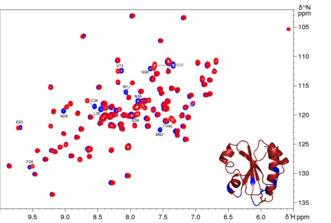
protein (DtrxH33G) was produced and purified as the wild type. The1H,15N HSQC spectrum was recorded and compared
with the Dtrx spectrum (supplemental Fig. S4). This compari-son shows that the mutation does not modify the structure of the protein because only residues close to the mutation undergo weak chemical shift variations. Next, we have deter-mined the activities and the redox potential of the DtrxH33G mutant. In the same experimental conditions as the wild type,
we have studied the ability of DtrxH33G to reduce insulin and, like Dtrx, DtrxH33G is unable to reduce insulin. It is thus not the presence of a histidine residue in the active site that induces this particular property of the protein. However, the DtrxH33G mutant shows a loss of the capacity of the protein to isomerize, or shuffle, incorrect disulfides of scrambled RNase A yielding 10% active RNase A only (Fig. 1B and supplemental Fig. S1). The histidine residue or the physicochemical properties
FIGURE 2. Titration of the redox potential of the disulfide bond of Dtrx and DtrxH33G. Changes in NMR signal intensities for the NH resonances of Cys34
under oxidizing or reducing conditions were reported in the function of half-cell potential of glutathione. Gray triangles and black circles represent signal intensity in the oxidized and reduced states, respectively. Redox potentials were calculated using the Nernst equation from the ratio of concentrations of reduced and oxidized glutathione. Experimental data were fitted against a sigmoidal decay (logistic) function.
FIGURE 3. pKadetermination of all Dtrx ionizable residues. A, 600 MHz two-dimensional CBCACO spectrum of reduced Dtrx at 298 K, pH 5.7, showing the cross-peaks for the C␣-CO and C-CO of all residues of the protein. Cross-peaks for the C␣-C␥and C-C␥of Asn and Asp, and for the C-C␦and C␥-C␦of Gln and Glu are also visible and connected by lines. The inset shows a close-up view of the pH-dependent chemical shift variations for the C-CO of Asp21(pH 4.9 (pink),
5.6 (red), 6.1 (orange), 6.6 (yellow), 7 (green), 7.8 (blue), and 9.3 (purple)). B, pKadetermination of the nucleophilic cysteine Cys31(black circle) and cysteine Cys34
(white circle) in the reduced form of Dtrx. C, pKadetermination of the histidine His
33in the oxidized form (white triangle) and reduced form (black circle) of Dtrx.
The pH-dependent chemical shift variation of the Ccarbons was measured, normalized, and fitted to one apparent pKavalue using the Henderson-Hasselbach
equation.
Structural Studies of Desulfothioredoxin
at CNRS on December 12, 2018
http://www.jbc.org/
induced by this histidine are essential for Dtrx to catalyze disul-fide isomerization. The determination of the redox potential of DtrxH33G shows effectively that His33plays an important role
in the Dtrx properties (Fig. 2). The redox potential of the active site cysteines at pH 7.0 for DtrxH33G (⫺226.4 ⫾ 0.6 mV) is 45 mV more reducing than for the wild type. This value is inter-mediate between Dtrx (⫺181.3 mV) and canonical Trx (⫺270 mV) redox potentials.
Dtrx Structure Analysis—From our studies, the activities, the redox potential, and the pKavalues of Dtrx are significantly
different from a canonical Trx. To gain insight into these atyp-ical properties of Dtrx and to propose a molecular catalytic mechanism, we have determined the three-dimensional struc-ture of the enzyme in the two redox states. The reduced and oxidized structures of Dtrx were calculated at pH 5.5 using interproton nOe-derived distance restraints in combination with the dihedral angle (21) and hydrogen bond restraints ( sup-plemental Table S1). We used the pKa values to define the
protonation state of the histidine and cysteine residues in the structure refinement. The resulting ensembles of solutions
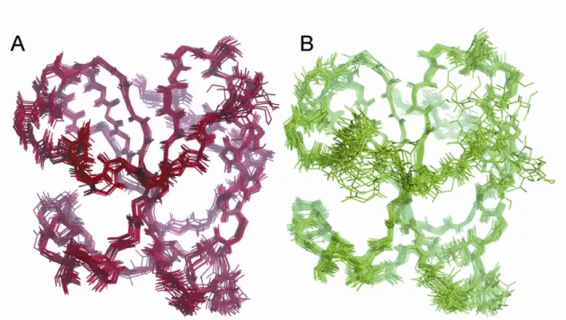
consisting of the 20 lowest energy structures of the oxidized and the reduced Dtrx are shown in supplemental Fig. S5. These ensembles have a backbone root mean square deviation relative to the average structure of 0.59⫾ 0.11 and 0.94 ⫾ 0.15 Å over the polypeptide chain for the oxidized and the reduced Dtrx, respectively. Detailed structure statistics are shown in supple-mental Table S1.
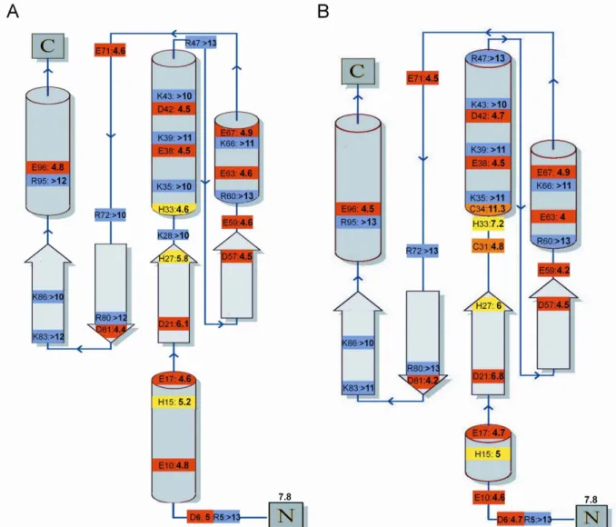
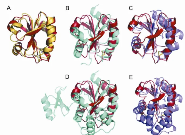
Oxidized and reduced Dtrx adopt a typical thioredoxin fold (3). The oxidized form consists of a four-stranded twisted central -sheet (residues 52–58 (1), 20–27 (2), 74–81 (3), and 83–90 (4)) surrounded by four ␣-helices (residues 12–17 (␣1), 33–46 (␣2), 61–69 (␣3), and 93–104 (␣4)) (Fig. 4A). The overall structure of the reduced Dtrx is similar to the one of the oxidized form. Dtrx displays high three-dimensional similarities to thioredoxins from other species such as E. coli Trx1 and the thioredoxin fold domain of other thiol disulfide oxidoreductases such as E. coli DsbA and DsbC (30, 31) (supplemental Fig. S6).
With the exception of the disulfide bond, the main structural differences between the two redox states of Dtrx concern the helix␣1 because of the number of restraints used in the
struc-FIGURE 4. Three-dimensional structure of Dtrx in both redox states. A, overlay of the three-dimensional solution structures of the reduced (green) and oxidized (red) forms of Dtrx calculated with CYANA (21). The NMR sample contained 1 mMprotein concentration (90% H2O, 10% D2O) in 100 mMNaCl, 50 mM
phosphate buffer, pH 5.7, at 290 K. For reduced Dtrx, the intramolecular disulfide bond was reduced by adding DTT to a final concentration of 10 mM, under argon atmosphere. The Dtrx typical thioredoxin fold is represented in cartoon, and the side chains of the cysteine residues are shown in sticks. B, local conformations of the active site in the reduced form of Dtrx. C, local conformations of the active site in the oxidized form of Dtrx. The active site residues Cys31,
Pro32, His33, and Cys34are shown and labeled. Sulfur atoms are shown in yellow, hydrogen atoms are in white, and nitrogen and oxygen atoms are in blue and
red, respectively. The figures were generated using the PyMOL Molecular Graphics System, Version 1.2r3pre, Schro¨dinger, LLC.
at CNRS on December 12, 2018
http://www.jbc.org/
ture calculation. The active site (31CPHC34) of Dtrx is formed
by a protruding loop between strand2 and the N termini of helix␣2. Between the two redox states, the cysteine side chain undergoes change in orientation (Fig. 4, B and C). In the dithiol form, the S atoms are at a mean distance of 4.8 Å. Moreover, a difference in orientation of the histidine His33is observed,
posi-tioning the cationic imidazole facing the thiolate anion of the reactive cysteine Cys31at a mean distance of 3.8 Å. In 14 of 20
conformers of the NMR structure, the thiolate anion of Cys31
forms two hydrogen bonds with the backbone amide and the HN␦of His33.
In the reduced Dtrx form, the Cys31side chain is exposed at
the protein surface, and the Cys34side chain is pointing toward the interior of the protein, as is the case in the active site of reduced E. coli Trx1. The electrostatic surface of Dtrx shows a different charge repartition compared with canonical Trx. Indeed, we observe a positive surface instead of the highly con-served hydrophobic patch found around the canonical active site (Fig. 5, A and B). The active site of Dtrx is perfectly super-imposable to the one of DsbA. Pro32and His33residues occupy
the same position when compared with the same residues in DsbA (1A2J) (Fig. 5C).
Thioredoxin and DsbA proteins generally contain the con-served residues Asp26and Glu24, respectively, which have been
described as involved in the activation of the second cysteine (Fig. 5D). As for DsbC, these residues are not found in Dtrx, and no acidic residue is found in the near environment of Cys34.
These structural data obtained on Dtrx active site and the strik-ing electrostatic surface potential around the active site strongly suggest a specific function of Dtrx in the anaerobe cytoplasm.
DISCUSSION
Properties of Dtrx—Based on sequence homologies, desulfo-thioredoxin (Dtrx) from D. vulgaris Hildenborough has been identified as a new member of the thioredoxin superfamily. Dtrx contains a particular active site consensus sequence, CPHC, found in the periplasmic DsbA. Dtrx does not show any disulfide reductase activity, being unable to reduce insulin. To date, no protein of the thioredoxin superfamily has shown
com-FIGURE 5. Comparative structural analysis of Dtrx. A, electrostatic surface potential representations of reduced Dtrx and reduced E. coli Trx, with blue representing basic residues, red representing acidic residues, and white representing neutral residues. The orientations of Dtrx and Trx are similar. The position of the sulfur atom of the N-terminal cysteine is indicated by a star. The hydrophobic patch observed in Trx is not conserved in Dtrx. Surface calculations were made using MolMol. B, superimposition of the redox active site of the reduced form of Dtrx (green) and E. coli DsbA (pink) in stick representation. The active site residues are labeled. C, sequence alignment of Dtrx, E. coli Trx, E. coli DsbA, and E. coli DsbC. Identical residues are in red boxes. Conserved residues are shown in red. The alignment was prepared with TCoffee (34) and ESPript (11).
Structural Studies of Desulfothioredoxin
at CNRS on December 12, 2018
http://www.jbc.org/
parable properties. According to the study of the DtrxH33G mutant, this property is not related to the particular active site of this protein. One explanation is the particular structural properties of Dtrx. The first step of the redox mechanism of Trx is the noncovalent interaction of the oxidized substrate with the hydrophobic surface of the active Trx (10). DsbA contains a helical insertion that forms a hydrophobic patch on the molec-ular surface, as well as a hydrophobic groove near the active site (32, 33). Rinaldi et al. (24) have observed a negative electrostatic surface of Xylella fastidiosa DsbA2, indicating that this protein versus DsbA has different substrate specificities. It is to be noticed that Dtrx contains a positive surface and no hydropho-bic patch around its active site. These differences are the main characteristic of Dtrx structure and may induce a strong spec-ificity for its substrate(s).
An in vitro assay involving refolding of scrambled RNase A showed an unusual disulfide isomerase activity of Dtrx. Consid-ering the nature and composition of the sequence in the active site, this activity can be explained. In fact, CXXC affects the standard redox potential of the particular proteins. In E. coli, the strongest reducing cytosolic Trx (CGPC) has a redox poten-tial of⌬E0⬘⫽ ⫺270 mV (25), and the strongest oxidizing agents,
the periplasmic DsbA (CPHC) and DsbC (CGYC), have a redox potential of⌬E0⬘⫽ ⫺122 mV (26). The redox potential value of human PDI (CGHC) is an intermediate,⌬E0⬘⫽ ⫺175 mV (27).
The redox potential observed for the cysteine active site of Dtrx at pH 7.0 is⫺181.3 mV. This characteristic shows that Dtrx is closer to oxidizing enzymes than canonical reductases and even closer to PDI isomerase. This property is in part due to the presence of histidine residue in the active site. Indeed, DtrxH33G lost the ability to catalyze the disulfide isomeriza-tion and presents a redox potential closer to disulfide reducta-ses (⫺226.4 mV).
The pKavalues of 4.8 and 11.3 obtained respectively for the
thiol group of Cys31and Cys34residues, are different from those found in all canonical Trx. In E. coli Trx1, the pKaof the thiol
group is 7.5 for the first cysteine and 9.5 for the second cysteine in the consensus site (11). The pKaof this thiol group reflects the stabilization of the thiolate anion of the accessible cysteine residue. Strong stabilization of the thiolate generates a low pKa
value and low stability for the disulfide bond; this phenomenon was observed for Dtrx. The instability of the disulfide bond is characteristic of oxidase or isomerase proteins because it is nec-essary for their catalytic mechanism (28). In summary, these physicochemical properties of Dtrx are in agreement with the unusual disulfide isomerase activity found in in vitro assays for Dtrx.
However, in vivo, Dtrx did not present any isomerase activity in the E. coli cytoplasm, because it has been observed for DsbA. Indeed, Xu et al. (23) showed that a functional expression of PalB in the cytoplasm of E. coli appeared to be limited by disul-fide bond formation, and a DsbA fusion tag was functional to enhance PalB folding in cytoplasm. Despite the observed in vitrodisulfide isomerase activity, Dtrx was not able to perform this activity in vivo in E. coli cytoplasm. Is it a problem of cyto-plasmic conditions or of substrate specificity?
The cytoplasm of most organisms is a highly reducing envi-ronment, in which protein cysteines are maintained in thiol/
thiolate form. However, in extremophile organisms, disulfide bond formation in cytosolic proteins is supposed to increase their thermodynamic stability. Several protein-disulfide oxi-doreductases in the disulfide-rich thermophiles were identified and could have dual functions in the cytoplasm, as oxidase and isomerase (29). Therefore we can suppose that oxidase activity could exist in the cytoplasm. Dtrx contains a positive surface and no hydrophobic patch around its active site. These differ-ences are the main characteristic of Dtrx structure and may induce a strong specificity for its substrate(s) and explain the absence of oxidation of lipase in vivo, where the ratio Dtrx: lipase is 1:1 in the cell.
Is Dtrx Really a Thioredoxin?—Considering the absence of in vitroreductase activity of Dtrx and its oxidizing physicochem-ical properties (redox potential, pKavalues of the active site), a
potential thioredoxin function is questionable. Nevertheless, glutaredoxin, which is a good reducing agent, was also reported to have oxidizing redox properties provided by its low pKaof
the active cysteine (30). Conversely, thioredoxins, which are potent reducing agents, can also act as oxidizing agents under particular conditions. Indeed, E. coli thioredoxin can be trans-located to the periplasm and then partially replaces the activity of DsbA in promoting the formation of disulfide bonds (31). Thioredoxin can function as a reductase or oxidase or isomer-ase, mainly depending on the redox environment.
For Dtrx, several aspects support a reductase activity: (i) the three-dimensional structure of Dtrx revealed an identical fold to canonical thioredoxins without a supplementary domain; (ii) the gene DVU_0378 encoding Dtrx is included in a polycis-tronic unit counting nine other genes, including DVU_0377, which encodes a thioredoxin reductase (desulfothioredoxin reductase) and is able to reduce Dtrx but not any other thiore-doxin (14); and (iii) Dtrx has a reductase activity on dithiobis-(nitrobenzoic acid) substrate (14).
Catalytic Mechanism of Dtrx—It is the first time that the pKa
values of the histidine of CPHC active site were determined. These atypical values vary from 7.2 to 4.6 for the reduced to the oxidized Dtrx, respectively.
On the basis of these results, we propose a catalytic mecha-nism for the reduction of the oxidized substrate by the reduced Dtrx (Fig. 6). The nucleophilic cysteine in the reduced Dtrx is deprotonated by water molecules because of its low pKa(4.8).
The Cys31thiolate of Dtrx nucleophilically attacks a disulfide sulfur atom of the substrate, leading to the formation of the so-called mixed disulfide intermediate in which Dtrx and sub-strate are covalently bound via a new disulfide bond. Next, the Cys34thiol group of Dtrx is deprotonated. However, the
mech-anism through which the buried Cys34 is deprotonated and
activated to perform the intramolecular nucleophilic attack on Cys31is still uncertain in canonical Trx. One hypothesis is that
an aspartic acid conserved in all canonical Trx is responsible for this deprotonation (32). It is to be noticed that a double alanine mutation of the conserved Asp26and Lys57in Trx and Glu24
and Lys58in DsbA did not change the pK
aof cysteines in both
enzymes. These data support the hypothesis that these residues are not involved in the properties of the dithiol active center (33). A second hypothesis involves the thiolate leaving group, which establishes S⫺…H-S hydrogen bonds with the thiol
at CNRS on December 12, 2018
http://www.jbc.org/
group of the buried cysteines (12). However, the mutation of the second cysteine of the substrate shows a dissociation of the intermolecular complex (13). Finally, in a last hypothesis, the cysteine is proposed to be activated for its nucleophilic attack by hydrogen bonds between this residue and the backbone amides of the tryptophan active site and of the N-terminal cys-teine (13). These hydrogen bonds explain the stabilization of thiolate group but not the deprotonation of this group. In Dtrx, the aspartic acid is not conserved, and analysis of pKavalue of
all residues does not present any acid group available for the activation of Cys34. Therefore, we centered the mechanism
around the second hypothesis where the thiolate of the sub-strate attacks the Cys34sulfur atom or any acid group of the
substrate. The protonated His33would stabilize by a salt bridge
successively Cys31and Cys34thiolates. In the second step, Cys34 thiolate would attack the Cys31 sulfur atom involved in the
disulfide bond, causing the rupture of the latter. At the same time, the imidazole group in the oxidized Dtrx being deproto-nated (pKa4.6), we propose that the thiolate of the reduced
substrate would attack His33and induce the release of the
prod-ucts of the reaction, an oxidized enzyme with deprotonated His33and a reduced dithiol substrate.
As for all thioredoxins, this mechanism is reversible. DsbA contains the same CPHC sequence motif, but no data are avail-able on the pKavalues for the imidazole group of this protein.
We suggest that the protonation state could be identical to Dtrx. In DsbA catalysis, the two-step bimolecular nucleophilic substitution mechanism described in the literature (34) can be completed by a first step substrate activation. In this case, the imidazole group of histidine being deprotonated (pKa 4.6)
attacks the thiol group of the reduced substrate and induces the activation of the substrate with the formation of a thiolate cys-teine. Effectively, in E. coli, it is unlikely that all of the 300 potential DsbA substrates present reactive cysteines (low pKa).
They most likely have to be activated by the deprotonated active site histidine of DsbA. In DsbA, mutations of the histi-dine located at the active site, diminished drastically the oxidase activity affecting the destabilization of the oxidized form of the enzyme (28, 35). According to the authors, the mutation affects the destabilization of the oxidized form of the enzyme. We think that the histidine mutant will affect not only the stability of disulfide bond but especially the activation of the substrates. In conclusion, the new thioredoxin Dtrx from anaerobe D. vulgaris Hildenborough presents several unusual structural
FIGURE 6. Model of the Dtrx catalytic mechanism. Substrate reduction (solid lines) by Dtrx occurs in four steps. In the first step, the Cys31thiolate of Dtrx
nucleophilically attacks sulfur atom of the substrate disulfide. In the second step, the thiolate of the substrate produces a base attack on Cys34sulfur atom. Next,
the Cys34thiolate attacks the Cys31sulfur atom involved in the disulfide bond, causing the rupture of the latter. At the same time, the imidazole group of His33
is deprotonated by a base attack of the thiolate of the reduced substrate. This reaction produces an oxidized Dtrx and a reduced dithiol substrate. Substrate oxidation (dotted lines) by Dtrx occurs in four steps. First, the deprotonated His33produces a base attack on the first cysteine of the substrate. After, this
activated cysteine of the substrate nucleophilically attacks the sulfur atom of Cys31of the oxidized Dtrx, leading to the formation of a disulfide-linked complex
between Dtrx and the substrate. In the next step, the second cysteine of the substrate is deprotonated probably by the Cys34thiolate of Dtrx and attacks the
sulfur atom of the substrate cysteine, which is disulfide-bonded with Cys31of Dtrx. This reaction results in the formation of a disulfide bond in the substrate and
the reduction of Dtrx with a protonated His33.
Structural Studies of Desulfothioredoxin
at CNRS on December 12, 2018
http://www.jbc.org/
features and contains a particular active site consensus sequence, CPHC, which gives oxidizing properties. Our data suggest a particular specificity for its substrates, and one cannot exclude that this specificity may be limited to one of the gene products of the operon it belongs to. Further studies of these gene products will be essential to determine the physiological function of Dtrx. The knowledge of the protonation state for the residues involved in the reaction mechanism is highly important, and the pKavalues for all residues of the protein were determined for the first time. pKavalue determination
allowed us to define the important function of the histidine residue at the catalytic site. Genome analysis of many bacteria reveals unusual thioredoxins. The structural and functional studies of such atypical systems will give new insights into TDOR catalytic mechanism.
Acknowledgments—We thank Dr. A. Dolla and Dr. B. Burlat for help-ful discussions and Dr. P. Barre´ for critical reading of the manuscript.
REFERENCES
1. Holmgren, A., and Bjo¨rnstedt, M. (1995) Thioredoxin and thioredoxin reductase. Methods Enzymol. 252, 199 –208
2. Rietsch, A., and Beckwith, J. (1998) The genetics of disulfide bond metab-olism. Annu. Rev. Genet 32, 163–184
3. Martin, J. L. (1995) Thioredoxin: a fold for all reasons. Structure 3, 245–250
4. Mo¨ssner, E., Huber-Wunderlich, M., and Glockshuber, R. (1998) Charac-terization of Escherichia coli thioredoxin variants mimicking the active-sites of other thiol/disulfide oxidoreductases. Protein Sci. 7, 1233–1244 5. Aslund, F., Berndt, K. D., and Holmgren, A. (1997) Redox potentials of
glutaredoxins and other thiol-disulfide oxidoreductases of the thioredoxin superfamily determined by direct protein-protein redox equilibria. J. Biol.
Chem. 272,30780 –30786
6. Chambers, J. E., Tavender, T. J., Oka, O. B., Warwood, S., Knight, D., and Bulleid, N. J. (2010) The reduction potential of the active site disulfides of human protein disulfide isomerase limits oxidation of the enzyme by Ero1␣. J. Biol. Chem. 285, 29200–29207
7. Kersteen, E. A., and Raines, R. T. (2003) Catalysis of protein folding by protein disulfide isomerase and small-molecule mimics. Antioxid. Redox
Signal. 5,413– 424
8. Quan, S., Schneider, I., Pan, J., Von Hacht, A., and Bardwell, J. C. (2007) The CXXC motif is more than a redox rheostat. J. Biol. Chem. 282, 28823–28833
9. Capitani, G., Rossmann, R., Sargent, D. F., Gru¨tter, M. G., Richmond, T. J., and Hennecke, H. (2001) Structure of the soluble domain of a membrane-anchored thioredoxin-like protein from Bradyrhizobium japonicum re-veals unusual properties. J. Mol. Biol. 311, 1037–1048
10. Holmgren, A. (1985) Thioredoxin. Annu. Rev. Biochem. 54, 237–271 11. Jeng, M. F., Holmgren, A., and Dyson, H. J. (1995) Proton sharing between
cysteine thiols in Escherichia coli thioredoxin: implications for the mech-anism of protein disulfide reduction. Biochemistry 34, 10101–10105 12. Carvalho, A. T., Swart, M., van Stralen, J. N., Fernandes, P. A., Ramos, M. J.,
and Bickelhaupt, F. M. (2008) Mechanism of thioredoxin-catalyzed disul-fide reduction: activation of the buried thiol and role of the variable active-site residues. J. Phys. Chem. B 112, 2511–2523
13. Roos, G., Foloppe, N., Van Laer, K., Wyns, L., Nilsson, L., Geerlings, P., and Messens, J. (2009) How thioredoxin dissociates its mixed disulfide. PLoS
Comput. Biol. 5,e1000461
14. Pieulle, L., Stocker, P., Vinay, M., Nouailler, M., Vita, N., Brasseur, G., Garcin, E., Sebban-Kreuzer, C., and Dolla, A. (2011) Study of the thiol/ disulfide redox systems of the anaerobe Desulfovibrio vulgaris points out pyruvate:ferredoxin oxidoreductase as a new target for thioredoxin 1.
J. Biol. Chem. 286,7812–7821
15. Garcin, E. B., Bornet, O., Pieulle, L., Guerlesquin, F., and Sebban-Kreuzer, C. (2010)1H,13C and15N backbone and side-chain chemical shift
assign-ments for oxidized and reduced desulfothioredoxin. Biomol. NMR Assign. 4,135–137
16. Holmgren, A. (1979) Thioredoxin catalyzes the reduction of insulin dis-ulfides by dithiothreitol and dihydrolipoamide. J. Biol. Chem. 254, 9627–9632
17. Hillson, D. A., Lambert, N., and Freedman, R. B. (1984) Formation and isomerization of disulfide bonds in proteins: protein disulfide-isomerase.
Methods Enzymol. 107,281–294
18. Bertini, I., Felli, I. C., Gonnelli, L., Pierattelli, R., Spyranti, Z., and Spyrou-lias, G. A. (2006) Mapping protein-protein interaction by13C-detected
heteronuclear NMR spectroscopy. J. Biomol. NMR 36, 111–122 19. Jeng, M. F., and Dyson, H. J. (1996) Direct measurement of the aspartic
acid 26 pKa for reduced Escherichia coli thioredoxin by 13C NMR.
Bio-chemistry 35,1– 6
20. Keller, R. L. J. (2004) Computer aided resonance assignment tutorial.
Cantina
21. Cornilescu, G., Delaglio, F., and Bax, A. (1999) Protein backbone angle restraints from searching a database for chemical shift and sequence ho-mology. J. Biomol. NMR 13, 289 –302
22. Shouldice, S. R., Cho, S. H., Boyd, D., Heras, B., Eser, M., Beckwith, J., Riggs, P., Martin, J. L., and Berkmen, M. (2010) In vivo oxidative protein folding can be facilitated by oxidation-reduction cycling. Mol. Microbiol. 75,13–28
23. Xu, Y., Yasin, A., Tang, R., Scharer, J. M., Moo-Young, M., and Chou, C. P. (2008) Heterologous expression of lipase in Escherichia coli is limited by fold-ing and disulfide bond formation. Appl. Microbiol. Biotechnol. 81, 79 – 87 24. Rinaldi, F. C., Meza, A. N., and Guimara˜es, B. G. (2009) Structural and
biochemical characterization of Xylella fastidiosa DsbA family members: new insights into the enzyme-substrate interaction. Biochemistry 48, 3508 –3518
25. Krause, G., Lundstro¨m, J., Barea, J. L., Pueyo de la Cuesta, C., and Hol-mgren, A. (1991) Mimicking the active site of protein disulfide-isomerase by substitution of proline 34 in Escherichia coli thioredoxin. J. Biol. Chem. 266,9494 –9500
26. Wunderlich, M., and Glockshuber, R. (1993) Redox properties of protein disulfide isomerase (DsbA) from Escherichia coli. Protein Sci. 2, 717–726 27. Lundstro¨m, J., and Holmgren, A. (1993) Determination of the reduction-oxidation potential of the thioredoxin-like domains of protein disulfide-isomerase from the equilibrium with glutathione and thioredoxin.
Bio-chemistry 32,6649 – 6655
28. Grauschopf, U., Winther, J. R., Korber, P., Zander, T., Dallinger, P., and Bardwell, J. C. (1995) Why is DsbA such an oxidizing disulfide catalyst?
Cell 83,947–955
29. Ladenstein, R., and Ren, B. (2006) Protein disulfides and protein disulfide oxidoreductases in hyperthermophiles. FEBS J. 273, 4170 – 4185 30. Fernandes, A. P., and Holmgren, A. (2004) Glutaredoxins:
glutathione-de-pendent redox enzymes with functions far beyond a simple thioredoxin backup system. Antioxid. Redox. Signal. 6, 63–74
31. Huber, D., Cha, M. I., Debarbieux, L., Planson, A. G., Cruz, N., Lo´pez, G., Tasayco, M. L., Chaffotte, A., and Beckwith, J. (2005) A selection for mu-tants that interfere with folding of Escherichia coli thioredoxin-1 in vivo.
Proc. Natl. Acad. Sci. U.S.A. 102,18872–18877
32. Chivers, P. T., and Raines, R. T. (1997) General acid/base catalysis in the active site of Escherichia coli thioredoxin. Biochemistry 36, 15810 –15816 33. Carvalho, A. T., Fernandes, P. A., and Ramos, M. J. (2006) Determination of the DeltapKa between the active site cysteines of thioredoxin and DsbA.
J. Comput. Chem. 27,966 –975
34. Kadokura, H., and Beckwith, J. (2010) Mechanisms of oxidative protein folding in the bacterial cell envelope. Antioxid. Redox Signal. 13, 1231–1246
35. Guddat, L. W., Bardwell, J. C., Glockshuber, R., Huber-Wunderlich, M., Zander, T., and Martin, J. L. (1997) Structural analysis of three His32 mutants of DsbA: support for an electrostatic role of His32 in DsbA sta-bility. Protein Sci. 6, 1893–1900
at CNRS on December 12, 2018
http://www.jbc.org/
Françoise Guerlesquin and Corinne Sebban-Kreuzer
Edwige B. Garcin, Olivier Bornet, Latifa Elantak, Nicolas Vita, Laetitia Pieulle,
Structural and Mechanistic Insights into Unusual Thiol Disulfide Oxidoreductase
doi: 10.1074/jbc.M111.288316 originally published online November 28, 2011 2012, 287:1688-1697.
J. Biol. Chem.
10.1074/jbc.M111.288316
Access the most updated version of this article at doi: Alerts:
When a correction for this article is posted
•
When this article is cited
•
to choose from all of JBC's e-mail alerts
Click here
Supplemental material:
http://www.jbc.org/content/suppl/2011/11/28/M111.288316.DC1 http://www.jbc.org/content/287/3/1688.full.html#ref-list-1This article cites 34 references, 7 of which can be accessed free at
at CNRS on December 12, 2018
http://www.jbc.org/
S-1
Edwige B. Garcin, Olivier Bornet, Latifa Elantak, Nicolas Vita, Laetitia Pieulle, Françoise
Guerlesquin and Corinne Sebban-Kreuzer
Table of contents
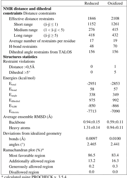
Table S1: NMR and refinement statistics for Dtrx structures. ... S-2
Figure S1: Scrambled RNaseA (ScRNase) refolding assay: the cleavage of RNA by RNaseA
was followed by 1D
1H-NMR. ... S-3
Figure S2: Titration of the redox potential of the disulfide bond of Dtrx. ... S-4
Figure S3: pKa of ionisable residues of reduced and oxidized Dtrx. ... S-5
Figure S4: Superimposition of
1H,
15N-HSQC spectra of Dtrx and DtrxH33G ... S-6
Figure S5: Solution structures of the reduced and oxidized forms of Dtrx. ... S-7
Figure S6: Structural comparisons. ... S-8
S-2
Table S1: NMR and refinement statistics for Dtrx structures. Structural statistics and restraint
violations of the 20 selected structures representative of Dtrx in solution at pH 5.7 and 290K.
Reduced
Oxidized
NMR distance and dihedral
constraints Distance constraints
Effective distance restraints
1846
2108
Short range
(|i-j|
≤ 1)
1152
1261
Medium range
(1 < |i-j| < 5)
276
415
Long range
(|i-j|
≥ 5)
418
432
Average number of restraints per residue
17
19
H-bond restraints
48
70
Dihedral angle restraints from TALOS
156
156
Structures statistics
Restraint violations
Distance >0,5Å
0
1
Dihedral >5°
0
5
Energies (kcal/mol)
E
total-2951
-2853
E
bond58
57
E
angle338
349
E
dihedral975
992
E
VdW-850
-866
E
electric-7713
-7090
Average ensemble RMSD (Å)
Backbone
0.94±0.15
0.59±0.11
Heavy atoms
1.31±0.14
0.94±0.11
Deviations from idealized geometry
bonds (Å)
0.0097
0.0100
angles (°)
2.465
2.441
Ramachandran plot (%)*
Most favorable region
86.5
83.4
Additionally allowed region
13.2
16.3
Generously allowed region
0.2
0.3
Disallowed region
0.0
0.0
S-3
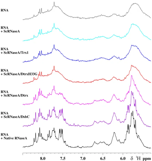
Figure S1: Scrambled RNaseA (ScRNase) refolding assay: the cleavage of RNA by RNaseA was
followed by 1D
1H-NMR.
1D 1H-NMR spectra of RNA with different mixture proteins: ScRNaseA (cyan), Trx1 and ScRNaseA
(blue), DtrxH33G and ScRNaseA (red), Dtrx and ScRNaseA (pink), DsbC and ScRNaseA (purple),
Native RNaseA (black). The assay was performed at 298K on a Bruker Avance III DRX 500 MHz
spectrometer after 30 min of reaction. The NMR sample of RNA was 2 mg/ml in phosphate buffer 100
mM at pH 7 and 10 % D
2O. For the determination of the percentage of the RNaseA activity of native
RNaseA and reshuffling of ScRNaseA after incubation with proteins, the mean intensity of several
isolated peaks in RNA spectrum was used relative to RNA spectrum in the presence of native
RNaseA. RNA spectrum (grey) in presence of scrambled RNaseA is used as blank.
S-4
Figure S2: Titration of the redox potential of the disulfide bond of Dtrx. Superimposition of the
1