HAL Id: hal-03030880
https://hal-cnrs.archives-ouvertes.fr/hal-03030880
Submitted on 3 Dec 2020
HAL is a multi-disciplinary open access
archive for the deposit and dissemination of
sci-entific research documents, whether they are
pub-lished or not. The documents may come from
teaching and research institutions in France or
abroad, or from public or private research centers.
L’archive ouverte pluridisciplinaire HAL, est
destinée au dépôt et à la diffusion de documents
scientifiques de niveau recherche, publiés ou non,
émanant des établissements d’enseignement et de
recherche français ou étrangers, des laboratoires
publics ou privés.
Surface Zwitterionization of Expanded
Poly(tetrafluoroethylene) via Dopamine-Assisted
Consecutive Immersion Coating
Peter Matthew Paul T. Fowler, Gian Vincent Dizon, Lemmuel Tayo, Alvin
Caparanga, James Huang, Jie Zheng, Pierre Aimar
To cite this version:
Peter Matthew Paul T. Fowler, Gian Vincent Dizon, Lemmuel Tayo, Alvin Caparanga, James Huang,
et al.. Surface Zwitterionization of Expanded Poly(tetrafluoroethylene) via Dopamine-Assisted
Con-secutive Immersion Coating. ACS Applied Materials & Interfaces, Washington, D.C. : American
Chemical Society, 2020, 12 (37), pp.41000-41010. �10.1021/acsami.0c09073�. �hal-03030880�
Any correspondence concerning this service should be sent
to the repository administrator: [email protected]
This is an author’s version published in:
http://oatao.univ-toulouse.fr/26986
To cite this version: Fowler, Peter Matthew Paul T. and Dizon,
Gian Vincent and Tayo, Lemmuel L. and Caparanga, Alvin R.
and Huang, James and Zheng, Jie and Aimar, Pierre and
Chang, Yung Surface Zwitterionization of Expanded
Poly(tetrafluoroethylene) via Dopamine-Assisted Consecutive
Immersion Coating. (2020) ACS Applied Materials and
Interfaces, 12 (37). 41000-41010. ISSN 1944-8244
Official URL
DOI : https://doi.org/10.1021/acsami.0c09073
Open Archive Toulouse Archive Ouverte
OATAO is an open access repository that collects the work of Toulouse
researchers and makes it freely available over the web where possible
Surface Zwitterionization of Expanded Poly(tetrafluoroethylene) via
Dopamine-Assisted Consecutive Immersion Coating
Peter Matthew Paul T. Fowler,
○Gian Vincent Dizon,
○Lemmuel L. Tayo, Alvin R. Caparanga,
James Huang, Jie Zheng, Pierre Aimar, and Yung Chang
*
ABSTRACT: Expanded polytetrafluoroethylene (ePTFE) is one of the materials widely used in the biomedical field, yet its application is being limited by adverse reactions such as thrombosis when it comes in contact with blood. Thus, a simple and robust way to modify ePTFE to be biologically inert is sought after. Modification of ePTFE without high-energy pretreatment, such as immersion coating, has been of interest to researchers for its straightforward process and ease in scaling up. In this study, we utilized a two-step immersion coating to zwitterionize ePTFE membranes. The first coating consists of the co-deposition of polyethylenimine (PEI) and polydopamine (PDA) to produce amine groups in the surface of the ePTFE for further functionalization. These amine groups from PEI will be coupled with the epoxide group of the zwitterionic copolymer, poly(GMA-co-SBMA) (PGS), via a ring-opening reaction in the second coating. The coated ePTFE membranes were physically
and chemically characterized to ensure that each step of the coating is successful. The membranes were also tested for their thrombogenicity via quantification of the blood cells attached to it during contact with biological solutions. The coated membranes exhibited around 90% reduction in attachment with respect to the uncoated ePTFE for both Gram-positive and Gram-negative strains of bacteria (Staphylococcus aureus and Escherichia coli). The coating was also able to resist blood cell attachment from human whole blood by 81.57% and resist red blood cell attachment from red blood cell concentrate by 93.4%. These ePTFE membranes, which are coated by a simple immersion coating, show significant enhancement of the biocompatibility of the membranes, which shows promise for future use in biological devices.
KEYWORDS: expanded polytetrafluoroethylene, dopamine, polyethylenimine, sulfobetaine methacrylate, glycidyl methacrylate
■
INTRODUCTIONPolytetrafluoroethylene (PTFE) is an interesting fluoropol-ymer in that its chemical nature, particularly the strength of its carbon−fluorine bonds and the smooth protective electron sheath provided by the fluorine atoms, results in exceptional chemical inertness, high thermal stability, and wear resistance.1 When subjected to anisotropic expansion, the resulting expanded PTFE (ePTFE) retains most of the desirable properties of bulk PTFE while introducing pores of control-lable size, known to imbue the polymer with additional strength and flexibility as well as potential for tissue and blood vessel in-growth.2These characteristics have made ePTFE the most commonly used fluoropolymer in medical applications, particularly in large-diameter, high-flow vascular grafts,3,4 and hemodialysis access grafts.5
Though chemically inert, ePTFE is not biologically inert, and this has severely limited its applications. For instance, small-diameter grafts made of ePTFE have not been successful in vivo due to the high thrombogenicity of the material and poor endothelialization,6,7 resulting from excessive serum protein and platelet adhesion;8 this poses the limit of only
using medium- to large-diameter ePTFE grafts to prevent occlusion by thrombi and subsequent complications. Bacterial infections have also been a pressing issue in ePTFE grafts and surgical meshes,9−11with Gram-positive Staphylococcus aureus being responsible for majority of these cases.12 This has been attributed to both the hydrophobicity of the surface and the accessibility of the micropores in the material.13 Most recent attempts at improving the biocompatibility of ePTFE are generally divided into two approaches: seeding endothelial cells in vitro or encouraging their in-growth in vivo,14−17 or surface modification by grafting molecules that confer biocompatibility.18−23 Surface modification is also used to encourage endothelialization.24
To modify the surface of ePTFE membranes, processes that are energetic enough to lyse the strong C−F bonds or introduce reactive groups have been used extensively. These include plasma treatment, atomic layer deposition, and irradiation by ions, γ rays, or UV light. All of these have the common features of complexity, expensive and delicate instrumentation, and high energy input.25,26 Wet-chemical modification, where the substrates are simply immersed in a reactive solution, has gained much attention recently primarily due to the need for more facile processes of surface modification, despite coming with the disadvantage of producing potentially toxic waste solutions; in the light of energy consumption and process simplicity, it is however a desired option. Previous approaches have used strong solutions such as oxidizing mixtures of nitric acid and potassium permanganate that introduced more reactive hydroxyl (C− OH) and carbonyl (CO) groups.27,28 More recently, biomimetic mussel-inspired approaches based on dopamine self-polymerization and adhesion have been successful at introducing various molecules onto the otherwise nonstick ePTFE surface.29 Progress since this discovery consists of altering the solvent for the coating solution,30co-deposition or conjugation of other molecules with dopamine, dopamine coating followed by grafting of a second coating via reactive groups in polydopamine,31or the combination of the last two methods. For example, dopamine−hyaluronic acid conjugates were successfully coated onto PTFE under mild conditions at room temperature and mildly basic pH, increasing its hydrophilicity and resistance to protein and macrophage cell adhesion and scarring in mouse tissue in vivo.32 Polyethyle-neimine (PEI) has also been co-deposited with dopamine to form a polydopamine/PEI (PDA/PEI) coating on porous ePTFE for use in microfiltration. This also improved the hydrophilicity and water flux through the membrane. More-over, the coating was found to be stable in a wide range of pH values.33 In a related study, a structurally similar compound, catechol, was used instead of dopamine. This was conjugated to PEI, then coated onto PTFE. A second step introduced a silane group by covalent linkage between the first layer and the reactive epoxy group of the silane-containing compound. This endowed the membrane with superior hydrophilicity, allowing higher water fluxes in their oil−water separation experiments.34 A similar approach used gallic acid (3,4,5-trihydroxybenzoic acid) instead, conjugated to PEI with subsequent covalent attachment of a silane moiety via the nucleophilic opening of an epoxy group. In addition to increased hydrophilicity, the coating was also effective against Escherichia coli and S. aureus.35 Despite the multitude of studies on this fast and
robust method of modifying ePTFE surfaces for increased hydrophilicity and antibacterial properties, no such effort has been made in studying the hemocompatibility of ePTFE membranes modified this way.
We present in this study a facile and low-energy two-step surface zwitterionization procedure using a mussel-inspired dopamine dip-coating approach that could successfully convert the hydrophobic ePTFE surface into a highly hydrophilic one with enhanced biocompatibility and that could be potentially used to improve the performance of ePTFE in biomedical applications. We tested different PEI molecular weights to determine an optimum coating procedure for the greatest improvement in biocompatibility. The modified membranes were tested for their hydrophilic character, surface morphol-ogy, surface chemistry, and resistance to fouling by bacterial and blood cell solutions in attachment tests. We have also attempted to provide meaningful correlations that could be referred to in future designs for hemocompatible biomaterials. To the best of our knowledge, this is a pioneering study on using a low-energy mussel-inspired coating procedure on a highly hydrophobic material to achieve both antibacterial property and hemocompatibility by surface zwitterionization.
■
EXPERIMENTAL SECTIONMaterials and Reagents.Chemicals such as branched PEI (0.6 and 10 kDa), dopamine hydrochloride, Tris base, and 2,2′-azobis-(2-methylpropionitrile) (AIBN) were purchased from Sigma-Aldrich; branched PEI (1.8 kDa) was purchased from Alfa Aesar; glycidyl methacrylate (GMA) was purchased from Tokyo Chemical Industry Co., Ltd.; 25 wt % aqueous solution of glutaraldehyde was purchased from Acros Organics and was diluted to 2.5 wt % with deionized water before use; and 1 M hydrochloric acid (HCl) was bought from Honeywell Fluka. Sulfobetaine methacrylate (SBMA) monomer was synthesized in accordance with Yue et al.36The ePTFE membranes
were purchased from EF-Materials Industries, Inc. HPLC-grade solvents such as methanol, n-butanol, and isopropanol (IPA) were purchased from Aencore Chemical Pty., Ltd. and Echo Chemical Co., Ltd. Phosphate-buffered saline (PBS) was first diluted to 1x from 10x concentration before use.
Preparation of ePTFE Membranes. The ePTFE membranes were cut into 0.85 cm disks and immersed in IPA to wash the membranes from dust and other dirt. The washing process was performed by putting the membranes under sonication three times for 15 min each time with replacement of the isopropanol every time. The membranes were then stored at 4 °C in IPA until further use.
Synthesis of Poly(GMA-co-SBMA). Poly(GMA-co-SBMA), which will be denoted as PGS, was synthesized according to the protocol of Chou et al.,37 where an optimal GMA/SBMA ratio of 20:80 was used. Briefly, predetermined amounts of the monomers were dissolved: SBMA in deionized water, and GMA in methanol.
These two solutions were then mixed, followed by addition of AIBN as a free-radical initiator. The solution was stirred for at least 20 min to ensure that the solution is completely homogeneous. The reaction solution was then placed in an oil bath controlled at 60 °C, and the reaction was kept running for 6 h. Polymerization was thermally terminated by transfer of the reaction flask to an ice bath. The copolymer was purified with methanol at least three times via reprecipitation. It was then dried under vacuum overnight to remove the bound solvent. Finally, freeze drying and grinding yielded the white crystalline PGS powder.
Coating with Dopamine and PEI.The overall coating process is shown inScheme 1. Tris buffer was initially prepared by dissolving Tris base in deionized water to a final concentration of 50 mM and then adjusting the pH of the solution to 8.5 using HCl. Dopamine and branched PEI were dissolved in the Tris buffer with concentrations of 2 and 1 mg/mL, respectively. This dopamine/PEI mass ratio has been adopted from Lv et al., whose study has shown that it was the optimum ratio based on the thickness of the coating produced.38 Upon dissolution, the dopamine immediately started undergoing self-polymerization into polydopamine (PDA), reacting also with PEI to form PDA/PEI conjugates. The ePTFE disks were immersed in a freshly prepared PDA/PEI coating solution in 2 mL Eppendorf tubes. The tubes were kept in a 37 °C oven for 24 h. Subsequently, the membranes were removed from the solution and washed three times with deionized water. Three different PEI molecular weights were used in this study: 0.6, 1.8, and 10 kDa.
Zwitterionization of ePTFE with PGS Copolymer.The coating solution for the zwitterionization process was prepared by dissolving the PGS copolymer in deionized water preheated to 60 °C, to a concentration of 10 mg/mL. The PDA/PEI-coated ePTFE disks were immediately immersed in the PGS coating solution immediately after washing with deionized water. The zwitterionization of the ePTFE disks was done and kept at 60 °C for 24 h. Membranes coated with only the PDA/PEI coating solution were labeled as D-P600, D-P1800, and D-P10000; membranes coated with both PDA/PEI and PGS coating solutions were labeled as P600-GS, P1800-GS, and D-P10000-GS. The coated samples were then stored immersed in deionized (DI) water until further use.
Physical Characterization of Coated Membranes. Coating density (mg/cm2) was measured as the difference in dry mass before
and after coating, normalized to the external surface area of each membrane. Water contact angle measurements were done with a DataPhysics OCA15EC goniometer to assess the surface hydro-philicity of the virgin and coated membranes. A 4 μL droplet was slowly added to the surface of each membrane. A dynamic measurement of the water contact angle was performed, keeping track of the change in contact angle while the droplet is being absorbed by the membrane. Measurements were done in triplicate for each condition. Differences in the slopes of the curves directly relate to hydrophilicity of the coatings. In conjunction with this, hydration tests were also performed to assess the hydrophilicity of the internal microstructure of the modified membranes. Preweighed samples were immersed in 1 mL of deionized water and stored for 24 h. The membranes were then gently pat-dried with laboratory-grade clean wipes to remove water adhered to the surface and weighed immediately; the difference in mass was assumed to be the mass of water absorbed by the membrane. Normalizing the water absorbed by the membrane to the membrane surface area gave hydration capacity (mg/cm2). A porosity test was performed to determine if the overall
procedure has affected the porosity of ePTFE, following a similar procedure except that the membranes were immersed in 1 mL of n-butanol. The alcohol was assumed to fill up the pores within the membrane. The following formula was used to estimate the porosity of the membranes ρ ρ ρ ρ = − − − m m m m porosity (%) ( ) ( ) M W D M W S M D
where ρMis the bulk density of the membrane (PTFE, 2.2 mg/cm3),
ρSis the density of the solvent used (n-butanol, 0.81 mg/cm3), and
mW and mD are the wet and dry masses of the membranes,
respectively. In both the hydration and porosity tests, triplicate measurements were performed, and the data obtained were averaged. Changes in the surface and internal microstructure were investigated by visualizing the surface and cross sections, respectively, using a scanning electron microscope (SEM). The samples were sputtered with gold and then visualized with a Hitachi S-3000 SEM at an accelerating voltage of 3 keV.
Surface Chemical Characterization of Coated Membranes. Coated membrane samples were subjected to X-ray photoelectron spectroscopy (XPS) analysis to confirm the presence of the PDA/PEI and PGS layers on the surface of the membranes. XPS analysis was performed using a Thermo Scientific K-Alpha X-ray photoelectron spectrometer System. A narrow scan in the C 1s, S 2p, and N 1s regions was done to confirm sulfur and nitrogen coming from the sulfobetaine functional group of PGS.
Biocompatibility Tests for the Coated Membranes.Fouling resistance tests were performed to evaluate biocompatibility. Membranes were exposed to biological fluids such as human whole blood and the erythrocyte-rich fraction, and their extent of attachment was investigated. Human whole blood collected from a pool of healthy individuals was provided by MacKay Memorial Hospital (Taipei, Taiwan). Virgin and coated membrane samples were individually placed in the wells of a sterile 24-well plate, immersed in PBS, and incubated at 37 °C for at least 4 h before each attachment test. The biological fluid samples were also kept at the same temperature just prior to use. After removal of the PBS in each sample, each membrane was immersed in 1 mL of the respective biological fluid for 1 h at 37 °C. The membranes were then washed with fresh PBS and fixed with 1 mL of a 2.5 wt % glutaraldehyde solution overnight.
Bacterial attachment tests were also performed to assess resistance to fouling by microorganisms forming biofilms on the samples. Gram-positive S. aureus and Gram-negative ATCC 23225 E. coli were used. E. coli cells were cultured by inoculating 1.8 mL of a preserved bacterial solution having a concentration of 8 × 10⁸cells/mL into 50
mL of sterile medium made up of 3 g/L beef extract and 5 g/L Bacto Peptone for 12 h. S. aureus cells were cultured in a 25 g/L Lysogeny broth (LB) medium and grown for 15 h. The solutions were both incubated at 37 °C with constant shaking at 100 rpm. Prior to the attachment tests, membranes were prepared in the same way as with the blood attachment tests. Briefly, the samples were incubated in PBS at 37 °C for at least 4 h prior to each test. The membranes were then immersed in 1 mL of each bacterial solution and incubated for 24 h at the same settings, with replacement of fresh bacterial solutions every 12 h. After the attachment experiment, the samples were washed with PBS three times, and then 1 mL of 2.5% glutaraldehyde was introduced in each sample to fix the attached cells. For all of the biocompatibility tests, the glutaraldehyde solution induces fluores-cence of the fixed cells, which renders them visible under a confocal laser scanning microscope (CLSM A1R+, Nikon), using excitation and emission wavelengths of 488 and 520 nm, respectively. Images were analyzed with ImageJ software, which allowed quantification of the relative area covered or occupied by the cells. Two images were captured for each membrane, and there were three replicates for each coating condition; thus, a total of six images were obtained and averaged for each condition. Virgin ePTFE membranes and SBMA hydrogels were used as negative and positive controls, respectively, in the biocompatibility tests.
■
RESULTS AND DISCUSSIONSurface Physical Characterization of Samples. The chemical inertness of PTFE is a direct consequence of the high bond dissociation energy of the C−F bonds, in addition to the protective electron sheath of the fluorine atoms covering the internal carbon backbone of the polymer molecules. This property makes it difficult to modify ePTFE using the common grafting procedures done for other polymeric surfaces. Surface modification using the strong adhesive properties of PDA
bypasses the need for a direct covalent linkage onto ePTFE, as it coats the polymer via a combination of hydrogen-bonding and pi−electron interactions. Because the procedure relies on the success of the first coating step, multiple chemical tests must be performed to confirm the presence of these coating layers.
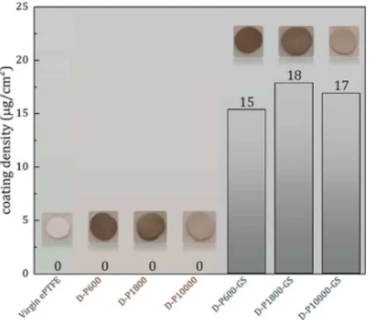
After the first coating with PDA/PEI, there was a negligible change in the membranes’ weights, as shown in Figure 1.
There were, however, notable changes in membrane color: from white for virgin ePTFE to brown for the coated samples seen in the images of the membranes inFigure 1. The darkest color was seen for D-P600, and with higher molecular weights of PEI, the color became less intense. It is worth noting that only PDA contains a chromophore and PEI does not; the variation in membrane color is therefore directly indicative of the amount of PDA adhering to the surface as a result of interacting with PEI.39 Additionally, since all of the coating solutions were also brown and transparent at the end of the coating, some undeposited PDA/PEI conjugates remain in solution. These direct observations point to the effect of the molecular weight of PEI used, where lower molecular weights were seen to be better at enhancing the deposition of PDA/ PEI conjugates onto the substrate surface. Although this same
trend has been observed in previous studies involving PDA/ PEI co-deposition on different surfaces,38,40 different explan-ations have been used to justify the effect of PEI molecular weight on deposition.
This is indeed an interesting outcome, considering that despite the differences in molecular weight, the usage of the same mass of PEI in the experiments should mean equivalent amounts of the reactive amine groups in each case, and consequently, a consistent reaction stoichiometry between the PDA and these amine groups. It must be noted, however, that with the longer chains of higher-molecular-weight PEI, there is an increasing degree of both intermolecular and intramolecular interactions, aside from the larger molecules having a much lower diffusivity in solution. Thus, despite following the same Schiff base or Michael addition stoichiometry as that in lower-MW PEI, the longer chains of higher-lower-MW PEI in the formed PDA/PEI conjugates encourage more interactions within the same molecule instead of with the substrate surface. This in effect “buries” the adhesive groups of PDA within the conjugates, causing stabilization in the aqueous phase and resulting in an overall decrease in the amount of PDA/PEI conjugates deposited.38This is reinforced by observations from succeeding physicochemical characterizations.
After the second coating, which formed the next layer consisting of PGS covalently linked to the first layer, the coating density was seen to increase drastically. Relative to the first layer, this second layer of PGS was thicker and more massive by several micrograms.
The SEM images shown in Figure 2 reveal the effect of coating on the surface morphology of the ePTFE; the cross-sectional views of the membranes are shown as inset images. Virgin expanded PTFE is characterized by nodes and fibrils as a result of the expansion process. Despite not having seen any change in weight after the PDA/PEI coating, SEM images reveal that there is an observable slight change in the surface morphology. The pores of the virgin ePTFE were slightly covered after the PDA/PEI coating but were virtually unchanged after the zwitterionization with PGS. More surface morphological changes are visible for the low-MW PEI after only the first coating, compared to the much larger 10 kDa PEI. This is consistent with the earlier explanation that there is less deposition of PDA/PEI conjugates with high-MW PEI. Further confirmation comes from the morphological changes after the second coating, where the greatest change is observed
Figure 1. Coating density measurements and actual color changes seen for ePTFE membranes subjected to the two-step coating procedure described. The average values of six independent samples are presented.
with D-P600-GS and the least with D-P10000-GS. This change obviously comes from the attachment of GS. With much less PDA/PEI deposited onto the surface of the D-P10000, less GS could attach; conversely, with the higher amount of PDA/PEI on the surface of D-P600, there are more sites for the attachment of GS. These SEM images, particularly those for the GS-coated samples, seem to suggest pore blockage at the surface, which could potentially alter the bulk physical properties of the membrane. To test whether these morphological changes are significant to the overall micro-structure of the membrane, porosity tests based on swelling by n-butanol were performed. Interestingly, the porosities were not found to be significantly different from the porosity of the virgin membrane, as presented in Table 1. In fact, the
cross-sectional SEM images show that aside from the deformities introduced by the tearing of the membranes to reveal the interior, the nodes are fibrils characteristic of ePTFE are still visible. For D-P1800-GS, these are visible at the top-right corner. The large “flat” region may have resulted from the tearing; porosity data for this membrane are not consistent with blockage. Good coats must only alter surface properties and not bulk properties; in this case, porosity, a representative bulk property, was not altered, and hence in this regard, the coats are not problematic.
Surface Chemistry of Modified Membranes. In the first coating, dopamine self-polymerizes and randomly reacts with amine moieties in PEI to form a co-deposited layer of PDA/ PEI on the membrane surface.38As mentioned previously, the stoichiometry of this reaction, in terms of moles of reactive amine groups, is unaffected by PEI molecular weight. In the second coating, the epoxy rings of the PGS are attacked and opened by the unreacted nucleophilic amine groups in PEI (mostly secondary and tertiary amines not participating in the Schiff base and Michael addition reactions earlier), forming a covalently linked second layer. The first layer, composed of co-deposited PDA and PEI, serves two functions: to form a tight noncovalent surface coating via the free catechol groups inherent in the polydopamine structure, and to introduce the nucleophilic amino group for the subsequent attachment of the PGS copolymer via a ring-opening nucleophilic attack on the glycidyl moieties shown in Scheme 2. This second layer presents free sulfobetaine groups, which are zwitterionic moieties. The PDA/PEI layer in itself has contributed polar functional groups (CO, C−OH, CN, R3−N, R2−NH) that could interact with water, but the full charges present in PGS render the layer even more hydrophilic by stronger Coulombic interactions with polar water molecules. It is thus reasonable to hypothesize that with more PGS on the surface, the more hydrophilic the surface should become, the stronger the hydration layer is held.
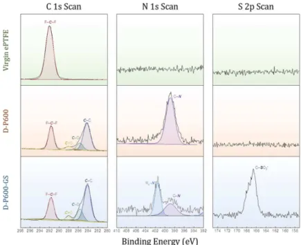
The success of the coating was also confirmed by XPS measurements on the virgin membrane and the samples D-P600 and D-D-P600-GS.Figure 3shows the C 1s, N 1s, and S 2p spectral scans. The C 1s spectrum for virgin PTFE shows a strong −CF2peak (291.7 eV). Addition of the PDA/PEI layer significantly enhanced the C−C band at 284 eV and introduced two new bands at 285.6 eV (C−O and C−N) and 288 eV (CO) as well as a peak in the N 1s spectrum at 398.9 eV.33The new carbon peaks come from the polydop-amine structure (Scheme 2), which contains oxygen both singly and doubly bonded to carbon in the catechol groups, Table 1. Porosity of Virgin and Coated Membranes
sample ID porosity (%) virgin ePTFE 45.05 ± 0.42 D-P600 45.41 ± 0.16 D-P1800 45.43 ± 0.48 D-P10000 45.20 ± 0.44 D-P600-GS 43.89 ± 0.49 D-P1800-GS 44.15 ± 0.35 D-P10000-GS 44.43 ± 0.49
and the nitrogen peak comes from the amine groups of both PDA and PEI. Finally, the second PGS layer further introduced a sulfur peak at 166.6 eV (−SO3−) and a new ammonium nitrogen (R4−N+) peak at 401.4 eV, which could only come from its sulfobetaine group and which confirms successful PGS grafting. In addition, relative to the PDA/PEI coating, the PGS coating has significantly decreased the intensity of the −CF2 peak as more C−C (mostly alkyl) carbons have been added. A summary of the elemental analysis is given inTable 2.
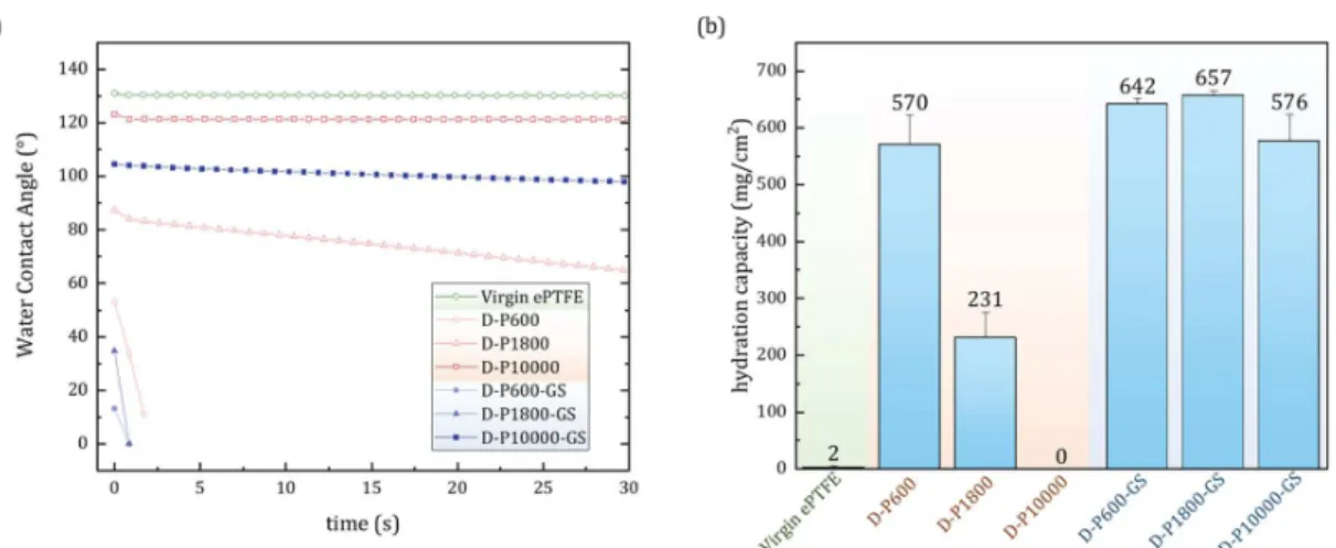
Evaluation of the Antifouling Properties of the Modified ePTFE Membranes. Dynamic water contact angle measurements, summarized in Figure 4a as a plot of contact angle versus time, show that virgin ePTFE is strongly hydrophobic, having a contact angle of 130°. The curves for D-P600-GS and D-P1800-GS are shown as highly hydrophilic as the water droplet was very quickly absorbed by the membranes, showing only a few contact angles before reaching a value of zero. Without PGS, hydrophilicity was strongly dependent on the PEI molecular weight used: D-P600 was seen to be the most hydrophilic based on the low contact angle, followed by D-P1800, then by D-P10000. This is again consistent with the hypothesis that high-MW PEI, despite possessing the same amount of reactive amine groups, interferes with the deposition process by promoting intra-molecular interactions that stabilize the conjugates in solution
instead of encouraging their deposition onto the surfaces. As a result, there are more PDA/PEI conjugates deposited on P600, slightly less on P1800, and so much less on D-P10000. In fact, both the value and the behavior of the contact angle for D-P10000 were close to those of the virgin PTFE, pointing to poor deposition on the surface. Additionally, the water droplet was most quickly absorbed through D-P600 and least through D-P10000. This means the pores in the interior of the membrane were more successfully coated with lower MW of PEI. This is confirmed by the hydration capacities (Figure 4b): more water is bound by the more hydrophilic pores of D-P600 and decreases with increasing PEI molecular weight. Addition of the PGS layer further improved the hydrophilicity, but for D-P10000-GS, this improvement was not as significant. The almost similar hydration capacities for all PGS-coated membranes point at saturation effects such that the differences in PDA/PEI density forming the first layer were normalized by the saturation of zwitterion−water interactions at the surface after the second coating. PGS is itself already a polymer, and as such, it requires only few covalent linkages per polymeric unit for it to remain attached to the first layer. The saturation effect is explained best when considering that as long as the PGS has been attached to the surface with at least a few covalent linkages, there would be no significant difference with a PGS unit being attached with more linkagesthe membrane has already been rendered zwitterionic. For example, despite being less hydrophilic than P600, D-P1800 has been rendered as hydrophilic after GS coating such that both D-P600-GS and D-P1800-GS have the same hydration capacity. The case for D-P1000 warrants more discussion. It must be noted that the time scales for both experiments differseconds for the contact angle test and a whole day for the hydration capacity. The dynamic water contact angle for D-P10000-GS has a nonzero slope, and it decreases very slowly, unlike that for D-P10000enough evidence that some GS has successfully attached. Besides, the coating density for D-P10000-GS was nonzero (Figure 1), although it must be emphasized that these values are
Figure 3.XPS analysis (C 1s, N 1s, and S 2p spectra) showing evidence for successful grafting of PDA/PEI and PGS layers onto ePTFE.
Table 2. Elemental Composition of Samples Obtained via XPS
sample composition (atomic %)
element virgin D-P600 D-P600-GS C 33.88 51.59 52.43 F 66.12 38.49 33.14 N 3.46 1.84 O 6.46 10.95 S 1.65
nevertheless very small, being in micrograms. With this nonzero slope, D-P10000-GS can still absorb as much water as it can overnight until it is saturated. In summary, the whole coating procedure successfully enhanced the hydrophilicity of the PTFE membranes, to only slightly different extents. The hydrophilicity of the material is a good indication of its antifouling property. The biofoulant would need to displace the tightly bound water molecules on the zwitterionic polymer chains. Thus, the samples were further subjected to experi-ments to evaluate their antifouling ability.
To assess how the modification affects the performance of the membrane against supporting biofilm formation, which would cause unwanted and sometimes fatal bacterial infections, bacterial attachment tests were performed. It has been hypothesized that biofilm formation is a stepwise process, beginning with the most important step of forming a conditioning film, which is composed of organic and inorganic compounds that immediately adhere to surfaces exposed to bacteria-contaminated solutions.41 This fouling was also seen to be more dominant on less hydrophilic surfaces,42 strongly suggesting that fouling could be reduced with more hydro-philic surfaces. The antifouling activity of the modified membranes was observed with both E. coli and S. aureus, as shown inFigure 5, representing the negative and Gram-positive groups of bacterial cells. Without any modification, virgin PTFE is prone to fouling by the microorganisms due to its hydrophobicity. In contrast, the SBMA hydrogel, which acted as the positive control for all biofouling experiments, shows essentially no attachment of either microorganism, owing to its high hydrophilicity forming a protective hydration layer.43 Results for both E. coli and S. aureus attachment for PDA/PEI-coated samples are consistent with the hydrophilic properties of the membranes. In fact, there is unquestionable consistency with the data from hydration capacity: D-P10000, being strongly hydrophobic and resisting water absorption, was the most fouled of the PDA/PEI-coated samples. Furthermore, it was also observed that after the PGS coating, the bacterial attachment was reduced significantly. With the PGS coating, a similar saturation effect is observed, as was the case for the hydration capacity of the modified membranes. The antifouling property provided by D-P10000GS seems to be in contra-diction when considering only the water contact angle. Prior to exposing the membranes to the bacterial solutions, they were
incubated in PBS at 37 °C for at least 4 h. As observed with the contact angle curve, the nonzero slope meant it could absorb water slowly. It is possible that by the time the D-P10000GS membranes were incubated with the bacterial solutions, they had already been saturated with water to provide the protective hydrating layer. The results so far confirm the direct consequence of hydrophilic character to antifouling tendencies, as it is known that hydrophilic surfaces more effectively prevent biofouling by promoting the formation of a protective hydrating layer. Furthermore, these results show that the coating is effective against both positive and Gram-negative microorganisms.
Successful prevention of bacterial attachment suggests the potential to prevent the attachment of other biological molecules. As ePTFE has consistently been used as materials for various grafts, which are constantly in contact with blood, the interaction of the modified membranes with blood was also studied. Such hemocompatibility studies are required for determining the biocompatibility of biomaterials designed for blood-interacting applications. To test this, attachment tests with whole blood and red blood cells (RBC)-rich samples were performed, and the results are shown inFigure 6. There is a
Figure 4.Hydrophilicity of the virgin ePTFE and coated membranes shown through (a) dynamic water contact angle measurements and (b) the average hydration capacity of six independent measurements via immersion in water with their standard deviations as error bars.
Figure 5.Bacterial attachment experiment for 24 h of E. coli (Gram-negative) and S. aureus (Gram-positive). Relative bacterial coverage (normalized using virgin ePTFE sample) was calculated using ImageJ software. The average values of nine CLSM images are presented with their standard deviation as error bars.
stark difference in the fouling of membranes by RBCs and whole blood. For RBC attachment, the surface modification reduced the attachment of the red blood cells relative to virgin ePTFE; additionally, the GS-coated membranes performed better compared to those coated only with PDA/PEI. The trends in the antifouling performance for both PDA/PEI and GS-coated membranes, however, were the opposite of those seen in hydrophilicity. Fouling with whole blood is remarkable. Although a trend consistent with hydrophilicity is seen in the PDA/PEI-coated and in the GS-coated membranes, some of them were worse than virgin ePTFE at preventing fouling, even enhancing fouling 5-fold.
Ideally, the resistance of surfaces to biofouling is enhanced when surface hydrophilicity is increased.44 This has been attributed to the prevention of stochastic protein adsorption, which is believed to be the priming step in surface fouling. In whole blood, fibrinogen, a plasma protein, makes up the majority of the quickly absorbed plasma proteins; this is followed by attachment of platelets, inducing thrombo-genesis.45 Since RBC concentrate is prepared from whole blood by centrifugation, which separates the much heavier cells from the much lighter plasma proteins, less to no fibrinogen is present in this RBC-rich fraction. Although membrane proteins do abound on RBCs, none of them are directly involved with the clotting process;46 instead, they are mostly involved in substrate transport and structural roles. However, it has been proposed that a group of these membrane proteins, called integrin-binding proteins because they bind to transmembrane integrin proteins responsible for cell adhesion to the extracellular matrix, presents a site for interaction with fibrinogen,47 suggesting that RBC attachment on surfaces is mediated by fibrinogen. Without much fibrinogen in this RBC-rich fraction, the fouling observed could be speculated to be less dependent on the priming ePTFE−fibrinogen interactions, or that these interactions are too few and weak such that a secondary factor affecting biocompatibility has taken control. Following this logic, the excessive biofouling with whole blood could then be partly accounted for due to the abundance of fibrinogen.
This secondary factor affecting biocompatibility could be surface roughness, which is directly related to the presence of
more surface irregularities and the total surface area exposed to the foulant.48Indeed, it has been shown that thicker PDA/PEI coatings are rougher as measured by atomic force micros-copy.38This has been attributed to the accumulation of surface defects as layers grow thicker. Thus, with more deposition seen for PDA and 600 Da PEI, the formed coating is rougher; with 1800 Da PEI, it is less rough, while for 10 000 Da PEI, having the least amount of deposition, the surface is smoother. It is thus reasonable to attribute the RBC fouling seen for the PDA/PEI-coated membranes primarily to surface roughness due to the very low concentration of fibrinogen. The cells could more easily be stuck on rougher surfaces than on smoother surfaces. We say fibrinogen is present at a very low concentration instead of zero because it can be seen that the attachment of GS has still improved their fouling resistance. The zwitterionic moieties introduced could be thought of as preventing any surface attachment of the very few fibrinogen remaining, in addition to destabilizing weak RBC-ePTFE interactions.
Whole blood presents a harsher environment for the membranes, with normal concentrations of plasma proteins, particularly fibrinogen at 0.2−0.45 g/dL. Surface hydrophilicity becomes significant due to their effect on the formation of the priming protein layer. The trend in resistance indeed compares well with hydrophilicity: coats formed with 600 Da PEI provided better fouling resistance due to higher hydrophilicity, and those formed from 10 000 Da PEI were the most fouled. The saturation effect seen in hydration capacity, invoked to account for the resistance of all GS-coated membranes toward bacterial attachment, no longer holds for blood. It is possible that the osmolarity of whole blood due to its protein content causes the water bound on and in the membranes to diffuse out and into the blood. Thus, a hydration layer that is very loosely held could easily be destroyed when in contact with blood, whereas a strongly held layer can oppose this osmotic force. This could explain why D-P10000-GS did not fare wellit could not hold water as strongly as D-P1800GS and D-P600GS. This is also exactly why it absorbs water so slowly, as pointed out by the contact angle and hydration capacity tests (Figure 4).
The higher fouling of P1800, P10000, and D-P10000GS by whole blood relative to unmodified, virgin ePTFE could not be accounted for by hydrophilicity arguments; virgin ePTFE is more hydrophobic, but it was fouled less. It could not also be explained by surface roughness arguments, because then, D-P600, having the roughest surface, should have been fouled more than virgin ePTFE. Instead, it has resisted fouling better. The difference could be accounted for by considering the incubation step prior to exposure to the blood solutions. Being strongly hydrophobic, the PBS solution used for the incubation has been prevented from entering and wetting the interior pores of virgin ePTFE, whereas the other membranes, owing to their different hydrophilicities, allowed wetting of their interiors by the same solution. Blood can more easily penetrate pores that have already been pre-wet by the PBS than those that have not. The higher fouling for D-P1800, D-P10000, and D-P10000GS could then be because of the greater total surface area of these membranes in contact with blood, including its pores. This reconciles well with the hydrophilicity trend as seen for the GS-coated samples. All of these membranes could be saturated with the PBS solution, but D-P600-GS has even its interior pores coated well with PDA/PEI and GS, whereas D-P10000-GS has not. Finally, it
Figure 6. Hemocompatibility tests on modified PTFE membranes with human whole blood and RBC-rich fraction. Relative blood cell attachment coverage (normalized using virgin ePTFE sample) was calculated using ImageJ software. The average values of nine CLSM images are presented with their standard deviation as error bars.
could also be assumed that the amount of water that can be absorbed by D-P10000 (Figure 4) is so low that it falls below the milligram range, but is nonzero to still allow partial wetting of its pores by PBS. With this assumption in place, the observations with whole blood are now accounted for.
Despite the complexity of the interactions seen in the blood tests, both hydrophilicity and biocompatibility tests point at D-P600-GS as the best modified sample in this study. The low molecular weight of PEI ensures more PDA/PEI conjugates are deposited onto the ePTFE surface, which then provide a strongly adhering coating. Although biocompatibility largely relies on hydrophilicity for the protective hydration layer, it was shown in this study that surface morphology also becomes a significant factor in determining biocompatibility in the absence of a large amount of fouling proteins.
It is interesting to note the differences in fouling between the bacterial solutions and whole blood. Both attachment processes in these cases are dependent on the formation of a priming or activating layer, which is mostly proteinaceous. It can be hypothesized that compared to the normal physiological concentration of fibrinogen in blood (0.2−0.45 g/dL), proteins are less abundant in the bacterial solutions prepared and used, explaining why even loosely held hydration layer assumed for D-P10000-GS was not affected the same way with whole blood
■
CONCLUSIONSA facile, low-energy, two-step coating procedure inspired by mussels was developed to construct first a PDA/PEI layer that strongly adhered to the ePTFE surface, followed by covalent attachment of PGS, which contains a zwitterionic moiety for enhanced biocompatibility. Without shaking, the layers were found to be extremely thin and lightweight, but their existence was confirmed immediately with color changes, and more accurately with the nitrogen and sulfur peaks in the XPS spectra. The effects of PEI molecular weight on hydrophilicity were confirmed in the first coating step: lower molecular weights were more preferred because although the same reactions could have occurred with the same number of reacting groups in PEI, longer chains are more susceptible to intramolecular interactions, effectively burying most of the catechol groups from PDA that are responsible for adhesion to the ePTFE surface. Despite these differences, hydrophilicity was enhanced in all cases, especially after the second coating step. It has turned out that while all PGS-coated membranes performed very similarly due to the saturating effect of zwitterion−water interactions at the surface, surface morphol-ogy had a significant contribution when the membranes came into contact with whole blood samples due to higher concentrations of blood plasma proteins. The extent of prewetting during PBS incubation also had a significant effect, with prewetted membranes allowing greater access to interior pores by the fouling solutions. These results provide a basis for further studies using the developed modification strategy with 600 Da PEI. It will be interesting to study the behavior of the membranes with respect to tissue adhesion, which generally prefers larger pore sizes, and cytotoxicity in vitro, which could possibly be used to determine an optimum set of coating parameters and further determine the modification’s overall effectiveness for grafted materials.
■
ASSOCIATED CONTENT*
sı Supporting InformationThe Supporting Information is available free of charge at
https://pubs.acs.org/doi/10.1021/acsami.0c09073.
Confocal laser scanning microscopy images of the bacterial and blood cell attachment experiments; representative confocal laser scanning microscope images for bacterial attachment experiment (Figure S1); representative confocal laser scanning microscope images for blood cell attachment experiment (Figure S2) (PDF)
■
AUTHOR INFORMATIONCorresponding Author
Yung Chang − R&D Center for Membrane Technology and Department of Chemical Engineering, Research Center for Circular Economy, Chung Yuan Christian University, Taoyuan 32023, Taiwan; orcid.org/0000-0003-1419-4478; Phone: +886-3-265-4113; Email:[email protected]
Authors
Peter Matthew Paul T. Fowler − School of Chemical, Biological and Materials Engineering and Sciences and School of Graduate Studies, Mapúa University, Manila 1002,
Philippines; orcid.org/0000-0001-6553-8221
Gian Vincent Dizon − R&D Center for Membrane Technology, Chung Yuan Christian University, Taoyuan 32023, Taiwan;
orcid.org/0000-0002-9424-1029
Lemmuel L. Tayo − School of Chemical, Biological and Materials Engineering and Sciences, Mapúa University, Manila 1002, Philippines
Alvin R. Caparanga − School of Chemical, Biological and Materials Engineering and Sciences, Mapúa University, Manila 1002, Philippines
James Huang − Yeu Ming Tai Chemical Industrial Co. Ltd., Taichung 407, Taiwan
Jie Zheng − Department of Chemical and Biomolecular Engineering, The University of Akron, Akron, Ohio 44325, United States; orcid.org/0000-0003-1547-3612
Pierre Aimar − Laboratoire de Génie Chimique, Université de Toulouse, CNRS, INPT, UPS, Toulouse 31062, France
Author Contributions ○
The manuscript was written through contributions of all authors. All authors have given approval to the final version of the manuscript. P.M.P.T.F. and G.V.D. contributed equally to this work.
Notes
The authors declare no competing financial interest.
■
ACKNOWLEDGMENTSThe authors acknowledge the projects of Distinguished Professorship at Chung Yuan Christian University, Ministry of Science and Technology and the Agence Nationale de la Recherche (MOST-ANR International Program: MOST 109-2923-E-033-001), and the National Science Foundation (NSF 1806138) for their financial support. P.M.P.T.F. extends his gratitude for the financial support from the Department of Science and Technology−Science Education Institute (DOST-SEI) and Engineering Research and Development for
Technology (ERDT) of the Philippines. The authors also express their gratitude to Professor Antoine Venault of the Chemical Engineering Department of Chung Yuan Christian University for the guidance and inspiration he provided for this research.
■
REFERENCES(1) Bunn, C. W.; Howells, E. R. Structures of Molecules and Crystals of Fluoro-Carbons. Nature 1954, 174, 549−551.
(2) Ebnesajjad, S. Properties, Characteristics, and Applications of Expanded PTFE (ePTFE) Products. In Expanded PTFE Applications Handbook; Ebnesajjad, S., Ed.; William Andrew Publishing: Oxford, 2017; pp 163−170.
(3) Faries, P. L.; LoGerfo, F. W.; Arora, S.; Hook, S.; Pulling, M. C.; Akbari, C. M.; Campbell, D. R.; Pomposelli, F. B. A comparative study of alternative conduits for lower extremity revascularization: All-autogenous conduit versus prosthetic grafts. J. Vasc. Surg. 2000, 32, 1080−1090.
(4) Kannan, R. Y.; Salacinski, H. J.; Butler, P. E.; Hamilton, G.; Seifalian, A. M. Current status of prosthetic bypass grafts: A review. J. Biomed. Mater. Res., Part B 2005, 74, 570−581.
(5) Li, L.; Terry, C. M.; Shiu, Y.-T. E.; Cheung, A. K. Neointimal hyperplasia associated with synthetic hemodialysis grafts. Kidney Int. 2008, 74, 1247−1261.
(6) Harris, J. R.; Seikaly, H. Evaluation of Polytetrafluoroethylene Micrografts in Microvascular Surgery. J. Otolaryngol. 2002, 31, 89−92. (7) Tseng, D. Y.; Edelman, E. R. Effects of amide and amine plasma-treated ePTFE vascular grafts on endothelial cell lining in an artificial circulatory system. J. Biomed. Mater. Res. 1998, 42, 188−198.
(8) Greisler, H. P. Interactions at the Blood/Material Interface. Ann. Vasc. Surg. 1990, 4, 98−103.
(9) Nassar, G. M.; Ayus, J. C. Infectious complications of the hemodialysis access. Kidney Int. 2001, 60, 1−13.
(10) Benrashid, E.; Youngwirth, L. M.; Mureebe, L.; Lawson, J. H. Operative and perioperative management of infected arteriovenous grafts. J. Vasc. Access 2017, 18, 13−21.
(11) Kirkton, R. D.; Prichard, H. L.; Santiago-Maysonet, M.; Niklason, L. E.; Lawson, J. H.; Dahl, S. L. M. Susceptibility of ePTFE vascular grafts and bioengineered human acellular vessels to infection. J. Surg. Res. 2018, 221, 143−151.
(12) Bachleda, P.; Kalinova, L.; Utikal, P.; Kolar, M.; Hricova, K.; Stosova, T. Infected Prosthetic Dialysis Arteriovenous Grafts: A Single Dialysis Center Study. Surg. Infect. 2012, 13, 366−370.
(13) Melman, L.; Jenkins, E. D.; Hamilton, N. A.; Bender, L. C.; Brodt, M. D.; Deeken, C. R.; Greco, S. C.; Frisella, M. M.; Matthews, B. D. Histologic and biomechanical evaluation of a novel macro-porous polytetrafluoroethylene knit mesh compared to lightweight and heavyweight polypropylene mesh in a porcine model of ventral incisional hernia repair. Hernia 2011, 15, 423−431.
(14) Lu, A.; Sipehia, R. Antithrombotic and fibrinolytic system of human endothelial cells seeded on PTFE: the effects of surface modification of PTFE by ammonia plasma treatment and ECM protein coatings. Biomaterials 2001, 22, 1439−1446.
(15) Begovac, P. C.; Thomson, R. C.; Fisher, J. L.; Hughson, A.; Gällhagen, A. Improvements in GORE-TEX vascular graft perform-ance by Carmeda bioactive surface heparin immobilization. Eur. J. Vasc. Endovasc. Surg. 2003, 25, 432−437.
(16) Tzchori, I.; Falah, M.; Shteynberg, D.; Levin Ashkenazi, D.; Loberman, Z.; Perry, L.; Flugelman, M. Y. Improved Patency of ePTFE Grafts as a Hemodialysis Access Site by Seeding Autologous Endothelial Cells Expressing Fibulin-5 and VEGF. Mol. Ther. 2018, 26, 1660−1668.
(17) Lei, Z. Y.; Li, J.; Liu, T.; Shi, X. H.; Fan, D. l. Autologous Vascularization: A Method to Enhance the Antibacterial Adhesion Properties of ePTFE. J. Surg. Res. 2019, 236, 352−358.
(18) Zhu, A. P.; Ming, Z.; Jian, S. Blood compatibility of chitosan/ heparin complex surface modified ePTFE vascular graft. Appl. Surf. Sci. 2005, 241, 485−492.
(19) Larsen, C. C.; Kligman, F.; Tang, C.; Kottke-Marchant, K.; Marchant, R. E. A biomimetic peptide fluorosurfactant polymer for endothelialization of ePTFE with limited platelet adhesion. Biomaterials 2007, 28, 3537−3548.
(20) Sisti, L.; Cruciani, L.; Totaro, G.; Vannini, M.; Berti, C.; Aloisio, I.; Di Gioia, D. Antibacterial coatings on poly-(fluoroethylenepropylene) films via grafting of 3-hexadecyl-1-vinyl-imidazolium bromide. Prog. Org. Coat. 2012, 73, 257−263.
(21) Venault, A.; Chang, Y.; Hsu, H.-H.; Jhong, J.-F.; Yang, H.-S.; Wei, T.-C.; Tung, K.-L.; Higuchi, A.; Huang, J. Biofouling-resistance control of expanded poly(tetrafluoroethylene) membrane via atmospheric plasma-induced surface PEGylation. J. Membr. Sci. 2013, 439, 48−57.
(22) Liu, Y.; Munisso, M. C.; Mahara, A.; Kambe, Y.; Fukazawa, K.; Ishihara, K.; Yamaoka, T. A surface graft polymerization process on chemically stable medical ePTFE for suppressing platelet adhesion and activation. Biomater. Sci. 2018, 6, 1908−1915.
(23) Liu, Y.; Munisso, M. C.; Mahara, A.; Kambe, Y.; Yamaoka, T. Anti-platelet adhesion and in situ capture of circulating endothelial progenitor cells on ePTFE surface modified with poly(2-methacry-loyloxyethyl phosphorylcholine) (PMPC) and hemocompatible peptide 1 (HCP-1). Colloids Surf., B 2020, 193, No. 111113.
(24) Zheng, W.; Liu, M.; Qi, H.; Wen, C.; Zhang, C.; Mi, J.; Zhou, X.; Zhang, L.; Fan, D. Mussel-inspired triblock functional protein coating with endothelial cell selectivity for endothelialization. J. Colloid Interface Sci. 2020, 576, 68−78.
(25) Feng, S.; Zhong, Z.; Wang, Y.; Xing, W.; Drioli, E. Progress and perspectives in PTFE membrane: Preparation, modification, and applications. J. Membr. Sci. 2018, 549, 332−349.
(26) Cheng, B.; Inoue, Y.; Ishihara, K. Surface functionalization of polytetrafluoroethylene substrate with hybrid processes comprising plasma treatment and chemical reactions. Colloids Surf., B 2019, 173, 77−84.
(27) Wang, S.; Li, J.; Suo, J.; Luo, T. Surface modification of porous poly(tetrafluoraethylene) film by a simple chemical oxidation treatment. Appl. Surf. Sci. 2010, 256, 2293−2298.
(28) Fu, C.; Liu, S.; Gong, T.; Gu, A.; Yu, Z. Investigation on surface structure of potassium permanganate/nitric acid treated poly-(tetrafluoroethylene). Appl. Surf. Sci. 2014, 317, 771−775.
(29) Xi, Z. Y.; Xu, Y. Y.; Zhu, L. P.; Wang, Y.; Zhu, B. K. A facile method of surface modification for hydrophobic polymer membranes based on the adhesive behavior of poly(DOPA) and poly(dopamine). J. Membr. Sci. 2009, 327, 244−253.
(30) Li, X.; Shan, H.; Cao, M.; Li, B. Mussel-inspired modification of PTFE membranes in a miscible THF-Tris buffer mixture for oil-in-water emulsions separation. J. Membr. Sci. 2018, 555, 237−249.
(31) Ma, Y.; Qiao, X. Y.; Lu, Q.; Li, R.; Bai, Y. J.; Li, X.; Zhang, S. P.; Gong, Y. K. Anchorable phosphorylcholine copolymer synthesis and cell membrane mimetic antifouling coating fabrication for blood compatible applications. J. Mater. Chem. B 2020, 8, 4299−4309.
(32) Lee, S.; Kim, S.; Park, J.; Lee, J. Y. Universal surface modification using dopamine-hyaluronic acid conjugates for anti-biofouling. Int. J. Biol. Macromol. 2019, 151, 1314−1321.
(33) Song, H.; Yu, H.; Zhu, L.; Xue, L.; Wu, D.; Chen, H. Durable hydrophilic surface modification for PTFE hollow fiber membranes. React. Funct. Polym. 2017, 114, 110−117.
(34) Xue, S.; Li, C.; Li, J.; Zhu, H.; Guo, Y. A catechol-based biomimetic strategy combined with surface mineralization to enhance hydrophilicity and anti-fouling property of PTFE flat membrane. J. Membr. Sci. 2017, 524, 409−418.
(35) Li, C.; Wang, J.; Luo, Y.; Wang, F.; Zhu, H.; Guo, Y. One-bath two step method combined surface micro/nanostructures treatment to enhance antifouling and antibacterial property of PTFE flat membrane. J. Taiwan Inst. Chem. Eng. 2019, 96, 639−651.
(36) Yue, W. W.; Li, H. J.; Xiang, T.; Qin, H.; Sun, S. D.; Zhao, C. S. Grafting of zwitterion from polysulfone membrane via surface-initiated ATRP with enhanced antifouling property and biocompat-ibility. J. Membr. Sci. 2013, 446, 79−91.
(37) Chou, Y. N.; Wen, T. C.; Chang, Y. Zwitterionic surface grafting of epoxylated sulfobetaine copolymers for the development of stealth biomaterial interfaces. Acta Biomater. 2016, 40, 78−91.
(38) Lv, Y.; Yang, S. J.; Du, Y.; Yang, H. C.; Xu, Z. K. Co-deposition Kinetics of Polydopamine/Polyethyleneimine Coatings: Effects of Solution Composition and Substrate Surface. Langmuir 2018, 34, 13123−13131.
(39) Yang, H. C.; Liao, K. J.; Huang, H.; Wu, Q. Y.; Wan, L. S.; Xu, Z. K. Mussel-inspired modification of a polymer membrane for ultra-high water permeability and oil-in-water emulsion separation. J. Mater. Chem. A 2014, 2, 10225−10230.
(40) Yang, H. C.; Wu, M. B.; Li, Y. J.; Chen, Y. F.; Wan, L. S.; Xu, Z. K. Effects of polyethyleneimine molecular weight and proportion on the membrane hydrophilization by codepositing with dopamine. J. Appl. Polym. Sci. 2016, 133, 43792.
(41) Matin, A.; Khan, Z.; Zaidi, S. M. J.; Boyce, M. C. Biofouling in reverse osmosis membranes for seawater desalination: Phenomena and prevention. Desalination 2011, 281, 1−16.
(42) Fane, A. G.; Fell, C. J. D. A review of fouling and fouling control in ultrafiltration. Desalination 1987, 62, 117−136.
(43) Holmlin, R. E.; Chen, X.; Chapman, R. G.; Takayama, S.; Whitesides, G. M. Zwitterionic SAMs that Resist Nonspecific Adsorption of Protein from Aqueous Buffer. Langmuir 2001, 17, 2841−2850.
(44) Rabe, M.; Verdes, D.; Seeger, S. Understanding protein adsorption phenomena at solid surfaces. Adv. Colloid Interface Sci. 2011, 162, 87−106.
(45) Wu, Y.; Simonovsky, F. I.; Ratner, B. D.; Horbett, T. A. The role of adsorbed fibrinogen in platelet adhesion to polyurethane surfaces: A comparison of surface hydrophobicity, protein adsorption, monoclonal antibody binding, and platelet adhesion. J. Biomed. Mater. Res., Part A 2005, 74, 722−738.
(46) Litvinov, R. I.; Weisel, J. W. Role of red blood cells in haemostasis and thrombosis. ISBT Sci. Ser. 2017, 12, 176−183.
(47) Lominadze, D.; Dean, W. L. Involvement of fibrinogen specific binding in erythrocyte aggregation. FEBS Lett. 2002, 517, 41−44.
(48) Vrijenhoek, E. M.; Hong, S.; Elimelech, M. Influence of membrane surface properties on initial rate of colloidal fouling of reverse osmosis and nanofiltration membranes. J. Membr. Sci. 2001, 188, 115−128.