Design, synthesis and structure of an amphipathic peptide with
pH-inducible haemolytic activity
Rudolf Moser
Biochemisches Institut der Universitat Zurich, Winterthurerstrasse 190, CH-8057 Zurich, Switzerland
A synthetic, 26-residue peptide having a strong helix forming potential in the protonated state was designed to interact with lipid bilayers in a pH-dependent way. On the basis of this concept a cluster of four glutamic acid residues was inserted in the central region of the amphipathic peptide to promote helix destabilization by mutual charge repulsion at neutral pH. Protonation of these residues might then bring about both a pH-mediated change in hydrophobicity and conformation forming a membrane-active amphiphilic helix. The sequence GLGTLLTLLEFLLEELLEFLKRKRQQamide produced by the design strategy induced pH-triggered lysis of human erythrocytes. A molecular model correlating the lytic activity to the formation of transmembrane pores which were detected by electron microscopy in erythrocyte membranes is discussed. Circular dichroism studies indicated a self-association of the monomeric random coil form with increasing peptide concentration leading to the apparent induction of strong a-helix formation (~ 100% helicity) in the fully aggregated state. However, no pH-dependent helix-random coil transition was observed, implying that interhelical hydrophobic and ionic interactions not only govern the self-association but also decisively influence the conformational stability of the peptide.
Key words: amphipathic helix/CD studies/molecular modelling/ pH-dependent haemolytic activity/transmembrane pores
Introduction
One of the key problems in molecular biology is to understand the question of how the primary amino acid sequence of a protein determines its specific three-dimensional structure and the pathway of folding. One approach to this problem involves the construction of model peptides designed to form a desired conformation (DeGrado, 1988). Peptides shorter than ~30 residues are, in general, predicted not to show marked conformational preferences, remaining unfolded and, rather than forming the medium and long-range interactions (typically hydrogen and ionic bonds and van der Waals interactions) that stabilize native protein structures, form preferential interactions with the solvent water. The presence of a well-defined structure in such peptides in aqueous solution is therefore unusual, although in contrast, organic solvents are known frequently to induce relatively stable conformations. The lack of stability of isolated secondary structure elements (especially intramolecular H-bonded a-helices) is sometimes considered as evidence against their participation as nucleation centres, forming individual folding units, in the initiation of the protein folding process.
Designing an a-helical peptide is greatly facilitated by the existence of considerable, detailed information about factors that stabilize this conformation in aqueous solution (De Grado et al.,
1989). Research is currently being directed towards establishing factors which influence the stabilization of secondary structure units, especially a-helices in short peptides. Recent studies on synthetic a-helical peptides have shown that a-helix stability is strongly influenced by the distribution of charged side chains which interact with the helix dipole and which can also form salt bridges (Baldwin and Eisenberg, 1987; Marqusee and Baldwin, 1987). In addition, hydrophobic side-chain interactions are partly responsible for the unexpectedly high helical content of short peptides and are thought to play a key role in the protein (peptide) folding process (Dill, 1990).
Relatively stable a-helical secondary structure units have been reported for several peptides in purely aqueous media or mixtures of water and organic solvents (the values in brackets being the number of residues in the peptide): (13) peptide analogues of C-peptide of ribonuclease A (Merutka and Stellwagen, 1989); (25) peptide fragments of bovine growth hormone (Brems et al., 1987); (37) antifreeze polypeptide from the winter flounder (Yang et al., 1988); (17) DNA binding helix F of cAMP receptor protein of Escherichia coli (Clore etal, 1985); (16/74) four-helix bundle proteins (Ho and DeGrado, 1987; Regan and DeGrado, 1988); (79) four-helix bundle protein 'Felix' (Hecht et al., 1990); (30) pH-dependent, amphiphilic a-helical peptide (Subbarao et al., 1987); (21) membrane-spanning, amphiphilic a-helical peptides (Lear et al., 1988); (16/17) alanine-based, salt bridge-stabilized, a-helical peptides (Marqusee and Baldwin, 1987); (28) a-helical peptide fragment of the N-terminus of human pituitary growth hormone (Roongta et al., 1989); (43) peptides corresponding to the isolated leucine zipper regions from Fos and Jun nuclear proteins (O'Shea etal., 1989); (17) monomeric, a-helical peptides containing 19 different central-residue replacements (Merutka etal., 1990); (17) peptide analogues (Padmanabhan et al., 1990) of an (16) alanine-based, monomeric, a-helical peptide (Marqusee et al., 1989); (17) cation stabilized a-helices (Ghadiri and Choi, 1990). The stability of the a-helix formed by many of the peptides mentioned above is much higher than would be predicted from the Zimm and Bragg theory (Zimm and Bragg, 1959). However, the stability of these a-helices should be considered in terms of a dynamic model, where molecules change rapidly between many different con-formations including unfolded states but exhibit, to a greater or lesser extent, strong preferences to adopt a low energy a-helical state, rather than a static rigid structure (Wright et al., 1988; Bradley et al., 1990).
In the present study I have examined whether it is possible to design a polypeptide which increases in hydrophobicity as the pH decreases from neutral to acidic and thereby folds into an amphiphilic a-helix. Such surface-active helices are known to promote strong protein/peptide-membrane interactions and are commonly found in membrane-associated proteins and membrane-active peptides. Thus, the peptide would be expected to induce pH-triggered haemolysis of human erythrocytes, and therefore I have adopted the name Helical Erythrocyte Lysing Peptide (HELP) for this substance.
Materials and methods
Design and structure-function relationship
In designing a putative, amphiphilic a-helix, the following factors have to be taken into account: (a) the helical propensity and the hydropathy of the amino acid residues used; (b) the length of the peptide; (c) the charge distribution on the a-helix surface; (d) the establishment of the amphiphilic character and (e) the solubility of the peptide in water. Although the contribution of single factors to the helix stability is partly understood, the strategy of using a well-characterized peptide as a structural template might be more successful than a de novo design ap-proach.
The three-dimensional structure of bee venom melittin from Apis mellifera (BVM), a small peptide toxin containing 26 amino acid residues (Eisenberg, 1984), has been determined by X-ray analysis (Terwilliger and Eisenberg, 1982) and by 'H 2D NMR spectroscopy (Bazzo etai, 1988; Ikura etai, 1991) and therefore, was taken as the main starting point for the construction of the 26-residue amphiphilic HELP helix. BVM and its analogues have been studied extensively under different condi-tions (Blondelle and Houghten, 1991) and it undergoes a pH, salt and peptide concentration dependent random coil/helix transi-tion upon tetramerizatransi-tion (Bello et al., 1982; Quay and Condie, 1983). Several models exist to explain the spontaneous interac-tion of BVM with lipid bilayers upon which it acts as an effec-tive lytic agent (Vogel and Jahnig, 1986; Dawson et al., 1978). Additional sequence and structure information for the design of HELP was obtained from studies on a synthetic model toxin (DeGrado et al., 1981) and on 5-haemolysin from Staphylococcus aureus (Tappin et al., 1988; Raghunathan et al., 1990; Thiaudiere etai., 1991).
The amino acid sequence of HELP was chosen by examining the three-dimensional structure of BVM and replacing most of the residues of the hydrophobic helical face by Leu (which is not only hydrophobic but also has a high a-helical potential) and some of the hydrophilic residues by Glu and Thr in such a way that the central region of the putative a-helix was composed purely of residues with high helix-forming propensities (Chou and Fasman, 1974; Argos and Palau, 1982; Palau et al., 1982). Residues with a high turn potential are often found at the N-terminus of a-helices (Richardson and Richardson, 1988) and therefore Thr was included in the initial turn of the helix as it has the added advantage of potentially H-bonding back onto the amide backbone and thereby aiding helix initiation (Palau et al., 1982). Strongly polar residues (mainly glutamic acid) were used when the design of the helix dictated the incorporation of a hydrophilic residue (i.e. for those residues forming the hydrophilic face of the helix) in order to ensure that the peptide would be soluble in water. The structure of HELP was completed by adding a short helix inducing sequence at the N-terminus and the basic C-terminal hexapeptide portion of BVM was added to the C-terminus since it is known that this hexapeptide is essential for the lytic activity of BVM. The C-terminal a-carboxyl group was blocked by an amide group whereas the N-terminal a-amino group remained unprotected. Thus, helix destabilization could occur at the N-terminus by unfavourable interactions of the positively charged amino group with the helix dipole. The negative pole of the a-helix dipole is localized near the C-terminus, and therefore the positively charged Lys and Arg residues close to the C-terminus would be expected to favour structure stabilization and to mitigate against aggregation. However, short peptides, including BVM, often show aggregation-induced structure formation because of their tendency 324
to be amphipathic. Such aggregated states or supersecondary structures (e.g. four a-helix bundles) arise from the alignment of and interaction between the hydrophobic residues of monomer units (Chothia et al., 1977; Chothia, 1984; Regan and DeGrado, 1988; Cohen and Parry, 1990).
Were a helix to form at neutral pH, the glutamate residues occupying the central part of the hydrophilic helical face would be expected to exhibit mutual charge repulsion and thereby destabilize the helix. Protonation of these residues (by lowering the pH) might then bring about a coil/helix transition, forming a membrane-active helix with both hydrophilic and hydrophobic faces. The folding of the primary sequence into a putative a-helix was simulated using computer-generated graphics; and energy minimization techniques were employed to optimize the design stepwise, leading to the final HELP sequence and to examine if a stable structure could indeed be adopted.
Computer modelling
The structural aspects of the putative a-helix were examined by the macromolecular computer modelling system INSIGHT and DISCOVER from Biosym Technologies. These programs were used to simulate a molecular model of HELP. The amino acid side chains were attached onto the a-carbon backbone of an idealized, right-handed a-helix with uniformly constant phi and psi angles ($ = - 5 7 ° , i/< = - 4 7 ° ) and 3.6 residues per helix turn. The a-helix was soaked with water molecules and the potential energy of the structure was minimized using a conjugate gradient minimization on a force field equation which is dependent on the positions of the atoms of the molecule. Molecular mechanics for energy minimization calculations were performed on a Micro Vax II (Digital Equipment Corporation) and for structure visualization of the minimal energy conformation displayed on an Evans and Sutherland PS390 system.
Synthesis and purification
The peptide was synthesized as a C-terminal amide on a/?-methyl-benzhydrylamine (poly sty rene/1% divinylbenzene) resin (150-200 mesh) by the solid-phase method of Merrifield as described by Stewart and Young (1984) using dichloromethane (DCM) as the solvent, 40% (v/v) trifluoroacetic acid (TFA)/DCM for deprotection and 10% (v/v) triethylamine/DCM for neutralization. Residual amino groups on the resin (Boc-Gln-resin loading: 0.30 mmol/g (Boc-Gln-resin) were blocked by the addition of acetic anhydride. The peptide was synthesized using DCC-activated derivatives (4.2-fold molar excess) of Boc-Arg (N6
-Tos), Boc-Glu (O^-Bzl), Boc-Gly, Boc-Leu, Boc-Lys (/V-2-CIZ), Boc-Phe, Boc-Thr (OBzl). For Boc-Gln, the HOBt/DCC coupling method was used (Konig and Geiger, 1970). The coupling reactions were performed twice for all residues. The standard HF method (scavenger: anisole) was used for side chain deprotection and cleavage of the peptide from the support. The peptide was extracted from the resin with 1 M acetic acid (HOAc) and lyophilized.
HELP was purified in a three step procedure by (a) gel filtration on Bio-Gel P-6 (200-400 mesh, 1000 x 20 mm) in 0.1 M HOAc, (b) preparative HPLC on a size exclusion column (Ultra Pac TSK-G 3000 SWG, TP 5000, 600 X 21.5 mm, LKB) in 0.2 M HOAc and (c) by semipreparative reverse-phase HPLC (Aquapore RP-300, C-8, 7 /tm, 250 X 7 mm, Brownlee Labs) equipped with a precolumn (Aquapore RP-8, 7 /xm, 15 x 3.2 mm), solvent system A: 0.1% (v/v) TFA in water, B: 0.08% (v/v) TFA in water : acetonitrile 20:80 (v/v) using a linear gradient from 60% B in A to 70% B in A generated in 45 min.
The homogeneity of the synthetic peptide was confirmed by (a) analytical reverse-phase HPLC (Nucleosil NS-300, C-8, 5 /*m, 250 X 4.0 mm, Macherey and Nagel), solvent systems A and B as above, (b) capillary electrophoresis, (c) complete amino acid sequence analysis, (d) amino acid analysis after peptide hydrolysis in 5.7 N HC1, 5% thioglycolic acid at 110°C for 48 h under vacuum [composition: Thr, 1.80 (2); Glx, 5.99 (6); Gly, 2.01 (2); Leu, 10.04 (10); Phe, 2.02 (2); Lys, 1.97 (2); Arg, 2.13 (2)] and (e) FAB mass spectral analysis [(M + H)+ peak at 3099.8, calculated = 3099.8].
Circular dichroism
Unless indicated otherwise, circular dichroism (CD) measurements were always performed at 22 °C on a Jasco model J-500C spectropolarimeter equipped with an Epson (model QX-10) computer for data processing using 10.0, 1.0, 0.2 and 0.1 mm pathlength cuvettes. The results were reported as mean residue ellipticity ([0]), in deg cm2/dmol. Peptide
concentra-tions were determined by amino acid analysis. Samples contain-ing guanidine hydrochloride (GuHCl) or urea used for denaturation studies were incubated overnight at room temperature before recording the spectra. Full-range, solvent subtracted CD spectra were recorded for all experiments over the wavelength range 300—185 nm in order to detect any changes in the spectral features associated with the a-helicity.
For a rough evaluation of the a-helical content of HELP from CD spectra the following formulae were used: (a) % a-helix = {(-[91208 - 4000)/29 000! * 100% (Greenfield and Fasman, 1969), (b) % a-helix = | ( - [ e ]2 2 2 - 2340)/30 300) X 100%
(Chen etal., 1972). The CD spectra were further analyzed between 190 nm and 240 nm using the method of Provencher and Glockner (1981) for the estimation of protein secondary structures based on CD spectra.
Haemolysis assay
Fresh human blood used for haemolysis experiments was obtained by venipuncture and coagulation was inhibited by the addition of sodium citrate. The blood was stored at 4°C for a maximum of two days. Following centrifugation, 2 ml packed human red blood cells (hRBC) were washed three times in 20 mM sodium acetate (NaOAc), pH 5.0 or 20 mM Tris(hydroxymethyl) amino-methane (Tris)-HCl pH 7.5 both containing 116 mM NaCl and recentrifuged. Packed cells were then resuspended in 100 ml of the same buffers. Morphological studies using elec-tron microscopy showed intact hRBC in the buffer at pH 7.5 and spherical, swollen, but unlysed cells at pH 5.0. For the lysis experiments 2 ml of this hRBC solution was incubated with 2 ml of buffer containing the test peptide for 30 min at 37 °C. After centrifugation of the sample for 5 min in a microfuge the absorbance of the supernatant was read at 578 nm.
Electron microscopy
hRBC were incubated with HELP in 20 mM NaOAc, 116 mM NaCl, pH 5.0 for 30 min at 37°C as described in the previous section. Cell membranes were recovered, resuspended in 1 ml 20 mM Tris-HCl, 116 mM NaCl, pH 7.5 and treated with 60 ng trypsin and chymotrypsin for 45 min at 37 °C to digest outer membrane proteins in order to reduce background staining while leaving the membrane structure intact (Bhakdi and Tranum-Jensen, 1987). Membranes were then washed several times with the same buffer, resuspended and placed on a carbon-coated copper grid and negatively stained with 2% (w/v) sodium phosphotungstate, pH 6.5, for electron microscopy. The specimens were examined with a transmission electron microscope (model TEM 420, Philips). Pores were detected
whereas, in control experiments at pH 7.5, no pores could be seen in proteolytically digested membranes of HELP-treated hRBC in 20 mM Tris-HCl, 116 mM NaCl, disrupted with a sonifier (model B-15, Branson).
Results
Design
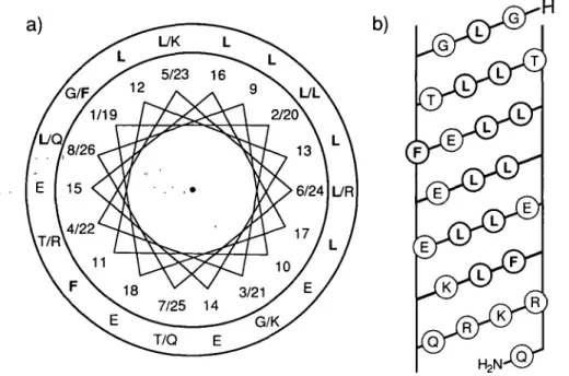
A sequential comparison between HELP and three other lyrically active peptides is illustrated in Figure 1. The distribution of the hydrophobic and hydrophilic amino acid residues in the putative helix are demonstrated in the helical-wheel plot and the helical net diagram (see Figure 2). The mean helical hydrophobic mo-ment ( < / i > ) and the mean hydrophobicity ( < H > ) of HELP (residues 12-22), which were calculated according to Eisenberg etal. (1984) using the normalized consensus hydrophobicity scale, were 0.736 and 0.022, respectively. The values obtained are typical for a 'surface-seeking' amphiphilic a-helix in peptides which promote cell lysis (Eisenberg etal., 1984). However, HELP did not have an amphiphilic a-helical structure in the strict sense because there are non-hydrophilic residues (Fll) on the hydrophilic side of the helix and vice versa (K23, R24) (see Figures 2 and 3). As a consequence, the values for </*> and < H > of the entire HELP helix (residues 1-26) were rather small, 0.348 and 0.043, respectively.
Secondary structure prediction methods in combination with computer modelling techniques have been used to investigate the conformation^ constraints. A molecular model of HELP simulated by computer graphics and energy minimization techniques indicated that well-defined a-helical conformations could be obtained. The structure of the putative helix generated by these studies (see computer modelling) is displayed in Figure 3. At neutral pH residues Glyl (a-NH3+); GlulO, 14, 15, 18;
Lys21, 23 and Arg22, 24 are in the ionized state. The analysis of the distribution of oppositely charged residues in the molecular model shows that at pH 7 only Glu"18/Lys+21 (i/i + 3) or
Glu~18/Arg+22 (i/i + 4) are properly positioned to form an
intrahelical salt bridge and could therefore contribute additional stability to the structure, whereas at acidic or basic pH, helix stabilization through singly charged H bonds between Glu°18/Lys+21 or Glu°18/Arg+22 and between GhT18/Lys°21
or Glu"18/Arg°22 respectively could occur. In an environment of low dielectric constant such as methanol (MeOH) or the hydrophobic interior of a membrane these electrostatic inter-actions between ionic residues even are enhanced.
A) H B) H 1 Gly Gly C) H Leu D) HCOMet 2 Leu lie Leu Ala 3 Gly Gly Gin Gin 4 Thr Ala Ser Asp 5 Leu Val Leu He 6 Leu Leu Leu lie Thr Lys Ser Ser 8 Leu Val Leu Thr 9 Leu Leu Leu He 10 Glu Thr Gin 11 Phe Thr Ser Gly Asp 12 Leu Gly Leu Leu 13 Leu Leu Leu Val 14 15 16 17 18 19 20 21 22 23 24 25 26 Glu Glu Pro Ala Ser Leu Glu Ser Gin Phe| Trp Leu lie TrpjLeu
Lys Trp llejllle Asp Thr Val Asn ^ y s Phe Thr Lys Lys OH NH2
NH2
NH2
Fig. 1. Sequential comparison between the lytic active peptides HELP (A), BVM from Apis mellifera (B), (Eisenberg, 1984), synthetic model toxin (C), (DeGrado el ai, 1981) and 5-haemolysin from Staphylococcus aureus (D), (Tappin et ai, 1988). Boxed amino acid residues in corresponding sequence positions are either identical or have similar properties.
b)
Fig. 2. (a) axial projection of the a-helical structure of HELP with the N-terminus to C-terminus orientation arranged from the top to the bottom. The
segregation of the hydrophilic and hydrophobic residues, printed in bold, is shown in the helical wheel diagram. The N-terminal residue Gly I is placed at position number one and the second position on the helical wheel, which is occupied by Leu 2 is located at an angle displaced by 100° from position number one and so on. (b) Surface net representation of the a-helical structure of HELP. The helical cylinder was split lengthwise along the hydrophilic face opposite the hydrophobic Leu area and the distribution of nonpolar residues, printed in bold, is illustrated.
Fig. 3. The wire frame structure of the minimal energy conformation of the putative a-helix obtained by molecular modelling studies of the conformation of
HELP at acidic pH (ionization state of the molecule, Gly + 1 (a-NH3 +); Glu°10, 14, 15, 18; Lys+21, 23 and Arg+22, 24) is displayed in a stereoview. For the sake of clarity H atoms are omitted in the representation.
However, the modelling studies also supported the pH-induced helix/coil transition model produced by the design strategy (see design and structure-function relationship) with a strong helix perturbation effect at pH 7 induced by charge repulsion of the central, ionized Glu residues: Glu~10/Glu~14 (i/i + 4), Glu"14/Glu"18 (i/i + 4) and Glu~15/GhT18 (i/i + 3). 326
Circular dichroism
Helix formation in HELP was studied under various conditions, and the CD spectra recorded were of qualitatively similar shape differing only in the absolute values of [ 9 ] . In the pH-range 2.0-12.0 HELP was soluble at high concentration in aqueous buffers. In the presence of 100 mM NaCl a sharp decrease in
260
200 220 240 Wavelength (nm)
260
Fig. 4. (a) The far-UV CD spectra represent a theoretical spectrum derived
from the CONTIN program (Provencher and Glockner, 1981) for a protein of 100% a-helix content ( • ) overlaid on the measured HELP spectrum (—) of the peptide (275 ^M) dissolved in 10 mM NaOAc, pH 4.2 (b) CD spectra of HELP dissolved in MeOH adjusted to 'pH' 3 0 (—), 5.0 (—) and 8.0 ( ).
the solubility was observed above pH 4.5. The CD spectral features and the absolute mean residue ellipticities were not significantly affected by the addition of 100 mM NaCl to HELP dissolved in buffers at different pH values. In addition, in the 'pH'-range 3.0-8.0 HELP was soluble in methanolic solution (MeOH adjusted to the desired pH with either 2 N aqueous HC1 or NaOH). In aqueous buffers, the helicity as derived from the ellipticity [9] at 222 nm was concentration dependent (see Figure 5) and an aggregation induced increase of the estimated a-helix content with increasing peptide concentration could be detected The increase in the helicity reached a plateau at ~ 50 /tM peptide with [9]222 = ~29 300 deg cm2/dmol. In the presence of
7.2 M GuHCl or 8.1 M urea, the concentration-dependence of the curve is shifted towards a higher peptide concentration indicating a decreased stability of the folded, aggregated form of HELP (see Figure 5). Thus, in the self-associated a-helical state, HELP exhibited considerable stability to high concentrations of chaotropic denaturants whereas at low peptide concentration the structure appears to be more susceptible to denaturation and it was under these conditions that HELP was found to exist in a predominantly random coil conformation.
The influence of charge effects on a-helix formation was evaluated by measuring CD spectra of the fully aggregated form of HELP as a function of side-chain ionization. The pH-dependence of helix formation, as estimated by [9]222> >s
displayed in Figure 6a. In aqueous buffers in the basic pH range the helicity increased slightly, indicating that this effect arose from titration of the €-amino group of lysine, whereas in methanolic solution HELP seemed to be most stable at acidic pH (see Figure 4b). It is interesting to note that HELP showed no significant, pH-triggered conformational changes (random coil/a-helix transition) as the CD spectra remained almost unaffected at different pH values.
The helical structure of HELP at acidic pH should be destabilized if the temperature is increased and, to investigate this, CD spectra were recorded as a function of temperature between 5.5 and
©
50 100 150
[peptide] |iM
Fig. 5. Peptide concentration dependence of the mean residue ellipticity
[G]222- CD spectral measurements were made at 22°C on HELP dissolved in 10 mM NaOAc, pH 4.2 ( • ) , 10 mM NaOAc, 8.1 M urea, pH 4.2 ( • ) , and 10 mM NaOAc, 7.2 M GuHCl, pH 4.2 (A).
1 3 5 7 9 11 13 0 10 20 30 40 50 60 70 pH Volume %TFE
Fig. 6. (a) pH dependence of the mean residue ellipticity [0)222 f°r HELP (200 fiM). (b) TFE concentration dependence of the mean residue ellipticity [O]222- CD spectral measurements were made on HELP (80 pM) dissolved in 10 mM NaOAc buffer, pH 4.2 with increasing TFE concentration.
95 °C. The helical content of HELP was linearly dependent on temperature as estimated from [9]208 an<J [9]222 values (see
Figure 7) and showed maximal helicity at low temperatures, while increasing the temperature gradually unfolded the helical structure, although a complete conformational transition was never observed. The thermal unfolding transition was broad and fully reversible, neither reaching 100% helix formation at low temperatures nor 100% random coil formation at high temperatures. As the temperature was increased the CD spectra showed an isodichroic point at 201 nm, which might indicate the existence of a transition between only two conformational forms, a-helix and random coil.
The characteristic CD spectrum of HELP is displayed in Figure 4. The spectrum in the 190-240 nm region with a maximum at 193 nm and local double minima at 208 nm and 222 nm is typical for all-a-helical peptides (Greenfield and Fasman, 1969) and is highly suggestive of the presence of a,unique folded, helical structure. To study the conformation of the peptide in a hydrophobic environment, CD spectral measurements were made on HELP dissolved in MeOH and in aqueous buffers containing increasing concentrations of 2,2,2-trifluoroethanol (TFE), which is known to enhance intramolecular interactions and therefore has been used as an a-helical structure-inducing and stabilizing solvent (Nelson and Kallenbach, 1986). In methanolic solution neither pH-triggered a-helix/coil transition (see Figure 4b) nor concentration dependent changes in the helicity were observed in the peptide concentration range of 1 —500 pM at acidic pH, indicating that HELP formed no aggregates under these conditions. Addition of TFE to HELP dissolved in 10 mM 327
20 40 60 Temperature (°C)
80 100
Fig. 7. Variation of the mean residue elhpticity [O]2o8 ( d ) a n^ [Q]222 (M) as a function of temperature. CD spectral measurements were made on
HELP (620 fiM) dissolved in 10 mM NaOAc, pH 4.2.
NaOAc buffer, pH 4.2 resulted in a further increase in the a-helical content which reached a plateau at - 4 0 % TFE with [e]2 2 2 = - 3 6 100 deg cm2/dmol (see Figure 6b).
A rough evaluation of the secondary structure content of HELP gave 100% a-helix [formula (a), (Greenfield and Fasman, 1969)] and 89% a-helix [formula (b), (Chen etal, 1972)], respectively.The CONTIN program of Provencher and Glockner has been thought to give the most accurate results of the common methods used for secondary structure calculation of peptides from CD spectra. Under aqueous solution conditions (10 mM NaOAc, pH 4.2, 22°C) and in the fully aggregated state ( > - 5 0 /*M, see Figure 5) HELP was calculated to be - 100% helical. The computer-simulated fit of the CD spectrum for a peptide containing 100% a-helix is in excellent agreement with the spectrum of HELP as illustrated in Figure 4a. However, several factors influence and complicate the estimation of the helical content in short peptides, including end effects produced by residues not H-bonded to the backbone at the distorted N- and C-termini of the a-helix, the contribution of aromatic amino acid residues and the lack of absolute reference spectra of a peptide in a complete helical or random coil state. In addition, CD signals for a-helices are known to be chain length dependent (Chen et al., 1974) and therefore the calculated values should be taken only as an approximation of the a-helix content of HELP. Haemolysis
The lytic activity of HELP on biological membranes can be conveniently monitored by the release of haemoglobin from hRBC. The percentage haemolysis was related to that of a completely lysed reference sample. In the assay no spontaneous haemolysis was observed in the absence of peptide. Comparative experiments were all performed on the same hRBC suspensions and BVM was always included as a positive control. At acidic pH, haemoglobin release by HELP appeared to follow kinetics comparable to those measured for BVM at neutral pH. In addition, the dose response curves for the extent of leakage are almost identical (see Figure 8). It was shown by titration experiments that, during the membrane disruption process, HELP was not released to act in a second lytic cycle and, once membrane associated or integrated, it was unable to lyse further cells. For HELP, BVM and a fragment of HELP (residues 9-26) a series of haemolysis assays were performed over a peptide concentration range of 0—50 /tM. Since HELP and BVM reached the same plateau value of 100% lytic activity at peptide
0.4 [peptide]
Fig. 8. The haemolytic activity of HELP compared with that of BVM using
hRBC as target. Lysis is expressed in z units by using the Poisson analysis (z = - I n [1 - y], where y is the fraction of lysed cells as determined by the haemoglobin release in the haemolytic assay at 37°C for HELP ( • ) , BVM ( • ) and HELP fragment (A) at pH 5.0 and for HELP ( • ) , BVM (O) and HELP fragment (A) at pH 7.5. The inset shows the HELP-induced leakage of hRBC at different pH values. Lysis measurements were made on HELP dissolved in the following buffers each containing 116 mM NaCl: 20 mM NaOAc, pH 4.8; pH 5.0; pH 5.5, 20 mM potassium phosphate, pH 6.0; pH 6.5; pH 7.0 and 20 mM Tris-HCI, pH 7.5; pH 8.0.
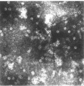
Fig. 9. Electron micrograph (magnification factor: 294 000) of pores in
membrane preparations of HELP-lysed hRBC. For details see electron microscopy section.
concentrations higher than — 1 /iM, only the concentration-dependent range of 0 - 1 /tM is displayed in Figure 8. Peptide-induced leakage of haemoglobin from hRBC was pH-dependent. The extent of leakage as a function of pH with a constant HELP/hRBC ratio was recorded (see Figure 8 inset). At pH 5.0 the lytic activity was maximal and dropped to zero as the pH increased. Incubation of hRBC and HELP at pH 7.5 did not lead to the release of haemoglobin from erythrocytes. In membrane preparations of HELP-lysed hRBC, round pores with an outer diameter of 8.3-10 nm and an inner diameter of 2.3-2.9 nm were detected by transmission electron microscopy (see Figure 9).
Discussion
Design and structure of HELP
A 26 residue peptide that induces pH-triggered haemolysis in hRBC at pH 5.0 has been designed, synthesized and characterized by several methods. Secondary structure prediction analysis (Argos et al., 1976, 1982) and computer modelling (see Figure 3) suggest that HELP would have a high a-helical structure content. Indeed, all results so far from CD measurements are consistent with the assumed secondary structure model. CD studies and analytical ultracentrifuge experiments (results not shown) performed to establish the aggregation state of the molecule strongly support the assumption that HELP forms a self-aggregation-induced, highly a-helical structure in purely aqueous solution at sufficiently high peptide concentrations ( > ~ 50 /tM) whereas, at lower concentrations the CD spectra indicate a considerably lower helical content (see Figure 5). Therefore, it appears to be reasonable to assume a dynamic model, with an interconversion process between the monomeric random coil and the aggregated a-helical state, where the HELP molecule most probably populates multiple low energy conforma-tions rather than occupying a single rigid structure, but preferen-tially adopts a well-defined a-helical conformation upon association. A possible explanation for the driving force of self-association, is that Leu residues with a high helical potential are tightly packed in an amphiphilic a-helix and are thus optimally shielded from the polar environment in the aqueous phase by aggregation along the hydrophobic faces. These strong inter-molecular attractive interactions between hydrophobic regions complemented by interhelical salt bridges (ion pairs) may also suppress the unfolding tendency of the helix at neutral pH and thus, might offer a rationale for the fact that HELP exhibits no pH-dependent random coil/helix transition. It is interesting to note that, despite the presence of only one intrahelical pair of amino acid residues positioned to form salt bridges (Glu~18/Lys+21
or Glu"18/Arg+22) or singly charged H bonds (Glu°18/Lys+21
or Glu°18/Arg+22) at pH 4.2 (Marqusee and Baldwin, 1987),
HELP demonstrates a considerable stability towards both chemical and thermal denaturation (see Figures 5 and 7). These observations underline the importance of strong HELP-HELP interactions which not only govern the monomer-aggregate equilibrium but also effectively stabilize the helical conformation even under strongly chaotropic conditions.
CD spectroscopy is a very sensitive tool with which to trace structural changes. As shown by CD measurements the helicity can be further increased by the addition of TFE (see Figure 6b). Although these findings are indicative of additional structure induction (e.g. in unordered regions at the N-, C-termini), helix stabilizing effects (enhanced H-bonding geometry) or an increased helix population (random coil-helix interconversion) cannot be ruled out.
The fact that the peptide concentration in MeOH did not affect the CD spectra strongly indicates that under these conditions HELP remained monomeric while maintaining a stable a-helical conformation. This assumption was further confirmed by NMR studies on the aggregation state of HELP in MeOH. Since the CD spectra of HELP dissolved in MeOH and in aqueous buffers are quite similar (see Figure 4) it is reasonable to assume that the molecular geometry of HELP is essentially the same in both solvents. The CD results, which reveal a unique folded a-helix, have been fully confirmed by the detailed investigations of the solution conformational properties of HELP using high-resolution 'H 2D NMR-spectroscopy and molecular dynamic simulations
(Klaus and Moser, 1992). These findings suggest that the hydro-carbon portion of MeOH could replace the interhelical hydrophobic contacts in aggregates and therefore the solution con-formation of HELP in MeOH, which might to some degree mimic the hydrophobic interior of a membrane, could also repre-sent a model for a single transmembrane HELP pore constituting building block.
Structure-junction relationship and comparison with models Amphiphilic a-helices are thought to represent the sites of interaction between haemolytic peptides and lipid bilayers and to be responsible for lytic function. Since haemolytic activity of HELP and BVM can be continuously monitored on going from peptide concentrations < 1 /iM to the concentration range of 1 - 5 0 fiM, where the activity curves of both peptides reach the same plateau value (100% lysis), it becomes of interest to explain how membrane disrupting activity could be related to the different conformations which, as shown by the CD studies (see Figure 5), are present in the monomer-aggregate equilibrium. HELP can be considered as a BVM analogue. Indeed, both peptides adopt an a-helical conformation in the self-aggregated state in water and exist as a monomeric a-helix in MeOH. However, it should be noted that HELP has a stronger tendency to form aggregates than BVM and therefore shows substantial helicity ([6]222 = —13 400 deg cm2/dmol) at a peptide concentration
of 1 nM (see Figure 5), whereas BVM at the same concentration at pH 7.5 is expected to be in a predominantly monomeric random coil state (Dawson et al., 1978; Brown etal, 1980). Furthermore, CD and NMR studies indicate that in aqueous solu-tion monomeric BVM preferentially exists in an unordered conformation, whereas in the presence of membranes or in MeOH, BVM monomers adopt an a-helical conformation similar to that in the tetrameric state (Brown et al., 1980; Bazzo et al., 1988). These findings, when considered together, support the assumption that monomeric HELP and BVM reflect structural similarities in that they probably form an amphiphilic a-helix in the membrane-active conformation when bound to the polar surface of the lipid bilayer. Thus, a preformed amphiphilic conformation seems not necessarily to be a basic requirement for lytic activity.
Since no membrane disrupting activity (transmembrane pore formation) was observed at neutral pH, the formation of the lytically active conformation seemed to be correlated to the increased hydrophobicity upon protonation of the Glu side chains, and gradual deprotonation of these side chains with increasing pH might prevent HELP from penetrating into the apolar lipid core. In this connection, it is interesting to note that BVM is known to induce fusion of lipid vesicles at neutral as well as at acidic pH values, whereas succinylated BVM cannot induce fu-sion at neutral pH but initiates rapid and efficient fufu-sion at acidic pH, suggesting that the membrane fusion activity of succinylated BVM is triggered by protonation of its carboxyl groups (Murata etal, 1987). Thus, HELP and succinylated BVM are both sensitive to pH-induced charge effects leading to altered func-tional properties. It is known that truncated forms of BVM (residues 7 - 2 6 and 8-26) are only slightly active whereas BVM (residues 1 —20) is completely inactive but is capable of binding to erythrocytes membranes (Schroder et al., 1971). A fragment of HELP (residues 9—26) was also studied in order to deter-mine the influence of chain length on the hRBC disruption process. This shortened HELP version, lacking the helix-inducing N-terminal sequence, showed a drastically decreased haemolytic potency at pH 5.0 (see Figure 8).
At present it is difficult to provide more than a phenomeno-logical description of the haemolytic activity of HELP, since peptide induced damage to cell membranes by both pore-forming and other leakage-inducing mechanisms are complex and remain to be established in detail (Bhakdi and Tranum-Jensen, 1987) even in the case of BVM, whose membrane-disrupting effects have been studied extensively. A possible model for an initial peptide^lipid complex and for peptide-induced membrane destabilization (disordering and reorientation of lipid molecules) could be described as follows: (1) the basic N-terminal hexa-peptide portion of the hexa-peptide anchors HELP to the hRBC surface through salt bridges between positively charged side chains (Lys+ and Arg+) and the phosphate groups of the phospholipids
in the hRBC membrane; (2) extensive structural rearrangements upon spontaneous incorporation of HELP into the lipid bilayer occur; (3) polar membrane-spanning pores are formed by HELP clusters, exposing the hydrophobic face of the amphipathic a-helices to the hydrocarbon chains of the hydrophobic lipid core; (4) the cell content leaks through the membrane-embedded pores which now have a polar inner surface.
Indeed, transmembrane pores in membrane preparations of hRBC treated with HELP at pH 5.0 are clearly visible in electron micrographs as characteristic ring-shaped structures partially buried within the lipid bilayer (see Figure 9), whereas in HELP-treated membrane preparations at pH 7.5 neither lytic activity nor pores could be detected. The electron microscopy results thus confirmed that pore formation (integration of HELP into the lipid bilayer) is critically governed by the pH-dependent ionization state of the molecule. Therefore, it appears to be reasonable to assume a direct correlation between the leakage (transmembrane passage) of haemoglobin from hRBC and the formation of peptide/protein-lined pores in the membrane. If the observed membrane structures are pores with a hydrophilic interior, then packing considerations imply that a minimal number of ~ 18—20 aggregated helices must be involved in the formation of this circular ultrastructure (calculated for a pore of 2.3—2.9 run inner diameter). A similar model, involving a cluster of four molecules in which the polar faces point inwards into the pore (ion channel), has been suggested by Vogel and Jahnig (1986) to explain the interaction of BVM with membranes upon which the toxin is thought to act by traversing the lipid bilayer and Lear et al. (1988) synthesized model ion channel peptides, which were designed to form pores composed of bundles of aggregated membrane-spanning amphiphilic a-helices. Alamethicin, a 20-residue peptide antibiotic, acts by forming voltage-gated transmembrane ion channels through aggregation in the lipid bilayer (Fox and Richards, 1982; Bernheimer and Rudy, 1986). Furthermore, a membranolytic Entamoeba histolytica peptide that causes pore formation by insertion into the target-cell membranes was found to form oligomers in solution or bound to membranes and its N-terminal sequence revealed a structural similarity to BVM (Leippe etai, 1991). As an, alternative transmembrane pore forming mechanism, aggregation of integral hRBC membrane proteins might be initiated by the amphipathic HELP molecule binding to these proteins and perturbating their structures. The formation of pores by aggregation of band 3, the major integral protein of erythrocyte membranes, has been discussed by Clague and Cherry (1988) who studied the p25 signal peptide- and BVM-induced hRBC haemolysis and band 3 aggregation.
The work presented here is a contribution towards a more coherent picture of the structure—function relationship of an amphipathic peptide specifically designed to interact with lipid bilayers in a pH-dependent fashion. Furthermore, the possibility
of constructing a peptide with a predetermined conformation and biological activity should enable the factors which influence peptide and protein folding to be better understood. Starting with model building, followed by synthesis, chemical and spectroscopical characterization of HELP and finally the comparison between the designed and the determined structure closes the circle and should allow the accuracy of model construction to be improved, and to design more sophisticated a-helices.
Acknowledgements
I thank Prof. B. Gutte for the support of this project and Dr S.Klauser for assistance in the computer modelling studies. I thank Dr A.Schaffer and M.Cavigelli for support in analytical ultracentrifuge experiments and Prof. P.Groscurth for advice, constant encouragement and the excellent electron micrographs. Also I would like to thank Dr R.Frank for his friendly support, Dr W.Klaus for nuclear magnetic resonance measurements and many stimulating discussions and dipl. Ing. H. Dirks for conducting FAB measurements. I am grateful to Dr R.M.Thomas and C.J.Henehan for critical discussions and careful reading of the manuscript. This work was supported by a grant of the Swiss National Science Foundation to B.Gutte.
References
Argos,P., Schwarz,J. and Schwarz,J. (1976) Biochim. Biophys. Acta, 439, 261-273.
Argos.P. and Palau.J. (1982) Int. J. Peptide Protein Res., 19, 380-393. Argos.P., RaoJ.K.M. and Hargrave.P.A. (1982) Eur. J. Biochem.. 128,
565-575.
Baldwin,R.L. and Eisenberg,D. (1987) In Oxender.D.L. and Fox.C.F. (eds),
Protein Engineering. Alan R. Liss, Inc., New York, pp. 127-148.
Bazzo.R., Tappin,M.J., Pastore,A., Harvey,T.S., CarverJ.A. and CampbellJ.D. (1988) Eur. J. Biochem., 173, 139-146.
Bello,J., Bello.H.R.and Granados,E. (1982) Biochemistry, 21, 461-465. Bernheimer.A.W. and Rudy.B. (1986) Biochim. Biophys. Acta, 864, 1 2 3 - 141. Bhakdi,S. andTranum-Jensen,J. (1987) Rev. Physiol. Biochem. Pharmacol., 107,
147-223.
Blondelle,S.E. and Houghten,R.A. (1991) Biochemistry, 30, 4671-4678. Bradley,E.K.,Thomason,J.F., Cohen,F.E., Kosen,P.A. and KuntzJ.D. (1990)
J. Mol. Bioi, 215, 607-622.
Brems.D.N., Plaisted,S.M., Kauffman.E.W., Lund.M. and Lehrman.S.R. (1987)
Biochemistry, 26, 7774-7778.
Brown.L.R., Lauterwein,J. and Wuthrich.K. (1980) Biochim. Biophys. Acta, 622, 231-244.
Chen,Y.-H., Yang,J.T. and Martinez.H.M. (1972) Biochemistry, 11,4120-4131. Chen,Y.-H., Yang.J.T. and Chau.K.H. (1974) Biochemistry, 13, 3350-3359. Chothia.C, Levitt,M. and Richardson.D. (1977) Proc. Natl Acad. Sci. USA,
74, 4130-4134.
Chothia.C. (1984) Annu. Rev. Biochem., 53, 537-572. Chou,P.Y. and Fasman.G.D. (1974) Biochemistry, 13, 211-221. Clague.M.J. and Cherry,R.J. (1988) Biochem. J., 252, 791-794.
Clore,G.M., Gronenborn.A.M., Briinger.A.T. and Karplus.M. (1985) J. Mol.
Bioi, 186,435-455.
Cohen.C. and Parry.D.A.D. (1990) Proteins, 7, 1-15.
Dawson,C.R., Drake.A.F., Helliwell,J. and Hider,R.C. (1978) Biochim. Biophys.
Acta, 510. 7 5 - 8 6 .
DeGrado.W.F. (1988) Adv. Protein Chem., 39, 51-118.
DeGrado,W.F., Kezdy.F.J. and Kaiser.E.T. (1981)/ Am. Chem. Soc, 103, 679-681.
DeGrado,W.F., Wasserman.Z.R. and Lear.J.D. (1989) Science, 243, 622-628. Dill.K.A. (1990) Biochemistry, 29, 7133-7155.
Eisenberg,D. (1984) Annu. Rev. Biochem., 53, 595-623.
Eisenberg.D., Schwarz.E., Komaromy.M. and Wall.E. (1984) J. Mol. Bioi.
179, 125-142.
Fox,R.O. and Richards.F.M. (1982) Nature, 300, 325-330.
Ghadiri.M.R. and Choi.C. (1990) J. Am. Chem. Soc, 112, 1630-1632. Greenfield.N. and Fasman,G.D. (1969) Biochemistry, 8, 4108-4116. Hecht,M.H., Richardson.J.S., Richardson.D.C. and Ogden.R.C. (1990) Science,
249, 884-891.
Ho,S.P. and DeGrado.W.F. (1987) J. Am. Chem. Soc. 109, 6751-6758. Ikura.T., Go,N. and Inagaki.F. (1991) Proteins, 9, 81-89.
Klaus,W. and Moser.R. (1992) Protein Engng, 5, 333-341. Konig.W. and Geiger.R. (1970) Chem. Ber., 103, 788-798.
Lear,J.D., Wasserman.Z.R. and DeGrado,W.F. (1988) Science, 240. 1177-1181.
Leippe.M., Ebel.S., Schoenberger.O.L., Horstmann,R.D. and Muller-Eberhard.H.J. (1991) Proc. Natl Acad. Sa. USA, 88, 7659-7663. Marqusee,S. and Baldwin,R.L. (1987) Proc. Natl Acad. Sci. USA, 84,
8898-8902.
Marqusee,S., Robbins.V.H. and Baldwin.R.L. (1989) Proc. Natl Acad. Sci. USA, 86, 5286-5290.
Merutka.G. and Stellwagen.E. (1989) Biochemistry, 28, 352-357. Merutka.G., Lipton.W., Shalongo.W., Parks,S.-H. and Stellwagen.E. (1990)
Biochemistry, 29, 7511-7515.
Murata.M., Nagayama.K. and Ohnishi.S. (1987) Biochemistry. 26, 4056-4062. NelsonJ.W. and Kallenbach,N.R. (1986) Proteins, 1, 211-217.
O'Shea.E.K., Rutkowski,R., Stafford.W.F. and Kim.P.S. (1989) Science, 245, 646-648.
Padmanabhan,S., Marqusee,S., Ridgeway.T., Laue.T.M. and Baldwin.R.L. (1990) Nature, 344, 268-270.
Palau.J., Argos.P. and Puigdomenech,P. (1982) Int. J. Peptide Protein Res.,
19, 394-401.
Provencher.S.W. and G16ckner,J. (1981) Biochemistry, 20, 3 3 - 3 7 . Quay.S.C. and Condie,C.C. (1983) Biochemistry, 22, 695-700.
Raghunathan,G., Seetharamulu.P., Brooks.B.R. and Guy.H.R. (1990) Proteins, 8, 213-225.
Regan.L. and DeGrado.W.F. (1988) Science, 241, 976-978.
Richardson.J.S. and Richardson,D.C. (1988) Science, 240, 1648-1652. Roongta.V , Powers,R., Jones,C, Beakage.M.J., Shields,J.E. and
Gorenstein.D.G. (1989) Biochemistry, 28, 1048-1054.
Schroder.E., Lubke.K., Lehmann,M. and Beetz.I. (1971) Experientia, 27, 764-765.
Stewart.J.M. and Young.J.D. (1984) Solid Phase Peptide Synthesis. Pierce Chemical Co., Rockford, Illinois, 2nd edn.
Subbarao,N.K., Parente.R.A., Szoka,Jr.,F.C, Nadasdi.L. and Pongracz,K. (1987) Biochemistry, 26, 2964-2972.
Tappin.M.J., Pastore.A., Nortnn.R.S., Freer,J.H. and Campbell.I.D. (1988)
Biochemistry, 27, 1643-1647.
Terwilliger,T.C. and Eisenberg.D. (1982) J. Biol. Chem., 257, 6016-6022. Thiaudiere.E., Siffert,O., Talbot,J.C, Bolard.J., Alouf,J.E. and Dufourcq.J.
(1991) Eur. J. Biochem., 195, 203-213.
Vogel.H. and Jahnig.F. (1986) Biophys. J., 50, 573-582.
Wright.P.E., Dyson.H.J. and Lemer.R.A. (1988) Biochemistry, 27, 7167-7175. Yang.D.S.C, Sax,M., Chakrabartty.A. and Hew.C.L. (1988) Nature, 333,
232-237.
Zimm.B.H. and Bragg.J.K. (1959)/ Chem. Phys., 31, 526-535.
Received on November 5, 1991; revised and accepted on February 20, 1992
![Fig. 5. Peptide concentration dependence of the mean residue ellipticity [G] 2 22- CD spectral measurements were made at 22°C on HELP dissolved in 10 mM NaOAc, pH 4.2 ( • ) , 10 mM NaOAc, 8.1 M urea, pH 4.2 ( • ) , and 10 mM NaOAc, 7.2 M GuHCl, pH 4.2 (A](https://thumb-eu.123doks.com/thumbv2/123doknet/14897504.652180/5.916.150.388.85.430/peptide-concentration-dependence-residue-ellipticity-spectral-measurements-dissolved.webp)