Publisher’s version / Version de l'éditeur:
Vous avez des questions? Nous pouvons vous aider. Pour communiquer directement avec un auteur, consultez la première page de la revue dans laquelle son article a été publié afin de trouver ses coordonnées. Si vous n’arrivez pas à les repérer, communiquez avec nous à PublicationsArchive-ArchivesPublications@nrc-cnrc.gc.ca.
Questions? Contact the NRC Publications Archive team at
PublicationsArchive-ArchivesPublications@nrc-cnrc.gc.ca. If you wish to email the authors directly, please see the first page of the publication for their contact information.
https://publications-cnrc.canada.ca/fra/droits
L’accès à ce site Web et l’utilisation de son contenu sont assujettis aux conditions présentées dans le site LISEZ CES CONDITIONS ATTENTIVEMENT AVANT D’UTILISER CE SITE WEB.
Journal of Lipid Research, 36, pp. 1905-1918, 1995
READ THESE TERMS AND CONDITIONS CAREFULLY BEFORE USING THIS WEBSITE. https://nrc-publications.canada.ca/eng/copyright
NRC Publications Archive Record / Notice des Archives des publications du CNRC : https://nrc-publications.canada.ca/eng/view/object/?id=5db8734f-3fcb-44a0-8bbe-5ede989cab25 https://publications-cnrc.canada.ca/fra/voir/objet/?id=5db8734f-3fcb-44a0-8bbe-5ede989cab25
NRC Publications Archive
Archives des publications du CNRC
This publication could be one of several versions: author’s original, accepted manuscript or the publisher’s version. / La version de cette publication peut être l’une des suivantes : la version prépublication de l’auteur, la version acceptée du manuscrit ou la version de l’éditeur.
Access and use of this website and the material on it are subject to the Terms and Conditions set forth at
Molecular characterization of two monoclonal antibodies specific for
the LDL receptor-binding site of human apolipoprotein E
Raffai, Robert; Maurice, Roger; Weisgraber, Karl; Innerarity, Thomas; Wang,
Xingbo; MacKenzie, Roger; Hirama, Tomoko; Watson, David; Rassart, Eric;
Milne, Ross
Molecular characterization of two monoclonal
antibodies specific for the
LDL
receptor-binding site
of human apolipoprotein E
zyxwvutsrqponmlkjihgfedcbaZYXWVUTSRQPONMLKJIHGFEDCBA
Robert
zyxwvutsrqponmlkjihgfedcbaZYXWVUTSRQPONMLKJIHGFEDCBA
-ai,*.** Roger Maurice,? Karl Weisgraber,Q Thomas Innerarity,§ XingboWang,+** Roger MacKenzie,tt Tomoko Hirama,tt David Watson,tt Eric Rassart,**
and Ross Mhelg*gt
zyxwvutsrqponmlkjihgfedcbaZYXWVUTSRQPONMLKJIHGFEDCBA
Lipoprotein and Atherosclerosis Group, University of Ottawa Heart Institute and Departments of Pathology and Biochemistry,* University of Ottawa, Ottawa, Canada; Institut de Recherches Cliniques de Montreal,? Montreal, Canada, Gladstone Institute of Cardiovascular Disease, Department of Pathology,§ Cardiovascular Research Institute, University of California, San Francisco, San Francisco, CA: Departement des Sciences Biologiques,** Universitt du Quebec & Montreal, MontrM, Canada; and Institute for Biological Sciences,?? National Research Council of Canada, Ottawa, Canada
Abstract Apolipoprotein E (apoE), a 299 amino acid pro- tein, is a ligand for the low density lipoprotein receptor (LDLr). It has been established that basic amino acids situated between apoE residues 136 and 150 participate in the inter- action of apoE with the LDLr. Evidence suggests that apoE is heterogeneous on lipoproteins in its conformation and in its ability to react with cell surface receptors. Our goal was to produce mAbs that could serve as conformational probes of the LDLr binding site of apoE. We used a series of apoE variants that have amino acid substitutions at residues 136, 140, 143, 144, 145, 150, 152, and 158 to identify the epitopes of two anti-human apoE monoclonal antibodies (mAbs), 1D7 and 2E8, that inhibit apoE-mediated binding to the LDLr. We show that most of the variants that have reduced reactivity
with the LDL receptor
zyxwvutsrqponmlkjihgfedcbaZYXWVUTSRQPONMLKJIHGFEDCBA
also have reduced reactivity with the mAbs. The epitopes for both mAbs appear to include resi-dues 143 through 150 and thus coincide with the LDLr-bind- ing site of apoE. It is notable that mAb 2E8, but not 1D7, resembles the LDLr in showing a reduced reactivity with
apoE
zyxwvutsrqponmlkjihgfedcbaZYXWVUTSRQPONMLKJIHGFEDCBA
(Arg*58+
Cys), While most of the receptordefective variants involve replacement of apoE residues directly impli-cated in binding, substitution of Arg'58 by Cys is thought to indirectly affect binding of apoE to the LDLr by altering the conformation of the receptor-binding site. To determine whether the similarity in specificities of the mAbs and the LDLr reflect structural similarities, we cloned and charac- terized the cDNAs encoding the light and heavy chains of
both mAbs. Primary sequence analysis revealed that, al- though these two antibodies react with overlapping epitopes, their respective complementarity determining regions (CDRs) share little homology, especially those of their heavy chains. The two mAbs, therefore, likely recognize different epitopes or topologies within a limited surface of the apoE molecule. Four negatively charged amino acids were present in the second CDR of the 2E8 heavy chain that could be approximately aligned with acidic amino acids within the consensus sequence of the LDLr ligand-binding domain. This could indicate that mAb 2E8 and the LDLr use a common mode of interaction with apoE.-Raffai, R, R Maurice, K.
Weisgraber, T. Innerarity,
X.
W a g , R MacKenzie,T.
Hi-rama, D. Watson,
E.
Rassart, and R. Milne. Molecular char- acterization of two monoclonal antibodies specific for the LDL receptor-binding site of human apolipoproteinE.
J.Lipid Res. 1995.36: 1905-1918.
Supplementary key words monoclonal antibodies LDL receptor surface plasmon resonance
Apolipoprotein (apo) E is a 34-kDa protein consisting of 299 amino acids and is a functionally important constituent of chylomicrons and very low, intermediate, and high density lipoproteins (1). As a ligand for the low density lipoprotein receptor (LDLr), apoE is a major modulator of lipoprotein metabolism. The most com- mon apoE isoforms, apoE2, apoE3, and apoE4, which are distinguishable by isoelectric focusing, are encoded by three codominandy expressed alleles. ApoE3, the most common of the three, is considered to be the wild type, whereas apoE2, apoE4, and the even rarer iso- forms are considered to be variants. The most fre- quently observed form of apoE2 differs from apoE3 by the substitution of a cysteine for an arginine at residue 158, whereas apoE4 differs from apoE3 by the replace- ment of a cysteine by an arginine at residue 112. Variant
Abbreviations: LDLr, low density lipoprotein receptor: CDR,
complementarity determining region: VLDL, very low density lipoprotein; SPR, surface plasmon resonance; DMPC, dimyristoyl- phosphatidylcholine.
'To whom correspondence should be addressed.
Journal of Lipid Research Volume 36, 1995 1905
at National Research Council Canada, on August 28, 2019
www.jlr.org
10
zyxwvutsrqponmlkjihgfedcbaZYXWVUTSRQPONMLKJIHGFEDCBA
8zyxwvutsrqponmlkjihgfedcbaZYXWVUTSRQPONMLKJIHGFEDCBA
6zyxwvutsrqponmlkjihgfedcbaZYXWVUTSRQPONMLKJIHGFEDCBA
-0zyxwvutsrqponmlkjihgfedcbaZYXWVUTSRQPONMLKJIHGFEDCBA
c 3 0 n b 4zyxwvutsrqponmlkjihgfedcbaZYXWVUTSRQPONMLKJIHGFEDCBA
e
a, c 2 01 D7
r* 10 8 6 U czyxwvutsrqponmlkjihgfedcbaZYXWVUTSRQPONMLKJIHGFEDCBA
3
- 45
a, c 2 03H1
&.
II ApoE3.
rApoE3 A Ser-136 v Ala-140 Ala-I43 Pro-144 C~S-145 ei Ala-150 A Pro-152 Cys-15810 100 lo00 lOo00 10 100 lo00 lo000 ng apo E protein I ml
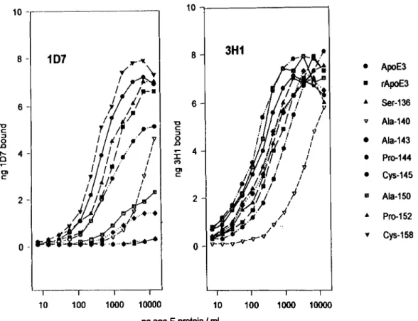
Fig. 1. Immunoreactivity of apoE variants with anti-apoE mAbs 1D7 and 3H1. Serial dilutions of apoE variants were incubated in polystyrene microwells to which anti-apoE mAb 6C5 had been adsorbed. The wells were then washed and exposed to either l*Wabeled 1D7 (left) or lP51-labeled 3H1 (right) and, after washing, the bound radioactivity was determined. The bound *P51-labeled mAb mass was calculated from the specific activity
of the labeled antibodies. Results from one experiment are shown with essentially identical results having been obtained in two other experiments.
forms of apoE, such as ap0E2(Argl~~
+
Cys), that show defective binding to the LDLr are, in part, responsible for the expression of type I11 dyslipoproteinemia. In addition, apoE polymorphism can influence plasma lipid levels (2), postprandial lipemia (3-5), apoB meta-bolism ( 6 ) , the lipoprotein subclass distribution of apoE
zyxwvutsrqponmlkjihgfedcbaZYXWVUTSRQPONMLKJIHGFEDCBA
(7-9), and susceptibility to Alzheimer’s disease (10). Limited proteolytic digestion of apoE has shown that the molecule is composed of two structural domains ( 11, 12), a 22-kDa amino-terminal domain that has relatively low affinity for lipid and includes the residues responsi- ble for binding to the LDLr and a 10-kDa carboxyl-ter- minal domain that binds lipid with high affinity (9). Several lines of evidence (1, 13-18) indicate that posi- tively charged amino acids within the region of residues 136-150 directly participate in the interaction of apoE with the LDLr. The three-dimensional crystal structure of the amino-terminal 22-kDa thrombolytic fragment of human apoE has been solved (19). The fragment is
folded into an elongated four-helix bundle with the positively charged amino acids implicated in receptor binding situated on the fourth helix and exposed to solvent.
The mAb 1D7 is one of a group of anti-human apoE mAbs that we first reported in 1981 (20). Of the six mAbs, only 1D7 is capable of blocking apoE-mediated binding to the LDLr (17). Antibody 1D7 has sub- sequently been used by ourselves and others to distin- guish between apoE- and apoB-mediated lipoprotein binding to cell surface receptors (2 1-23) and as a probe for the identification of the receptor-binding (17) and heparin-binding sites of apoE (24). To define the fine specificity of 1D7, we have now determined its reactivity with a series of apoE variants that differ in their affinities for the LDLr. In parallel with 1D7, we have similarly analyzed the reactivity of a second anti-apoE mAb, ZES, which also blocks binding of apoE to the LDLr. In both cases the respective epitopes of 1D7 and 2E8 appear to coincide with the LDLr-binding site on apoE. We have furthermore determined the nucleotide sequence of the cDNAs coding both antibodies and have demonstrated that the deduced amino acid sequence of the antibody combining site of 2E8 shows homology to the ligand binding consensus sequence of the LDLr. The results are discussed in terms of the potential mechanisms of
binding of apoE to both antibodies and the LDLr.
1906 Journal of Lipid Research Volume 36, 1995
at National Research Council Canada, on August 28, 2019
www.jlr.org
n a
zyxwvutsrqponmlkjihgfedcbaZYXWVUTSRQPONMLKJIHGFEDCBA
E3 rE3zyxwvutsrqponmlkjihgfedcbaZYXWVUTSRQPONMLKJIHGFEDCBA
. . . ~ Ser 136 Ala 140 . . .-
Ala 143 . . . . . .zyxwvutsrqponmlkjihgfedcbaZYXWVUTSRQPONMLKJIHGFEDCBA
J
Pro 144 . . . . . .4
145 . . . . . .-
Ala 150 . . .zyxwvutsrqponmlkjihgfedcbaZYXWVUTSRQPONMLKJIHGFEDCBA
1
152 158zyxwvutsrqponmlkjihgfedcbaZYXWVUTSRQPONMLKJIHGFEDCBA
In triglyceride-rich lipoproteins, apoE appears to be heterogeneous with respect to its conformation or ac- cessibility, with only a subpopulation of molecules being capable of mediating binding to the LDLr (25). It has been reported that expression of the 1D7 epitope on very low density lipoproteins (VLDL) may correlate with the ability of the particle to bind to the LDLr (26). Thus, 1D7 may be specific for a conformational'state of apoE that permits interaction of apoE with the LDLr or may react with a subpopulation of molecules whose receptor binding site is exposed. Our goal is to identify or pro- duce mAbs that would recognize the same conforma- tional structure on apoE as is recognized by the LDLr. These antibodies will be used as conformational probes to study the physical and chemical parameters that modulate expression of the LDL receptor binding site of apoE.
EXPERIMENTAL PROCEDURES
zyxwvutsrqponmlkjihgfedcbaZYXWVUTSRQPONMLKJIHGFEDCBA
Monoclonal antibodies
The production and characterization of mAbs 1D7,
3H1, and
zyxwvutsrqponmlkjihgfedcbaZYXWVUTSRQPONMLKJIHGFEDCBA
6C5 have been described previously (17, 20,24). Hybridoma 2E8 was the product of a fusion be- tween spleen cells of a mouse immunized with purified human apoE3 and the non-secreting plasmacytoma cell line SP2-0. The mAb 2E8 was characterized according
Fig. 2. Relative reactivity of mAb 1D7 with apoE
variants. The relative immunoreactivity of apoE vari-
zyxwvutsrqponmlkjihgfedcbaZYXWVUTSRQPONMLKJIHGFEDCBA
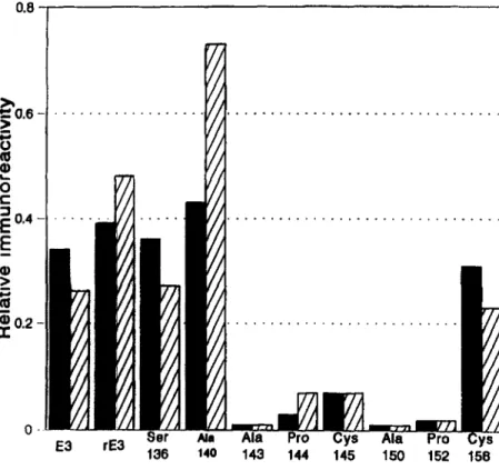
ants (m) with mAb ID7 was calculated from the results presented in Fig. 1 according to the formula: ng apoE necessary to have 3 ng of 1S51-labeled 3H1 bound/ng apoE necessary to have 3 ng of lP51-labeled ID7 bound. Results are also presented from the same experiment for apoE variants that had been incorporated into DMPC vesicles (cross hatched bars).
to the same criteria that were used for the other anti- apoE mAbs (17). The IgG fraction containing the mAbs was purified from the ascites of hybridoma-bearing mice by affinity chromatography on Protein A-Sepharose
(27). IgG was labeled with 1251 as previously described (28).
Preparation of apoE and production of apoE variants
ApoE3, apoE2(Arg158
+
Cys), and apoE2(Arg145 4Cys) were purified from plasma as reported by Weisgra- ber, Rall, and Mahley (29). The generation of the apoE variants apoE(Arg136
+
Ser), a p ~ E ( H i s l ~ ~+
Ala),a p o E ( L y ~ I ~ ~
+
Ala), apoE(Leu144+
Pro), apoE(ArgIS0zyxwvutsrqponmlkjihgfedcbaZYXWVUTSRQPONMLKJIHGFEDCBA
3 Ala), and apoE(Ala15*
+
Pro), their expression in E.coti, and their purification have also been described (18). For comparison of purified plasma apoE and apoE produced by E. coli, purified, bacterially expressed apoE3 was included in some experiments (30). For certain experiments, the isolated apoE was incorporated into dimyristoylphosphatidylcholine (DMPC) vesicles (31)-
Sandwich apoE radioimmunometric assay
Polystyrene wells (Removawells, Dynatech Laborato- ries, Alexandria, VA) were coated by an overnight incu- bation at room temperature with 100 pl of 6C5 IgG at a concentration of 10 pg/ml in 5 mM glycine, pH 9.2.
After washing with 0.15 M NaCl containing 0.025%
zyxwvutsrqponmlkjihgfedcbaZYXWVUTSRQPONMLKJIHGFEDCBA
Raffuf et ul. Anti-apE monoclonal antibodies 1907
at National Research Council Canada, on August 28, 2019
www.jlr.org
0.40
zyxwvutsrqponmlkjihgfedcbaZYXWVUTSRQPONMLKJIHGFEDCBA
0.35 0.30 0.25zyxwvutsrqponmlkjihgfedcbaZYXWVUTSRQPONMLKJIHGFEDCBA
-0 CzyxwvutsrqponmlkjihgfedcbaZYXWVUTSRQPONMLKJIHGFEDCBA
zyxwvutsrqponmlkjihgfedcbaZYXWVUTSRQPONMLKJIHGFEDCBA
a m W CzyxwvutsrqponmlkjihgfedcbaZYXWVUTSRQPONMLKJIHGFEDCBA
z
0.20 0.15 0.10 0.05 0.00 II
/
2E8I
I I I I 10 100 1000 10000ng apo
zyxwvutsrqponmlkjihgfedcbaZYXWVUTSRQPONMLKJIHGFEDCBA
E protein I mlTween 20 (NaC1-Tween), the wells were saturated by
zyxwvutsrqponmlkjihgfedcbaZYXWVUTSRQPONMLKJIHGFEDCBA
a30-min incubation with a 1% (w/v) solution of bovine serum albumin in phosphate-buffered saline (PBS-BSA). The wells were emptied and 100 pl of dilutions of apoE or apoE-DMPC complexes in PBS-BSA were added and allowed to incubate overnight at room temperature. After washing with NaC1-Tween the wells were filled with 100 p1 of 1251-labeled 1D7 or lZ5I-labeled 3H1 di- luted to 100 ng/ml in PBS-BSA. After an overnight incubation, the wells were washed and bound radioac-
tivity was determined. The calculation of IgG mass bound was based on the specific activity of the l25I-la- beled IgG.
Surface plasmon resonance
The kinetics of 3H1, 1D7, and 2E8 binding to apoE and apoE variants were determined by surface plasmon resonance (SPR) using a BIAcore biosensor system (Pharmacia Biosensor). This technology allows for inter- actions to be continuously monitored in real time and generates binding profiles, or sensorgrams, from which rate constants can be derived (32). Primary amine groups of apoE3 and five variants (apoE(Arg136
+
Ser), a p o E ( H i ~ ' ~ ~+
Ala), a p o E ( L y ~ l ~ ~-+
Ala), apoE(Arg150+
Ala), and a p ~ E ( A r g l ~ ~+
Cys)) were covalently cou- pled to research grade CM5 sensor chips (Pharmacia Biosensor) using the amine coupling kit supplied by the manufacturer. The proteins were diluted to 20 pg/ml in 10 mM sodium acetate, pH 4.5, and 40-p1 aliquots were0 apoE3 Ser-136 A Ala-140 V Ala-143
+
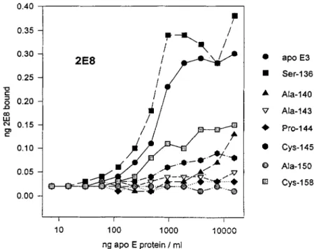
Pro-144 cys-145 8 Ala-150 FJ cys-15aFig. 3. Immunoreactivity of apoE variants with anti-apoE mAbs 2E8. Serial dilutions of apoE vari- ants were incubated in polystyrene microwells to which anti-apoE mAb 6C5 had been adsorbed. The wells were then washed and exposed to '251-labeled 2E8 and, after washing, the bound radioactivity was determined. The bound '2SI-labeled mAb mass was calculated from the specific activity of the labeled mAb.
injected over the activated chip surface. Unreacted moieties were blocked with ethanolamine. All measure- ments were carried out in HEPES-buffered saline (HBS) which contained: 10 mM HEPES, pH 7.4, 150 mM NaC1, 3.4 mM EDTA, 0.005% Surfactant P-20 (Pharmacia Biosensor). Analyses were performed at 25°C and the binding of each antibody was tested at seven concentra- tions. Immobilizations and binding assays were carried out at a flow rate of 3 pl/min. Sensor chip surfaces were regenerated with 100 mM HC1 and surface integrity was periodically checked with control antibody.
Association and dissociation rate constants were cal- culated by non-linear fitting of the sensorgram data (33) using the BIAevaluation 2.0 software (Pharmacia Biosensor). The dissociation rate constant is derived using the equation:
Rt = Rto e-koff(t-to)
where Rt is the response at time t, Rt, is the amplitude of the response, and kos is the dissociation rate constant. The association rate constant can be derived using the equation:
where Rt is the response at time t, R,, is the maximum
response, C is the concentration of ligate in solution, k,
zyxwvutsrqponmlkjihgfedcbaZYXWVUTSRQPONMLKJIHGFEDCBA
1908 Journal of Lipid Research Volume 36, 1995
at National Research Council Canada, on August 28, 2019
www.jlr.org
s
zyxwvutsrqponmlkjihgfedcbaZYXWVUTSRQPONMLKJIHGFEDCBA
c
zyxwvutsrqponmlkjihgfedcbaZYXWVUTSRQPONMLKJIHGFEDCBA
P
E
zyxwvutsrqponmlkjihgfedcbaZYXWVUTSRQPONMLKJIHGFEDCBA
m ac
P
zyxwvutsrqponmlkjihgfedcbaZYXWVUTSRQPONMLKJIHGFEDCBA
I
m 3H1 1 D7 2E8 3H1 1 D7 2E0zyxwvutsrqponmlkjihgfedcbaZYXWVUTSRQPONMLKJIHGFEDCBA
=E
lo00 3H1 1 D7 2E8 lo00 2E8zyxwvutsrqponmlkjihgfedcbaZYXWVUTSRQPONMLKJIHGFEDCBA
TinW (-1 "0 (-1Fig. 4.
zyxwvutsrqponmlkjihgfedcbaZYXWVUTSRQPONMLKJIHGFEDCBA
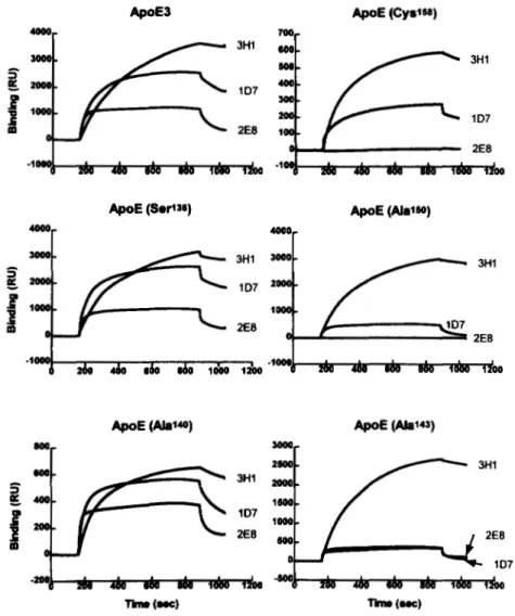
Sensorgrams of apoE variants with 1D7,2E8, and 3H1 using SPR. These sensorgrams depict the kineticsof 1D7, 2E8, and 3H1 binding to apoE3, apoE(Argl36 -+ Ser), apoE(HisI4O -+ Ala), a p o E ( L y ~ ~ * ~ -+ Ala), apoE(ArgIw -+ Ala), and apoE(ArgIS8 -+ Cys). The association phase of each sensogram begins at approximately 180 sec and the dissociation phase at 850 sec. The results that are illustrated were obtained with concentrations of 150 nM, 150 nM, and 50 nM for 1D7,2E8, and 3H1, respectively.
is the association rate constant, and
kg
is the dissocia- tion rate constant.Molecular cloning and characterization of 1D7 and 2E8 heavy and light chain cDNAs
Total RNA from 1D7 and 2E8 hybridoma cells was extracted according to the acid-guanidinium thiocy- anate phenol chloroform method (34) and poly(A+) RNA was purified by oligo-dT affinity chromatography. Northern blot analysis was performed to ensure the presence of light and heavy chain immunoglobulins (35). The constant region of a mouse kappa light chain (36) and the first constant region of an IgGla heavy chain (37) were used as molecular probes for hybridiza- tion. A 1D7 cDNA library was created by using 5 pg of poly(A+) mRNA as template for reverse transcription
with an oligo(dT) primer. Following second strand syn- thesis, Notl/EcoRl adaptors (Pharmacia) were added and the products were cloned into pUC18. Recombi- nant plasmids were transformed into DH5a competent bacteria by heat shock treatment. Colonies were screened by colony lift hybridization using the same radiolabeled IgG l a and kappa constant region probes that were used for northern analysis (38).
The cDNA encoding the 2E8 light chain was amplified by the polymerase chain reaction (PCR) (39), after re- verse transcription of hybridoma mRNA, using one primer complementary to the 3' end of the kappa trans- lated sequence (5'-GCG CCG TCT AGA A'IT AAC ACT CAT TCC TGT TGA A-3'), and the other corresponding to the nucleotides encoding the amino terminal residues
of the mature light chain (5'-CCA GGT CCG AGC TCG
zyxwvutsrqponmlkjihgfedcbaZYXWVUTSRQPONMLKJIHGFEDCBA
Raffai' et
zyxwvutsrqponmlkjihgfedcbaZYXWVUTSRQPONMLKJIHGFEDCBA
al. Anti-apoE monoclonal antibodies 1909at National Research Council Canada, on August 28, 2019
www.jlr.org
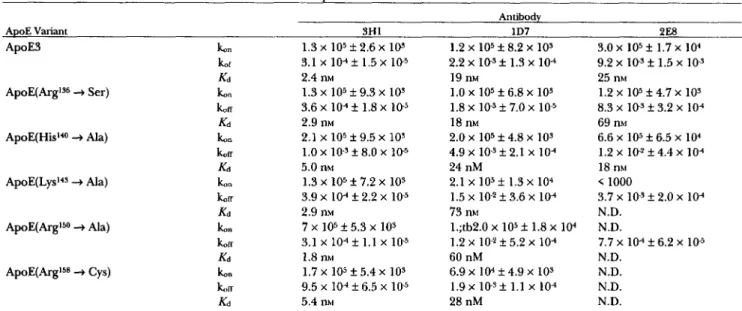
TABLE 1. Association (16.)
zyxwvutsrqponmlkjihgfedcbaZYXWVUTSRQPONMLKJIHGFEDCBA
and dissociation ( 1 6 ~ ) rate constants of mAbs 3H1, 1D7, and 2E8zyxwvutsrqponmlkjihgfedcbaZYXWVUTSRQPONMLKJIHGFEDCBA
with apoE variants as determined by surface plasmon resonanceAntibody
ApoE Variant 3H1
zyxwvutsrqponmlkjihgfedcbaZYXWVUTSRQPONMLKJIHGFEDCBA
1D7 2E8 ApoE3ApoE(Argls6
+
zyxwvutsrqponmlkjihgfedcbaZYXWVUTSRQPONMLKJIHGFEDCBA
Ser)t" 1.3 x lo5 f 2.6 x los 1.2 x 10⁵ f 8.2 x
zyxwvutsrqponmlkjihgfedcbaZYXWVUTSRQPONMLKJIHGFEDCBA
103 3.0 x 10⁵ f 1.7 x 10⁴tf K d 2.4 nM 19 nM 25 nM t"
zyxwvutsrqponmlkjihgfedcbaZYXWVUTSRQPONMLKJIHGFEDCBA
koa Kd 2.9 nM 18 m 69 nhi Ib"zyxwvutsrqponmlkjihgfedcbaZYXWVUTSRQPONMLKJIHGFEDCBA
K i 5.0 nM 24 nM 18 nhi 3.1 x 10⁴ f 1.5 x 10⁻⁵ 1.3 x lo5 f 9.3 x los 3.6 x lo4 f 1.8 x lo5 2.1 x 10⁵ f 9.5 x 10³ 2.2 x i o - s f 1.3 x 10⁴ 1.0 x 10⁵ f 6.8 x 10⁹ 1.8 x los f 7.0 x 2.0 x 105 f 4.8 x 10s 9.2 x lo3 f 1.5 x 10-s 1.2 x 10⁵ f 4.7 x 10s 8.3 x lo5 f 3.2 x lo4 6.6 x lo5 k 6.5 x lo4 ApoE(His140+
Ala) koff 1.0 x 1 0 . 9 8.0 x 10⁻⁵ 4.9 x 10⁻³ 2.1 x 10⁴ 1.2 x 1 0 - 2 f 4.4 x 10⁴ koa 3.9 x 10⁴ 2.2 x 10⁻⁵ 1.5 x lo2 f 3.6 x lo4 3.7 x 10⁻³ 2.0 x 10⁴koa 7.7 x 10⁴ f 6.2 x 10⁻⁵
A p o E ( L y ~ l ~ ~ 4 Ala) k o n 1.3 x lo5 f 7.2 x 10s 2.1 x 10⁵ f 1.3 x 10⁴ < 1000
K d 2.9 nM 73 nhi N.D.
ApoE(ArgI5O
+
Ala) ko" 7 x 105*
5.3 x 103 l.;tb2.0 x lo5 f 1.8 x 10.' N.D.zyxwvutsrqponmlkjihgfedcbaZYXWVUTSRQPONMLKJIHGFEDCBA
K d 1.8 nM 60 nM N.D.
ApoE(.bgI5* Cys) ko" 1.7 x 10⁵ f 5.4 x 10s 6.9 x 10⁴ f 4.9 x 10s N.D.
LIT 9.5 x 10⁻⁴ 6.5 x 1 0 5 1.9 x i o - ~ f 1.1 x 10⁴ N.D.
zyxwvutsrqponmlkjihgfedcbaZYXWVUTSRQPONMLKJIHGFEDCBA
ILd 5.4 nhl 28 nM N.D.
3.1 x 10⁴ 1.1 x 10⁵ 1.2 x lo2 f 5.2 x lo4
The values t. and t u represent the means calculated from determinations at seven different antibody concentrations; N.D., association and/or dissociation rate constants could not be calculated.
TGA TGA CCC ACT CTC CA-3'). Both the 5' and 3' region primers were based on previously compiled se- quences (40) and contain, respectively, Sac1 and Xbal restriction sites indicated in bold, which facilitate sub- sequent cloning of PCR amplification products for even- tual expression. After amplification, the material was gel purified and a 700 base-pair fragment corresponding to two immunoglobulin domains was extracted using Gene Clean (Bio 101, La Jolla, CA) and subcloned, after Klenow and kinase treatment, into p-Bluescript(=+) (Stratagene, San Diego, CA) digested with SmaI (38).
After reverse transcription of total RNA that was iso- lated from the 2E8 hybridoma, the 2E8 heavy chain cDNA was amplified by anchored PCR (41). Terminal deoxynucleotidyl transferase was used to add a 5' over- hang consisting of guanosine nucleotides to a first strand cDNA template. A first round of amplification using an oligo(dC) and a 3' primer complementary to codons 224-232 located in the hinge region (5'-AGG CTT ACT ACT ACA ATC CCT GGG CAC A-3') and which contains an Spel site indicated in bold, was carried out and the products were separated on a low melting point agarose gel (FMC Bioproducts, Rockland, ME). The gel containing fragments with molecular weights ranging from 300 to 800 base pairs was excised and the products were used directly as template for a second round of PCR using the oligo dC and an internal 3'constant region primer (5'-GGC AGC AGA TCC AGG GGC-3') corresponding to codons 125-130. This second amplification gave a specific 500 base pair product that was isolated and sub-cloned into p-Bluescript. All the
cloned cDNAs were sequenced using the dideoxy chain termination method (42).
Amino acid sequence sequence analysis and cleavage with
CNBr
at methionine residuesProtein and peptide samples for sequencing were purified by SDS-PAGE and electroblotted onto se- quencer stable PVDF membrane as described previously (43). Automated gas-phase sequencing was performed on an Applied Biosystems 475A protein sequencing system (Foster City, CA) incorporating a model 470A gas-phase sequencer equipped with an on-line model 120A PTH analysis module. Cleavage of peptide bonds adjacent to methionine residues was effected by using CNBr. Freeze-dried protein was dissolved in 500 pl of 88% (v/v) formic acid followed by the addition of solid CNBr. The reaction vial was flushed with argon, sealed and incubated in the dark at room temperature for 24 h. The reagent and solvent were removed under a stream of argon and the remaining material was sus- pended in Milli-Q water and freeze dried.
RESULTS
Fine specificity of mAb 1D7
We have previously shown that mAb 1D7 reacts with a cyanogen bromide fragment of apoE composed of residues 126-218 and with a synthetic peptide that includes apoE residues 139-169 (17). To more precisely map the 1D7 epitope, we have taken advantage of a
1910 Journal of Lipid Research Volume 36, 1995
at National Research Council Canada, on August 28, 2019
www.jlr.org
panel of recombinant apoE variants produced by site-di- rected mutagenesis and synthesized in a bacterial ex- pression system (18). For these studies, a solid phase apoE sandwich immunometric assay was developed.
The mAb 6C5 was used
zyxwvutsrqponmlkjihgfedcbaZYXWVUTSRQPONMLKJIHGFEDCBA
as the anchor and bound apoEwas detected with either "Wlabeled ID7 or 125I-labeled
3H1
zyxwvutsrqponmlkjihgfedcbaZYXWVUTSRQPONMLKJIHGFEDCBA
(Fig. 1). Elsewhere, evidence has been presentedthat the epitopes for anti-apoE mAbs 6C5 and 3H1 are entirely or partially situated between residues 1-13 and 243-272, respectively (24). Antibodies 6C5 and 3H1 do not compete with each other for binding to immobilized apoE nor do they compete with 2E8 and 1D7 (R. Milne
and R. Raffai, unpublished results).
As
zyxwvutsrqponmlkjihgfedcbaZYXWVUTSRQPONMLKJIHGFEDCBA
the mutagenesiswas confined to the receptor-binding region of apoE, the introduced amino acid substitutions should not influence binding of the variants to either 6C5 or to '25I-labeled 3H1. Recombinant apoE3 is somewhat less immunoreactive with both 1D7 and with 3H1 than is purified plasma apoE3 (Fig. 1). This could be attribut- able to some protein denaturation or a decreased bind- ing of recombinant apoE to 6C5 due to the presence of
an amino-terminal methionine that is not present in mature plasma apoE. The differences in immunoreac- tivity observed between native and recombinant apoE3 are, however, minor in comparison with the decreases in 1D7 immunoreactivity that resulted from amino acid substitutions in apoE. Amino acid substitutions at resi- dues 143, 144, 150, or 152 almost totally eliminated binding of 1D7 and, as reported previously, the natural
apoE variant apoE(Arg145
+
zyxwvutsrqponmlkjihgfedcbaZYXWVUTSRQPONMLKJIHGFEDCBA
Cys) showed reduced 1D7immunoreactivity. In contrast, normal 1D7 reactivity was seen with a p ~ E ( A r g l ~ ~
+
Ser) and apoE(Arg158+
Cys) variants. ApoE(Hislm+
Ala) showed a large de- crease in 1D7 binding compared to apoE3 (Fig. l); however, when normalized for 3H1 binding (Fig.2),
thisdifference was lost. While it is possible
zyxwvutsrqponmlkjihgfedcbaZYXWVUTSRQPONMLKJIHGFEDCBA
that a substitu-tion at residue 140 could modulate both the 1D7 and 3H1 epitopes, it is more probable that there is reduced binding to the anchor mAb, 6C5, perhaps as a result of denaturation or amino-terminal proteolysis of the mole- cule. While lipid-free apoE is not a ligand for the LDLr, binding activity is restored when the apoE is incorpo-
( 1 )
zyxwvutsrqponmlkjihgfedcbaZYXWVUTSRQPONMLKJIHGFEDCBA
AsnzyxwvutsrqponmlkjihgfedcbaZYXWVUTSRQPONMLKJIHGFEDCBA
C y s Val Met Thr G l n Thr Pro Lys Phe Leu Leu Val Ser Ala G l ya a t t g c g t g a t g a c c cag a c t ccc aaa t t c c t g c t t g t a t c a gca gga
* * * *** * * * I * * * * * * * * *** * l a
___ _ _ _
a-- tcc ac-__-
_ t __ _ _
Met Ser Thr --- Val --- Asp Ile
_ _ _ _ _ _ _ _ _ _ _ _
Ser G l n_ _ _
_ _ _
(17) A s p A r g V a l Thr Ile Thr C y s L p Ala Ser Qln Ser V a l Ser &n
*
g a c agg g t t a c c a t a a c c t g c aag goo aqt org aqt qto aqt ut qat
---
---
--c -g- --c --- ------
---
---
_--
-a- --Q q-- -0- -0-A s p A r g Val Thr
___ ___ ___
-__
___
___
_--
b n___
-__
___
Ala( 3 3 ) V a l Ala Trp Tyr Gln Gln Lys Pro Gly Gln Ala Pro Lys Leu Leu Ile
qta got t g g t a c caa cag aaa cca ggg cag g c t c c t aaa c t g c t g a t a
---
--e --- --t -----_
_-- - _ _ - _ a - _ a t _ - ---_ _ _
--a a-- --t___
___
_ _ _ ___
--__ _ _ _ _ _ _ _ _ _ _ _ _ _ _
ser_ _ _
_ _ _ _ _ _
M e t_ _ _
(49) TYK Tyr Ala Ser A.n &q Tyr Thr Gly Val Pro Asp AKg Phe Thr Gly
t a c t a t qoa too .at ago tmo a o t gga g t c c c t g a t cgc t t c a c t ggc --a ---
--(I
_--
--- _ _ -
--__-_
_ _ _ _ _ _
- _ _
_ _ _
sir -09---
---
---
---
---
---
___
_--
---
_ _ - _ _ ___-
___
_-_
_ _ _
- _ __ _ _
_ _ _
( 6 5 ) Ser Gly Tyr Gly Thr Asp Phe Ser Phe Thr Ile Ser Thr Val Gln Ala
a g t gga t a t ggg acg g a t t t c t c t t t c a c c a t c agc a c t g t g cag g c t
___ _ _ _
_c-___
--a --- --- a _ - c_- ------
--t _ a - a-___-
t _ _ Thr Leu --- --- --- Asn Met --- Ser___ _ _ _
ser_--
---
--- ---(81) Glu Asp Leu Ala Val Tyr Phe Cys Qln Qln !JSA Tyr Arg Ser Pro Pro
gaa g a c c t g gca g t t t a t t t c t g t o8q orq qat t a t ago t o t w t oat
--c
---
--a t-- .go a-- -1----
-to---
Tyr S i r Sir Tyr---
Leu___
_ _ _ _ - ____
Asp___
_-- ------
_-_
___
--- --- -a-_ _ _
_--(97) Thr Phe Gly Ala Gly Thr Lys Leu Glu Leu Lys acg
___
t t c g g t g c t ggg a c c aag c t g gag c t g aaa_ _ _ ___ _ _ _
---
--- ---
t-- --- --- ---___
_ _ _
_--
_ _ _
-----_
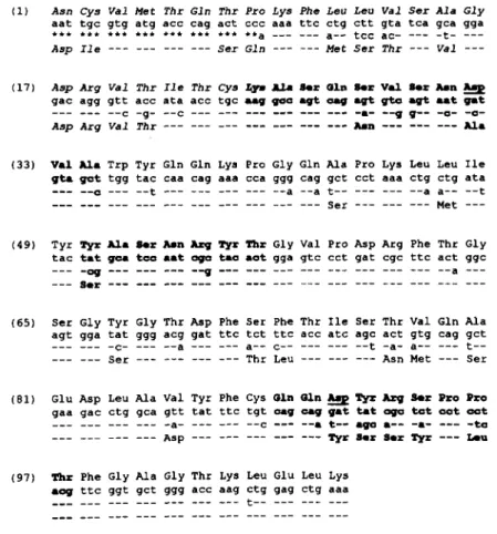
_-- --- --- --- ---Fig. 5. Nucleotide and deduced protein sequences of 1D7 and 2E8 light chains. The nucleotide and deduced
protein sequences of the variable domains of 1D7 (upper) and 2E8 (lower) light chains are presented. CDRs are indicated in bold print and negatively charged residues within the CDRs are underlined. Amino acid residues represented in italics were confirmed by protein sequence determination. The asterisks indicate the nucleotides within the degenerate PCR primer that were used for cloning the 2E8 light chain cDNA. The corresponding amino acids were determined by direct protein sequencing. Codons are numbered and CDRs are identified according to Wu and Kabat (47).
Rafai et al. Anti-apoE monoclonal antibodies 191 1
at National Research Council Canada, on August 28, 2019
www.jlr.org
Gln Val His Leu Lys Glu Ser Gly Pro Gly Leu Val Ala Pro Ser Gln cag gtg cac ctg aag gag tca gga cct ggc ctg gtg gcg ccc tca cag
g-- --t --g
zyxwvutsrqponmlkjihgfedcbaZYXWVUTSRQPONMLKJIHGFEDCBA
--- c-- c-- --t -- g g-a -ag g-t --- ag- t-a ggg gcczyxwvutsrqponmlkjihgfedcbaZYXWVUTSRQPONMLKJIHGFEDCBA
G l u ---
zyxwvutsrqponmlkjihgfedcbaZYXWVUTSRQPONMLKJIHGFEDCBA
G l n ---zyxwvutsrqponmlkjihgfedcbaZYXWVUTSRQPONMLKJIHGFEDCBA
G l n G l n --- --- AlazyxwvutsrqponmlkjihgfedcbaZYXWVUTSRQPONMLKJIHGFEDCBA
G l u Val --- A r qzyxwvutsrqponmlkjihgfedcbaZYXWVUTSRQPONMLKJIHGFEDCBA
Ser G l y AlaSer Leu Ser Ile Thr Cys Thr Val Ser Gly Phe Ser Leu Thr Qly Tyr
agc ctg tcc atc aca tgc acc gtc tca ggg ttc tca tta acc ggc
zyxwvutsrqponmlkjihgfedcbaZYXWVUTSRQPONMLKJIHGFEDCBA
t a ttca g-c aag t-g t-c --- --a -ct --t --c --- aac a-t -aa -a- t-c
- - _ V a l L y s L e u Ser --- --- Ala --- --- --- Asn Ile Lys
*
---
Qly Val A m Trp Val Arg Gln Pro Pro Gly Thr Gly Leu Glu Trp Leu
gPt gt8 a8C tgg gtt cgc cag cct cca gga acg ggt ctg gag tgg ctg
Tyr Ilr H i m --- --- Lys --- Arg --- Glu Lys --- --- --- --- Ile Gly Leu Ilr Tlp Ala Qly Thr
a
Tyr A.n I e r N a Lmuh- .-e e-- --- --g aaq --- agg --t -a- -a- --c _-- --- --_ a-t gga t% at. g& gat -1 .Q. aoI g.0 t a t 8.t toI gCt C t C
--- -q- --t 9.t cct -a8 a t t --t gat --t --a
---
ptC C-q aag t----- Trp
---
Pro Ilc---
9---
e
---
Val Pro Lys Phc Lys Ser Arg Leu Ser Ile Ser Lys Asp Asn Ser Lys Ser Gln Val Phe u a tcc aga ctg agc atc agc aag gac aac tcc aag agc caa gtt ttcc-g qq- -ag gcc -ct -tg -ct gca --- -ca --- tcc -a- ac- -cc -a-
Qln Qly Lys Ala Thr Met Thr Ala --- Thr --- Ser Asn Thr Ala Tyr
Leu Lys Met A s n Ser L e u G l n T h r A s p Asp
zyxwvutsrqponmlkjihgfedcbaZYXWVUTSRQPONMLKJIHGFEDCBA
T h r A l a A r g Tyr Tyr Cystta aaa atg Lac agt ctg caa act gat gac aca gcc agg tac tac tgt c-g c-- c-c -4- --c --- ac- t-- --g --- --t --- gtc --t _ _ - ---
--- Gln Leu Ser --- --- Thr Ser Glu --- --- --- Val --- --- ---
Ala Arg E Qly Val Qly Tyr Pro Phe
a
Tyr Trp Gly Gln gcc aga gag qtt ggt t a t ccc ttt gac tac tgg ggc caa aat gc- -q- cat -8- trc g-c agq ma cgg --c cct---
--- --- ---Asn Ala Qly H i m Tyr
*
k q Qly---
pro---
--- --- ---Gly Thr Thr Leu Thr Val Ser Ser ggc aac act ctc aca gtc tcc tca --g -ct ctg g-- --t --- -- t g--
_ _ _
--- Leu Val --- --- --- AlaFig. 6. Nucleotide and deduced protein sequences of 1D7 and 2E8 heavy chains. The nucleotide and deduced protein sequences of the variable domains of 1D7 (upper) and 2E8 (lower) heavy chains are presented. CDRs are indicated in bold print and negatively charged residues within the CDRs are underlined. Amino acid residues represented in italics were confirmed by protein sequence determination. Codons are numbered and CDRs are identified according to Wu and Kabat (47).
rated into lipid vesicles (44). As it has been suggested that binding to lipid may induce a conformational change in apoE, we compared the 1D7 immunoreactiv- ity of the apoE variants in a lipid-free and a lipid-bound form. However, as shown in Fig. 2, incorporation of the apoE into DMPC vesicles did not significantly change the observed fine specificity of 1D7.
Fine specificity mAb 2E8
A second anti-human apoE mAb, 2E8, has been iden- tified that blocks binding of apoE to the LDLr (T. Innerarity and K. Weisgraber, unpublished results). While the affinity of 2E8 for apoE is lower than that of 1D7, it can compete with ID7 for binding to immobi- lized apoE and shows a pattern of reactivity similar to that of 1D7 with apoE proteolytic fragments (T. Inner- arity and K. Weisgraber, unpublished results). We have
used the apoE variants to determine the fine specificity of 2E8 as was done for 1D7. The results in Fig. 3 confirm the lower affinity of 2E8 for apoE compared to 1D7 but also demonstrate that, in general, the two antibodies have similar specificity with respect to the apoE variants. One notable difference, however, is that 2E8, unlike 1D7, shows decreased reactivity with apoE2(Arg15*
+
Cys). Antibody 2E8 does not discriminate between lipid- bound and free apoE nor does incorporation of apoE into lipid vesicles change its relative reactivities with apoE3 and apoE2(Arg15*+
Cys) (results not shown).Surface plasmon resonance
In order to confirm the binding specificities of 1D7 and 2E8 that were determined by sandwich RIA, and to establish association and dissociation rate constants for the antibodies with the different mutants, we have stud-
1912 Journal of Lipid Research Volume 36, 1995
at National Research Council Canada, on August 28, 2019
www.jlr.org
ied their interactions using surface plasmon resonance
(SPR). ApoE3 and the five variants, apoE(Arg136
+
zyxwvutsrqponmlkjihgfedcbaZYXWVUTSRQPONMLKJIHGFEDCBA
Ser),apoE(His'40
+
Ala), apoE(Ly~I*~+
Ala), apoE(Arg150+
Ala), and apoE(Arg158+
Cys) were immobilized and were subjected to a pulse of antibody. Sensorgrams depicting the binding of the antibodies to the apoEvariants as a function of time are shown in Fig.
zyxwvutsrqponmlkjihgfedcbaZYXWVUTSRQPONMLKJIHGFEDCBA
4. Thecontrol antibody 3H1 reacted well with a l l of the variants including a p ~ E ( H i s l ~ ~
+
Ala), indicating that the re- duced reactivity observed by RIA may well have been due to a reduced capture of this mutant by the anchorantibody
zyxwvutsrqponmlkjihgfedcbaZYXWVUTSRQPONMLKJIHGFEDCBA
6C5. The binding of 1D7 to the variants re-vealed the same fine specificity as
zyxwvutsrqponmlkjihgfedcbaZYXWVUTSRQPONMLKJIHGFEDCBA
that determined usingthe sandwich RZA. Again 1D7 had reduced reactivity with apoE(Lys143
+
Ala) and apoE(Arg150+
Ala). In the case of 2E8, the results obtained using SPR are also consistent with those obtained using the sandwich RIA. Notably, the reduced of reactivity of 2E8 with apoE(Lysl43+
Ala), apoE(Arg150+
Ala), and a p ~ E ( A r g ' ~ ~+
Cys) have been confirmed.This methodology has also allowed us to determine the association and dissociation rate constants as well as the affinity of the antibodies for the individual apoE variants (Table 1). From Table 1, it is apparent that the
lower affinity of 2E8 compared to 1D7 and 3H1 for apoE3 is the result of a high dissociation rate constant. Moreover, the reduction of affinity of 1D7 and 2E8 for individual apoE variants can reflect changes in the asso- ciation and/or the dissociation rate constants. For ex- ample, the reduced affinities of 1D7 for a p ~ E ( L y s ' ~ ~
+
Ala) and apoE(ArgI5O+
Ala), compared to that for apoE3, are largely due to an increased dissociation rate constant whereas the reduced affinity of 2E8 for a p o E ( L y ~ ' ~ ~-+
Ala) reflects a reducedken.
Reactivity of 2E8 with certain of the variants was too low to allowcalculation of
zyxwvutsrqponmlkjihgfedcbaZYXWVUTSRQPONMLKJIHGFEDCBA
rate constants and affinities. As expected,the binding kinetics of 3H1 were similar with all of the apoE variants.
LDL
receptor
zyxwvutsrqponmlkjihgfedcbaZYXWVUTSRQPONMLKJIHGFEDCBA
195 210
Cys & Gly Gly Pro & Cys Lys & Lys Ser ASD Glu Glu Asn Cys
Trp Ile & Pro Tyr Val
50 60
Glu Ile Gly & Thr
2E8 heavy
chain
CDR2
Fig. 7. Comparison of the putative ligand binding sites of the LDLr and the heavy chain CDR2 of 2E8. Residue numbers 195 to 210 (cysteine-rich repeat number 5) of the human LDLr (upper) are compared to those present in the heavy chain CDR2 of mAb 2E8. Negatively charged residues are underlined.
Molecular cloning and characterization of the heavy
and light chain
cDNA
of mAbs1D7
and2E8
The cDNAs encoding the entire light and the Fd regions (variable and first constant domains of the heavy chains) of mAbs 1D7 and 2E8 have been cloned. A series of cDNA light chain clones were identified that had an identical nucleotide sequence and an intact reading frame (Fig. 5). In the case of the 1D7 heavy chain, a single message was cloned from the cDNA library and corresponded to the expected protein (Fig. 6). This was
also the case for the 2E8 light (Fig. 5) and heavy chains (Fig. 6) that were cloned using PCR. Partial protein sequences were obtained from the variable domains of each of the antibodies and, in each case, the amino acid sequence corresponded to the amino acid sequence deduced from the nucleotide sequence of the respective cDNA clones. In the process of cloning the 1D7 light chain from a cDNA library, two non-functional light chains were identified.
Both
had deleterious frame shifts within the nucleotides encoding the variable region and would represent products of non-productive kappa gene rearrangements (45).Comparison of the variable region sequences of the two mAbs with those in the database of Kabat et al. (46) indicated that the rearranged 1D7 heavy chain is a member of the mouse heavy chain variable region sub- group I(B) and that codons 103-117 were derived from
the mouse heavy chain J-minigene, MUSJH2 (47). Its D-minigene could not be identified. The 2E8 heavy chain gene is a member of the mouse heavy chain variable region subgroup II(C) and includes sequences derived from the DSP2.2R + 2 D-minigene (codons 99
to 103) (47) and from the MUSJH3 J-minigene (codons 106- 120) (47). Sequencing of the nucleotides encoding the CHI showed that both heavy chains were of the 7-1
subclass, thus confirming serological identification. The 1D7 and 2E8 light chains are both members of the mouse kappa variable subgroup V, and both contain sequences derived from the MUSJK5 kappa J-minigene As the two anti-apoE mAbs have similar fine specifici- ties, it may be expected that they share homology in the six hypervariable or complementarity determining re- gions (CDRs). Amino acids that constitute the three CDRs of the light chain and the three CDRs of the heavy chain interact directly with the epitope and are respon- sible for antibody specificity. Comparison of the primary structure of the light chains of 1D7 and 2E8 shows considerable homology for the CDRl and CDR2 with most of the differences occurring in CDR3. The 1D7 light chain contains two negatively charged residues in CDRl and CDR2, respectively, whereas the 2E8 light chain has no negatively charged residues within its CDRs. There is little homology between the heavy chain
(47).
zyxwvutsrqponmlkjihgfedcbaZYXWVUTSRQPONMLKJIHGFEDCBA
RaJai et al. Anti-apoE monoclonal antibodies 1913
at National Research Council Canada, on August 28, 2019
www.jlr.org
CDRs of the two mAbs, and CDR2 and CDR3 of 2E8 contain one and two additional amino acids, respec-
tively,
zyxwvutsrqponmlkjihgfedcbaZYXWVUTSRQPONMLKJIHGFEDCBA
as compared to the corresponding CDRs of 1D7.The heavy chain CDRs of 1D7 include four acidic amino acids whereas those of the 2E8 heavy chain contain seven acidic residues.
DISCUSSION
We present evidence that the 1D7 and 2E8 epitopes coincide with the LDLr-binding site on apoE that is situated between residues 136 and 150. We cannot, however, exclude the possibility that the epitopes of ID7 and 2E8 are located elsewhere on the molecule and may be disrupted by changes in conformation due to the amino acid substitutions that characterize the different apoE variants that we have tested. Nevertheless, it
should be noted that a recent study that evaluated site-directed mutagenesis as a method for epitope map- ping revealed that mutations which caused a greater than l0-fold change in apparent antibody binding affin- ity (using methods analogous to those presented in Fig. 2) *were correctly assigned to the epitope when con- firmed by X-ray crystallography (48). From results pre- sented here and elsewhere (16, 49), it is probably that Arg142, Lys143, Arg145, Lys146, and Arg'50 form part of the 1D7 and 2E8 epitopes. Furthermore, the lack of
reactivity of apoE(Leu144
zyxwvutsrqponmlkjihgfedcbaZYXWVUTSRQPONMLKJIHGFEDCBA
+
Pro) and apoE(Alal52+
Pro) may indicate that either Leu144 and Ala152 are also directly implicated in the epitopes or that a local confor- mational change induced by the introduction of a proline at these positions eliminates mAb binding. The relatively moderate decrease in reactivity of the apoE(Arg145
+
Cys) variant would be consistent with the Arg145 side chain making only a minor contribution to the binding energy of the mAb-apoE complex in the case of both mAbs. While the fine specificity of 2E8 is similar to that of 1D7, it is nevertheless notable that 2E8 and ID7 differ in their relative affinities for apoE2(Arg158-+
Cys) and apoE3, with 2E8 having little affinity for apoE2(Arg15*+
Cys) (Figs. 1 and 3 and Table 1). Our results, therefore, indicate that the epitopes for 1D7 and 2E8 overlap with each other and with the LDLr binding site on apoE.Incorporation of apoE into lipid vesicles does not change its immunoreactivity with either of the mAbs. It has been proposed that, in the presence of lipid, the four-helix bundle of the amino terminal domain of apoE opens up to generate the receptor-active conformation (50). While this would represent a major alteration in tertiary structure, the changes in the secondary struc- ture of the individual a-helices may be minor. The reactivity of ID7 with apoE synthetic peptides as short
as 30 amino acids (17) would indicate that its epitope
zyxwvutsrqponmlkjihgfedcbaZYXWVUTSRQPONMLKJIHGFEDCBA
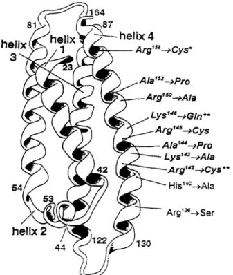
Fig. 8. Comparison of apoE variants with respect to their binding to mAbs 1D7 and 2E8 and to the LDLr. The positions of amino acid substitutions that have been shown to decrease affinity of apoE for the LDLr are indicated with respect to their position in the four-helix bundle that constitutes the amino terminal 22-kDa fragment of apoE. Those variants that also have reduced reactivity with mAbs 1D7 and/or 2E8 are represented in bold italics. Of these, a p ~ E ( A r g l ~ ~ Cys) (*) showed reduced reactivity with 2E8 but not with 1D7. A p ~ E ( A r g l ~ ~ + Cys) and a p o E ( L y ~ ' ~ ~ + Gln) (**) showed reduced reactivity with 1D7 but were not tested with 2E8.
does not require elaborate tertiary structure and is not composed of disparate regions of apoE primary struc- ture. In the case of 1D7, the epitope may thus be continuous or, as we have suggested earlier (17), com- posed of one face of the a-helix that constitutes the secondary structure of this region.
It has been determined that the ligand-binding do- main of the LDLr is situated at the amino-terminus and is made up of seven imperfect repeats of a 40-amino acid cysteine-rich sequence that is characterized by a con- served cluster of acidic amino acids in the carboxy-ter- minal portion of the repeat (51). Repeats 3 through 7
appear to contribute to apoB-mediated binding of LDL, whereas only repeat 5 appears to be critical for apoE- mediated binding of P-VLDL (52). Binding of apoE and apoB-100 to the LDLr is thought to involve ionic inter- actions between basic amino acids of the apolipoprote- ins and acidic residues of the receptor (53,54). Chemical modifications of arginine and lysine residues in apoB-
100 and apoE (13,54) and substitution of neutral amino
zyxwvutsrqponmlkjihgfedcbaZYXWVUTSRQPONMLKJIHGFEDCBA
1914 Journal of Lipid Research Volume 36, 1995
at National Research Council Canada, on August 28, 2019
www.jlr.org
acids for basic residues in the putative receptor-binding region of apoE reduce affinity of the apolipoproteins for the LDLr. Reductive methylation of lysine residues in apoE and apoB, which maintains the positive charge of the modified residues, also eliminates binding to the LDLr. This would suggest that not only the positive charge but also the structure of the basic residues con- tribute to the recognition of the ligands by the receptor. This is further supported by the observation that cys- teamine treatment of apoE(Arg”*, Cys14*) restores a positive charge at residue 142 without rendering the molecule capable of binding to the LDLr (49). More- over, the acid-mediated dissociation between the recep tor and PVLDL is not dependent on a simple titration of charged residues within the ligand binding domain While basic residues located within the LDLr binding site of apoE contribute to the epitopes of both mAbs 1D7 and 2E8, the lack of similarity between the primary structures of the respective CDRs of the two mAbs indicates that they recognize a similar region on apoE differently. It has been observed that oriented dipoles rather than counter charges are preferentially used in stabilizing charged residues in the formation of antigen- antibody complexes (56). Nevertheless, both mAbs do contain acidic residues within their respective CDRs that could potentially contribute to the binding energy of the antibody-antigen complex through the formation of ionic bonds with basic residues of apoE in a manner similar to that which has been proposed for the interac- tion between apoE and the LDLr. The two mAbs differ in the distribution of these negatively charged amino acids amongst their respective CDRs. In the case of lD7, the six acidic residues are distributed in the CDRs of both the heavy and light chains whereas, in 2E8, the seven acidic residues are restricted to the heavy chain CDRs. Assuming that aspartic and glutamic residues of the two antibodies do make a major contribution to the binding energy of their respective immune complexes, this could indicate that 1D7 makes use of both chains for binding apoE, whereas, in 2E8, it is primarily the heavy chain that is responsible for antigen binding. It should, however, be emphasized that the surface of contact between antigen and antibody is large, and
hydrogen bonds, van der Waals forces,
zyxwvutsrqponmlkjihgfedcbaZYXWVUTSRQPONMLKJIHGFEDCBA
as well as elec-trostatic interactions likely contribute to the binding energy.
ApoB, the major protein of LDL, can compete with apoE for binding to the LDLr. Comparison of the primary structures of apoE and apoB has revealed a short sequence of apoB composed of residues 3359 through 3367 that resembles residues 140-150 of apoE with respect to the relative positions of basic amino acids (57). It has been proposed that this characteristic distri- (55).
bution of basic residues could indicate that both apolipo- proteins may be forming similar ionic interactions in their association with the ligand binding site of the LDLr. We have made an analogous comparison of the primary structures of the ligand-binding repeats of the LDLr and the CDRs of mAbs 1D7 and 2E8. Of the seven acidic residues in the heavy chain CDRs of 2E8, four are clustered in CDR2. The spacing of the four aspartic and glutamic residues within CDR2 of the 2E8 heavy chain bears some resemblance to that of the acidic residues in
the ligand-binding repeats of the LDLr
zyxwvutsrqponmlkjihgfedcbaZYXWVUTSRQPONMLKJIHGFEDCBA
(Fig.zyxwvutsrqponmlkjihgfedcbaZYXWVUTSRQPONMLKJIHGFEDCBA
7). There-fore, the CDR2 of the 2E8 heavy chain may mimic the postulated ionic interaction between the consensus se- quence repeats of the LDLr and apoE. While the overall architecture of an antibody and that of the LDL receptor are clearly different, it is interesting to note that an anti-idiotypic mAb prepared against a neutralizing anti- reovirus hemagglutinin mAb recognized the mammal- ian cell surface reovirus receptor and, in this case, the molecular mimicry resulted from homology in primary structure between the CDRs of the anti-idiotypic mAb and the viral hemagglutinin (58). In addition, it has been shown that certain anti-integrin antibodies that bind to the ligand binding site of platelet integrin a I l p 3 contain
the “Arg-Gly-Asp” integrin recognition motif in their antigen binding sites (59). Moreover, the activity of one such inhibitory antibody could be emulated by synthetic peptides whose synthesis was based on the primary structure of the heavy chain CDR3 of the mAb.
It is interesting to compare the structural require- ments for apoE-mediated binding to the LDLr with those for 1D7 and 2E8 immunoreactivity (Fig. 8). Sub- stitutions that result in the loss of a positive charge at residues 142, 143, 145, 146, or 150 produce a decrease in the ability of the variant to bind to both antibodies and to the receptor (17, 18, 49, 60, 61). Similarly, the introduction of proline residues at positions 144 and 152 decreases both antibody and receptor binding by either the replacement of a critical residue for binding or by disruption of apoE secondary or tertiary structure (18). In contrast, several of the variants (e.g.,
a p ~ E ( A r g ’ ~ ~
+
Ser) and a p ~ E ( A r g ’ ~ ~zyxwvutsrqponmlkjihgfedcbaZYXWVUTSRQPONMLKJIHGFEDCBA
+
Cys)) are defec-tive with respect to LDLr binding but bind to ID7 with high affinity (18, 60). The binding of antibody 2E8 to a p ~ E ( A r g ’ ~ ~
+
Cys), on the other hand, is severely impaired (Fig. 3, Table 1). It is thought that ArglSs is not directly implicated in binding to the LDLr but its re- placement by a Cys in a p ~ E ( A r g ’ ~ ~+
Cys) induces a conformational change in the receptor binding domain, probably situated between residues 136 and 150 (31). In the crystal structure of the amino terminal domain of apoE3, Arg158 forms salt bridges with Ghg6 and Asp154 and may help to stabilize the pairing of helices 3 and 4(19). In apoE(ArgI5*
+
Cys), ArgI5O forms a salt bridgezyxwvutsrqponmlkjihgfedcbaZYXWVUTSRQPONMLKJIHGFEDCBA
&#ai’
zyxwvutsrqponmlkjihgfedcbaZYXWVUTSRQPONMLKJIHGFEDCBA
et al. Anti-apoE monoclonal antibodies 1915at National Research Council Canada, on August 28, 2019
www.jlr.org
with Asp154 (62). The putative conformational change responsible for the decrease in binding affinity of
a p o E ( A ~ p l ~ ~
+
Cys) for the LDLr may result from thesezyxwvutsrqponmlkjihgfedcbaZYXWVUTSRQPONMLKJIHGFEDCBA
reorganized salt bridges. LDLr-binding and 1D7 im- munoreactivity can also be distinguished by other crite- ria. Rat and mouse apoE bind with high affinity to the human LDLr but have little 1D7 immunoreactivity.
Finally,
zyxwvutsrqponmlkjihgfedcbaZYXWVUTSRQPONMLKJIHGFEDCBA
as discussed above, only lipid-bound apoE isrecognized by the LDLr (44) whereas neither mAb differentiates between free and lipid-bound apoE. Thus, while the apoE LDLr-binding domain and the 1D7 and 2E8 epitopes may coincide or overlap, the respective conformational elements that are recognized by the antibodies and the receptor are not identical.
It appears that apoE can be present in two forms of triglyceride-rich lipoproteins, one of which is resistant to cleavage by proteases and is not recognized by the LDLr and a second that is protease-sensitive and capable
of binding to the LDLr
zyxwvutsrqponmlkjihgfedcbaZYXWVUTSRQPONMLKJIHGFEDCBA
(25). ApoE may be convertedfrom the first to the second form by either change in conformation or in accessibility that is induced by lipolysis of the particle (23). As it has been proposed that 1D7 may distinguish between these two forms (26), it
may be a useful probe of the LDLr-binding site of apoE. Here we have identified a number of apoE residues that are critical for binding of the 1D7 and 2E8 mAbs and have demonstrated that the epitopes for both mAbs overlap with the LDLr-binding site on apoE. How each of these residues contributes to the antibody-antigen interaction should become more apparent once the three-dimensional crystal structures of apoE mAb com- plexes have been solved. By mutagenesis of the two anti-apoE mAbs, we are now attempting to identify residues within the CDRs that are important in antigen binding and to produce variants that even more closely resemble the LDLr in their interactions with apoE. These antibodies will be used to determine the physical and chemical properties of lipoproteins that modulate the conformation of the apoE LDL receptor binding site. In addition, they will be used as immunogens in order to produce anti-idiotypic mAbs that will cross
react with the LDLr.
zyxwvutsrqponmlkjihgfedcbaZYXWVUTSRQPONMLKJIHGFEDCBA
IWe thank Mr. Marc Deaorges, Ms. Bingyi Han, Mr. Christo- phe Marcel, and Ms. Thanh Dung Nguyen for excellent tech- nical assistance and Drs. Benoit Barbeau, David Gould, Dan Sparks, and Zemin Yao, and Mr. Xingyu Wang for helpful discussions and advice. This investigation was supported by grants from the Heart and Stroke Foundation of Ontario and the Foundatioii des Maladies du Coeur du QuCbec. Ross Milne
is a Scientist of the Medical Research Council of Canada.
zyxwvutsrqponmlkjihgfedcbaZYXWVUTSRQPONMLKJIHGFEDCBA
Manuscrip received 14 December 1994 and in revised form
zyxwvutsrqponmlkjihgfedcbaZYXWVUTSRQPONMLKJIHGFEDCBA
31 May 1995.REFERENCES
1. Mahley, R. W. 1988. Apolipoprotein E cholesterol trans- port protein with expanding role in cell biology. S c d e .
2. Davignon, J., R. E. Gregg, and C. F. Sing. 1988. Apolipo- protein E polymorphism and atherosclerosis. Arteria&- 3. Brenninkmeyer, B. J., P. M. J. Stuyt, P. N. M. Demacker,
A. F. H. Stalenhoef, and A. van't b a r . 1987. Catabolism of chylomicron remnants in normolipidemic subiects in
240: 622-630.
T O S ~ ~ . 8: 1-21.
relation to the apoprotein E phen0he.J. Lipid
h.
2 8361-370.
4. Weintraub, M. S., S. Eisenberg, and J. L. Breslow. 1987. Dietary fat clearance in normal subjects is regulated by genetic variation in apolipoprotein E.J. Clin. Invest. 80:
5. Brown, A. J., and D. C. K. Roberts. 1991. The effect of fasting triacylglyceride concentration and apolipoprotein E polymorphism on postprandial lipemia. Artm'ascl~r.
Thromb. 11: 1737-1744.
6. Demant, T., D. Bedford, C. J. Packard, and J. Shepard. 1991. Influence of apolipoprotein E polymorphism on
apolipoprotein B-100 metabolism in normolipemic
zyxwvutsrqponmlkjihgfedcbaZYXWVUTSRQPONMLKJIHGFEDCBA
sub- jects.J Clin. Invest. 88: 1490-1501.7. Gregg, R. E., L. A. Zech, E. J. Schaefer, D. Stark, D. Wilson, and H. B. Brewer, Jr. 1986. Abnormal in vivo metabolism of apolipoprotein in humans. J. Clin. Invest. 78: 8. Steinmetz, A., C. Jakobs, S. Motzny, and H. Kaf€arnik. 1989. Differential distribution of apolipoprotein E iso- forms in human plasma lipoproteins. Arteriosclerosis. 9 9. Weisgraber, K. H. 1990. Apolipoprotein E distribution
among human plasma lipoproteins: role of the cyste-
ine-arginine interchange at residue 112. J. Lipid
zyxwvutsrqponmlkjihgfedcbaZYXWVUTSRQPONMLKJIHGFEDCBA
Res. 31:10. Strittmatter, W. J., A. M. Saunders, D. Schmechel, M. Pericak-Vance, J. Enghild, G. S. Salvesen, and A. D. Roses. 1993. Apolipoprotein E high-avidity binding to pamyloid and increased frequency of type 4 allele in lateanset familial Alzheimer disease. h o c . Nutl. Acud. Sci. USA. 90:
11. Wetterau, J. R., L. P. Aggerbeck, S. C. Rall, Jr., and K. H. Weisgraber. 1988. Human apolipoprotein E3 in aqueous solution. I. Evidence for two structural d0mains.J Biol.
Chem. 263: 6240-6248.
12. Aggerbeck, L. P., J. R. Wetterau, K. H. Weisgraber, CS. C. Wu, and F. T. Lindgren. 1988. Human apolipoprotein E3 in aqueous solution. 11. Properties of the amino-and carboxyl-terminal domains. J. Biol. Chem. 263: 6249-6258. 13. Mahley, R. W., T. L. Innerarity, R. E. Pitas, K. H. Weisgra- ber, J. H. Brown, and E. Gross. 1977. Inhibition of lipo- protein binding to cell surface receptors of fibroblasts following selective modification of arginyl residues in arginine-rich and B apoproteins. J. Biol. Chem. 252:
14. Weisgraber, K. H., T. L. Innerarity, and R. W. Mahley. 1978. Role of the lysine residues of plasma lipoproteins in high affinity binding to cell surface receptors on human fibrob1asts.J. Biol. Chem. 253: 9053-9062.
15. Innerarity, T. L., E. J. Friedlander, S. C. Rall, Jr., K. H.
1571- 1577. 815-82 1. 405-41 1. 1503-1511. 1977- 1981. 7279-7287.
1916 Journal of Lipid Research Volume 36, 1995
at National Research Council Canada, on August 28, 2019
www.jlr.org
