ORIGINAL ARTICLE
The contractile properties of vaginal myofibroblasts:
Is the myofibroblasts contraction force test a valuable
indication of future prolapse development?
S. Meyer&C. Achtari&P. Hohlfeld&
L. Juillerat-Jeanneret
Received: 27 October 2007 / Accepted: 17 March 2008 / Published online: 30 May 2008
# International Urogynecology Journal 2008
Abstract Using a specific myofibroblast contraction test, we try to predict future utero-vaginal prolapse development in young primiparae women. We compare myofibroblast cultures of the vaginal wall in primiparae women (group 1), young multiparae women (group 2) and older multiparae women (group 3) who were operated on for severe utero-vaginal prolapse. A myofibroblast-mediated collagen gel contraction assay determined a contraction factor that was compared in the three groups of women. The myofibro-blasts contraction factor after 24 and 48 hours was significantly higher in group 1 women (2.4±0.6/4.4±1.9) compared to group 2 (1.6±0.3/ 1.8±0.1) andgroup 3 (1.6± 0.3/1.8 ±0.3), but showed no differences in group 1 women without (2.1 ± 0.5/3.5 ± 1.9) and with (2.7 ±0.6/5.1 ±1.7) cystocoele. Vaginal myofibroblasts of young women show better contraction forces than young women with severe utero-vaginal prolapse. The latter have a myofibroblast
contraction factor similar to those of older post-menopausal women operated for the same condition.
Keywords Utero-vaginal prolapse . Pelvic floor myofibroblasts
Introduction
Although there have been very few prevalence studies using the International Continence Society-validated pelvic organ prolapse test for the objective assessment of utero-vaginal prolapse disorders, these disorders are very common. A study by Swift et al. [1] demonstrated that the prevalence of stage 1 prolapse was greater than 93% in a population of 497 women seen in an outpatient clinic, with a mean age of 44 years seen in an outpatient clinic, the prevalence of stage 2 being 48% and that of stage 3, 3%. A Swedish study based on a non-validated genital examination of a population of 487 women (mean age 39 years) found a prevalence of 31% for prolapse of any stage and a prevalence of 1.6% for prolapse to introitus [2].
Fibroblasts are found in urothelial tissues [3] and have the potential to transform into myofibroblasts, which are rich in actin/myosin filaments. We were interested in whether such cells may be important factors in healing the anchoring tissues in the genital area after the“birth trauma” of vaginal delivery.
We previously carried out a study of the contractile force of these cells in prolapse and non-prolapse situations [4]. The first aim of the present study was to determine and compare the contractile force of pelvic floor myofibroblasts in a population of young primiparae women to that of a population of young women who had given birth several years previously before undergoing an operation for severe
S. Meyer (*)
:
C. AchtariUrogynecology Unit, Obstetrics & Gynecology Department, CHUV, Route du Bugnon 46,
1011 Lausanne, Switzerland e-mail: [email protected] C. Achtari
Urogynaecology Unit, Gynecology & Obstetrics Department, CHUV, Lausanne, Switzerland
C. Achtari
:
P. HohlfeldGynecology & obstetrics Department, CHUV, Lausanne, Switzerland
C. Achtari
:
L. Juillerat-JeanneretUniversity Institute of Pathology, Bugnon 25, CH1011 Lausanne, Switzerland
utero-vaginal prolapse and in a population of older women operated on for the same condition. The second aim of the study was to correlate the contractile force of myofibro-blasts in the young primiparae women with the presence or absence of some degree of utero-vaginal prolapse evaluated 6 months after delivery, trying to find a specific test to predict future utero-vaginal prolapse development when comparing the results obtained in women without and in women with some degrees of prolapse.
Materials and methods
The subjects consisted of three groups of women:
Group 1: 15 primiparous women, 29 ± 4-years-old, who underwent spontaneous non-instrumented delivery. Group 2: four women, 37 ± 2-years-old (P:< 0.001) para 2, operated on for severe (i.e., grade 3) utero-vaginal prolapse, four to six years after their delivery. Group 3: nine women, 64 ± 6-years-old, (P:< = 0.0001) para 2 ± 1, operated on for severe (i.e., grade 3) utero-vaginal prolapse.
Clinical examination
Clinical examination of subjects in groups 2 and 3 was carried out with the women in a 45 degree position, such as in birthing chair, according to the modified POP-Q ICS classification [5]: all showed utero-vaginal grade 3 pro-lapse. Clinical examination of group 1 was carried out 6– 12 weeks after vaginal delivery and a clinical grading of vaginal wall prolapse was made using the same classifica-tion; seven women had no anterior vaginal wall prolapse, eight women had grade anterior vaginal wall prolapse grade 1 and 2 (six women: grade 1, two women grade 2).
Human tissues
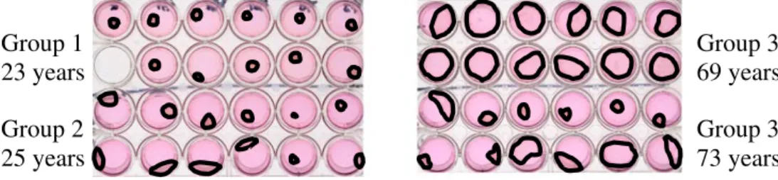
Vaginal samples were obtained from surgical biopsy samples of the posterior vaginal wall during episiotomy repair in group 1 or during uterovaginal prolapse elective surgical repair in groups 2 and 3, according to a protocol approved by the Ethics Committee of the University Hospital of Lausanne (protocol number 4902) and after pre-delivery consent by group 1 patients and pre-operative consent by patients in groups 2 and 3. During the 3 months before surgery, all women in group 3 received intravaginal estriol (Ortho-Gynest-Dr, 3 mg) 1 to 2 times a week. The fresh tissues were used to prepare cultures of myofibro-blasts. The following are the methods for cell culture and the collagen gel contraction assay [6]: Fifty thousand cells were added to 500 mL of a 1:1 neutralized solution of rat tail collagen (BD Biosciences, San Jose) in culture medium that contained 4.5 g/L glucose, 10% fetal calf serum, and 6% methylcellulose (Sigma Chemical Company). At the end of incubation, culture plates were scanned, and the contraction factor of the myofibroblast-collagen gel was quantified by measurement of the mean diameter of the contracted plug after 24 hours or 48 hours of incubation. The contraction factor was calculated as the ratio of the diameter of the contracted plug to the initial diameter of the well, i.e., 16 mm (Fig.1).
Statistical methodology
Means and standard deviation for duplicate wells were calculated. Statistical significance was assessed with one-way analysis of variance and LSD test for post hoc comparisons. A P value of <0.05 two-tailed was considered as significant.
Prior to calculations, force test data after 24 and 48 hours were tested for normality using the Kruskal–Wallis test.
Group 1
23 years
Group 2
25 years
Group 3
69 years
Group 3
73 years
•
50’000 cells are seeded in 500 µl of collagen and treated with antagonists to ETR or endothelin-1.•
Every treatment is done in duplicate on cell cultures of women with prolapse (RX 34, SD 26) or without (GE 80, GS 78).•
Contraction of collagen gel is measured after 24 and 48 hours.Fig. 1 The myofibroblast contraction force test in four women: on the left side, two young women of group 1 (without prolapse) and on the right side, two older women of group 3: the contraction factor of the myofibroblast-collagen gel is quantified by measure-ment of the mean diameter of the contracted plug after 24 hours or 48 hours of incubation (black circles). The contraction factor is determined as the ratio of the diameter of the contracted plug to the initial diameter of the well
Results
After 24 hours, the contractile force test gave a significant higher value for myofibroblasts from group 1 (2.4±0.6) than for those from group 2 (1.6±0.3, P = 0.004) or group 3 (1.6±0.3, P = 0.000).
After 48 hours, the contractile force was greater, but the differences were still significant between groups 1 and group 2 (4.4±1.9 and 1.8±0.3, respectively; P = 0.002) and groups 1 and 3 (4.4±1.9 and 1.8±0.3, respectively; P = 0.000) (Fig.2) (Table1).
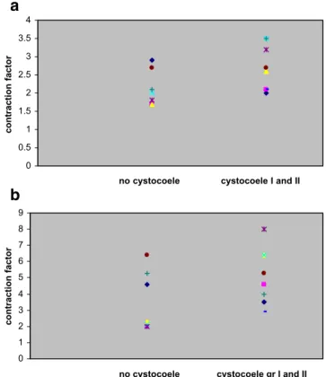
Post-partum examination of group 1 showed absence of prolapse in seven women and the presence of grade 1 and 2 anterior vaginal prolapse in eight women. Comparison of the myofibroblast contractile force between the seven women without prolapse and the eight with grade 1 and 2 anterior vaginal wall prolapse showed no significant difference. Women with grade 1 or 2 anterior vaginal wall prolapse showed similar contractile force values at 24 hours (2.1±0.5 and 2.7±0.6, respectively; P = 0.91) and at 48 hours (3.5± 1.9 and 5.1±1.7, respectively; P = 0.93) (Fig.3a,b).
Discussion
Numerous studies have demonstrated an association of prolapse disorders with pregnancy and vaginal delivery. One study found that pregnancy itself is responsible for some degree of prolapse, with a prevalence of 48% for stage 2 prolapse determined by pelvic organ prolapse quantification in pregnant primiparae women compared to 0% in a group of non-pregnant primiparae women [7].
Furthermore, vaginal delivery is considered the greatest risk factor for the development of prolapse in women under
the age of 59, the relative risk being 8.4 for women with two children and 10.9 for women with four children or more compared to nulliparous women, after adjusting for age [8]. Apart from spontaneous single delivery itself, other vaginal delivery parameters (instrumental delivery, episiot-omy, birth weight, management, and duration of the second phase of labor) have been suggested as a possible etiology of future prolapse disorders.
Consequently, “normal” vaginal delivery is responsible for trauma to the tissues of the birth canal responsible for utero-vaginal prolapse in about 50% of parous women, with 10% to 20% being symptomatic [9], and is the greatest risk factor for the development of pelvic organ prolapse, with an adjusted relative risk of 10.85 (95% CI, 4.65–33.81) [8,10].
0 0.5 1 1.5 2 2.5 3 3.5 4 4.5 5
group 1 group 2 group 3
contraction factor
after 24 hours after 48 hours
Fig. 2 Myo-fibroblast contraction force test values in the three groups of women: young delivered primiparae women of group 1 have significant higher values when compared to young women with severe utero-vaginal prolapse of group 2 and to older women of group 3 with similar pathology
Table 1 Myo-fibroblasts contraction force test values in women of group 1, women of group 2 and women of group 3 operated on for severe utero-vaginal prolapse
Contraction factor Group 1 (N = 15) (29±4 years) Group 2 (N =4) (37±2 years) Group 3 (N =13, 64±6 years) P Gr.1 vs. Gr 2/3 After 24 hours 2.4±0.6 1.6±0.3 1.6±0.3 0.004/0.000 After 48 hours 4.4±1.9 1.8±0.3 1.8±0.3 0.002/0.000 0 0.5 1 1.5 2 2.5 3 3.5 4 no cystocoele 0 1 2 3 4 5 6 7 8 9
no cystocoele cystocoele gr I and II
contraction factor
contraction factor
cystocoele I and II
a
b
Fig. 3 a,b Contraction factor after 24 hours in group 1 women, considering the absence (no cystocoele) and presence of grade I and II anterior vaginal wall prolapse
Race and hereditary factors are also implicated in the genesis of such disorders due to heritable intrinsic connec-tive tissue abnormalities: there is a higher risk of prolapse in women with a mother (OR 3.2) or a sister (OR 2.4) who have had a prolapse [11].
The etiology of the problem is poorly understood. One hypothesis is that these utero-vaginal defects are related to abnormal repair of the wounded tissue after the birth injury of delivery [12]. Connective tissue abnormalities have been described in many studies, which have shown a lower collagen content [13], collagen abnormalities [14], and higher expression of genes responsible for collagen breakdown and lower expression of genes responsible for collagen synthesis [15,16]. These last studies showed that the differences between patients with advanced pelvic organ prolapse and controls may be related to the differential expression in the pubococcygeus muscle of genes for structural proteins related to actin and myosin and for extracellular matrix proteins [16,17].
These studies were all retrospective and were performed on material obtained during pelvic floor surgery.
On the other hand, we know that fibroblasts transform into myofibroblasts, which contain a highly developed contractile apparatus similar to that in smooth muscle cells, contain alpha-smooth muscle actin (α-SMA), and are essential for establishing tension during repair phase and are important in the synthesis of the extracellular matrix [18,19]. Myofibroblasts can temporarily acquire contractile properties and the capacity to synthesize large amounts of extracellular matrix components, among which collagen type I predominates [20]. The participation of neo-expressed contractile proteins, such as α-SMA, in force generation by myofibroblast cells is now well accepted [21]. However, no study had assessed the functions of these supporting and contractile cells (i.e., fibroblasts/myofibro-blasts) in the pelvic floor of newly delivered women and compared the fibroblast contractile force of this group to that of young women who had undergone an operation for grade 3 utero-vaginal prolapse several years after delivery or that of older women operated on for the same pathology. For obtaining tissues specimen, the posterior vaginal wall was chosen for practical and evident reasons: it was more easy and acceptable for the young women of group 1 to accept the idea of tissues biopsies in a place where there is already or a wound (the episiotomy) or a tear who will be surgically repaired. Subsequently, we took biopsies speci-men in the same region when operating wospeci-men of group 2 and 3 with severe prolapse.
The following could be another question: Is the vaginal wall really the right structure to study in order to find a factor of predictability of pelvic organ prolapse? All biopsies specimens were deep excised taking each time the sub-mucous layer, the latter being rich in fibroblasts and
being involved in the scarring process of birth trauma. For obvious ethical reasons, we have not proposed biopsies in other sites, such as the pelvic floor.
Other questions remain unresolved: Is the posterior vaginal wall of young pregnant women representative of normal myofibroblasts function? Is it possible to use these specimens of vaginal wall as controls? Can their higher contractile force be only a pregnancy effect? Can we exclude that non-pregnant young women may have the same results as prolapsed patients of groups 2 and 3? To rule out this possibility, another study should be carried out on biopsies taken from the posterior vaginal wall of young non-pregnant women, but it is doubtful that such a study would be accepted by an ethics committee.
Similarly, we cannot conclude that the lesser contractility is the only cause of the prolapse in group 2 and group 3 women; this lowered contractility may perhaps simply be what happens to myofibroblasts in the vaginal epithelium when it has prolapsed or stretched over time. Up to now, nobody has answeredthis question, and a control group of women of same age as the women in group 3 should be carried out with biopsies of posterior vaginal walls. This too, however, would like be rejected by an ethics committee. In a previous publication [4], we demonstrated that myofibroblasts express the complete endothelin system, but do not secrete endothelin-1, and that in 3-D collagen gels, the spontaneous contraction of myofibroblasts from estrogen-treated women with prolapse is significantly lower than that of myofibroblasts from young primiparous women. We concluded that the genital myofibroblasts of women with uterovaginal prolapse are poorly contractile, and that endo-thelin-1 further decreases vaginal myofibroblast contraction, which contrasts with observations on skin myofibroblasts [21].
In the present study, we compared the contractile force of myofibroblasts in newly delivered women (group 1) and in other young women who had undergone an operation for severe utero-vaginal prolapse 4 to 6 years after spontaneous delivery of normal weight babies (group 2). At 24 hours, the contractile force of the myofibroblasts from group 2 was significantly lower than that of the group 1 women of the same age (1.6±0.3 and 2.4±0.6, respectively; p = 0.004) Furthermore, the contractile force for group 2 was the same as that for group 3 (1.6±0.3; p = 0.91 NS) consisting of older women operated on for the same pathology, suggest-ing a congenital weakness of these tissues.
We also showed that the myofibroblast contractile force for group 1 increased with time in the test, ranging from 2.4±0.6 at 24 hours of incubation and 4.4±1.9 at 48 hours (p=0.002), whereas the contractile force for the young women in group 2 and the older women in group 3 was significantly lower at 24 h (1.6±0.3 and 1.6±0.3, respec-tively, p=0.004 and p=0.000) and did not increase with
time (1.8±0.3 and 1.8±0.3, respectively; p = 0.30 NS and p=0.08 NS) These results lead us to hypothesize that myofibroblasts of young women with severe prolapse are less able to affect tissue repair, explaining the development of prolapse after delivery.
But it is not also possible to conclude that the lesser contractility of group 2 and 3 is a cause of the prolapse; it may simply be what happens to myofibroblasts in the vaginal epithelium when it has prolapsed or stretched over time.
However, we were unable to demonstrate significant differences between the myofibroblasts of group 1 women with or without some degree of anterior vaginal wall mobility during post-partum examination. This may be explained by the small number of women and the low grade of anterior vaginal wall prolapse (i.e., grade 1) in six of the eight women studied. This finding should be investigated in a study involving more patients. Another possibility could be a close follow-up of these group 1 young women with another clinical and biopsy one to two years after delivery (we did controls six months after delivery); this aspect of the study was discussed with these women, but none of them accepted the idea of a second biopsy outside the pregnancy-delivery-lactation period. This fact is responsible for some limitations of our significant findings when comparing the tissues of young delivered women with those of young and older patients with established prolapse. Why do some women have high myofibroblast contrac-tile forces, while others have low ones? The myofibroblast contractile force test is a global test, taking into account many different factors; other cellular pathways are possible pathways for explaining such differences. They have not been investigated up to now.
In the field of screening tests for future prolapse development, the study of the different factors responsible for the contractile force of myofibroblasts may lead to the definition of biomarkers allowing the development of a specific test capable of replacing the expensive and sophisticated myofibroblast contractile force test.
Conflicts of interest None.
References
1. Swift SE (2000) The distribution of pelvic organ support in a population of female subjects seen for routine gynecologic health care. Am J Obstet Gynecol 183:277–85
2. Samuelsson EC, Arne Victor FT, Tibblin G, Svardsudd KF (1999) Signs of genital prolapse in a Swedish population of women 20 to 59 years of age and possible related factors. Am J Obstet Gynecol 180:299–305
3. Fry CH, Brading AF, Hussain M, Lewis SA, Takeda M (2005) Cell biology. In: Abrams P, Cardozo L, Khoury S, Wein A (eds)
Incontinence, volume 1, Basics & evaluation. Health Publication, Paris, pp 313–362
4. Poncet S, Meyer S, Richard C, Aubert JD, Juillerat-Jeanneret L (2005) The expression and function of the endothelin system in contractile properties of vaginal myofibroblasts of women with uterovaginal prolapse. Am J Obstet Gynecol 192:426–432 5. Swift S (2002) Current opinion on the classification and definition
of genital tract prolapse. Curr Opin Obstet Gynecol 14(5):503– 507
6. Lee YR, Oshita Y, Tsuboi R, Ogawa H (1996) Combination of insulin-like growth factor (IGF)-1 and IGF-binding protein promotes fibroblast-embedded collagen gel contraction. Endocri-nology 137:5278–5283
7. O’Boyle AL, Woodman PJ, O’Boyle JD, Davis GD, Swift SE (2002) Pelvic organ support in nulliparous pregnant and nonpreg-nant women: a case control study. Am J Obstet Gynecol 187:99– 102
8. Mant J, Painter R, Vessey M (1997) Epidemiology of genital prolapse: observations from the oxford family planning associa-tion study. Brit J Obstet Gynecol 104:579–585
9. Moalli PA, Klingensmith WL, Meyn LA, Zyczynski HM (2002) Regulation of matrix metalloproteinase expression by estrogen in fibroblasts that are derived from the pelvic floor. Am J Obstet Gynecol 187:72–79
10. Arnold EP, Burgio K, Diokno AC, Herzog AR, Mallet VT (1998) Epidemiology and natural history of urinary incontinence. In: Abrams P, Khoury S, Wein A (eds) Incontinence: 1st international consultation on incontinence. Plymbridge Distributors, Plymouth, pp 165–202
11. Chiaffarino F, Chatenoud L, Dindelli M, Meschia M, Buonaguidi A et al (1999) Reproductive factors, family history, occupation and risk of urogenital prolapse. Eur J Obstet Gynecol & Reprod Biol 82:63–67
12. Meyer S, Schreyer A, De Grandi P, Hohlfeld P (1998) The effects of birth on urinary continence mechanisms and other pelvic floor characteristics. Obstet Gynecol 92:613–618
13. Wong MY, Harmanli OH, Agar M, Dandolu V, Grody MH (2003) Collagen content of nonsupport tissue in pelvic organ prolapse and stress urinary incontinence. Am J Obstet Gynecol 189:1597– 1599
14. Goepel C, Hefler L, Methfessel HD, Koelbl H (2003) Periurethral connective tissue status of postmenopausal women with genital prolapse with and without stress incontinence. Acta Obstet Gynecol Scand 82:659–664
15. Chen BH, Wen Y, Li H, Polan ML (2002) Collagen metabolism and turnover in women with stress urinary incontinence and pelvic prolapse. Int Urogynecol J 13:80–87
16. Visco AG, Yuan L (2003) Differential gene expression in pubococcygeus muscle from patients with pelvic organ prolapse. Am J Obstet Gynecol 189:102–112
17. Chen BH, Wen Y, Zhang Z, Wang H, Warrington JA et al (2003) Menstrual phase-dependent gene expression differences in peri-urethral vaginal tissue from women with stress incontinence. Am J Obstet Gynecol 189:89–97
18. Hinz BG, Gabbiani G (2003) Mechanisms of force generation and transmission by myofibroblasts. Curr Opin Biotechnol 14:538–546 19. Tomasek JJ, Gabbiani G, Hinz B, Chaponnier C, Brown RA (2002) Myofibroblasts and mechanoregulation of connective tissue remodeling. Nature Rev Mol Cell Biol 3:349–363 20. Dugina V, Alexandrova A, Chaponnier C, Gabbiani G (1998) Rat
fibroblasts cultured from various organs exhibit differences in alpha-smooth muscle actin expression, cytoskeletal pattern, and adhesive structure organization. Exp Cell Res 238:481–490 21. Serini G, Gabbiani G (1999) Mechanisms of myofibroblast