Research Article
Enhanced heparan sulfate proteoglycan-mediated uptake of
cell-penetrating peptide-modified liposomes
C. Martya, C. Meylana, H. Schottb, K. Ballmer-Hoferaand R. A. Schwendenera,*
aPaul Scherrer Institut, Biomolecular Research, Molecular Cell Biology, 5232 Villigen-PSI (Switzerland),
Fax: +41 56 310 2080, e-mail: reto.schwendener@psi.ch
bInstitute of Organic Chemistry, Eberhard-Karls University, Auf der Morgenstelle 18, 72076 Tübingen (Germany)
Received 16 April 2004; received after revision 13 May 2004; accepted 25 May 2004
Abstract. Protein transduction domains (PTDs) are used to enhance cellular uptake of drugs, proteins, polynu-cleotides or liposomes. In this study, functionalized An-tennapedia (Antp, aa 43 – 58) and HIV Tat (aa 47 – 57) peptides were coupled to small unilamellar liposomes via thiol-maleimide linkage. Modified liposomes showed higher uptake into a panel of cell lines including tumor and dendritic cells than unmodified control liposomes. Liposome uptake was time and concentration dependent as analyzed by flow cytometry and live-cell microscopy. At least 100 PTD molecules per small unilamellar lipo-some (100 ± 30 nm) were necessary for efficient
translo-cation into cells. Cellular uptake of PTD-modified lipo-somes was 15- to 25-fold increased compared to unmod-DOI 10.1007/s00018-004-4166-0
© Birkhäuser Verlag, Basel, 2004
CMLS
Cellular and Molecular Life Sciencesified liposomes and was inhibited by preincubation of li-posomes with heparin. Glycosaminoglycan-deficient CHO cells showed dramatically reduced cell association of PTD-modified liposomes, confirming the important role of heparan sulfate proteoglycans in PTD-mediated uptake. Antp-liposomes used as carriers of the cytotoxic drug N4-octadecyl-1-
b-D-arabinofuranosylcytosine-(5¢-5¢)-3¢-C-ethinylcytidine showed a reduction of the IC50by
70 % on B16F1 melanoma cells compared with unmodi-fied liposomes. PTD-functionalized liposomes, particu-larly Antp-liposomes, represent an interesting novel car-rier system for enhanced cell-specific delivery of a large variety of liposome-entrapped molecules.
Key words. Antennapedia; HIV-Tat; peptide-liposome; heparan sulfate proteoglycan; cytotoxic liposome.
Uptake of pharmaceutically active molecules into living cells is hampered by the lipophilic nature of the plasma membrane. Large molecules such as proteins, peptides and oligonucleotides are generally poorly taken up by cells since they do not cross the lipid bilayer of the plasma membrane efficiently. Strategies to improve membrane permeability play an important role in the development of new drug delivery systems. Specific domains of several proteins, so-called protein transduction domains (PTDs), efficiently pass through biological membranes [1]. PTDs were first identified while investigating the spontaneous
*Corresponding author.
cell entry of the HIV Tat protein and its subsequent translocation to the nucleus. A short sequence rich in ba-sic amino acids is responsible for cellular uptake of this protein [2, 3]. Similar properties were found for Anten-napedia (Antp), a Drosophila homeodomain transcrip-tion factor [4] and in the Herpes virus protein VP22 [5]. The peptide sequence Antp (43 – 58), corresponding to the third helix of the homeodomain of Antennapedia, has been shown to promote translocation across cellular membranes. Due to their basic sequence, these and re-lated basic peptides are rapidly internalized by mam-malian cells and therefore used as delivery vectors [6]. Colloidal drug carriers such as liposomes and target-spe-cific immunoliposomes are widely used as delivery
sys-tems for a broad spectrum of agents including chemo-therapeutics, imaging agents, antigens, lipids and DNA [7 – 10]. These delivery systems have several therapeutic advantages, such as the ability to transport large drug concentrations to a specific site, sparing healthy tissue from toxic effects and increasing the systemic drug cir-culation time. Therapeutic molecules are protected by the lipid bilayer of the liposome from metabolism and enzy-matic degradation, a feature that is particularly important for in vivo applications. To improve cellular uptake of li-posomes, we developed a simple and versatile method to link C-terminally functionalized Antp and Tat peptides via a covalent cysteine-maleimide coupling reaction to the outer surface of liposomes [11].
There is still controversy about the mechanism responsi-ble for cell membrane translocation of PTDs and it may vary among the various PTDs and depend on the structure and composition of the target cell membranes. Recent publications have proposed that PTD-mediated mem-brane translocation is an energy-dependent process that may be a direct consequence of peptide association with heparan sulfate proteoglycans (HSPGs) [12]. Drin et al. [13] showed that internalization of SynB5, a peptide de-rived from protegrin and the Antp peptide is a tempera-ture- and energy-dependent process with endosomal transport as a key component of the mechanism. We [14] and others [15] have demonstrated that PTDs facilitate cell uptake of cargo molecules via endocytosis and re-quire the expression of negatively charged glycosamino-glycans on the cell surface. Another detailed study demonstrated that the Tat peptide interferes with the re-lease of newly synthesized HPSGs from cells [16]. One elegant study showed recently that the most likely route for PTD-mediated cellular uptake is via lipid raft-medi-ated macropinocytosis [17]. Most studies involving PTD-mediated cellular uptake have been carried out with Tat peptide. Tat-modified nanoparticles, such as cross-linked iron oxide for cell tracking by magnetic resonance [18] or polymeric drug carriers [19] showed improved uptake properties when compared to unmodified particles. The Tat peptide was also used to investigate PTD-mediated li-posome uptake. As shown by Torchilin et al. [20], con-ventional and polyethyleneglycol-modified Tat-lipo-somes were taken up by different cell types, provided that 100 – 500 peptide molecules were attached per liposome. In a subsequent paper, the same authors described the cell transfection properties of Tat-liposome/DNA complexes and reported high rates of green fluorescent protein ex-pression at lower cytotoxicity compared with common commercial transfection reagents [21]. The Antp peptide was also used to deliver a poorly antigenic MHC class I-restricted peptide to antigen-presenting cells to elicit a cytotoxic T lymphocyte immune response. A recombi-nant Antp antigenic fusion peptide was encapsulated in li-posomes to prevent peptide degradation, enhance
cytoso-lic delivery and antigen presentation on dendritic cells [22, 23].
The work we present here illustrates the properties of Antp- and Tat-modified small unilamellar liposomes (Antp-L, Tat-L) and their ability to bind to and translocate into different cell types, such as CHO, B16 melanoma, F9 teratocarcinoma and dendritic cells. We show that at least 110 – 136 peptide molecules per liposome are necessary for effective cell translocation and that cell uptake of the PTD-modified liposomes is strongly dependent on gly-cosaminoglycan expression on the target cells, confirm-ing the important role of HSPGs in PTD-dependent cel-lular uptake. Thus, PTD-modified liposomes, and partic-ularly Antp-L, provide a novel system for enhanced delivery of a large variety of liposome-entrapped mole-cules into cells.
Materials and methods
Chemicals
Soy phosphatidylcholine (SPC) was obtained from L. Meyer (Hamburg, Germany). Cholesterol (CHOL) was purchased from Fluka (Buchs, Switzerland). 2-Dipalmi-toyl-sn-glycerol-3-phosphatidylethanolamine (DPPE) and methoxy-polyethyleneglycol-phosphatidylethanolamine (PE-PEG2000-OMet) were from Sygena (Liestal,
Switzer-land) and amino-polyethyleneglycol-phosphatidyl-ethanol-amine (PE-PEG2000-NH2) from Shearwater Polymers
(En-schede, The Netherlands). The bifunctional coupling reagent sulfosuccinimidyl 4-[N-maleimidoethyl]-cyclo-hexane-1-carboxylate (sulfo-SMCC) was from Pierce (Lausanne, Switzerland). The fluorescent dye 3,3
¢-dioc-tadecyl-oxacarbocyanine perchlorate (DiO) and Alexa Fluor 546 transferrin were from Molecular Probes (Eu-gene, Ore.). WST-1 reagent was from Roche Diagnostics (Mannheim, Germany). The new cytotoxic heterodinucleo-side dimer N4-octadecyl-1-
b-D-arabinofuranosylcytosine-(5¢-5¢)-3¢-C-ethinylcytidine (NOAC-ETC) was synthesized
as described for similar dimers [24]. C-terminal cysteine-modified Antp- and TAT-peptides and correspondingly FITC-labeled Antp and TAT were synthesized by Neosys-tems (Strasbourg, France). The C-terminal cysteine-modi-fied Tat peptide was also synthesized as described by Con-sole et al. [14]. Peptides without FITC contained N-termi-nal biotin. Heparin (a heparan sulfate fraction) was a kind gift from H. P. Wessels (Hoffmann-La Roche, Basel, Switzerland). Dulbecco’s modified Eagle’s medium (DMEM) was from Sigma (Buchs, Switzerland). Opti-MEM-1, fetal bovine serum (FBS) and all culture media supplements were from Invitrogen (Basel, Switzerland). Ham’s F12 medium was from Bioconcept (Allschwil, Switzerland). All buffer salts and other chemicals were of analytical grade and obtained from Fluka, Sigma (Buchs, Switzerland) or Merck (Darmstadt, Germany).
Cells
Murine B16F1 melanoma, murine F9 teratocarcinoma, human HeLa and W-38 fibroblast cells were maintained in DMEM containing 10 % FBS, 1 % L-glutamine, 100 U/ml penicillin, 100 mg/ml streptomycin and
50 U/ml nystatin. Wild-type Chinese hamster ovary cells CHO K1, and the mutants CHO pgs A-677, and CHO pgs A-745 were from ATCC (Manassas, Va.) and maintained in Ham’s F12 medium. Mouse dendritic cells (DCs) were a kind gift from B. Ludewig (Kantonsspital, St. Gallen, Switzerland) and purified as described elsewhere [25]. Liposome preparation, modification and labeling The basic composition of the liposomes was SPC:CHOL:D,L a-tocopherol at a molar ratio of
1:0.2:0.001. All additional compounds were added in mol parts refering to SPC as the main lipid. For maleimide modification, DPPE was added at 0.035 mol parts. For fluorescence-labeled liposomes, the lipophilic dye DiO (0.004 mol parts) was included, whereas for the cytotoxi-city studies, the duplex drug NOAC-ETC (0.058 mol parts) was added to the lipid mixture. PEG-liposomes were prepared by addition of 0.035 mol parts PE-PEG2000-OMet and 0.035 mol parts PE-PEG2000-NH2to
the basic lipid mixture. Small unilamellar liposomes (SUVs) were prepared by repeated sequential filter ex-trusion of multilamellar liposomal preparations in phos-phate buffer (PB, 67 mM, pH 7.4) through Nuclepore membranes (Sterico, Dietikon, Switzerland) of 0.2- and 0.1-mm pore diameter with a Lipex extruder (Lipex
Bio-membranes, Vancouver, Canada) [11]. Size and stability of the liposomes were analyzed with a Nicomp particle sizer (Model 370; Santa Barbara, Calif.).
Preparation of peptide-modified liposomes
Liposomes containing 0.035 mol parts DPPE or PE-PEG-NH2in PB were incubated with crystalline sulfo-SMCC at
a molar ratio of liposomal amino to succinimide groups of 1:5 for 30 min at 30 °C. Excess sulfo-SMCC was removed by overnight dialysis at 4 °C against PB. Peptides (Antp or Tat) were incubated under stirring with 1 ml maleimide-modified liposomes (80 mg/ml SPC) in PB buffer for 48 h at 25 °C. Control liposomes (cys-L) were modified with a tenfold molar excess of cysteine to block all maleimide groups. To prevent dimerization of the peptides, the coupling reaction was made in the presence of the reduc-ing agent tributylphosphine (2 mM) and kept under a nitrogen atmosphere. Non-reacted peptides were removed by extensive dialysis at 4 °C against PB.
Determination of the number of peptide molecules attached to liposomes
FITC trace-labeled peptides were incubated with maleimide-modified liposomes at molar ratios of maleimide to peptide-thiol groups of 1:0.12, 1:0.24,
1:0.60 and 1:1.20 as described above. The coupling effi-ciency was measured in 96-well plates using a Tecan mul-tiplate reader fluorometer (Tecan Ultra Evolution; Tecan, Männedorf, Switzerland) using 485-nm excitation and 535-nm emission filters. The numbers of coupled peptide molecules per liposome were determined by applying the calculation method as described by Marty and Schwen-dener [26] for liposomes of a mean diameter of 100 nm. Briefly, model calculations of numbers of liposomes and reactive groups are based on experimentally determined mean hydrodynamic diameters of the liposomes and from assumptions on spherical vesicle geometry parameters. Using these parameters, one can approximate liposome numbers per volume and, e. g. the numbers of amino groups available for peptide modification on one lipo-some of a given composition and mean diameter. Flow cytometry
All cells (105cells per well in 12-well plates) were
cul-tured in growth medium for 24 h at 37 °C and 5 % CO2.
The medium was removed and cells were incubated in serum-free OptiMEM-1 medium for 30 min at 37 °C. DiO-labeled liposomes (Antp-L, Tat-L, cys-L; 500 nmol total lipid/ml each) were incubated at 37 °C and 5 % CO2
for 90 min with cells at different peptide-coupling ratios (1:0.24; 1:0.6 and 1:1.2) or with liposomes containing the same total number of peptide molecules attached at dif-ferent molar maleimide to peptide ratios. Cells were also incubated with dilutions of peptide liposomes prepared at the 1:1.2 ratio (15 – 500 nmol lipid/ml medium) or incu-bated for different time periods (5 min – 24 h). Liposome association to different cell lines was assessed by incuba-tion of the cells with liposomes at 500 nmol lipid/ml medium for 90 min. Heparin-mediated inhibition of lipo-some association was investigated by preincubation of the liposomes with 20 mg/ml heparin for 15 min before
incubation with cells. Liposomes were removed and cells were kept in growth medium for 1 h at 37 °C and 5 % CO2.
Then, the cells were washed with PBS (0.137 M NaCl, 2.68 mM KCl, 8.09 mM Na2HPO4, 1.76 mM KH2PO4, pH
7.4), detached with 10 mM EDTA in PBS and fixed in 3.7 % formaldehyde in PBS. In some additional experi-ments, cells were treated with trypsin (0.5 mg/ml) before flow cytometric analysis. This treatment reduced the flu-orescence intensity of cell-associated liposomes up to 70 %, presumably due to loss of cell surface-bound lipo-somes. Cell-associated fluorescence of the liposomes was measured using a FACScan (Becton Dickinson, Basel, Switzerland) using the CELLQuest software. Experi-ments were carried out in triplicate by analysis of 10,000 cells per sample.
Live cell fluorescence microscopy
HeLa cells were grown on glass coverslips to 50 – 80 % confluency. DiO-labeled liposomes and Alexa Fluor 546
transferrin added as a marker for receptor-mediated en-docytosis were incubated with cells in OptiMEM-1 for 1 h at 37 °C in a CO2incubator. Cellular uptake was
mon-itored in live cells on an Olympus Biosystems widefield fluorescence microscope (OBS, Munich, Germany). Twenty-four images in the z-axis were acquired and de-convoluted with the Autodeblur software (Autoquant, Watervliet, N. Y.). Imaris image-processing software (Bit-plane, Zürich, Switzerland) was used for further image analysis.
Cytotoxicity tests
B16F1 cells (10,000 cells per well in 96-well plates) were seeded in growth medium for 24 h and cultured at 37 °C and 5 % CO2. Liposomes containing NOAC-ETC at
con-centrations of 1.6 – 100 mM in PB were diluted in DMEM
medium 1:1 (v/v) and added to the cells for 30 min, 2 and 4 h. Liposomes and heparin (20 mg/ml) were preincubated
for 15 min before being added to the cells in the corre-sponding assay. After the indicated incubation times, the liposome-containing medium was removed and replaced with growth medium for 24 h. Cell viability was deter-mined with the WST-1 test using a Dynatech MR 4000 plate reader. All measurements were carried out in qua-druplicate. The 50 % growth-inhibitory concentration IC50 was calculated from interpolations of the graphical
data.
Results
Preparation and characterization of peptide-modified liposomes
We studied cellular uptake of liposomes modified with the two highly basic peptide sequences derived from Antp (SGRQIKIWFQNRRMKWKKC) and HIV Tat (SGY-GRKKRRQRRRC), respectively. The peptides were chemically synthesized, biotinylated at the amino termi-nus and functionalized at the carboxy termitermi-nus with a cysteine residue to allow covalent coupling to liposomes. Small unilamellar liposomes were of 100 ± 30 nm mean
diameter and produced by a filter extrusion technique de-scribed before [11]. In a first set of experiments, we ana-lyzed whether the total number of peptide molecules at-tached per liposome influences the binding and uptake properties of the liposomes. Liposomes containing vari-ous numbers of peptide molecules linked to the surface were prepared. The coupling efficiency was determined with FITC-labeled peptide included as trace label. As summarized in table 1, coupling reactions performed at molar ratios of peptide to maleimide groups on liposomes of 0.12, 0.24, 0.6, and 1.2 resulted in an average of 0, 36, 69 and 110 Antp peptide molecules and 0, 45, 60 and 136 Tat peptide molecules, respectively, attached per lipo-some.
Next, we investigated cell binding of Antp- and Tat-mod-ified liposomes carrying different numbers of coupled peptides by flow cytometry. Flow-cytometric analysis cannot distinguish between cell surface-associated and internalized liposomes and allows only the determination of average fluorescence values in a large cell population. Thus, we use the term ‘association’ for the description of the liposome cell interaction measured by flow cytome-try. Increasing numbers of peptide molecules linked to li-posomes (table 1) effectively enhanced association to B16F1 cells (fig. 1 A). Liposomes modified with an aver-age of 110 Antp molecules, corresponding to the 1 to 1.2 molar coupling reaction ratio of maleimide to peptide, re-sulted in the highest association to B16F1 cells. Cell binding of liposomes modified with approximately 136 copies of Tat peptide was reduced by two-thirds relative to Antp-L. Lower peptide concentrations used for the coupling reaction, i. e. liposomes modified with less than 100 peptides, were taken up at considerably lower effi-ciency (fig. 1 A).
To further analyze the binding properties of the two types of liposomes, cells were incubated with two and five times the amount of liposomes that were modified with 0.6 and 0.24 moles of peptide, corresponding to 60 – 70 and 35 – 45 peptide molecules per liposome, respectively. As shown in figure 1 B, increasing the liposome number did not result in equivalent cell association. Thus, for all fur-ther experiments, the liposomes were modified with the highest molar liposome to peptide ratio of 1 to 1.2 result-ing in 110 ± 14 Antp and 136 ± 5 Tat peptides per
lipo-some. Time- and concentration-dependent association to B16F1 cells are shown in figure 2 . Association of Antp-L with B16F1 cells was considerably more efficient than with Tat-L or cys-L (fig. 2). Taken together, our data show that a minimal number of peptide molecules per liposome is required for efficient cellular uptake. The addition of more liposomes modified with fewer peptide molecules did not result in comparable cell association (fig. 1 B). Similar results were obtained with F9 and W38 cells (data not shown). Fixation of cells did not influence the fluo-rescence signal in flow cytometry (data not shown). Table 1. Determination of peptide numbers attached to liposomes. Maleimide:peptide 1:0.12 1:0.24 1:0.6 1:1.2 (mol ratio)
Antp peptide per n.d. 36 ± 9 69 ± 16 110 ± 14 liposome
Tat peptide per liposome n.d. 45 ± 1 60 ± 4 136 ± 5 Sulfo-SMCC-modified liposomes were incubated with cysteine-modified peptides containing trace labeled FITC-peptide as de-scribed in Materials and methods. After removing non-reacted pep-tides by dialysis, the number of peptide molecules attached to the surface of a liposome was determined by fluorescence spectroscopy and application of the calculation parameters as described by Marty and Schwendener [26]. n.d., not detectable.
Microscopic analysis of liposome uptake
Cellular uptake of PTD-modified liposomes was further investigated microscopically in live cells. As shown in figure 3 A, cys-L were internalized into HeLa cells and accumulated in intracellular vesicular structures, presum-ably early and late endosomes that were enriched in trans-ferrin added as a marker for endocytosis. Due to the fact that cell association of cys-L was less efficient than PTD-liposomes (cf. fig. 2), the exposure time in figure 3 A was approximately ten times longer to visualize equally the cell distribution of cys-L and peptide-modified lipo-somes. Antp- and Tat-L accumulated to a lower extent in endosomes and remained associated with the cell sur-face and with intracellular vesicular structures devoid of transferrin (fig. 3 B, C). At late time points, Tat-L accu-mulated also in large aggregates on the cell surface while
Antp-L were enriched in small intracellular vesicular structures (data not shown). This suggests alternative routes of cell entry for cys- and PTD-derivatized lipo-somes.
Mechanism of cellular uptake
We next compared the association properties of Antp-L and Tat-L in various cell lines. As summarized in figure 4, the fluorescence intensity of Antp-L was highest in B16F1 melanoma cells, followed by DC and W38 fibro-blasts. In contrast, Tat-L fluorescence was reduced by about 60 % in B16F1, 30 % in DCs and 24 % in W38 cells, whereas in CHOK1 cells, Tat-mediated liposome associ-ation was similar to the level determined for Antp-L in B16F1 cells. This finding suggests that cell surface ex-pression of HSPGs differs between cell lines either in the number of molecules per cell or in the structure of the HSPGs exposed on the cell surface.
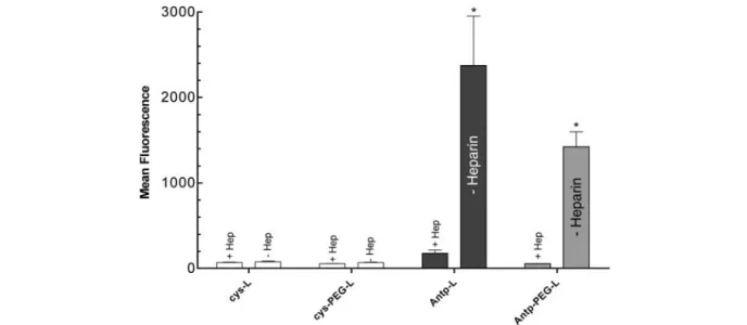
To investigate further the putative mechanism responsible for PTD-mediated cellular uptake, we preincubated two types of Antp-L, conventional liposomes and PEG-lipo-somes, with 20 mg/ml heparin prior to addition to B16F1
cells. As shown in figure 5 heparin inhibited cellular up-Figure 1. Dependence of liposome association with B16F1 cells on
peptide derivatization. (A) Association of liposomes modified with different numbers of peptide molecules depending on the incuba-tion ratio of maleimide groups to peptide (1:1.2, 1:0.6, 1:0.24 and 1:0.12 mol) at a concentration of 500 nmol total lipid/ml medium. *, ** p = 0.001. (B) Association of Antp-L and Tat-L modified with different numbers of peptides and incubated at a constant peptide to cell ratio. * p = 0.0005; * p = 0.0111; *** p < 0.0001. The data are expressed as mean ± SD (n = 3), Cys-L, cys-liposomes; Antp-L,
Antp-liposomes; Tat-L, Tat-liposomes.
Figure 2. Concentration (A) and time (B)-dependent association of different liposome formulations with B16F1 cells. Cells (105per
well) were incubated with fluorescence-labeled Antp-L (squares), Tat-L (triangles) or cys-L (circles) at a final concentration of 15 – 900 nmol total lipid /ml for 90 min (A) or at 250 nmol lipid/ml for 5 min to 24 h (B). Data are expressed as the mean ± SD (n = 3).
take of both types of Antp-modified liposomes. PEG-modified liposomes bound less efficiently to cells, pre-sumably due to the interference of PEG with binding to the HSPGs. Comparable results were obtained with Tat-L (data not shown). Cell association of liposomes was also inhibited by preincubation of liposomes with dextran sul-fate and chondroitin sulsul-fate A as shown before [14]. The involvement of HSPG in binding of liposomes to cells was further demonstrated with two mutant CHO cell
lines. CHO pgs A-745 cells lack UDP-D-xylose:serine beta-1,3-D-xylosyltransferase and are therefore deficient in the synthesis of all proteoglycans. CHO pgs D-677 cells express a reduced amount of HSPG due to a tenfold reduction of N-acetyl-glucosaminyl- and glucuronosyl-transferase activities. As shown in figure 6, reduced up-take of both Antp-L and Tat-L was observed with the mu-tants CHO pgs 677 and CHO pgs 745 compared with the CHO K1 cells. We conclude that cell surface-expressed HSPGs play an important role in the association and sub-sequent uptake of PTD-modified liposomes by various cell types, the exact mechanism remaining to be deter-mined.
Cytotoxic activity of peptide-modified liposomes Due to their rapid cellular uptake (fig. 2), PTD-modified liposomes might be a useful tool to deliver drugs into cells. To demonstrate the advantage of increased cellular uptake of a cytotoxic compound, we compared the effect of Antp-L with cys-L loaded with NOAC-ETC on B16F1 cells. NOAC-ETC is a heterodimeric drug composed of the lipophilic derivative of cytosine arabinoside (NOAC) and the highly active antitumor drug ethinylcytidine [27, 28]. By treating B16F1 cells with drug-containing Antp-L, the IC50of NOAC-ETC was reduced after 2 h
incuba-tion by 40 % from 38 to 24 mM and after 4 h by 70 % from
17 to 5 mM compared to cys-L. Unexpectedly, the
reduc-tion of the IC50observed with Antp-L was lower than
an-Figure 3. Cell association and internalization of fluorescence-labeled liposomes studied in live HeLa cells. Cells were incubated at 37 °C with DiO-labeled cys-L (A), Antp-L (B) and Tat-L (C) for 1 h. Alexa 546 transferrin (25 mg/ml) was added as a marker for endocytosis.
DiO and transferrin fluorescence are shown in green and red, respectively. The square panel in each figure represents a representative x/y plane, the rectangular pictures at the bottom and on the right of each figure show x/z and y/z sections, respectively. The exposure time of
A was approx. ten times longer than of B and C. Bar, 20 mm.
Figure 4. Liposome association with different cell types. Cells were incubated with fluorescence-labeled cys-L, Antp-L or Tat-L at concentrations of 500 nmol total lipid/ml medium for 90 min at 37 °C. Cell association of liposomes was analyzed by flow cytome-try. Data are expressed as the mean ± SD (n = 3). Due to the low
flu-orescence, the bars of cys-L are not visible in the B16F1, DC, W38 and F9 cells.
ticipated from the flow cytometry experiments (see fig. 4), where the difference of cell-associated liposomes be-tween cys-L and Antp-L on B16F10 cells was more pro-nounced. The reduced cytotoxic effect of Antp-L could be explained by the prodrug nature of NOAC-ETC requiring cleavage of the dimeric molecule for cytotoxic activation. Preincubation of Antp-L or Tat-L loaded with NOAC-ETC with heparin reduced the cytotoxic effect on B16F1 cells as shown in figure 7B. These results further corro-borate the importance of HSPGs on cellular uptake of PTD-modified liposomes.
Figure 5. Inhibition of liposome uptake by heparin. Antp-L and Antp-PEG-liposomes (500 nmol total lipid/ml) were preincubated with 20 mg/ml heparin for 15 min before incubation with B16F1 cells for 60 min at 37 °C. Cell association of fluorescence-labeled liposomes
was analyzed by flow cytometry. Data are expressed as the mean ± SD (n = 3); * p = 0.0526.
Figure 6. Involvement of HSPGs in the cell association of PTD li-posomes. Antp-L (open bars), Tat-L (black bars) and cys-L (hatched bars) at 500 nmol total lipid/ml were incubated for 90 min with CHO-K1 cells and the mutants CHO-677 and CHO-745. Cell asso-ciation of fluorescence-labeled liposomes was analyzed by flow cy-tometry. Data are expressed as the mean ± SD (n = 3). Due to the
low fluorescence, the bars of cys-L are not visible in the CHO mu-tant cells.
Figure 7. Cytotoxicity of drug-loaded liposomes in B16F1 melanoma cells. (A) B16F1 cells were incubated with cys-L and Antp-L loaded with NOAC-ETC for 2 and 4 h. Cell viability was measured using the WST-1 test after 24 h incubation at 37 °C. The IC50values were extrapolated from graphical plots of the viability
curves. Data are expressed as the mean ± SD (n = 3); * p < 0.0001;
** p < 0.002. (B) Cys-L, Tat-L and Antp-L loaded with NOAC-ETC were preincubated with heparin (20 mg/ml) for 15 min followed by
addition to B16F1 cells for 30 min. Cell viability was measured us-ing the WST-1 test after 24 h incubation at 37 °C and 5 % CO2.
Discussion
In this study we show that small nanosized cargo particles such as liposomes functionalized with PTD peptides pro-mote efficient delivery of entrapped molecules such as lipophilic fluorescent dyes and cytotoxic drugs into live cells. In this system, the liposome functions as a nanocage providing a protected compartment carrying bioactive molecules, while the peptide bound to the sur-face of liposomes facilitates cell association and pro-motes transport across the plasma membrane. We used a simple, versatile and effective site-specific coupling strategy where peptides are modified at their carboxy ter-minus with cysteine residues. By reaction with male-imide groups introduced on the liposome surface, pep-tides are attached to the outer liposome bilayer membrane via a stable thioether bond endowing the peptide with a high degree of motional freedom to interact with target molecules expressed on the surface of cells. The number of peptide molecules attached to a small liposome of 100 nm average diameter seems to be very important. Whereas an average of 110 – 136 peptide molecules linked per liposome promoted strong cell association and internalization, liposomes decorated with less than 70 peptides (fig. 1) were not taken up efficiently.
Since there are no reports in the literature that Antp pep-tides have been linked to liposomes, our findings can only be evaluated against Tat-modified liposomes as re-ported by Torchilin et al. [20, 21]. These authors devel-oped Tat peptide-liposome-DNA complexes as transfec-tion vehicles for gene therapy. In vitro transfectransfec-tion of dif-ferent cell types with these vehicles was effective and less toxic when compared with commercially available trans-fection reagents. Because Tat peptides were attached to the liposome surface by a non-specific method where peptide amino groups reacted randomly with a p-nitro-phenylcarbonyl-phosphatidyl-ethanolamine lipid, up to 500 copies of Tat peptide per liposome were necessary to obtain effective cell translocation [20]. Thus, we con-clude that our site-specific thiol-maleimide coupling method has several advantages over methods where a high fraction of the peptides may be sterically hindered from binding to cellular target proteins due to unordered attachment to the liposome surface.
Our microscope data obtained in live cells show that li-posomes are internalized by cells. Live-cell imaging was chosen to exclude possible fixation artifacts. The distrib-ution of liposomes on the cell surface and in intracellular vesicles apparently varies among the different types of li-posomes used in this study. Images taken from control cys-L were exposed approximately ten times longer to achieve equal fluorescence intensity, reflecting the fact that the PTDs promoted accelerated binding to and up-take into cells. Cys-L accumulated in large vesicles re-sembling late endosomes that also contained transferrin,
as indicated by the fact that green and red vesicles colo-calized, giving rise to orange cellular staining (fig. 3A). PTD-derivatized liposomes, on the other hand, accumu-lated in small vesicle-like structures with a large propor-tion of material still associated with the plasma mem-brane even after prolonged exposure (data not shown). As in the controls, the transferrin marker accumulated in large intracellular vesicles that are visible in the red chan-nel in figure 3B, C. Whether the PTD-liposomes released their content at the plasma membrane without entering the endocytotic pathway as the cys-liposomes is an inter-esting possibility that is the subject of ongoing work. PTD-liposomes might bypass endocytosis and therefore favor cytoplasmic drug delivery into cells. To define the localization and uptake mechanism of PTD and control liposomes, a detailed analysis of liposome trafficking in live cells is underway. This might open new opportunities for liposome targeting to specific cellular compartments. The mechanism responsible for PTD translocation across membranes is still the subject of controversial discus-sions and may vary among the various PTDs. In earlier publications, the mechanism was proposed as energy in-dependent and preferentially mediated via direct interac-tion of PTDs with the lipid bilayer of cell membranes [13, 29]. In a recent study, Drin et al. [30] and Letoha et al. [31] proposed that for Antp (aa 43 – 58) and derivatives thereof cell binding is not only promoted by HSPGs. Such peptides also directly bind to lipids and may thereby cause membrane association due to their amphipathic properties. However, there is increasing evidence that HSPGs play a paramount role as the cellular entry gate-way of positively charged molecules and synthetic deliv-ery vehicles. HSPGs function as a natural entry mecha-nism for polyamines [32], viruses [33], polybasic pep-tides and polycation nucleic acid complexes [4] and, as shown here, for liposomes carrying positively charged peptides. Independently of the mechanism of internaliza-tion, the potential of PTD-conjugated carrier systems to deliver pharmacologically active molecules into almost any cell type in vitro and in vivo suggests the existence of a ubiquitous transport pathway that can be exploited for cargo delivery [34].
We reported earlier the influence of HSPGs on cellular up-take of Tat and Antp peptides and peptide-avidin com-plexes [14] and our data were recently confirmed by a re-port showing that entry is mediated by lipid rafts in a process called macropinocytosis [17]. Here, we demon-strate that the uptake of peptide-modified liposomes is also inhibited upon blocking of cellular HSPGs with solu-ble heparin. In addition, uptake of Antp-L and Tat-L is dra-matically reduced in mutant cells defective in gly-cosaminoglycan synthesis. Interestingly, we found a dis-tinct pattern of cell association between Antp-L and Tat-L on different cell types. These peptides conceivably follow different binding and uptake mechanisms depending on
the variability of the HSPG expression pattern in various cell types. Furthermore, the distinct physicochemical properties of Antp and Tat, such as hydrophobicity and amphipathicity of Antp in contrast to the high polarity of Tat, may also contribute to the characteristic pattern of up-take and translocation observed in different cells (fig. 4). A particularly interesting application of Antp-L and Tat-L could be their utilization as carriers for antigens in vac-cine development. Vacvac-cines are predominantly applied via subcutaneous or intradermal injection routes. We demonstrated the feasibility of conventional, unmodified antigen-carrying liposomes as vaccines in the lympho-cytic choriomeningitis virus model [25]. After cutaneous application of antigen-carrying Antp-L, such vesicles may be taken up at enhanced rates by antigen-presenting dendritic cells located in the dermal tissue. For parenteral application, liposomes are routinely prepared with PEG-modified lipids to prolong their blood circulation time. We modified PEG-liposomes by attaching Antp peptide molecules at the end of the PEG chain. Such Antp-PEG-liposomes were still able to associate with cell mem-branes, however, with reduced efficiency, as shown in figure 5. For in vivo use of such cargo vehicles, the cova-lent attachment of antibodies or antibody fragments to li-posomes may be further required for target-specific deliv-ery, because PTD-modified cargo vehicles do not discrim-inate between the various cell types they encounter after parenteral application. The likelihood that PTD-modified liposomes bind in an unspecific manner to HSPGs ex-pressed on endothelial cells of blood vessels, in the extra-cellular matrix or on other cell surfaces is high. Therefore, the construction of a drug carrier device combining high target specificity with fast and efficient cell surface asso-ciation and membrane translocation is an attractive possi-bility. The liposome scaffold offers a large surface area to accommodate a high number of different functional groups allowing polyvalent coupling reactions. We previously showed improved targeting of scFv antibody-modified li-posomes to tumor tissue in animals using the thiol-maleimide coupling strategy [11]. We are therefore pursu-ing the possibility to simultaneously modify liposomes with PTD peptides and target cell-specific antibodies. Such multifunctional liposomes might allow enhanced de-livery of pharmaceuticals for therapeutic and diagnostic in-tervention into diseased tissue. The exact mechanism of HSPG function in PTD-peptide-modified liposomes or nanoparticle cell binding and translocation remains to be further investigated and offers exciting opportunities for future research in the field of targeted drug delivery.
Acknowledgements. We gratefully thank C. Garcia-Echeverria
(No-vartis Pharma, Basel, Switzerland) for Tat peptide synthesis and B. Ludewig (Kantonsspital, St. Gallen, Switzerland) for the dentritic cells. C. M. was supported by a fellowship from Oncoswiss, Bern, Switzerland.
1 Lindsay M. A. (2002) Peptide-mediated cell delivery: applica-tion in protein target validaapplica-tion. Curr. Opin. Pharmacol. 2: 587 – 594
2 Schwarze S. R., Ho A., Vocero-Akbani A. and Dowdy S. F. (1999) In vivo protein transduction: delivery of a biologically active protein into the mouse. Science 285: 1569 – 1572 3 Derossi D., Joliot A. H., Chassaing G. and Prochiantz A. (1994)
The third helix of the Antennapedia homeodomain translocates through biological membranes. J. Biol. Chem. 269: 10444 – 10450
4 Derossi D., Chassaing G. and Prochiantz A. (1998) Trojan pep-tides: the penetratin system for intracellular delivery. Trends Cell Biol. 8: 84 – 87
5 Lindgren M., Hallbrink M., Prochiantz A. and Langel U. (2000) Cell-penetrating peptides. Trends Pharmacol. Sci. 21: 99 – 103
6 Fischer P. M., Krausz E. and Lane D. P. (2001) Cellular deliv-ery of impermeable effector molecules in the form of conju-gates with peptides capable of mediating membrane transloca-tion. Bioconjug. Chem. 12: 825 – 841
7 Sapra P. and Allen T. M. (2003) Ligand-targeted liposomal an-ticancer drugs. Prog. Lipid Res. 42: 439 – 462
8 Cattaneo-Pangrazzi R. M., Schott H., Wunderli-Allenspach H., Rothen-Rutishauser B., Guenthert M. and Schwendener R. A. (2000) Cell-cycle arrest and p53-independent induction of apoptosis in vitro by the new anticancer drugs 5-FdUrd-P-Fd-CydOct and dCydPam-P-FdUrd in DU-145 human prostate cancer cells. J. Cancer Res. Clin. Oncol. 126: 247 – 256 9 Mastrobattista E., Koning G. A. and Storm G. (1999)
Immuno-liposomes for the targeted delivery of antitumor drugs. Adv. Drug Deliv. Rev. 40: 103 – 127
10 Barratt G. (2003) Colloidal drug carriers: achievements and perspectives. Cell Mol. Life Sci. 60: 21 – 37
11 Marty C., Odermatt B., Schott H., Neri D., Ballmer-Hofer K., Klemenz R. et al. (2002) Cytotoxic targeting of F9 teratocarci-noma tumours with anti-ED-B fibronectin scFv antibody mod-ified liposomes. Br. J. Cancer 87: 106 – 112
12 Belting M. (2003) Heparan sulfate proteoglycan as a plasma membrane carrier. Trends Biochem. Sci. 28: 145 – 151 13 Drin G., Cottin S., Blanc E., Rees A. R. and Temsamani J.
(2003) Studies on the internalisation mechanism of cationic cell-penetrating peptides. J. Biol. Chem. 278: 31192 – 31201 14 Console S., Marty C., Garcia-Echeverria C., Schwendener R.
and Ballmer-Hofer K. (2003) Antennapedia and HIV transacti-vator of transcription (TAT) „protein transduction domains“ promote endocytosis of high molecular weight cargo upon binding to cell surface glycosaminoglycans. J. Biol. Chem.
278: 35109 – 35114
15 Ou J., Geiger T., Ou Z., Ackerman A. W., Oldham K. T. and Pritchard K. A. (2003) AP-4F, antennapedia peptide linked to an amphipathic alpha helical peptide increases the efficiency of lipofectamine-mediated gene transfection in endothelial cells. Biochem. Biophys. Res. Commun. 305: 605 – 610
16 Sandgren S., Cheng F. and Belting M. (2002) Nuclear targeting of macromolecular polyanions by an HIV-Tat derived peptide. Role for cell-surface proteoglycans. J. Biol. Chem. 277: 38877 – 38883
17 Wadia J. S., Stan R. V. and Dowdy S. F. (2004) Transduci-ble TAT-HA fusogenic peptide enhances escape of TAT-fusion proteins after lipid raft macropinocytosis. Nat. Med. 10: 310 – 315
18 Koch A. M., Reynolds F., Kircher M. F., Merkle H. P., Weiss-leder R. and Josephson L. (2003) Uptake and metabolism of a dual fluorochrome Tat-nanoparticle in HeLa cells. Bioconjug. Chem. 14: 1115 – 1121
19 Nori A., Jensen K. D., Tijerina M., Kopeckova P. and Kopecek J. (2003) Tat-conjugated synthetic macromolecules facilitate cytoplasmic drug delivery to human ovarian carcinoma cells. Bioconjug. Chem. 14: 44 – 50
20 Torchilin V. P., Rammohan R., Weissig V. and Levchenko T. S. (2001) TAT peptide on the surface of liposomes affords their ef-ficient intracellular delivery even at low temperature and in the presence of metabolic inhibitors. Proc. Natl. Acad. Sci. USA
98: 8786 – 8791
21 Torchilin V. P., Levchenko T. S., Rammohan R., Volodina N., Papahadjopoulos-Sternberg B. and D’Souza G. G. (2003) Cell transfection in vitro and in vivo with nontoxic TAT peptide-li-posome-DNA complexes. Proc. Natl. Acad. Sci. USA 100: 1972 – 1977
22 Chikh G. G., Kong S., Bally M. B., Meunier J. C. and Schutze-Redelmeier M. P. (2001) Efficient delivery of Antennapedia homeodomain fused to CTL epitope with liposomes into den-dritic cells results in the activation of CD8+T cells. J. Immunol.
167: 6462 – 6470
23 Chikh G., Bally M. and Schutze-Redelmeier M. P. (2001) Char-acterization of hybrid CTL epitope delivery systems consisting of the Antennapedia homeodomain peptide vector formulated in liposomes. J. Immunol. Methods 254: 119 – 135
24 Cattaneo-Pangrazzi R. M., Schott H. and Schwendener R. A. (2000) The novel heterodinucleoside dimer 5-FdU-NOAC is a potent cytotoxic drug and a p53-independent inducer of apop-tosis in the androgen-independent human prostate cancer cell lines PC-3 and DU-145. Prostate 45: 8 – 18
25 Ludewig B., Barchiesi F., Pericin M., Zinkernagel R. M., Hen-gartner H. and Schwendener R. A. (2000) In vivo antigen load-ing and activation of dendritic cells via a liposomal peptide vac-cine mediates protective antiviral and anti-tumour immunity. Vaccine. 19: 23 – 32
26 Marty C. and Schwendener R. A. (2004) Cytotoxic tumor tar-geting with scFv antibody modified liposomes. Methods Mol. Biol. in press
27 Schwendener R. A., Friedl K., Depenbrock H., Schott H. and Hanauske A. R. (2001) In vitro activity of liposomal N4
-oc-tadecyl-1-b-D-arabinofuranosylcytosine (NOAC), a new lipo-philic derivative of 1-b-D-arabinofuranocylcytosine on biop-sized clonogenic human tumor cells and hematopoietic precur-sor cells. Invest New Drugs 19: 203 – 210
28 Ludwig P. S., Schwendener R. A. and Schott H. (2003) A new laboratory scale synthesis for the anticancer drug 3
¢-C-ethynyl-cytidine. Synthesis 16: 2387 – 2392
29 Vives E., Brodin P. and Lebleu B. (1997) A truncated HIV-1 Tat protein basic domain rapidly translocates through the plasma membrane and accumulates in the cell nucleus. J. Biol. Chem.
272: 16010 – 16017
30 Drin G., Mazel M., Clair P., Mathieu D., Kaczorek M. and Tem-samani J. (2001) Physico-chemical requirements for cellular uptake of pAntp peptide: role of lipid-binding affinity. Eur. J. Biochem. 268: 1304 – 1314
31 Letoha T., Gaal S., Somlai C., Czajlik A., Perczel A. and Penke B. (2003) Membrane translocation of penetratin and its deriva-tives in different cell lines. J. Mol. Recognit. 16: 272 – 279 32 Mislick K. A. and Baldeschwieler J. D. (1996) Evidence for the
role of proteoglycans in cation-mediated gene transfer. Proc. Natl. Acad. Sci. USA 93: 12349 – 12354
33 Gratton J. P., Yu J., Griffith J. W., Babbitt R. W., Scotland R. S., Hickey R. et al. (2003) Cell-permeable peptides improve cellu-lar uptake and therapeutic gene delivery of replication-defi-cient viruses in cells and in vivo. Nat. Med. 9: 357 – 363 34 Tyagi M., Rusnati M., Presta M. and Giacca M. (2001)
Inter-nalization of HIV-1 tat requires cell surface heparan sulfate pro-teoglycans. J. Biol. Chem. 276: 3254 – 3261