ORIGINAL ARTICLE
Proximal direct composite restorations and chairside CAD/
CAM inlays: Marginal adaptation of a two-step self-etch
adhesive with and without selective enamel conditioning
T. Bortolotto&I. Onisor&I. Krejci
Received: 30 January 2006 / Accepted: 31 August 2006 / Published online: 10 October 2006 # Springer-Verlag 2006
Abstract The aim of this study was to evaluate the marginal adaptation of CEREC ceramic inlays, CEREC composite inlays and direct composite restorations in unbeveled proximal slot cavities under artificial aging conditions. Two groups of each restoration type were prepared (n=6), one group with a self-etch adhesive, the other group with H3PO4enamel etching before the self-etch
adhesive application. Replicas were generated before and after long-term thermo-mechanical loading under dentinal fluid simulation and margins were evaluated at ×200 magnification in the scanning electron miscroscope (SEM). Statistically, significant differences were found before and after loading with respect to the percentages of “continuous margins”, the direct composite filling with H3PO4 enamel etching giving the lowest percentages of
“continuous margins” after loading (p<0.05). The highest percentage of “continuous margin” was attained by com-posite inlays without H3PO4 enamel etching. However,
these results were not significantly different from ceramic inlays after stressing. Polymerization shrinkage is still one
critical property of composite restorative materials. The marginal adaptation of indirect adhesive proximal slot restorations without enamel bevels both fabricated out of composite and ceramic is better than that of directly placed composite restorations.
Keywords Slot . Marginal adaptation . Ceramic inlay . Layering technique . Wall flexibility
Introduction
Minimal intervention dentistry is considered the modern style of operative dentistry, and the rationale behind this concept is the maximum preservation of sound tooth tissues [52]. It is based on conservative principles such as remineralization of early lesions, reduction in cariogenic bacteria, repair of defective restorations, disease control and minimum surgical intervention (MSI) [55]. In this regard a number of MSI restorative techniques such as preventive resin restorations [14,36,51,59], preventive glass-ionomer restorations [21, 38, 50], ART restorations [7, 8], tunnel restorations [17, 19] and posterior approximal miniboxes and microchips [18] have been proposed for the treatment of decayed teeth.
Resin composite restorative materials have witnessed a tremendous development since their first application in dentistry in 1950 [3]. Due to their improved esthetic qualities, strength, wear resistance and reduced water sorption with respect to earlier versions, composite restora-tions are being increasingly placed in both anterior and posterior regions of the mouth [61]. Nevertheless, poly-merization shrinkage [27,42,53] and microleakage [45] of resin-based restorative materials is still an unsolved problem in clinical dentistry. This is especially of concern
Clinical significance: Polymerization shrinkage is still one critical property of composite restorative materials. The marginal adaptation of indirect adhesive proximal slot restorations without enamel bevels both fabricated out of composite and ceramic is better than that of directly placed composite restorations.
T. Bortolotto
Division of Cariology and Endodontology, School of Dental Medicine, University of Geneva, Geneva, Switzerland
I. Onisor
:
I. Krejci (*)Division of Cariology and Endodontology, School of Dental Medicine, University of Geneva, 19, Rue Barthélemy-Menn,
CH-1205 Geneva, Switzerland e-mail: [email protected]
when evaluating the marginal adaptation of these restora-tions as it depends, among other factors, on the capability of the bonding agent and tooth structure to withstand the stresses resulting from the polymerization contraction of the composite [47].
Efforts in the scientific field have been undertaken to diminish the adverse effects of polymerization shrinkage in resin composites. In addition to modifications of light-curing protocols [4] and chemical or structural modifica-tions of methacrylate-based materials [58], refinement of clinical application techniques have also been proposed to solve or at least minimize the shrinkage problem. It is in this context where incremental techniques for direct composite restorations as well as indirect techniques have been investigated during the last years [5, 32,34]. It was shown that indirect techniques with both composite and ceramic materials may optimize marginal adaptation in large posterior cavities. Similarly, a sophisticated incremen-tal technique was introduced in 1986 by Lutz and collaborators, with the idea to reduce the effects of polymerization shrinkage and to provide a similar quality of marginal adaptation in large box-shaped posterior cavities to indirect techniques [30,60].
However, in minimally invasive proximal restorations the situation might be more complex. A recent study by Hugo et al. [15] substantiated the use of enamel beveling to improve marginal adaptation. Nevertheless, their results evidenced an increase in the cavity size when bevels were performed, rendering the cavity preparation less conserva-tive. In addition, the risk of damaging the neighbor tooth during the preparation of such a bevel might exist unless ultrasonic pre-shaped instruments [26], scarcely used by the general practitioners, are employed [62].
It might be of interest to evaluate how marginal adaptation can be improved in conservative non-beveled proximal cavities. There are no studies at present prescrib-ing the use of CAD/CAM technology [35] for the restoration of conservative proximal lesions. The Cerec 3D CAD/CAM system (Sirona, Bensheim, Germany) is well recognized in scientific literature, and the construction of a ceramic or composite inlay, onlay, or crown from material blocks in one appointment is possible [37]. A survival rate of around 95% for bonded all-ceramic inlays for up to 10 years has been achieved by the use of this technology [44]. In addition, the prefabricated blocks are industrially conceived and highly homogene, which should improve the mechanical properties and therefore the performance of the restoration over time [46].
Therefore, the purpose of this in vitro study was to evaluate and compare the marginal adaptation of ceramic and compos-ite slot inlays fabricated with the Cerec 3 CAD/CAM technology and directly filled fine hybrid composite Class II restorations before and after thermal and mechanical fatigue
testing. A two-step self-etch adhesive system in combination with a fine hybrid composite was used for both filling and luting procedures. As conventional phosphoric acid etching is considered the most reliable enamel conditioning agent available at present [49] and self-etching systems described as more user-friendly and less technique-sensitive [20], a two-step self-etch adhesive system used with two different bonding approaches, i.e., with and without prior H3PO4
enamel conditioning, was selected to be used in this study. The null hypothesis tested was that no difference existed between both inlay materials and the direct restorative technique in their ability to provide gap-free margins before and after artificial aging conditions.
Materials and methods
The experimental setup of this investigation is schemati-cally represented in Fig. 1. Thirty-six intact, caries-free human molars with completed root formation, which had been stored in 0.1% thymol solution between the time of extraction and use, were selected for this in vitro test. After scaling and pumicing, the teeth were prepared for the simulation of intratubular fluid flow according to a protocol described before by Krejci et al. [25]. This intrapulpal pressure was maintained at 25 mmHg throughout the experiment, i.e., during cavity preparation, restoration placement, finishing, and thermo-mechanical loading. A non-beveled class II slot was prepared in the mesial part of each tooth (Fig. 2) using coarse diamond burs (Diatech Dental, Coltène-Whaledent, Altstätten, Switzerland) and then to be able to perfectly standardize the size and shape of the cavities (height 4.1 mm, width 4 mm, mesio-distal depth 2.5 mm, distance from the CEJ 1.5 mm approxi-mately), they were finished by the use of U-shaped ultrasonic instruments (PCS, EMS Dental, Nyon, Switzer-land) without a bevel (Fig. 2).
Ceramic and composite inlays were fabricated with the Cerec 3D system (Sirona, Bensheim, Germany). The soft-ware version V2.60 was used for this purpose, and the restorations were digitally designed in Dental Database construction mode. The milling was performed with both 1.2-mm cylindrical and standard cone-shaped burs. The polishing of the external surface of both ceramic and composite materials was done by the use of polishing burs (Cerec Set, Intensiv, Switzerland) and Soflex discs (3M/ ESPE, Seefeld, Germany).
Each tooth was randomly assigned to one of six experimental groups. Groups, manufacturers, colors, and batch numbers of the products tested are described in detail in Table1.
In groups 2, 4, and 6, 35% phosphoric acid enamel conditioning was carried out for 20 s. The cavity was then
rinsed and air-dried before the application of Clearfil SE bond (SE). In groups 1, 3, and 5 the adhesive system was applied without previous H3PO4 enamel etching. In all
groups, the self-etching primer was applied for 20 s and air-dried. Then the bonding agent was painted in the cavity’s internal surface and light-cured for 20 s (Astralis 10, Turbo mode, Ivoclar Vivadent, Schaan, Liechtenstein). The power output was continuously tested with a curing radiometer and proved to be higher than 1,000 mW/cm2.
For the placement of the indirect restorations (groups 1 to 4) the luting sequence differed according to the restorative material that was used, i.e., ceramic or compos-ite blocks. The internal surface of the ceramic restorations was etched for 60 s with 5% hydrofluoric acid gel (Ceramics Etch, Vita Zahnfabrik, Germany). After rinsing and drying, a silane was applied (Monobond S, Ivoclar Vivadent, Liechtenstein), left undisturbed for 60 s and consecutively air-dried. Finally, a thin layer of bonding
Fig. 1 Schematic representation of the setup of the study
Fig. 2 Shape and dimensions of the mesial slot preparations with the unbeveled margins located completely in enamel
resin (Clearfil SE Bond, Kuraray, Japan) was applied and the inlay was protected from direct light until being placed into the cavity. A fine hybrid light-cured composite was selected for the luting procedure, applied and light-cured for 60 s each from occlusal, buccal, and oral (Astralis 10, Turbo mode).
With respect to composite inlays, the internal surface was sand-blasted using aluminum oxide (Al2O3) powder of
50μm particle size. Next, a silane solution (Monobond S, Ivoclar Vivadent, Liechtenstein) was applied onto the surface, left undisturbed for 60 s and air-dried. Finally, a thin layer of bonding resin (Clearfil SE Bond, Kuraray, Japan) was applied. A fine hybrid composite was selected for the luting procedure, applied and light-cured for 60 s from occlusal, buccal, and oral (Astralis 10, Turbo mode).
The incremental three-sited light-curing technique was used in groups 5 and 6. For this purpose a layer of approximately 1.0 mm of restorative composite was placed in the gingival’s preparation floor and cured from the cervical margin with a light-transmitting wedge for 60 s. A second increment of restorative composite was placed in the lingual portion of the box and cured from the lingual side for 60 s. The last and buccal portion of the box was restored and cured from the facial direction for 60 s. All restorations were polished immediately after polymeriza-tion. For this purpose fine diamond burs (Diatech Dental, Coltène-Whaledent, Altstätten, Switzerland) and finishing discs (Soflex, 3M–ESPE, St. Paul, MN, USA) were used. After completion of the finishing procedure, the teeth were stored in a 0.9% saline solution at 37°C in the dark for 7 days before loading.
The restored teeth were simultaneously loaded with repeated thermal and mechanical stresses in a chewing machine [23–25]. Thermal cycling was carried out in flushing water with temperatures changing 3,000× from 5 to 50°C and vice versa with a dwelling time of 2 min at 5 and 50°C. The mechanical stress comprised 1,200,000 load cycles transferred on the occlusal center of the mesial slot restoration with a frequency of 1.7 Hz and a maximal load of 49 N applied by using a natural lingual cusp taken from an extracted human premolar. The dentinal fluid simulation was permanently maintained throughout the loading proce-dure. Immediately after completion of the polishing procedure and after stressing, respectively, impressions of each restoration were made with a polyvinylsiloxane impression material (President light body, Coltène AG, Switzerland).
Subsequently, epoxy replicas were prepared for the computer assisted quantitative margin analysis in a scanning electron microscope (Philips XL20, Philips, Eidhoven, Netherlands) at ×200 magnification. In the inlay groups the tooth–composite (TC) and composite– inlay (CI) interfaces were evaluated separately. The statistical evaluation was performed with SPSS 14.0 for Windows (SPSS, Chicago, IL, USA). The scores of marginal adaptation were non-normally distributed, as was shown by Kolmogorov–Smirnov test. Therefore, non-parametric tests were performed for pairwise compar-isons among groups (Kruskal–Wallis and Mann–Whitney U) and for detection of initial/terminal differences within a group (Wilcoxon signed rank test). The confidence level was set to 95% (p < 0.05).
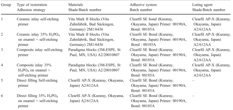
Table 1 Description of the experimental groups
Group Type of restoration Materials Adhesive system Luting agent
Adhesion strategy Shade/Batch number Batch number Shade/Batch number
1 Ceramic inlay self-etching
primer
Vita Mark II blocks (Vita Zahnfabrik, Bad Säckingen, Germany) 2M1/6436
Clearfil SE bond (Kuraray, Okayama, Japan) Primer: 00190A, Bond: 00185A
Clearfil AP-X (Kuraray, Okayama, Japan) A2/612AA
2 Ceramic inlay 35% H3PO4
on enamel + self-etching primer
Vita Mark II blocks (Vita Zahnfabrik, Bad Säckingen, Germany) 2M1/6436
Clearfil SE Bond (Kuraray, Okayama, Japan) Primer: 00190A, Bond : 00185A
Clearfil AP-X (Kuraray, Okayama, Japan) A2/612AA
3 Composite inlay self-etching
primer
Paradigma blocks (3M-ESPE, St Paul, MN, USA) A2/20010807
Clearfil SE Bond (Kuraray, Okayama, Japan) Primer: 00190A, Bond: 00185A
Clearfil AP-X (Kuraray, Okayama, Japan) A2/612AA
4 Composite inlay 35%
H3PO4on enamel +
self-etching primer
Paradigma blocks (3M-ESPE, St Paul, MN, USA) A2/20010807
Clearfil SE Bond (Kuraray, Okayama, Japan) Primer: 00190A, Bond: 00185A
Clearfil AP-X (Kuraray, Okayama, Japan) A2/612AA
5 Direct filling Self-etching
primer
Clearfil AP-X (Kuraray, Okayama, Japan) A2/612AA
Clearfil SE Bond (Kuraray, Okayama, Japan) Primer: 00190A, Bond: 00185A
–
6 Direct filling 35% H3PO4
on enamel + self-etching primer
Clearfil AP-X (Kuraray, Okayama, Japan) A2/612AA
Clearfil SE Bond (Kuraray, Okayama, Japan) Primer: 00190A, Bond: 00185A
Results
The marginal integrity of the groups with H3PO4 enamel
pretreatment (groups 2, 4, and 6) was negatively affected by thermo-mechanical loading (Table 2), and lower percen-tages of continuous margins were observed with respect to the original application of the self-etching adhesive (groups 1, 3, and 5).
Indirect composite restorations with SE used on a self-etch approach (group 3) attained a percentage of 80.2 (14.3) continuous margins after loading, and this was the best result obtained in course of the present investigation. The lowest result was observed for direct composite restorations with SE and H3PO4enamel pretreatment (group 6), with a
percentage of continuous margins of only 21.9 (10.8) after loading.
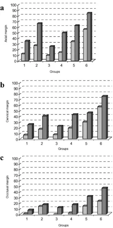
No significant differences could be detected after loading between the marginal adaptation of ceramic and composite inlays in both adhesive approaches tested (group 1 vs group 3 and group 2 vs group 4), although a tendency towards a better adaptation of composite inlays was observed com-pared to ceramic. Nevertheless, when non-continuous margins were taken into consideration, ceramic inlays presented an increased amount of enamel fractures com-pared to composite inlays (Table3). The specific distribu-tion of enamel fractures at the axial, cervical, and occlusal margins of all groups tested is detailed in Fig.3.
The behavior of ceramic and composite inlays was different at the composite–inlay interface (Table 4). A significant degradation between the initial and terminal values was observed for the ceramic inlays in groups 1 and 2. With composite inlays, the luting composite–inlay interface remained stable throughout the loading procedure and almost 100% of gap-free margins could be observed after stressing.
The lowest marginal performance was observed for the direct composite groups (5 and 6) despite the use of an incremental filling technique. The situation was even more adverse when enamel was etched with phosphoric acid before the application of SE bond and almost 80% of
marginal openings were observed in this group after loading. Scanning electron microscopic micrographs, as detailed in Fig.4, represent the most common observations during the marginal microscopic evaluation.
Discussion
In this study, the long-term behavior of ceramic inlays, composite inlays and direct composite filled class II slot restorations was evaluated by the use of thermo-mechanical artificial aging methodology and scanning electron microsco-py (SEM) for the assessment of marginal adaptation. The marginal quality, expressed in percentages of “continuous margins,” was reported for the total marginal length at both tooth–composite (TC) and composite–inlay (CI) interfaces and was used as a descriptive mean of the marginal integrity that can be obtained with any given restorative material. “Non-continuous margins” were investigated at the TC interface as well, enamel fractures being the main parameter considered.
Both thermo-mechanical loading and intrapulpal pres-sure were used in an attempt to simulate the oral environment [31]. Stressing the restorations up to 1.2 million cycles in vitro may simulate approximately 5 years of clinical use [22]. Therefore, it could be assumed that the results of this study might have a certain clinical relevance. The materials used in this investigation, i.e., feldspathic ceramic blocks, composite blocks and direct fine hybrid composite were settled on for performance assessment, all of them being widely used as restorative materials in modern conservative dentistry [12].
No marginal bevels were placed in the present study despite existing scientific evidence that recommends this finishing procedure [10,39]. However, it has to be stated that including a bevel in a conservative proximal cavity is not an easy procedure in the clinical reality. Unless pre-shaped beveled instrumentation is used, the preparation of a bevel in small proximal cavity margins becomes burden-some due to the presence of the neighbor tooth, which risks to be damaged during bur preparation [28, 29, 41]. In addition, beveling of the tooth preparation margins for CEREC restorations might not improve marginal adaptation
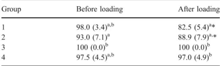
Table 3 Percentages of enamel fractures at the Tooth–Composite interface, before and after thermal and mechanical stressing (Mann– Whitney in superscript letters, Wilcoxon Signed Rank in asterisks, p<0.05)
Group Before loading After loading
1 98.0 (3.4)a,b 82.5 (5.4)a*
2 93.0 (7.1)a 88.9 (7.9)a,*
3 100 (0.0)b 100 (0.0)b
4 97.5 (4.5)a,b 97.0 (4.9)b
Table 2 Percentages of continuous margins at the tooth–composite interface before and after thermal and mechanical stressing (Mann– Whitney in superscript letters, Wilcoxon signed rank in asterisks, p<0.05)
Group Before loading After loading
1 91.2 (3.3)a 66.2 (10.3)a* 2 77.2 (13.7)b,c 51 (10.5)b,d* 3 85.8 (9.4)a,c,d 80.2 (14.3)a* 4 92.0 (7.0)a 58.8 (14.6)a,d,e* 5 74.0 (10.8)b,d 49.4 (13.8)b,e* 6 55.5 (19.4)b 21.9 (10.8)c*
but increase the width of the luting space, adversely affecting the marginal integrity of the restoration [6,48].
As a result of the SEM margin analysis, significant differences in marginal adaptation were found between the test groups (p<0.05). The highest percentage of“continuous margin” after stressing was observed for the composite inlays [80.2(14.3)] and the worse for direct composite fillings [21.9 (10.8)]. Because none of the materials evaluated was able to provide completely gap-free margins before and after artificial aging conditions, the null hypothesis had to be rejected. However, these results are in contrast to the findings of Manhart et al. [33], where almost perfect marginal adaptation in class II MOD enamel cavities were reported after loading when direct composite, composite inlays, and ceramic inlays were used as filling materials.
These differences could account for loading conditions (only 50,000 cycles against 1.2 million in the present study) and to differences in dentinal substrate, i.e., dentinal fluid simulation, which might partially explain their better results. In addition, different cavity configurations were considered, which may play an important role in the absorption and distribution of mechanical stresses. Class II MOD cavities exhibit a lower configuration factor, i.e., the ratio between the bonded and non-bonded or free surface, together with larger wall flexibility (due to the absence of the marginal ridges), which could partially compensate for the stresses generated during polymeriza-tion and fatigue challenge.
The present study also found that the luting composite– inlay interface was not stable under load conditions, as increased percentages of marginal gaps could be observed in ceramic restorations after loading. Our results are supported by similar clinical findings [9]. The use of both silane and bonding agent from different manufacturers, as accomplished in our study, might have negatively influ-enced the results probably due to chemical incompatibility between products. The manufacturers of Clearfil SE bond also provide a silane coupler (Clearfil Porcelain bond activator) that, when needed, can be mixed with the bonding-agent component and applied in a single step,
Fig. 3 Graphical representation of the distribution of enamel fractures (expressed in percentages) at the axial (a), cervical (b), and occlusal (c) margins of the six experimental groups before (light grey) and after (dark grey) thermo-mechanical stressing. An interesting observation is that increased amount of enamel fractures was observed in the direct composite groups (5 and 6), despite the use of an incremental filling technique. Regarding inlay groups, this situation was even more pronounced when enamel was previously etched with H3PO4, as can
be visualized in groups 2 and 4. The most favourable situation was for ceramic and composite inlays with original application of SE Bond (groups 1 and 3). Ceramic might be more aggressive to axial and cervical margins under loading as the presence of enamel fractures is increased with respect to composite inlays
Table 4 Percentages of continuous margins at the Composite–Inlay interface before and after thermal and mechanical stressing (Mann– Whitney in superscript letters, Wilcoxon Signed Rank in asterisks, p<0.05)
Group Before loading After loading
1 7.1 (4.7)a 22.8 (9.0)a* 2 20.8 (13.8)b,d,e 46.4 (12.2)b,d* 3 10.9 (8.8)a,d 18.7 (14.5)a* 4 5.6 (5.2)a 35.8 (15.3)a,d,e* 5 25.7 (10.8)c,e 46.8 (13.8)b,e* 6 43.8 (19.4)c,e 73.1 (9.8)c*
which might contribute to a better adhesion between ceramic and luting composite material [2].
Very low scores of“continuous margin” were attained by the direct composite groups (5 and 6). These results are in agreement with those of recent investigations [13, 16]. A reasonable explanation could be found again in the above-mentioned “wall flexibility” concept. In slot cavities, wall flexibility is almost inexistent; hence, polymerization shrink-age of the composite cannot be compensated by the elastic deformation of the walls. Recently, different cavity models were investigated by the use of Finite Element Analysis [57], and it was concluded that in class I and small class II MO cavities maximum stresses were generated along the tooth-restoration interface basically due to this lack of wall deformation. These observations were also confirmed in vivo [11,40,43,54]. In addition to the C-factor, lack of wall elasticity seems to be a critical issue that should be addressed
in further investigations. Even if the clinical realization of such small proximal inlays might be more time-consuming and thus more expensive, it might represent the best restorative option in non-beveled slot cavities.
Although no significant differences could be detected between both composite and ceramic inlays, an increased amount of enamel fractures perpendicular to the cervical margin was observed in ceramic inlays after loading (Fig. 4d). The rigidity of dental restorative materials is considered a very important issue when evaluating the adhesive tooth-restoration interface. Composite materials are more resilient than ceramics, and this could have an effect on the stress transferred to the margin walls. In a recent paper [1], the distribution of stresses in ceramic and composite class II inlays was evaluated with Finite Element Analysis. It was observed that ceramic FEA models transmitted higher stresses to the cavity walls than
Fig. 4 SEM micrographs (×200) of a composite inlay (group 3) after thermo-mechanical loading. The luting composite interface is almost indistinguishable from the composite inlay and from enamel. b, c Direct composite filling (group 6) after thermo-mechanical loading. The presence of enamel fractures close to the margin might be the result of phosphoric acid conditioning in non-beveled enamel. d Ceramic inlay micrograph (group 1) after thermo-mechanical loading. Enamel fractures
can be observed below the cervical enamel margin, most probably due to the high stresses resulting from mastication forces that are transferred to the margins. Ceramic, as stiff material, may not able to follow the deformation of the tooth during mastication function. DC Direct composite, Cer I ceramic inlay, Cpr I composite inlay, LC luting composite, E enamel, EF enamel fractures
composite models and that resin composite had a greater stress-dissipating effect than ceramic. Even if there is lack of scientific evidence correlating these enamel fractures and the clinical long-term behavior of ceramic restorations, they may be interpreted as a sign of early failure.
Contrary to recent findings [56] for both inlay and direct-filled groups, enamel conditioning with phosphoric acid negatively affected the marginal integrity after loading. One explanation could be that depending on how enamel prisms are cut, i.e., transversally or longitudinally, shearing off of the subsurface enamel prisms together with cohesive fractures within enamel might be produced [39,49]. This is probably the reason why in our study enamel fractures were mostly found close to both axial and cervical margins as illustrated in Fig.4b,c. The parallel orientation of enamel prisms in the axial walls in combination with the weakened region of non-beveled enamel after phosphoric acid conditioning could be an explanation for these findings. The advantage of self-etching systems with respect to phosphoric acid may be that as weaker acids are incorpo-rated into the primer, less risk of over-etching and weakening enamel is present. Therefore, our results confirm those of other papers that self-etching primers can be used for bonding composite to ground enamel [20].
Conclusions
Within the limitations of this in vitro study, the following conclusions can be drawn:
1. None of the materials tested and restorative techniques used could prevent the formation of marginal gaps in enamel both before and after loading.
2. Enamel conditioning with self-etching primers appeared to be less destructive to unbeveled cavo-surface margins than phosphoric acid conditioning. 3. Marginal adaptation of direct composite restorations in
unbeveled cavities was suboptimal despite the use of a sophisticated layering technique.
4. Computerized restorations appeared to be an interesting alternative for the restoration of class II slot cavities. Enabling the construction of slot inlays in one single appointment highlights the use of this technology for the restoration of conservative proximal lesions.
References
1. Ausiello P, Rengo S, Davidson C, Watts D (2004) Stress distributions in adhesively cemented ceramic and resin-composite Class II inlay restorations: a 3D-FEA study. Dent Mater 20:862– 872
2. Blatz MB, Sadan A, Kern M (2003) Resin-ceramic bonding: a review of the literature. J Prosthet Dent 89:268–274
3. Buonocore M (1955) A simple method of increasing the adhesion of acrylic filling materials to enamel surfaces. J Dent Res 34:849– 853
4. Burgess JO, Walker RS, Porche CJ, Rappold AJ (2002) Light curing—an update. Compend Contin Educ Dent 23:889–892 5. Burke FJ, Watts DC, Wilson NH, Wilson MA (1991) Current
status and rationale for composite inlays and onlays. Br Dent J 7:269–273
6. Fett H, Mörmann W, Krejci I, Lutz F (1991) Marginal adaptation of Cerec-MOD inlays in vitro. In: Proceedings of the international symposium on computer restorations. Zurich, Switzerland, pp 393–404
7. Frencken JE, Pilot T, Songpaisan Y, Phantumvanit P (1996) A traumatic restorative treatment (ART): rationale, technique and development. J Public Health Dent 56:135–140
8. Frencken JE, Makoni F, Sithole WD, Hackenitz E (1998) 3 year survival of ART restorations and glass-ionomer sealants in a school oral health program in Zimbabwe. Caries Res 32:119–126 9. Friedl KH, Hiller KA, Schmalz G, Bey B (1997) Clinical and quantitative marginal analysis of feldspathic ceramic inlays at 4 years. Clin Oral Investig 1:163–168
10. Göhring TN, Peters OA, Lutz F (2001) Marginal adaptation of inlay-retained adhesive fixed partial dentures after mechanical and thermal stress: an in vitro study. J Prosthet Dent 1:81–92 11. Hasselrot L (1993) Tunnel restorations. Swed Dent J 17:173–182 12. Hickel R, Manhart J (2001) Longevity of restorations in posterior
teeth and reasons for failure. J Adhes Dent 3:45–64
13. Hilton T, Schwartz R, Ferracane J (1997) Microleakage of four Class II resin composite insertion techniques at intraoral temper-ature. Quintessence Int 28:135–144
14. Houpt M, Fuks A, Eidelman F (1994) The preventive resin (composite resin/sealant) restoration. Nine-year results. Quintes-sence Int 25:155–159
15. Hugo B, Lussi A, Hotz P (1992) The preparation of enamel margin beveling in proximal cavities. Schweiz Monatsschr Zahnmed 102:1181–188 (in German)
16. Hugo B, Stassinakis A, Hofmann N, Schmitz B, Klaiber B (2001) Marginal adaptation of small Class II resin restorations. An in vitro evaluation. Schweiz Monatsschr Zahnmed 111:19–27 (in German)
17. Hunt PR (1984) A modified class II cavity preparation for glass ionomer restorative materials. Quintessence Int 10:1011–1018 18. Hunt PR (1994) Rational cavity design principles. J Esthet Dent
6:245–256
19. Jinks GM (1963) Fluoride-impregnated cements and their effect on the activity of interproximal caries. J Dent Child 30:87–92 20. Kaaden C, Powers JM, Friedl KH, Schmalz G (2002) Bond
strength of self-etching adhesives to dental hard tissues. Clin Oral Investig 6:155–160
21. Knight GM (1984) The use of adhesive materials in the conservative restoration of selected posterior teeth. Aust Dent J 29:324–331
22. Krejci I, Lutz F (1990) In-vitro test results of the evaluation of dental restoration systems. Correlation with in-vivo results. Schweiz Monatsschr Zahnmed 100:1445–1449 (in German) 23. Krejci I, Reich T, Lutz F, Albertoni M (1990) In-vitro test
procedure for evaluation of dental restorative systems: 1. Computer-controlled chewing simulator. Schweiz Monatsschr Zahnmed 100:953–960 (in German)
24. Krejci I, Albertoni M, Lutz F (1990) In-vitro test procedure for evaluation of dental restorative systems: 1. Computer-controlled chewing simulator. 2. Toothbrush-/toothpaste abrasion and chem-ical degradation. Schweiz Monatsschr Zahnmed 100:1164–1168 (in German)
25. Krejci I, Kuster M, Lutz F (1993) Influence of dentinal fluid and stress on marginal adaptation of resin composites. J Dent Res 72:490–495
26. Krejci I, Lutz F, Krejci D (1995) Sonic/ultrasonic diamond coating instruments for cavity preparation, for contouring and finishing. ZWR 104:781–786 (in German)
27. Lu H, Stansbury JW, Dickens SH, Eichmiller FC, Bowman CN (2004) Probing the origins and control of shrinkage stress in dental resin-composites: I. Shrinkage stress characterization technique. J Mater Sci Mater Med 15:1097–1103
28. Lussi A (1995) Damage to neighboring teeth during the preparation of proximal cavities. An in-vivo study. Schweiz Monatsschr Zahnmed 10:1259–1264 (in German)
29. Lussi A, Kronenberg O, Megert B (2003) The effect of magnification on the iatrogenic damage to adjacent tooth surfaces during Class II preparation. J Dent 31:291–296
30. Lutz F, Krejci I, Oldenburg TR (1986) Elimination of polymer-ization stresses at the margins of posterior composite restorations: a new restorative technique. Quintessence Int 17:777–784 31. Lutz F, Krejci I, Barbakow F (1991) Quality and durability of
marginal adaptation in bonded composite restorations. Dent Mater 7:107–113
32. Lutz F, Krejci I (1999) Resin composites in the post-amalgam age. Compend Contin Educ Dent 12:1138–1144
33. Manhart J, Schmidt M, Chen HY, Kunzelmann KH, Hickel R (2001) Marginal quality of tooth-colored restorations in class II cavities after artificial aging. Oper Dent 4:357–366
34. Manhart J, Chen H, Hamm G, Hickel R (2004) Buonocore Memorial Lecture. Review of the clinical survival of direct and indirect restorations in posterior teeth of the permanent dentition. Oper Dent 29:481–508
35. Mehl A, Hickel R (1999) Current state of development and perspectives of machined-based production methods for dental restorations. Int J Comput Dent 2:9–35
36. Mertz-fairhust EJ, Curtis JW, Ergle JW, Rueggeberg FA, Adair SM (1998) Ultraconservative and cariostatic sealed restorations: results at year 10. J Am Dent Assoc 129:55–66
37. Mormann W, Bindl A (2000) The Cerec 3-A quantum leap for computer-aided restorations: initial clinical results. Quintessence Int 10:699–712
38. Mount GJ (1986) Longevity of glass ionomer cements. J Prosthet Dent 55:682–685
39. Munechika T, Suzuki K, Nishiyama M, Ohashi M, Horie K (1984) A comparison of the tensile bond strengths of composite resins to longitudinal and transverse sections of enamel prisms in human teeth. J Dent Res 8:1079–1082
40. Nordbo H, Leirskar J, Von Der Fehr F (1993) Saucer-shaped cavity preparations for composite resin restorations in class II carious lesions: three year results. J Prosthet Dent 69:155–159 41. Opdam NJM, Roeters JJM, Van Berghem E, Eijsvogels E,
Bronkhorst E (2002) Microleakage and damage to adjacent teeth when finishing Class II adhesive preparations using either a sonic device or bur. Am J Dent 15:317–320
42. Peutzfeld A, Asmussen E (2004) Determinants of in vitro gap formation of resin composites. J Dent 32:109–115
43. Pyk N, Mejare I (1999) Tunnel restorations in general practice. Influence of some clinical variables on the success rate. Acta Odontol Scand 57:195–200
44. Reiss B, Walther W (2000) Clinical long-term results and 10 year Kaplan Meier Analysis of Cerec restorations. Int J Comp Dent 3:9–23
45. Roulet JF, Salchow B, Wald M (1991) Margin analysis of posterior composites in vivo. Dent Mater 7:44–49
46. Rusin R (2001) Properties and applications of a new composite block for CAD/CAM. Compendium 22:35–41
47. Sarrett DC (2005) Clinical challenges and the relevance of materials testing for posterior composite restorations. Dent Mater 21:9–20
48. Schmalz G, Federlin M, Reich E (1995) Effect of dimension of luting space and luting composite on marginal adaptation of a class II ceramic inlay. J Prosthet Dent 73:392–399
49. Shimada Y, Tagami J (2003) Effects of regional enamel and prism orientation on resin bonding. Oper Dent 28:20–27
50. Sidhu SK, Watson TF (1995) Resin-modified glass ionomer materials. A status report for the American Journal of Dentistry. Am J Dent 8:59–67
51. Simonsen RJ (1989) Cost effectiveness of pit and fissure sealant at 10 years. Quintessence Int 20:75–84
52. Staehle HJ (1999) Minimally invasive restorative treatment. J Adhes Dent 3:267–284
53. Stavridakis MM, Lutz F, Johnston WM, Krejci I (2003) Linear displacement and force induced by polymerization shrinkage of resin-based restorative materials. Am J Dent 16:431–438
54. Strand GV, Nordbo H, Tveit AB, Espelid I, Wikstrand K, Eide GE (1996) A three-year clinical study of tunnel restorations. Eur J Oral Sci 104:384–389
55. Tyas M, Anusavice K, Frencken J, Mount GJ (2000) Minimal intervention dentistry-a review. Int Dent J 50:1–12
56. Van Landuyt KL, Kanumilli P, de Munck J, Peumans M, Lambrechts P, Van Meerbeek B (2006) Bond strength of a mild self-etch adhesive with and without prior acid-etching. J Dent 34:77–85
57. Versluis A, Tantbirojn D, Pintado M, Delong R, Douglas W (2004) Residual shrinkage stress distributions in molars after composite restoration. Dent Mater 20:554–564
58. Weinmann W, Thalacker CH, Guggenberger R (2005) Siloranes in dental composites. Dent Mater 21:68–74
59. Welbury RR, Walls AW, Murray JJ, Mccabe JF (1990) The management of occlusal caries in permanent molars. A 5-year clinical trial comparing a minimal composite with an amalgam restoration. Br Dent J 169:361–366
60. Wilder A, May K, Bayne S, Taylor D, Leinfelder K (1999) Seventeen-year clinical study of ultraviolet-cured posterior composite class I and II restorations. J Esthet Dent 3:135– 142
61. Willems G, Lambrechts P, Braem M, Vanherle G (1993) Composite resins in the 21st century. Quintessence Int 24:641–658
62. Yip HK, Samaranayake LP (1998) Caries removal techniques and instrumentation: a review. Clin Oral Invest 2:148–154