HAL Id: hal-01779754
https://hal.archives-ouvertes.fr/hal-01779754
Submitted on 26 May 2020
HAL is a multi-disciplinary open access
archive for the deposit and dissemination of sci-entific research documents, whether they are pub-lished or not. The documents may come from teaching and research institutions in France or abroad, or from public or private research centers.
L’archive ouverte pluridisciplinaire HAL, est destinée au dépôt et à la diffusion de documents scientifiques de niveau recherche, publiés ou non, émanant des établissements d’enseignement et de recherche français ou étrangers, des laboratoires publics ou privés.
considerations and transcriptional modulation during
mycoparasitic association with Fusarium solani olive
trees
Maroua Ben Amira, Robin Mom, David Lopez, Hatem Chaar, Ali Khouaja,
Valérie Pujade-Renaud, Boris Fumanal, Aurelie Gousset, Gisèle Bronner,
Philippe Label, et al.
To cite this version:
Maroua Ben Amira, Robin Mom, David Lopez, Hatem Chaar, Ali Khouaja, et al.. MIP diversity from Trichoderma: Structural considerations and transcriptional modulation during mycoparasitic association with Fusarium solani olive trees. PLoS ONE, Public Library of Science, 2018, 13 (3), pp.23. �10.1371/journal.pone.0193760�. �hal-01779754�
MIP diversity from Trichoderma: Structural
considerations and transcriptional modulation
during mycoparasitic association with
Fusarium solani olive trees
Maroua Ben Amira1,2☯, Robin Mom1☯, David Lopez1, Hatem Chaar3, Ali Khouaja4, Vale´rie Pujade-Renaud1,5, Boris Fumanal1, Aure´lie Gousset-Dupont1, Gisèle Bronner6,
Philippe Label1, Jean-Louis Julien1, Mohamed Ali Triki7, Daniel Auguin8*, Jean-Ste´phane Venisse1
*
1 UCA, UMR PIAF, Clermont-Ferrand, France, 2 Faculte´ des Sciences de Bizerte, Zarzouna, Tunisia, 3 National Institute of Agronomy of Tunisia (INAT), Crop Improvement Laboratory, INRAT, Tunis, Tunisia, 4 National Institute of Agronomy of Tunisia (INAT), Sylvo-Pastoral Laboratory of Tabarka, Tabarka,Tunisia, 5 CIRAD, UMR AGAP, Clermont-Ferrand, France, 6 Universite´ Clermont Auvergne, UMR CNRS 6023 Laboratoire Microorganismes: Ge´nome et Environnement, Clermont-Ferrand, France, 7 Institut de l’Olivier, LR: Ame´lioration et Protection des Ressources Ge´ne´tiques de l’Olivier-Universite´ de Sfax, Sfax, Tunisia, 8 Universite´ d’Orle´ans, Laboratoire de Biologie des Ligneux et des Grandes Cultures, UPRES EA 1207, INRA-USC1328, Orle´ans, France
☯These authors contributed equally to this work.
*[email protected](DA);[email protected](JSV)
Abstract
Major intrinsic proteins (MIP) are characterized by a transmembrane pore-type architec-ture that facilitates transport across biomembranes of water and a variety of low molecu-lar weight solutes. They are found in all parts of life, with remarkable protein diversity. Very little is known about MIP from fungi. And yet, it can legitimately be stated that MIP are pivotal molecular components in the privileged relationships fungi enjoy with plants or soil fauna in various environments. To date, MIP have never been studied in a mycopara-sitism situation. In this study, the diversity, expression and functional prediction of MIP from the genus Trichoderma were investigated. Trichoderma spp. genomes have at least seven aquaporin genes. Based on a phylogenetic analysis of the translated sequences, members were assigned to the AQP, AQGP and XIP subfamilies. In in vitro and in planta assays with T. harzianum strain Ths97, expression analyses showed that four genes were constitutively expressed. In a mycoparasitic context with Fusarium solani, the caus-ative agent of fusarium dieback on olive tree roots, these genes were up-regulated. This response is of particular interest in analyzing the MIP promoter cis-regulatory motifs, most of which are involved in various carbon and nitrogen metabolisms. Structural analy-ses provide new insights into the possible role of structural checkpoints by which these members transport water, H2O2, glycerol and, more generally, linear polyols across the
membranes. Taken together, these results provide the first evidence that MIP may play a key role in Trichoderma mycoparasitism lifestyle.
a1111111111 a1111111111 a1111111111 a1111111111 a1111111111 OPEN ACCESS
Citation: Ben Amira M, Mom R, Lopez D, Chaar H, Khouaja A, Pujade-Renaud V, et al. (2018) MIP diversity from Trichoderma: Structural considerations and transcriptional modulation during mycoparasitic association with Fusarium
solani olive trees. PLoS ONE 13(3): e0193760. https://doi.org/10.1371/journal.pone.0193760
Editor: Vijai Gupta, Tallinn University of Technology, ESTONIA
Received: July 13, 2017 Accepted: February 17, 2018 Published: March 15, 2018
Copyright:© 2018 Ben Amira et al. This is an open access article distributed under the terms of the
Creative Commons Attribution License, which permits unrestricted use, distribution, and reproduction in any medium, provided the original author and source are credited.
Data Availability Statement: All relevant data are within the paper and its Supporting Information files.
Funding: This work was supported by the PHC program “Uthic” from Campus France (grant 34861PF) under the joint aegis of the Tunisian Ministry of Foreign Affairs and the French Ministry of Higher Education and Scientific Research.
Competing interests: The authors have declared that no competing interests exist.
Introduction
Most crop farmers are confronted with the need to control various diseases (physiological or parasitic), while trying to meet strong consumer demands to use environment-friendly farm-ing methods. One option is to use members of the fungus genusTrichoderma, most of which are now known to be effective antagonists of a broad array of soil-borne pathogens [1]. We recently showed that a strain ofT. harzianum (Ths97), isolated from Tunisian farmlands, expressed antagonist activities against a strain ofFusarium solani (Fso14), which causes severe dieback of olive roots in Tunisia [2]. Fusarium root rot diseases are steadily expanding world-wide in nurseries and young olive groves, and disease control is still limited to systemic fungi-cide treatments and prophylactic actions. By analogy with different tripartite pathosystems that include someTrichoderma spp. as mycopesticides, Ths97 is thought to act as a necro-trophic myco-hyperparasite, stoppingFso14 growth in in vitro through the development of contact structures, namely helicoidal structures around its host, papilla-like structures and the collapse of severalFso14 septa. Furthermore, on olive trees, Ths97 develops substantial protec-tive activity againstFusarium root infestation. This bioprotection is correlated with the up-reg-ulation of an array of plant defense-related pathways byThs97.
Trichoderma spp. occur as ubiquitous common agents in most soils, and in a few cases, they are also competitive saprotrophs, opportunistic parasites of other organisms (animals, plants or fungi), and possibly endophytes/symbionts of plants [3]. In a competitive context within an rhizospheric ecosystem, like other chemo-heterotroph mycoparasites (whether or not they are classified as a biological control agent or BCA),Trichoderma spp. weave an intimate network of nutritional links with their close partners, most relevantly here the plant [the olive tree] and its prey [the phytopathogenF. solani]. Even today, most studies on mycoparasites (lato sensu) focus exclusively on the mechanisms of attack and/or self-defense in plants [4]. Feed mecha-nisms are rarely mentioned or only very cursorily. Yet they are crucial to the relationship between myco-hyperparasites and other living organisms. Some aspects of this physiological pathway need to be more fully understood.
Parasite growth depends on the retrieval of a countless number and variety of nutrients from host organisms. They are mainly water, inorganic solutes, and a plethora of nitrogen and carbon organic precursors, such as carbohydrates, amino acids, fatty acids, and nucleosides. Internalizing external food, when it occurs without membrane deformation (ie endocytosis), is made possible by an abundant arsenal and diverse protein groups of plasma membrane trans-porters. This group includes the major intrinsic proteins (MIP) [5]. MIP are a large transporter superfamily generically designated as “aquaporins” (AQP). They facilitate the selective bidirec-tional transport of water and small uncharged molecules across biological membranes [6]. Structurally, AQP share classic folded topology and channel architecture lending them an hourglass shape. The overall three-dimensional design of the integral membrane region has a two-fold symmetry consisting of six transmembraneα-helices with five internal loops. A sev-enth pseudo-transmembrane helix is formed by two smaller hemi-helical segments (in the middle of loop B and loop E segments) that project opposing “NPA” boxes (Asn-Pro-Ala) at the center of the structure. The pore formed by the packing of these seven helices displays this hourglass aspect, in which the narrow constriction determines transport selectivity based on solute size and hydrophobicity [7]. A second major determinant for substrate specificity is located in the outer channel vestibule, and is referred to as the ar/R (aromatic/arginine) selec-tivity region [8,9]. This feature consists of a tetrad of amino acid residues, one from each of the transmembrane helices 2 [H2] and 5 [H5], and two from the inter-helical loop containing the second “NPA” box [LE1 and LE2]. “NPA” boxes and the ar/R filter regulate the single-file conductance of water and molecules by acting as a cation- and proton-excluding selectivity
filter. These physicochemical and thermodynamic contexts determine which molecules can cross the pore.
With an increasing number of genome sequences available, MIP genes have now been fully described across all living organisms, except for some thermophilic Archaea and intracellular bacteria [10]. Despite its overall diversity, the MIP family can be functionally divided into two major phylogenetic divisions, separating the water-selective AQP (i.e. the water-specific and the orthodox AQP) from the glycerol facilitators or aquaglyceroporin (GlFp) [11]. In fungi, MIP nomenclature is established on that of yeasts, and resembles that of vertebrates [12,13]. Phylo-genetic analysis finds three main groups with possible subdivisions: classical aquaporins (AQP), fungal XIP, and aquaglyceroporins (AQGP) subdivided into Fps-like AQGP, Yfl054-like AQGP and "other" aquaglyceroporins [12,14,15,16].
While aquaporins have been the subject of intensive study mostly in vertebrates and plants concerning their transport specificity and their direct or indirect involvement in controlling homeostasis, their precise role in various challenged environments is still not entirely under-stood in most eukaryotes. This is particularly true for fungi for which MIP structure, functions and regulation are less studied, beyond several closely related models ofSaccharomyces cerevi-siae yeasts [12], two Basidiomyceta and ectomycorrhiza fungusGlomus intraradices [16,17] andLaccaria bicolor [15], and the AscomycetaAspergillus glaucus [18]. Even so, the general lack of fungus MIP data is surprising, given the large number of fungus species and their diverse physiology and ecology niches that are always connected with water and a broad range of solutes. Remarkably, MIP from fungal mycoparasites have never been comprehensively and specifically explored.
In this study, the tripartite myco-phytopathosystem [T. harzianum Ths97 –F. solani Fso14 –olive trees] was used to gain insight into the molecular mechanisms involved in cell uptake of essential nutrients, by focusing specifically on the MIP route. We first investigated MIP diversity in the genusTrichoderma, and monitored the transcriptional expression pat-terns of these MIP in a situation of mycoparasitism involving theT. harzianum Ths97 strain andF. solani Fso14 strain, both in vitro and in olive trees (either preventively in primed plants or curatively in diseased plants). Second, we depicted the protein structure of the MIP expressed by modeling, and hypothesized its ability to transport water, H2O2and
glyc-erol. In addition, the possibility that particular solutes such as small carbohydrates may be transported across these MIP is discussed. In brief, our data provide the first comprehensive information concerning the MIP superfamily in the Ascomyceta genusTrichoderma and their potential involvement in a mycoparasitism context. We go on to discuss our findings with a special focus on the trophic behaviors thatTrichoderma sets up in its habitat, which remain almost unknown in a situation of mycoparasitism.
Materials and methods
Fungal strains and plant material
Both theTrichoderma harzianum strain Ths97 and the soil-borne Fusarium solani strain Fso14 (accession number KU863548) were isolated from private Tunisian farmlands, with the per-mission and the help of the owner of the land, and registered at the "Institut de l’Olivier" (Uni-versity of Sfax, Tunisia) [19]. Fungi were grown on PDA plates at 25˚C and 27˚C forFso14 and Ths97, respectively. The cultivar Olea europaea cv. Chemlali obtained from herbaceous cuttings of two-year-old plants were used for assays because of their high susceptibility toFso14 [20,2]. Plants were planted in plastic bags containing autoclave-sterilized sandy clay soil, and kept in a controled growth chamber with the following growth parameters: 16h photoperiod, 26/23˚C (day/night), relative humidity around 70%, and regular irrigation.
Root inoculations were performed for 1 hour by placing the roots in the conidia suspen-sions (S2 Fig). After inoculation, plants were replanted in plastic bags containing new soil. For the confrontation assays,Ths97 and Fso14 were inoculated successively with 6 days apart. The preventive assay corresponded to plants inoculated withThs97 in the first step, and the cura-tive assay to plants inoculated withThs97 in the second step. The number of biological repeti-tions was:n = 6 for water control plants, n = 9 each for Ths97 and Fso14 infested plants, and n = 18 each for curative and preventive treatments. After eight weeks of infestation, roots were carefully harvested and randomly pooled in three samples in terms of biological conditions, rapidly frozen in liquid nitrogen and stored at -80˚C until needed for molecular analyses. Con-cerning thein vitro confrontation tests, two mycelial plugs (8 mm diameter) were cut from the edge of actively growing cultures ofThs97 and Fso14 respectively, and placed 4-cm apart in a new PDA plate (S3 Fig). The paired cultures and control cultures (Fso14 alone) were incubated at 27˚C in the dark and sealed with Parafilm. The biological repetitions were done in triplicate, and each zone of interest was carefully harvested, rapidly frozen in liquid nitrogen and stored at -80˚C until needed for molecular analyses. Statistical analyses ofin planta and in vitro dual tests were carried out using rank-based non-parametric and ANOVA parametric methods, respectively. These analyses are detailed in [2]. All experiments for this study with the strains were done at the "Institut de l’Olivier" under the supervision of Dr TRIKI Mohamed Ali.
Bioinformatic analysis
Protein sequences homologous to MIP transporters fromTrichoderma spp. were retrieved at the Joint Genome Institute (http://genome.jgi-psf.org/). Some new sequences were also identi-fied by tBLASTn searches against the NCBI GenBank GSS databases (http://www.ncbi.nlm. nih.gov/). These investigations were conducted using keyword queries (IPR000425; Major Intrinsic Protein; Aquaporin) and tblastn searches (with conservative criteria requiring a cut-off ofE-value of 1.0−5). For allin silico analyses on T. harzianum, T. harzianum strain CBS 226.95 v1.0 (from JGI) was used as reference. Protein names and accession numbers are listed inS1 Table. During retrieval, each MIP member was verified by predicting the transmembrane topology with Interproscan from EMBL (http://www.ebi.ac.uk/Tools/pfa/iprscan/). Molecular modeling of MIP fromT. harzianum was performed with the I-TASSER (Iterative Threading ASSEmbly Refinement) program suite [21,22,23]. Electrostatic potentials were established in a PARSE forcefield [24] using the Adaptive Poisson-Boltzmann Solver [25] in PyMOL [26], which was used to analyze and illustrate the molecular models. Structural alignment was gen-erated with mulPBA [27]. MOLE-2 was used to define the central pores in terms of radius and polarity. Amino acid sequences were aligned using MUSCLE [28]. Phylogenetically informa-tive positions were selected using Gblocks [29], and maximum likelihood phylogenetic recon-structions were made with PhyML (v3.0) [30] using the WAG substitution model, bootstrap supports with 500 replicates and default parameters. Tree was carried out using maximum likelihood and the phylogenetic tree was visualized with TreeDyn [31]. Theoretical isoelectric point (pI) and molecular weight (Mw) were calculated with the Compute pI/Mw tool (expasy. org/compute_pi/). Putative transcription factor binding sites (TFBSs) were analyzed on MIP genes fromT. harzianum that were expressed in our biological conditions. Promoters were retrieved by searching the JGI database on sequences fromTrichoderma harzianum CBS 226.95 that corresponded to the 1.5Kb of the genomic sequence upstream of the initiation codon. TFBSs were detected with the Promoter Database ofSaccharomyces cerevisiae, SCPD (http://rulai.cshl.edu/SCPD/; [32]), and the putative biological processes (GO) were identified with SCPD and the Universal Protein Resource Uniprot (http://www.uniprot.org/).
RNA isolation and quantitative real-time PCR (qRT-PCR) analysis
Total RNA was extracted as previously described by [33]. Mycelia fromin vitro cultures and infected root tissues were ground to a fine powder in liquid nitrogen and transferred to 1 ml of lysis CTAB extraction buffer (bromide cetyltrimethylammonium). The homogenate was incu-bated for 5 min at 65˚C, and treated twice with 1 volume of chloroform:isoamyl alcohol (24:1). The supernatant was collected and treated overnight in 2M of LiCl at -20˚C. The precipitate was collected by centrifugation (16,000g for 45 min) and washed with 80% ethanol. The pellet was dissolved in 25μl of water (DEPC) and then treated with 1U of RNase-free RQ1 DNase (Pro-mega, Madison, WI, U.S.A.) for 30 min to remove contaminating genomic DNA. After two chloroform:isoamyl alcohol (24:1) washes, total RNA was precipitated with 100% ethanol (2V) for 2 hours at -20˚C. After centrifugation at 16,000g for 30 min, the pellet was washed with 80% ethanol, dissolved in 50μl of water (DEPC), and stored at -80˚C for later analysis. RNA concen-trations were determined by spectrophotometry at OD 260/280 (spectrophotometer ND-1000, Nanodrop, France), and quality was checked by using 2% TAE/agarose electrophoresis. Twoμg of total RNA were reverse-transcribed into cDNA with Oligo-dT using the SuperScript1III First-Strand Synthesis System for RT-PCR (Invitrogen). cDNA was diluted 10-fold with sterile water, and used as a template for qPCR. The abundance of MIP-related transcripts was deter-mined by real-time qPCR with a MyiQ instrument (Bio-Rad). MIP gene expression levels were calculated by the 2-ΔΔCTmethod [34]. PCR amplifications were done in 15μL of PCR reaction using MESA GREEN qPCR MasterMix Plus (Eurogentec) from 2μl of cDNA template. Cycle parameters were 94˚C for 30s, followed by 35 cycles at 94˚C for 15s, at 58˚C for 15s, and 72˚C for 20s. PCR reactions were ended by generating a dissociation curve to confirm the amplifica-tion of PCR single bands. Geometric mean ofCtrelated to genes encoding totubulin (Th,
pro-tein ID: 516507;Fs, 98894; JGI; [35]),18SrRNA (Th, sequence ID: KT897696.1; Fs, JQ837837.1; NCBI) and according to the strain, genes encoding actin (Fs, protein ID: 63567; JGI) or elonga-tion factor 1-alpha (Th, protein ID: 146236; JGI; [36]) were used as internal references to nor-malize MIP expression for their stable constitutive expressions during fungus development and infestation. Specific primer pairs for each MIP member were designed in consensus zones after alignment of MIP sequences retrieved fromTrichoderma spp. or Fusarium spp. with Primer3-plus application (http://www.bioinformatics.nl/primer3plus). Specific amplification of only one desired band was observed using each primer combination for qRT-PCR analysis. Primer pairs are listed inS2 Table. All PCR technical samples were assayed in triplicate, and reactions were carried out with three biological replicates. For statistical analysis, data were analyzed using a parametric method on STATISTIX V8 software where aquaporin steady-state gene expression levels were computed by a one-way analysis of variance (ANOVA) followed by a Tukey’s honest significant difference (HSD)post hoc test (p < 0.05).
Results and discussion
Originality of the topic
In major ongoing research in molecular plant pathology, the characterization of virulence fac-tors that underpin host-pathogen interactions is still a topical issue. Pathogens deploy an array of effectors that intrinsically constitute their "cell architecture" or which are secreted into the surrounding environment to interfere with host cell processes. By extension, during a situation of pathogenicity, all solute transporters may be regarded as virulence factors, since they are involved in controlling the entry into the cell of molecules with nutritive value, notably when they originate from the host prey. Because MIP play major roles in numerous physiological processes, it is meaningful to consider these channels as pathogenic factors. The genus
Trichoderma offers us an opportunity to study this subfamily in a tripartite mycoparasitic con-text [T. harzianum/F. solani/olive trees] [2].
MIP diversity
In order to characterize the MIP family fromTrichoderma, the genome databases NCBI and JGI were searched using the previously described MIP translated sequences fromTrichoderma virens and Nectria haemotococca (asexual name of Fusarium solani) [15]. The different Tricho-derma strains encode six to eight predicted MIP, while Fusarium strains encode six predicted MIP except forF. solani with five members, and F. oxysporum and F. oxysporum f. sp. lycoper-sici with seven members each (S1 Table).
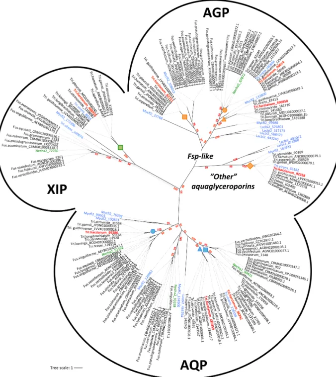
T. harzianum and F. solani (Nehca2) exhibited eight and five predicted MIP, respectively. A random analysis of this MIP family from diverse fungi (JGI) showed an average of five MIP members, placingTrichoderma among those species that share the broadest range. Phyloge-netic relationships among theTrichoderma and Fusarium MIP proteins were analyzed with classified orthologs fromLaccaria bicolor and Mycosphaerella fijiensis [15] as a reference. Sequences fall into three major clades: the classical aquaporins (AQP), aquaglyceroporins (AQGP) and X-intrinsic proteins (XIP) (Fig 1). Specifically,Trichoderma shows three classical AQP, three AQGP (two Fps-like and one "other" AQGP) and a single XIP. Amino acid conser-vation ranges from 40% to 54% sequence identity in AQP, from 40% to 54% sequence identity within AQGP, and from 76% to 87% sequence identity in XIP. By comparison,Fusarium exhibits three classic AQP, one AQGP (Fps-like) and likewise a single XIP. However, unlike Fusarium, AQP present in Trichoderma were split into three sub-groups, and AQGP into four sub-groups with three Fps-like and one "Other aquaglyceroporin" branches. All the XIP coa-lesced into a major clade, which can be divided into two branches. Although fewer subgroups are met in fungi than in plants, the emergence of a structural diversity is highlighted. More-over, whatever the number of MIP members from each species, there is invariantly a genus-dependent subfamily distribution. Despite the common lineage of these two fungi (class of Sordariomycetes), these MIP differences in each subgroup may result from independent rounds of gene events such as duplications, but without excluding possible gene losses. For the Trichoderma genus, however, the limited number of differences between MIP sequences has not provided a clear-cut answer to the question of MIP expansion. At least one duplicated event seems to have occurred inT. harzianum and concerns the aquaglyceroporin 82211, absent in the ancestral speciesT. reesei. Gene duplication plays a key role in increasing genetic variability (driving an increase in the sizes of gene families, andin fine, the genome expansion of species), but most importantly, these genomic events create novel genes, which may confer potential new adaptation abilities. Here, such a relative conservation in a MIP subfamily in the Trichoderma genus suggests that each MIP member is devolved to transporting particular sol-utes that are pivotal in the full cycle of fungus development.
Additionally, insofar as these subfamilies (AQP, AGP and XIP) are expected to transport different solutes [16], the strong diversity and the large number of AQGP specifically observed inTrichoderma probably reflect the divergences in the adaptation of this fungi to contrasting niches and/or infection processes in a specific host range of organisms completely different from that forFusarium. This differentiates Trichoderma from Fusarium in their respective mycoparasitic and necrotrophic lifestyles. While still hypothetical, it is nevertheless possible that a versatile arsenal of aquaglyceroporins may help the mycoparasite extract particular mol-ecules at the hyphae of a broad range of potential host prey. Some examples in an amplified spectrum of genes have also been found for virulence factors (chitinases, hydrophobins, etc) in certain BCAs, which seem correlated with their strong mycoparasitic abilities [37,38]. With
the availability of the MIP gene sequences, this work lays a firm phylogenetic foundation from Fig 1. Unrooted phylogeny of MIP protein sequences from generaTrichoderma and Fusarium genus. AQP, classical aquaporins; AGP,
aquaglyceroporins; XIP, X-intrinsic protein. The bootstrap values indicated at the nodes are based on 500 bootstrap replicates. Branch values lower than 50% are hidden. The distance scale denotes the evolutionary distance expressed in number of amino acid substitutions per site. MIP sequences fromT. harzianum (CBS 226.95 v1.0 as reference, JGI) are highlighted in red. MIP sequences from F. solani (Nehca2 for Nectria haematococcae, JGI) are highlighted in green. The reference sequences used to give the MIP sub-group nomenclatures are highlighted in blue
(Lacbi2,Laccaria bicolor V2; Mycfi2, Mycosphaerella fijiensis V2; Tri.virens, Trichoderma virens V2; JGI). Accession numbers of proteins are
attached after each species name; both are listed inS1 Table. Protein sequences are given inS1 Fig. Orange, blue and green squares, circles and triangles indicate nodes that include specificT. harzianum MIP members. This code refers toFig 2andS1 Table.
which to investigate this possibility by means of respective knock-out strains, and to assess possible gene regulatory network resetting linked to MIP.
MIP structure
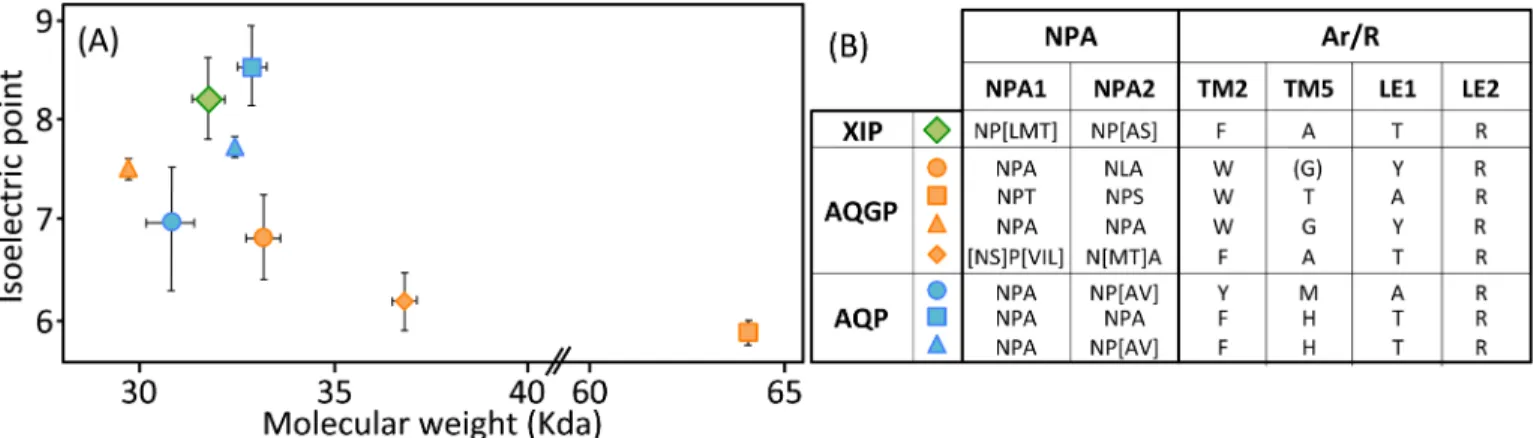
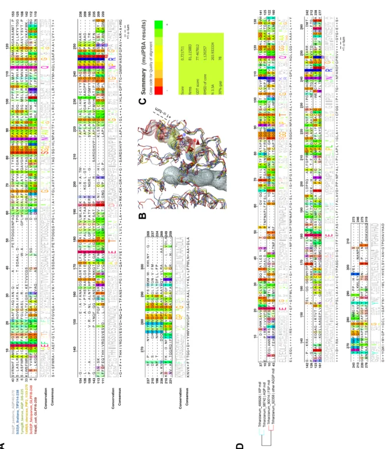
The MIP protein family in fungi contains a large number of highly divergent proteins. Apart from being assessed by their sequence identity, MIP diversity can be monitored not only through their biochemical features such as isoelectric points (pI) and molecular weights (MW), but also and most importantly, by modeling their three-dimensional profiles. (Fig 2;S1 Table). Except for the AQGP subgroup with Fps-like and "Other", MIP inTrichoderma spp. show a mean of 303 amino acids and a mean MW of 32 kDa. These features cover expected value ranges [39]. An analysis of their overall structure shows that most AQP are neutral or basic, the XIP are basic, and the majority of AQGP are neutral or acidic (Fig 2). Their distribu-tions are in line with what has been observed for a broad range of MIP from other fungi [40].
However, this analysis may be too simplistic, as these distributions do not reveal subtle fea-tures, especially as regards to potential sites of regulation such as loops and specific residues or motifs inside the pore. Further molecular structure analysis by modeling shows that the central channel polarization seems conserved, with almost the same distribution of charges along the z-axis. The positive charge of the guanidinium group of the characteristic arginine in the con-striction region is strongly expressed, and radiates over a long portion of the light of the pore (Figs3B,3C,3D,4B,4C,4D,5B,5C and 5D) in the absence of an immediate counterion. In fact, most of the differences in size and charge of the MIP mentioned here stem from the polymor-phism of their amino and carboxy terminal extensions, whose role has not yet been completely characterized (Figs3A,4Aand5A). We focused our interest on the MIP that are constitutively expressed (ie Fsp-like-90014Fig 3; "Other AQGP"-92358 membersFig 4; AQP-98742,Fig 5; XIP-488926,Fig 5;cf section “MIP expression”), and inspection of the alignments by phyloge-netic group shows that, aside from those variable extensions at both ends, we are facing two groups of MIP in terms of their putative transport capabilities. On one hand, we have what resembles water -and by extension H2O2- facilitators in the case of the AQP-98742 member and
the XIP-488926 member, and on the other hand, we have probable glycerol facilitators in the case of the "Other AQGP"-92358 member and the Fsp-like-90014 member, whose family is also known to group glycerol facilitators regulated by osmotic changes [16]. This segregation is Fig 2. Biochemical features ofTrichoderma MIP. (A) Relationship between isoelectric point and molecular weight for Trichoderma spp. MIP clusters. Plot
showing isoelectric point versus molecular weight for XIP (X), aquaglyceroporins (X) and aquaporins (X). Subgroups are detailed in the phylogeny inFig 1and in theS1 Table. Means± SE according to number of MIP members from each subgroup. (B) Amino acid diversity in NPA boxes and Aromatic/arginine selectivity filters in the different MIP subgroups fromTrichoderma spp. Exact ar/R locations on MIP proteins are detailed in Figs3A,4Aand5A.
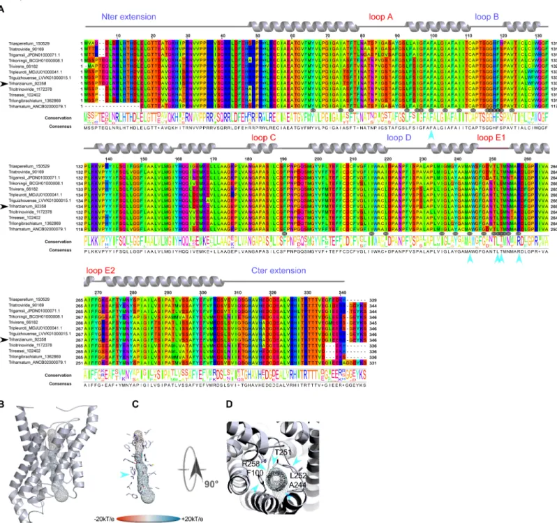
confirmed on a Newick tree when comparing the four models on multiple structural alignments with MulPBA [27]. This could be extended to the other members of each subfamily or group considered here because of their intra-proximity. The primary difference justifying this segrega-tion is located at the principal constricsegrega-tion site in the central pore, the so-called ar/R filter, which is slightly smaller in the water-specific AQP and composed of four residues, and slightly larger in the aquaglyceroporin and composed of four residues of which one is small (alanine) or by only three residues (the fourth is absent, and instead a glycine is found in its position). In our case, the constriction site is composed of F65, H185, T194 and R200 for the AQP-98742 member, of N81, S211, Q225 and R230 for the XIP member, of F100, A244, T251 and R258 for the "other AQGP"-92358 member, and W63, Y212, and R218 for the Fsp-like-90014 member (Figs2,3A,4Aand
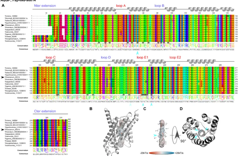
5A). The associated diameters measured at this site on our models with the MOLE 2.0 approach Fig 3. Structural analysis of the expressed fungal Fsp-like-90014 MIP. (A) Multiple sequence alignments (MSA) were generated from MIP homologs of different groups by group from various fungi computed with Muscle WS in Jalview, and colored by the Taylor color code. HomologousT. harzianum strains CBS 226.95 of the
expressed members fromThs97 are indicated by a black arrow before their names. Topology of each type is indicated by a ribbon diagram above the sequences on which
the different segments are labeled in blue for those in the inner compartment, and red for the outer compartment. The positions of the residues exposed to the light of the channel are designated by a target symbol formed of three black circles under the MSA. The conservation and consensus sequence are given and marked by blue arrows to indicate the positions at the constriction site. (B) Models out of an I-Tasser computation (after different runs to improve the confidence range) are shown in PyMOL scenes. The C-score (estimating the quality of the prediction) is positive for this model used (Cscore = 1.18), suggesting a good level of confidence in all the predictions (the normal range of C-scores being between−5 and 2). The pore established with "MOLE- 2" is materialized by a grid on which the electrostatic potential calculated by APBS with the PARSE forcefield is reported to compare the physicochemical nature of the channels. (C) Focus on the residues of the pore. A blue arrow indicates the ar/R region. (D) Sidechains of the amino acids constricting the channel after both NPA motifs.
were 1.28Å, 1.8Å, 3.78Å and 2.54Å, respectively, which is consistent with the reasonable assump-tion that the aperture in the glycerol transporters will be larger than in strict water transporters.
The second difference arises from the extracellular loops (A, C and E), which present vari-able lengths. Loop A with 14 residues (D42-P55) is prominent as expected for the AQP-98742 Fig 4. Structural analysis of the expressed fungal "other AQGP"-92358 MIP. (A) Multiple sequence alignments (B) Models out of an I-Tasser computation. The C-score (0.51) is positive, suggesting a good level of confidence in all the predictions. The pore is materialized by a grid on which the electrostatic potential is reported to compare the physicochemical nature of the channels. (C) Focus on the residues of the pore. A blue arrow indicates the ar/R region. (D) Sidechains of the amino acids constricting the channel after both NPA motifs. Technical procedures for each item are detailed in theFig 3caption.
member. This is also found on the "other AQGP" member, where it measures 13 residues (N80-S92); loop A is found to be slightly shorter with 10 residues (L65-G74) for the XIP, and is substantially halved with 7 residues (S49-D55) for the Fsp-like-90014 member. This criterion does not seem to be discriminating in terms of molecules to be transported. Conversely and more remarkably, loops C and E seem to permit a distinction in the nature of the transport ensured by the MIP, suggesting a possible coupling with a third-party effector, as it could pro-vide an interacting site for one. We note that both putative glycerol facilitators share a com-mon topology concerning their long loop C, which fits the model of an alpha hairpin as found in the GlFp, for which the archetype namely theE. coli Glycerol Facilitator structure was released [41]. The second alpha helix of the hairpin is mostly hydrophobic and ends with a cys-teine, which is also found in the XIP member at the same position near the pore entry. In both putative glycerol transporters, this segment is 20 residues longer than its homolog in the AQP-98742 fold: loop C is 19 residues long in the AQP member, 24 residues in the XIP member, 38 residues in the "other AQGP" member, and 36 residues in the Fsp-like-90014 member. On the intracellular side, we note a last subtle but still remarkable difference between the two GlFp candidates expressed concerning the net charge of loop B. In the first segment of this long loop, prior to the short NPA helix and at the very beginning of the loop, a lysine conserved in the Fsp-like subgroup (K76) is found instead of a conserved threonine (T114) as in the "other AQGP" members.Post hoc, the characteristic asparagine residue of the NPA motif is replaced by a serine in most of the "Other AQGP" members. In the second part of this loop, a conserved arginine (R93) is present in the Fsp-members, while this position is occupied by a glutamine (Q131) in most of the "other AQGP" members. This loop ends in both groups in a basic motif of two successive lysines in the "other AQGP" and an arginine followed by a lysine in the Fsp-members. To sum up, this loop is more basic in the Fsp candidates in addition to the presence of a supernumerary and conditional positive charge of a histidine (H95 instead of the neutral F133). This could have implications in how the two kinds of pores function, for example in their ability to interact with possible regulators, or in their ability to favor one circulating direction for the polyols they can tunnel across the membrane. The impact of such a subtle dif-ference will need to be addressed in further investigations.
Even more interesting, in both GlFp-like proteins we found an internal salt bridge between the conserved aspartate next to the ar/R filter arginine with another arginine on the helical turn immediately following, possibly helping to regulate aperture size by tilting the short NPA helix (Fig 6). This also occurs in generic GlFp, where this hemihelix is also one turn longer than its homolog from the AQP-98742 member. This is currently apparent in two available structures, an aquaglyceroporin fromPlasmodium falciparum (pdb code 3C02) and the first in the series of theE. coli glycerol facilitator (pdb code 1FX8). It implies translocation of the argi-nine of the filter, and its Cbeta moves about 1Å away from its canonical position on classical aquaporins. Thisin silico data can provide intuitive insight into the potential permeability properties of the channel in transporting not only polyhydroxyl alcohols (or polyols such as glycerol), but also more voluminous polyols such as erythritol, arabitol, sorbitol and mannitol as observed forpfAQP and ApAQP2, two multifunctional aquaglyceroporin channels from Plasmodium falciparum and Acyrthosiphon pisum, respectively [42,43]. These polyol Fig 5. Structural analysis of the expressed fungal AQP-98742 and XIP-488926 MIPs. (A) Multiple sequence alignments (B) Models out of an I-Tasser computation. The C-score are positive for all two models used (Cscore = 1.39 for AQP-98742; Cscore = 1.26 for XIP-488926), suggesting a good level of confidence in all the predictions. The pore is materialized by a grid on which the electrostatic potential is reported to compare the physicochemical nature of the channels. (C) Focus on the residues of the pore. A blue arrow indicates the ar/R region. (D) Sidechains of the amino acids constricting the channel after both NPA motifs. Technical procedures for each item are detailed in theFig 3caption.
Fig 6. Structural alignments of MIP to highlight noticeable differences in glycerol facilitatorsversus standard AQP. (A) Structural alignment of different MIP
based on the coordinates of resolved structures made with MulPBA on a narrow but still representative sample of MIP of different classes from different kingdoms. The name of the proteins and their relative pdb code is written with distinctive colors on the left of the alignment, itself colored by the Taylor color code in Jalview. A
transporters, alongside specific sucrose transporters, could be expected to feed the fungal car-bohydrate metabolism, which provides energy for hyphal growth and supplies carbon skeleton to other metabolisms. However, and again most importantly, they may participate in the con-tinuous process of generating hydrostatic pressure used by the pathogenic hyphae to break the hyphae cell wall surface of its host and penetrate it. Because polyols make a major contribution to the osmotic ballast, water and polyols are two interplaying components essential for hyphae integrity when fungi move in fluctuating environments [42,15,16].
Finally, we used the Glycopred prediction method to examine the differences in terms of numbers of potential glycosylation sites in these external loops. All of them are potentially gly-cosylated except for loop A of the FSP member, loop C of the AQP and loop E of the XIP and both putative glycerol facilitators. Most of the sites are far from the central pore. In the putative glycerol facilitators, glycosylation sites are found in the descending hydrophobic helix of loop C.
To conclude, on the basis of these structural and possible functional considerations, eluci-dating the physiological role of MIP inTrichoderma spp. through in-depth functional studies with MIP variants in key residues will answer these important unanswered questions. How-ever, this approach will not be applied onThs97: systematic of T. harzianum appears to be complex with many cryptic species, making it quite difficult to work with. Mutagenesis tech-nologies require double cross-over homologous recombination around 1,5kb up- and down-stream of the target gene, and therefore a thorough knowledge of intergenic regions, which are highly diversified and complex betweenT. harzianum spp. in contrast to the transcribed regions, which are highly conserved as shown byMIP genes. Thus, our hypothesis will need to be confirmed in the future by mutagenesis ofMIP from Trichoderma species whose genomes are sequenced.
MIP expression
The transcriptome ofTrichoderma is still the subject of several molecular studies, leading to the identification of pathways involved in the different aspects of biocontrol mechanisms [1,
44,45,46]. From these studies, however, no MIP information has yet been provided. To com-plete thein silico identification of candidates for MIP channels, their expression profile was addressed at transcript level using real-time quantitative reverse transcription-polymerase chain reaction (qRT-PCR) with MIP gene-specific primers. Molecular analysis is aimed pri-marily atTrichoderma under non-mycoparasitic conditions (mycelial growth or infestation without its hostF. solani, corresponding to the control samples) or under mycoparasitic condi-tions in the presence ofF. solani (corresponding to the assays). Additionally, two different bio-logical contexts were studied: on “artificial substrates” with PDA on Petri dishes (in vitro), and in roots from young olive trees (in planta). Similarly, MIP expressions from F. solani were studied in the same biological conditions. Results demonstrated that of the eight MIP genes conservation threshold of 50% is applied to highlight the conservation by groups. From this comparison emerges the particular meaning of the conserved GlFp motif NPARD: the conserved negatively-charged residue aspartate makes a salt bridge with an equally conserved residue at exactly oneα-turn from it. This bridge quenches both charges by mutual neutralization, allowing their presence in a quite hydrophobic environment for folding purposes (first quarter ofα-6). (B) PyMOL scene of the superimposition results from mulPBA displayed as a wireframe diagram of the main chain colored with respect to the sequence name coloring. The channel is shown as a transparent volume to materialize the localization. The sidechain of the conserved arginine from the NPAR motif is shown as sticks, as also are both charged residues occurring only in the GlFp proteins (light and dark red). A red arrow shows the relative displacement (concomitant with this type of electrostatic bridge within the shortα-helix of loop E) responsible, at least in part, for a larger pore aperture at its constriction site. Only the NPA α-helices are shown as transparent colored coils (C) Summary of the superimposition score from mulPBA. (D) Structural alignment of MIP from theT. harzianum strain CBS 226.95
homologous to those expressed fromThs97 and based on the coordinates of good quality I-Tasser homology models. The MSA is colored by the Taylor color code in
Jalview. On the left, the Newick tree established by mulPBA is given showing the relative proximity of both XIP-488926 and AQP-98742 members on one side, and both "other AQGP"-92358 and Fsp-like 90014 members on the other side. Models are consistent with previous data obtained on experimental structures. A conservation threshold of 50% is also applied to highlight the conservation by groups.
present in the genome ofT. harzianum, only four were transcribed with significant differential modulation during mycelial growth on an artificial medium and on olive roots (Fig 7). In detail, the steady-state level of transcript abundance of AQP-98742, AQGP-92358 and XIP-488926 was higher during the mycelial growthin planta than in vitro, while AQGP-90014 was slightly less abundant. These diverging expressions between “growth environments” are not surprising and have already been mentioned [47]. They could result from the presence of vari-ous chemical factors in plant tissues that may be lacking in artificial substrates. Similarly, these contrasting expressions could be linked to a subtle difference observed between the net charges of their respective loop B that would determine a specific ability to favor one circulating direc-tion of particular solutes across the membrane.
Concerning the confrontation situations, and irrespective of the biological contexts (i.e. preventive or curative), the expressions of these four MIP were significantly modulated by the presence ofF. solani. It is of note that the onset of a differential expression of MIP genes is a rather early event during the interaction with host prey: it occurs during the first stage of mycelial growth, whenT. harzianum is in physical contact with its prey. MIP transcript abun-dance then increased considerably over the contact area betweenT. harzianum and F. solani.
Fig 7. Relative transcription ratios of the MIP genes fromT. harzianum and F. solani. Relative expression levels of MIP genes
fromT. harzianum (Ths97) and F. solani (Fso14) cultivated separately or together in artificial culture (after 6 days of inoculation)
or on roots of olive trees (after 8 weeks of inoculation). Plantsvs Plates: constitutive steady state level of MIP expression from T.
harzianum and F. solani cultivated separately in plants or on PDA medium. In vitro assay: (A) T. harzianum individually, (B) area
of confrontation between hyphae, (C) area of overlap ofT. harzianum on F. solani (detailed inS2 Fig).In planta assay: inoculated
separately in roots or in curative and preventive contexts (root symptoms detailed inS3 Fig). Transcript levels for each gene were estimated using real-time qRT-PCR analyses, and normalized by the expression of three housekeeping genes specific for each fungal strain. Relative transcript abundance rates were obtained by theE-ΔΔCtmethod with transcript abundances from individual
in vitro culture or in planta inoculation. Data correspond to means of three independent biological experiments. Bars represent
the biological standard error. Data not sharing the same letter are significantly different (Tukeypost-hoc test after one-way analysis
of variance (ANOVA),p < 0.05). Nd, transcript non-detectable significantly. https://doi.org/10.1371/journal.pone.0193760.g007
Unexpectedly, MIP patterns contrasted sharply between the artificial andin planta dual cul-ture contexts, except for 92358, which remained up-regulated. AQP-98742, AQGP-90014 and XIP-488926 were up-regulated in artificial substrates, but down-regulatedin planta. Very few functional studies have been carried out on fungal MIP. However, AQP-98742 belongs to a MIP cluster that comprises MIP with putative water channels [12,14,47]. AQGP-90014 belongs to a "facultative Fsp-like aquaglyceroporin" cluster including MIP with putative water, glycerol and small neutral molecule transport channels [13,17,48], whereas AQGP-92358 belongs to the "other-aquaglyceroporin" cluster made up of MIP that present glycerol and small neutral molecule transport capacities [13,15]. Concerning XIP, no biochemical vali-dation has been reported in fungi. However, three inputs can be exploited to gain a better understanding of this unorthodox cluster: (i) the MIP JQ412059 fromGlomus intraradices, a relatively proximate phylogenetic homolog of fungal XIP [16], exhibits a water transport chan-nel [49], (ii) its transcriptional kinetics parallel that of AQP-98742 (Fig 7), and (iii) three-dimensional structure analysis suggests a tighter channel, particularly at the constriction zone approaching the level seen in the AQP-98742 channel (Fig 5). This would indicate a plausible ability to channel water and possibly other small polar molecules like H2O2, but not glycerol as
previously observed for certain XIP from plants [50,51,52]. Despite these putative biochemi-cal extrapolations, and because evidence of how MIP take part in fungal lifestyles is still scant and speculative, further interpretations concerning the putative involvement of each member during mycoparasitism ofThs97 would be premature. However, data do suggest that F. solani has a direct influence onThs97 genome reprogramming, and this is significant when we read the MIP expression from the ‘in planta’ biological context. This takes place invariantly whether Ths97 has colonized healthy plant tissues prior to a F. solani infestation (preventive treatment) or an infested fusarium environment (curative treatment). Thein vitro and in planta environ-ments are not comparable, and it is difficult to envisage howF. solani can directly up- or down-regulate someThs97 genes in specific environments, unless we consider the possibility thatThs97 displays a direct mycoparasitic activity on F. solani. The interaction of T. harzianum withF. solani is described as mycoparasitic [35], and this feature was observedin vitro between Ths97 and Fso14 (S2andS3Figs) [2]. This overall adjustment may be supplemented by the release of cell-wall-degrading enzymes, known to be directly involved in the mycoparasitism interaction, and whose production is influenced by various ambient factors [53,54,55]. These fine and complex molecular adjustments generate specific metabolized-products (i.e. oligo-mers) that may themselves become secondary inducers of cell responses forTrichoderma [56,
57]. This would explain the differential expression patterns of transcripts encoding MIP pro-teins observed during the different biological contexts and stages of confrontation.
Two other interesting scenarios should be considered. The first one is that the biochemical environments of the intercellular space change fundamentally. This event is mainly due to the virulent activity ofF. solani and also its ability to secrete an arsenal of hydrolytic enzymes [58,
59]. Certain particular plant residues generated byF. solani could interfere here with Ths97 cel-lular responses. Such residues are inevitably absent in thein vitro context, but could possibly be produced whenF. solani infests its plant host. In the second scenario, although no information is available about competition and defense reactions ofF. solani as a host, F. solani would is be able to develop a differential toxicogenic activityin planta compared with the in vitro context (likeThs97, F. solani senses and responds differentially to contrasting environments) [60,61], and specific secreted mycotoxins (possibly in a "growth medium"-dependent manner) could affect certain gene responses inThs97 without, however, upsetting its mycoparasitic behavior. To the best of our knowledge, there is no evidence to support these two last suppositions, but whatever the case, the transport machinery reprogramming forThs97 is governed by environ-mental changes, probably due to the presence of exudates released from the host mycelium (F.
solani), whose priority remains to meet nutritional needs. As for F. solani MIP expression pat-terns, four MIP out of the six in its genome were transcribed and differentially modulated. Interestingly, none of them was significantly detectable in infested plants treated withThs97. This result provides new evidence suggesting the ability of the beneficial partner to drastically reduce the population of its host target.
MIP regulation
In line with previous findings, we showed here thatThs97 seems able to sense the presence of its host prey and respond by modulating a set of genes that could be involved in its mycopara-sitism. In our work on MIP, we are aware that correlations alone do not allow a causal link to be established. In addition, the transcriptional level does not represent what happens at the protein level. However, there are good indications that MIP transcript regulations may imply assigned functions of isoforms in mycelia trans-cellular solute flows. Thus whatever the biolog-ical contexts, we can intuitively expect the expression of a broad range of genes to depend pre-ponderantly on solute sources (carbon, nitrogen, minerals, etc) available in the environment. One of the major challenges facing biologists is to unravel the complex networks that govern these gene expressions. One clue could come from the establishment of the existence of meta-bolons. Transcriptional regulation relies to a large extent on molecular mechanisms that allow nucleic acid binding proteins or transcription factors (TF) to recognize specific sets of nucleic acids in DNA, known as transcription factor binding sites (TFBSs) orcis-regulatory sequences. Identifying these regulatory elements in non-coding regions is an interesting key step in understanding gene regulation and ultimately in inferring regulatory networks.
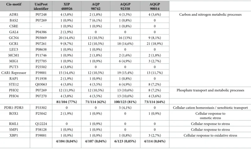
We scanned 1.5kb upstream of the start codon of the four expressed MIP using the yeast Saccharomyces cerevisiae dedicated promoter database SCPD [32]. Conscious of the limitations inherent in such a systematic analysis on TFBSs, which are usually very short and statistically often highly degenerate, the fact remains that results showed an over-representation of motifs targeted by TFs known to be involved in various carbon, nitrogen sulfur and phosphate meta-bolic processes (Tables1andS3). Between 62% and 81% of motifs constituting the four MIP promoters are in promoters of genes encoding proteins involved in carbohydrate, fatty acid and sterol, amino acid or nucleotide metabolisms. Unexpectedly, motifs involved in the cellu-lar responses to stress (osmosensing and ion homeostasis pathways, drug metabolization and exportation, oxidative stress) were poorly represented (<0.05%). This contrasts notably with plant MIP promoters, which contain a large number of TFBSs related to cellular responses to abiotic and biotic stress [62,63,64]. The remaining motifs control mRNA transcription, cell growth and division, and DNA synthesis (meiosis process) (S3 Table). We hypothesize that thiscis-element provides indications about the potential involvement of these MIP in estab-lishing a trophic relationship thatTrichoderma creates with its surroundings, and especially here withF. solani, with which Ths97 initiates a competitive relationship. This functional trend corroborates previous findings where functional annotations of different wide-transcript libraries linked to a mycoparasitic context indicated a substantial over-represented category related to various metabolic processes [35,53,55,65]. Finally, ifF. solani really influences Ths97 genes expression in some way, then it would be relevant to identify the TFs network fromThs97, which could be in direct relation with certain virulence effectors secreted by F. solani during its plant tissue infestation phase, or related metabolized products in the case of effectors with intrinsic hydrolase activities. To further test our hypothesis of a plausible involvement of MIP in the competition machinery ofTrichoderma against various pathogens, and the establishment of its trophic network, suppression of MIP gene function within non-encoding and non-encoding regions would have to be addressed in future experiments.
Conclusion
Our present results bring us nearer to understanding one molecular mechanism potentially involved in the mycoparasitic process ofT. harzianum (with the example of Ths97 here) with the involvement of the MIP family. Modulated transcript abundance of members belonging to the three sub-classes representative of the fungal MIP family suggests the importance of trans-porting certain specific solutes during hyphae development and possibly self-defenses. How-ever, owing to the complexity of the underlying mycoparasitism mechanisms, an in-depth understanding of the functional characterization of the MIP genes reported here is essential, and this will be improved by future studies of their subcellular localization, post-translational regulation and precise roles in signaling and solute transporting processes in such "myco-phy-toparasitic" tripartite interactions. Part of this effort will require focusing on key residues shown in this study to be responsible for the specialization of the two GlFp and subsequently testing these by mutagenesis approaches. Lastly, if we consider -by definition- that MIP are vir-ulent factors in this (myco)parasitic interaction, the manipulation of candidate MIP genes linked to virulence activity remains a pertinent approach to improve theT. harzianum strain. Table 1. Proportion of putative transcription factor binding sites (TFBSs) on the 1.5kb promoter region of the expressed MIP genes fromTrichoderma harzianum.
MIP promoter sequences fromT. harzianum CBS 226.95 v1.0 (JGI) were used as reference. TFBSs were detected with the "Promoter Database of Saccharomyces cerevisiae"
(http://rulai.cshl.edu/SCPD/), and biological processes (GO) analyzed using «Uniprot» (http://www.uniprot.org/). TFBS nucleotide sites on 1.5kb of each promoter are detailed in theS3 Table.
Cis-motif UniProt identifier XIP 488926 AQP 98742 AQGP 92358 AQGP 90014
ADR1 P07248 4 (3,8%) 2 (1,8%) 4 (3,3%) 4 (3,6%) Carbon and nitrogen metabolic processes
BAS2 P07269 1 (0,9%) 7 (6,1%) 1 (0,8%) 0 CSRE - 1 (0,9%) 1 (0,9%) 1 (0,8%) 0 GAL4 P04386 2 (1,9%) 0 0 0 GCN4 P03069 20 (14,4%) 12 (10,5%) 16 (13%) 9 (8,1%) GCR1 P07261 9 (8,7%) 12 (10,5%) 18 (14,6%) 21 (18,9%) LEU3 P08638 1 (0,9%) 1 (0,9%) 0 0 MCM1 P11746 1 (0,9%) 2 (1,8%) 2 (1,6%) 2 (1,8%) MIG1 P27705 1 (0,9%) 1 (0,9%) 6 (4,9%) 3 (2,7%) PUT3 P25502 4 (3,8%) 0 0 0 CAR1 Repressor P39001 15 (14,4%) 12 (10,5%) 19 (15,4%) 13 (11,7%) RAP1 P11938 2 (1,9%) 1 (0,9%) 1 (0,8%) 0 STE12 Q03063 4 (3,8%) 4 (3,5%) 6 (4,9%) 8 (7,2%)
PHO2 P07269 12 (11,9%) 12 (10,5%) 13 (10,6%) 8 (7,2%) Phosphate transport and metabolic processes PHO4 P07270 4 (3,8%) 4 (3,5%) 13 (10,6%) 4 (3,6%)
81/104 (77%) 71/114 (62%) 100/123 (81%) 73/114 (64%)
PDR1-PDR3 P33302 0 0 5 (4,1%) 0 Cellular cation homeostasis / xenobiotic transport
ROX1 P25042 2 (1,9%) 1 (0,9%) 0 1 (0,9%) Cellular response to
osmotic stress
RML1 Q12224 0 1 (0,9%) 0 0 Cellular response to stress
SMP1 P38128 1 (0,9%) 1 (0,9%) 0 0 Cellular response to stress
XBP1 P39001 1 (0,9%) 1 (0,9%) 1 (0,8%) 3 (2,7%) Cellular response to oxidative stress 4/104 (0,04%) 4/107 (0,04%) 6/123 (0,05%) 4/114 (0,04%)
Supporting information
S1 Fig. Detail of all MIP protein sequences used in this work.
(PDF)
S2 Fig. Symptoms of fusarium root rot disease on root system from olive trees. Preventive
treatment:Ths97-treated plants subject to Fso14 infestation; Curative treatment: Fso14 infested plants treated withThs97. Dual inoculation contexts were set up with a 10-day delay between each fungal inoculation. Fungi were inoculated on roots.
(PDF)
S3 Fig. Culture ofF. solani (Fso14 strain) and T. harzianum (Ths97 strain) cultivated sepa-rately, or together in a dual growth context related to a mycoparasitic situation. Mycelial
were grown in Petri dishes on PDA medium. Slides show 6 days of growth at 27˚C. Letters A, B, and C on dual culture assay correspond to area sampled for molecular experiments, with (A)Ths97 individually, (B) area of confrontation between mycelia, and (C) area of overlap of Ths97 on Fso14.
(PDF)
S1 Table. Features of the non-redundant representative fungal MIP proteins from Tricho-derma and Fusarium species used in the phylogenetic analysis. Reference species for MIP
nomenclature:Mycosphaerella fijiensis (Mycfi) and Laccaria bicolor (Lacbi). AQP, aquaporins; AQGP, aquaglyceroporins; XIP, X-intrinsic proteins.
(PDF)
S2 Table. Primers used for qPCR amplification.
(PDF)
S3 Table. Detail of the TFBS nucleotide sites found on 1.5kb of each promoter of the four expressed MIP.
(PDF)
Acknowledgments
The authors wish to thank the Institut de l’Olivier (Department of “Ame´lioration et Protection des Ressources Ge´ne´tiques de l’Olivier”, University of Sfax, Tunisia) for the two strains of fungi used in this study and for carrying out all the experiments on olive trees. We are grateful to Ce´line Sac and Dominique Marcon for their technical assistance in molecular biology and photographic editing, respectively. This work was supported by the PHC program “Uthic” from Campus France (grant 34861PF) under the joint aegis of the Tunisian Ministry of For-eign Affairs and the French Ministry of Higher Education and Scientific Research. The authors also wish to acknowledge Christie Nielsen Chaar for generously providing the final linguistic revision of the manuscript. We are also grateful to the anonymous reviewers for their con-structive comments.
Author Contributions
Conceptualization: Daniel Auguin, Jean-Ste´phane Venisse. Data curation: Jean-Ste´phane Venisse.
Formal analysis: Jean-Ste´phane Venisse. Funding acquisition: Jean-Ste´phane Venisse.
Methodology: Maroua Ben Amira, Robin Mom, David Lopez, Hatem Chaar, Daniel Auguin,
Jean-Ste´phane Venisse.
Project administration: Hatem Chaar, Ali Khouaja, Vale´rie Pujade-Renaud, Philippe Label,
Jean-Louis Julien, Mohamed Ali Triki, Jean-Ste´phane Venisse.
Resources: Philippe Label, Daniel Auguin, Jean-Ste´phane Venisse.
Software: Robin Mom, Boris Fumanal, Gisèle Bronner, Daniel Auguin, Jean-Ste´phane Venisse.
Supervision: Daniel Auguin, Jean-Ste´phane Venisse.
Validation: Aure´lie Gousset-Dupont, Daniel Auguin, Jean-Ste´phane Venisse. Visualization: Boris Fumanal.
Writing – original draft: Maroua Ben Amira, Robin Mom.
Writing – review & editing: Hatem Chaar, Boris Fumanal, Aure´lie Gousset-Dupont, Gisèle Bronner, Philippe Label, Daniel Auguin, Jean-Ste´phane Venisse.
References
1. Srivastava M, Pandey S, Shahid M, Kumar V, Singh A, Trivedi S, et al. Trichoderma: A magical weapon against soil borne pathogens. African Journal of Agricultural Research 2015; 10: 4591–4598.
2. Ben Amira M, Lopez D, Triki MA, Khouaja A, Chaar H, Fumanal B, et al. Beneficial effect of Trichoderma harzianum strain Ths97 in biocontrolling Fusarium solani causal agent of root rot disease in olive trees. Biological Control 2017; 110: 70–78.
3. Waghunde RR, Shelake RM, Sabalpara AN (2016) Trichoderma: A significant fungus for agriculture and environment. Afr J Agric Res. 2016; 11: 1952–1965.
4. Druzhinina IS, Seidl-Seiboth V, Herrera-Estrella A, Horwitz BA, Kenerley CM, Monte E, et al.
Tricho-derma: the genomics of opportunistic success. Nat Rev Microbiol. 2011; 9: 749–759.https://doi.org/10. 1038/nrmicro2637PMID:21921934
5. Finn RN, Cerda J. Evolution and Functional Diversity of Aquaporins. Biol. Bull. 2015; 229: 6–23.https:// doi.org/10.1086/BBLv229n1p6PMID:26338866
6. Madeira A, Moura TF, Soveral G. Aquaglyceroporins: implications in adipose biology and obesity. Cell Mol Life Sci. 2015; 72: 759–771.https://doi.org/10.1007/s00018-014-1773-2PMID:25359234 7. Wree D, Wu B, Zeuthen T, Beitz E. Requirement for asparagine in the aquaporin NPA sequence
signa-ture motifs for cation exclusion. FEBS J. 2011; 278: 740–748.https://doi.org/10.1111/j.1742-4658. 2010.07993.xPMID:21205205
8. Wu B, Steinbronn C, Alsterfjord M, Zeuthen T, Beitz E. Concerted action of two cation filters in the aqua-porin water channel. EMBO J. 2009; 28: 2188–2194.https://doi.org/10.1038/emboj.2009.182PMID: 19574955
9. Almasalmeh A, Krenc D, Wu B, Beitz E. Structural determinants of the hydrogen peroxide permeability of aquaporins. FEBS J. 2014; 281: 647–656.https://doi.org/10.1111/febs.12653PMID:24286224 10. Angel A, Stahlberg H. Aquaglyceroporins: channel proteins with a conserved core, multiple functions,
and variable surfaces. Int Rev Cytol 2002; 215: 75–104. PMID:11952238
11. Abascal F, Irisarri I, Zardoya R. Diversity and evolution of membrane intrinsic proteins. Biochim Biophys Acta 2014; 1840: 1468–481.https://doi.org/10.1016/j.bbagen.2013.12.001PMID:24355433
12. Pettersson N, Filipsson C, Becit E, Brive L, Hohmann S. Aquaporins in yeasts and filamentous fungi. Biol. Cell 2005; 97: 487–500.https://doi.org/10.1042/BC20040144PMID:15966864
13. Soveral G, Prista C, Moura TF, Loureiro-Dias MC. Yeast water channels: an overview of orthodox aqua-porins. Biol Cell 2010; 103: 35–54.https://doi.org/10.1042/BC20100102PMID:21143194
14. Gupta AB, Sankararamakrishnan R. Genome-wide analysis of major intrinsic proteins in the tree plant
Populus trichocarpa: characterization of XIP subfamily of aquaporins from evolutionary perspective.
BMC Plant Biol. 2009; 9: 134.https://doi.org/10.1186/1471-2229-9-134PMID:19930558
15. Dietz S, von Bu¨low J, Beitz E, Nehls U. The aquaporin gene family of the ectomycorrhizal fungus
Lac-caria bicolor: lessons for symbiotic functions. New Phytol. 2011; 190: 927–940.https://doi.org/10.1111/ j.1469-8137.2011.03651.xPMID:21352231
16. Xu H, Cooke JEK, Zwiazek JJ. Phylogenetic analysis of fungal aquaporins provide insight into their pos-sible role in water transport of mycorrhizal associations. Botany 2013; 91: 495–504.
17. Li T, Hu YJ, Hao ZP, Li H, Chen BD. Aquaporin genes GintAQPF1 and GintAQPF2 from Glomus
intrar-adices contribute to plant drought tolerance. Plant Signal Behav 2013; 8: e24030.https://doi.org/10. 4161/psb.24030PMID:23435173
18. Nehls U, Dietz S. Fungal aquaporins: cellular functions and ecophysiological perspectives. Appl Micro-biol Biotechnol 2014; 98: 8835–8851.https://doi.org/10.1007/s00253-014-6049-0PMID:25213914 19. Triki MA, Priou S. L’utilisation des traitements chimiques et biologiques pour re´duire le risque de fuite
de la pomme de terre cause´e par Pythium aphanidermatum en Tunisie. Potato Res. 1997; 40: 391– 398.
20. Triki MA, Rhouma A, Khbou W, Boulila M, Ioos R. Recrudescence du de´pe´rissement de l’olivier cause´ par les champignons telluriques en Tunisie. Proceeding International Conference of Olive tree and Olive Products, Olive bioteq, Sfax, Tunisia; 2009. pp. 142–147.
21. Zhang Y. I-TASSER server for protein 3D structure prediction. BMC Bioinformatics 2008; 9: 40.https:// doi.org/10.1186/1471-2105-9-40PMID:18215316
22. Roy A, Kucukural A, Zhang Y. I-TASSER: a unified platform for automated protein structure and func-tion predicfunc-tion. Nature Protocols 2010; 5: 725–738.https://doi.org/10.1038/nprot.2010.5PMID: 20360767
23. Yang J, Yan R, Roy A, Xu D, Poisson J, Zhang Y. The I-TASSER Suite: protein structure and function prediction. Nature Methods 2015; 12: 7–8.https://doi.org/10.1038/nmeth.3213PMID:25549265 24. Tang CL, Alexov E, Pyle AM, Honig B. Calculation of pKas in RNA: on the structural origins and
func-tional roles of protonated nucleotides. Journal of Molecular Biology 2007; 366: 1475–1496.https://doi. org/10.1016/j.jmb.2006.12.001PMID:17223134
25. Baker NA, Sept D, Joseph S, Holst MJ, McCammon JA. Electrostatics of nanosystems: application to microtubules and the ribosome. Proc Natl Acad Sci USA 2001; 98: 10037–10041.https://doi.org/10. 1073/pnas.181342398PMID:11517324
26. DeLano WL. PyMOL User’s Guide. DeLano Scientific, San Carlos, California; 2004
27. Le´onard S, Joseph AP, Srinivasan N, Gelly JC, de Brevern AG. MulPBA: an efficient multiple protein structure alignment method based on a structural alphabet. J Biomol Struct Dyn. 2014; 32: 661–668. https://doi.org/10.1080/07391102.2013.787026PMID:23659291
28. Edgar RC. MUSCLE: multiple sequence alignment with high accuracy and high throughput. Nucleic Acids Res. 2004; 32: 1792–1797.https://doi.org/10.1093/nar/gkh340PMID:15034147
29. Castresana J. Selection of conserved blocks from multiple alignments for their use in phylogenetic anal-ysis. Mol Biol Evol. 2000; 17: 540–552.https://doi.org/10.1093/oxfordjournals.molbev.a026334PMID: 10742046
30. Guindon S, Dufayard JF, Lefort V, Anisimova M, Hordijk W, Gascuel O. New Algorithms and Methods to Estimate Maximum-Likelihood Phylogenies: Assessing the Performance of PhyML 3.0. Systematic Biology 2010; 59: 307–321.https://doi.org/10.1093/sysbio/syq010PMID:20525638
31. Chevenet F, Brun C, Banuls AL, Jacq B, Chisten R. TreeDyn: towards dynamic graphics and annota-tions for analyses of trees. BMC Bioinformatics. 2006; 10: 439.
32. Zhu J, Zhang MQ. SCPD: A promoter database of yeast Saccharomyces cerevisiae. Bioinformatics 1999; 15: 607–611. PMID:10487868
33. Chang S, Puryear J, Cairney J. A simple and efficient method for isolating RNA from pine trees. Plant Mol. Biol. Report 1993; 11: 113–116.
34. Livak KJ, Schmittgen TD. Analysis of relative gene expression data using real-time quantitative PCR and the 2(-Delta Delta C(T)) Method. Methods 2001; 25: 402–408.https://doi.org/10.1006/meth.2001. 1262PMID:11846609
35. Vieira PM, Siqueira Guedes Coelho A, Steindorff SA, Linhares de Siqueira SJ, do Nascimento Silva R, Ulhoa CJ. Identification of differentially expressed genes from Trichoderma harzianum during growth on cell wall of Fusarium solani as a tool for biotechnological application. BMC Genomics 2013; 14: 177. https://doi.org/10.1186/1471-2164-14-177PMID:23497274
36. Seidl V, Song L, Lindquist E, Gruber S, Koptchinskiy A, Zeilinger S, et al. Transcriptomic response of the mycoparasitic fungus Trichoderma atroviride to the presence of a fungal prey. BMC Genomics 2009; 10; 567.https://doi.org/10.1186/1471-2164-10-567PMID:19948043
37. Seidl V, Huemer B, Seiboth B, Kubicek CP. Complete survey of Trichoderma reveals three distinct sub-groups of family 18 chitinases. The FEBS J. 2005; 272: 5923–5939. https://doi.org/10.1111/j.1742-4658.2005.04994.xPMID:16279955