HAL Id: hal-01860011
https://hal.archives-ouvertes.fr/hal-01860011
Submitted on 22 Aug 2018
HAL is a multi-disciplinary open access archive for the deposit and dissemination of sci-entific research documents, whether they are pub-lished or not. The documents may come from teaching and research institutions in France or abroad, or from public or private research centers.
L’archive ouverte pluridisciplinaire HAL, est destinée au dépôt et à la diffusion de documents scientifiques de niveau recherche, publiés ou non, émanant des établissements d’enseignement et de recherche français ou étrangers, des laboratoires publics ou privés.
of menstrual cycle and associations with related amino
acids and nuclear factor kB activation
Gernot Faustmann, Andreas Meinitzer, Christoph Magnes, Beate Tiran,
Barbara Obermayer-Pietsch, Hans-Jürgen Gruber, Josep Ribalta, Edmond
Rock, Johannes M. Roob, Brigitte M. Winklhofer-Roob
To cite this version:
Gernot Faustmann, Andreas Meinitzer, Christoph Magnes, Beate Tiran, Barbara Obermayer-Pietsch, et al.. Progesterone-associated arginine decline at luteal phase of menstrual cycle and associations with related amino acids and nuclear factor kB activation. PLoS ONE, Public Library of Science, 2018, 13 (7), pp.e0200489. �10.1371/journal.pone.0200489�. �hal-01860011�
Progesterone-associated arginine decline at
luteal phase of menstrual cycle and
associations with related amino acids and
nuclear factor kB activation
Gernot Faustmann1,2, Andreas Meinitzer3, Christoph Magnes4, Beate Tiran3,
Barbara Obermayer-Pietsch5, Hans-Ju¨ rgen Gruber3, Josep Ribalta6, Edmond Rock7, Johannes M. Roob2, Brigitte M. Winklhofer-Roob1*
1 Human Nutrition & Metabolism Research and Training Center, Institute of Molecular Biosciences,
Karl-Franzens University, Graz, Austria, 2 Clinical Division of Nephrology, Department of Internal Medicine, Medical University, Graz, Austria, 3 Clinical Institute of Medical and Chemical Laboratory Diagnostics, Medical University, Graz, Austria, 4 Institute for Biomedicine and Health Sciences, HEALTH, Joanneum Research Forschungsgesellschaft m.b.H., Graz, Austria, 5 Division of Endocrinology, Department of Internal Medicine, Medical University, Graz, Austria, 6 Unitat de Recerca de Lipids I Arteriosclerosi, Facultat de Medicina, Universitat Rovira I Virgili, Facultat Medicina i Ciències de la Salut, Reus, Spain, 7 Unite´ de Nutrition Humaine, Centre Auvergne Rhoˆne-Alpes, Institut National de la Recherche Agronomique, Saint-Gènes-Champanelle, France
*brigitte.winklhoferroob@uni-graz.at
Abstract
Background/Objectives
Given their role in female reproduction, the effects of progesterone on arginine and related amino acids, polyamines and NF-κB p65 activation were studied across the menstrual cycle.
Methods
Arginine, ornithine and citrulline as well as putrescine, spermidine, spermine, and N-acetyl-putrescine were determined in plasma, NF-κB p65 activation in peripheral blood mononu-clear cells and progesterone in serum of 28 women at early (T1) and late follicular (T2) and mid (T3) and late (T4) luteal phase.
Results
Arginine and related amino acids declined from T1 and T2 to T3 and T4, while progesterone increased. At T3, arginine, ornithine, and citrulline were inversely related with progesterone. Changes (ΔT3-T2) in arginine, ornithine, and citrulline were inversely related with changes (ΔT3-T2) in progesterone. Ornithine and citrulline were positively related with arginine, as were changes (ΔT3-T2) in ornithine and citrulline with changes (ΔT3-T2) in arginine. At T2, NF-κB p65 activation was positively related with arginine. Polyamines did not change and were not related to progesterone. All results described were significant at P<0.001.
a1111111111 a1111111111 a1111111111 a1111111111 a1111111111 OPEN ACCESS
Citation: Faustmann G, Meinitzer A, Magnes C,
Tiran B, Obermayer-Pietsch B, Gruber H-J, et al. (2018) Progesterone-associated arginine decline at luteal phase of menstrual cycle and associations with related amino acids and nuclear factor kB activation. PLoS ONE 13(7): e0200489.https://doi. org/10.1371/journal.pone.0200489
Editor: Franc¸ois Blachier, National Institute for
Agronomic Research, FRANCE
Received: January 15, 2018 Accepted: June 27, 2018 Published: July 10, 2018
Copyright:© 2018 Faustmann et al. This is an open access article distributed under the terms of the
Creative Commons Attribution License, which permits unrestricted use, distribution, and reproduction in any medium, provided the original author and source are credited.
Data Availability Statement: The relevant data of
this study cannot be de-identified. Therefore, the data is restricted to only available upon request. These restrictions were approved by the Ethics Committee of the Medical University of Graz. Data are made available for researchers who meet the criteria for access to confidential data. Data requests may be sent tobioclaims@uni-graz.at.
Funding: This work was carried out with financial
Conclusions
This study for the first time provides data, at the plasma and PBMC level, supporting a pro-posed regulatory node of arginine and related amino acids, progesterone and NF-κB p65 at luteal phase of the menstrual cycle, aimed at successful preparation of pregnancy.
Introduction
The menstrual cycle is characterized not only by a strong increase between follicular and luteal phase in basal and resting metabolic rate [1,2], but also in amino acid oxidation [2] and nitro-gen excretion [3], suggesting a rise in whole body protein turnover at luteal phase. In this con-text, amino acids that play a functional role in the preparation for successful pregnancy are of particular interest. While these amino acids may be essential at luteal phase, substantial utiliza-tion could reduce their availability. On the other hand, amino acids with specific immune reg-ulatory functions could be kept low at luteal phase on purpose.
So far, only small-scale studies reported lower plasma arginine, citrulline, and ornithine concentrations at luteal compared to follicular phase [4,5]. While these changes have been pos-tulated to be due to changes in progesterone levels [4,5], which show a sharp increase at luteal phase [6], direct evidence that these changes occur in response to elevated progesterone con-centrations at luteal phase has not been provided. Several mechanisms, including transport, synthesis and recycling, have been proposed to be involved in maintaining arginine concentra-tions within a physiological range, which was reported as being 80–120μmol/L [7], in the absence of published reference values.
Arginine is a conditionally essential amino acid for most mammals including humans [7], with the availability of citrulline being the limiting factor forde novo synthesis [8]. Arginine is a substrate for two competing enzymes, i.e., arginase, producing urea and ornithine [9], and nitric oxide (NO) synthase, producing NO and citrulline [10]. At luteal phase, high arginase expression was shown in endometrium [11], as was endothelial NO synthase (eNOS) expres-sion in endometrium [12] and corpus luteum [13,14]. In female fertility, NO plays important roles in angiogenesis [15,16], endothelial function [17], endometrial receptivity and implanta-tion [18].
A temporary suppression of the immune response at luteal phase of the menstrual cycle as well as during pregnancy is critical for materno-fetal tolerance [19–21]. Arginine exerts immune modulatory functions [22,23] by specifically up-regulating the expression of the T cell antigen receptor zeta chain (CD3z) [24,25], which in turn induces the TCR-to-nuclear factor kappa B (NF-κB) pathway [26], resulting in nuclear translocation of NF-κB [27], known as a central regulator of immune responses [28]. The activation of the NF-κB p65 subunit in
peripheral blood mononuclear cells (PBMC) was reduced not only during pregnancy, but also already at luteal phase [20,29], along with a shift from TH1- to TH2-type cytokines [19–21],
which has been implicated in the preparation of the endometrium for implantation [30,31]. Arginine, via ornithine, is also a precursor of polyamines [9], which play a critical role in the preparation of the endometrium for implantation, including endometrial cell proliferation [32]. Polyamines are synthesized from ornithine with ornithine decarboxylase as the rate-lim-iting enzyme, catalyzing the conversion of ornithine to putrescine. Putrescine, spermidine, and spermine are interconverted by highly regulated enzymatic reactions, including back-con-version via intermediate acetylated polyamines, catalyzed by acetyltransferases and oxidases [33,34].
Programme FP7 2007-2013 under grant agreement nº 244995 (BIOCLAIMS Project) to Karl-Franzens University, Graz, Austria (BWR), and Medical University, Graz, Austria (JMR) and of the Federal Ministry of Science, Research and Economy of Austria under grant agreement GZ 651.483/0001-II/2/2010 to Karl-Franzens University, Graz, Austria (BWR), and grant agreement GZ 651.484/0001-II/2/2010 to Medical University, Graz, Austria (JMR) for the BIOCLAIMS Project, and of the European Union’s 5th Framework Programme under grant agreement nº QLK1-CT-1999-00830 (VITAGE Project) to Karl-Franzens University, Graz, Austria (BWR), Institut National de la Recherche´ Agronomique, Clermont-Ferrand, France (ER), and Universitat Rovira I Virgili, Tarragona, Spain (JR), and of the Federal Ministry of Science, Research and Economy of Austria under grant agreement GZ 650.874/1-VI/2/ 2003 to Karl-Franzens University, Graz, Austria (BWR), for the VITAGE Project. The funders had no role in study design, data collection and analysis, decision to publish, or preparation of the manuscript.
Competing interests: The authors have declared
Data on polyamine plasma concentrations in healthy subjects are generally limited in num-bers of subjects and individual polyamines analyzed [35–39]. Only two of these studies addressed longitudinal changes across the menstrual cycle, which were restricted to spermi-dine and spermine in 4 women, showing individually different fluctuations [37], and spermine in 9 women, reaching peaks at late follicular phase [39], while putrescine and N-acetyl-putres-cine have not been studied.
Considering available evidence as described above, we established the working concept of a possible progesterone-controlled regulatory node that is physiologically relevant in female reproduction, including (i) enhanced arginase and NO synthase activities, resulting in increased arginine utilization, (ii) reduced arginine-dependent T cell receptor CD3z expres-sion, leading to reduced NF-κB p65 activation and, finally, to a shift from TH1- to TH2
immune response required for materno-fetal immune tolerance, and (iii) arginase-induced enhanced conversion of arginine to ornithine, enhancing the production of polyamines to ensure endometrial growth, all of which, in concert, are aimed at successful preparation of pregnancy.
Using a comprehensive approach, the present study focused on investigating, in healthy women at the plasma and PBMC level, (i) longitudinal changes in arginine and arginine-related amino acids, including ornithine and citrulline, and polyamines, including putrescine, spermidine, spermine and N-acetyl-putrescine, across a given menstrual cycle, as well as (ii) associations at luteal phase of circulating arginine and related amino acids and polyamines with progesterone and (iii) associations of changes thereof between follicular and luteal phase, and, last but not least, (iv) relations of arginine with NF-κB p65 activation, with the aim to pro-vide data that could support the postulated physiological node at luteal phase of the menstrual cycle as described above.
Materials and methods
Study design and study subjects
Healthy women of the BIOCLAIMS cohort (1310 Austrian study subjects, 606 men and 704 women, established 2011–2014 within the European Commission’s Framework 7 collaborative project entitled “Biomarkers of Robustness of Metabolic Homeostasis for Nutrigenomics-derived Health Claims Made on Food”) not using hormonal contraceptives and with self-reported stable lengths of the individual menstrual cycles, were eligible for the study.
The individual time points of each investigation were determined for each woman based on basal body temperature records over at least two menstrual cycles prior to study entry, which helped to estimate the individual lengths of the follicular and luteal phases. The first day of menstrual bleeding was considered as the first day of the menstrual cycle (T0). Investigations were performed at T1, early follicular phase, day 5.8± 1.0; T2, late follicular phase, day 11.9± 1.8; T3, mid luteal phase, day 19.6 ± 2.4; and T4, late luteal phase, day 25.3 ± 2.1. The individual timing of blood drawings in the mornings of the 4 investigation days of each woman was kept the same in order to minimize the effects of possible circadian variability of the biomarkers under investigation.
Data on arginine concentrations of 292 healthy men of the VITAGE cohort (295 Austrian, Spanish and French study subjects, established 2000–2004 within the European Commission’s Framework 5 collaborative project entitled “Fat-soluble vitamin status and metabolism during ageing: functional and nutritional consequences”), included in reference ranges of four age groups published previously [40] were used for calculation of reference ranges (5th-95th per-centile) for comparison with the results obtained in women across the menstrual cycle.
The study was conducted in accordance with the Helsinki Declaration and the study proto-col of the BIOCLAIMS study was approved by the Ethics Committees of the Medical Univer-sity, Graz, Austria (reference number 23–306 ex 10/11), and the Karl-Franzens UniverUniver-sity, Graz, Austria (reference number GZ. 39/23/63 ex 2011/12), the study protocol of the VITAGE study by the Ethics Committees of the Medical University of Graz (reference number 10–149 ex 99/00), the Unite´ de Nutrition Humaine, Centre Auvergne Rhoˆne-Alpes, Institut National de la Recherche Agronomique, Clermont-Ferrand, France (CCPPR-Auvergne, Clermont Fer-rand, France, reference number PR-AU342), and Unitat de Recerca de Lipids I Arteriosclerosi, Facultat de Medicina, Universitat Rovira I Virgili, Tarragona, Spain (CEIC-HUSJR of the Uni-versitat Rovira I Virgili, Reus, Spain, reference number 02-03-21_4NO7). Written informed consent was obtained from all women prior to study entry.
Methods
Basal body temperature was determined by the study participants at rest in the morning under the tongue, using Cyclotest lady1 ovulation thermometer (UEBE Medical, Wertheim, Ger-many), after receiving detailed instructions on how to perform the measurements and record-ings on chart templates. Absence of pregnancy was confirmed prior to each investigation using the human chorionic gonadotropin (hCG) Pregnancy Rapid Test (Mexacare, Heidel-berg, Germany). Blood was collected after an overnight fast, using Vacuette1 (Greiner Bio-One, Frickenhausen, Germany) blood collection tubes and centrifuged immediately. Plasma and serum were obtained and aliquots stored at -80˚C until analysis. Particular attention was paid to careful sample processing, given that elevated arginase activity in hemolytic blood sam-ples resulted in reduced plasma arginine concentrations [41]. Samples of all 4 time points of individual study participants were analyzed within the same analytical run, given that analyti-cal within-day variability is usually smaller compared to between-day variability.
Serum progesterone concentrations were determined using an ELISA from DiaMetra S.r.I. (Segrate, Italy). Plasma concentrations of arginine and citrulline were measured based on methods previously described by Roth [42] and Schwarz et al. [43] with modifications. After precipitation of plasma with perchloric acid following neutralization of the supernatant with sodium carbonate, the extracted amino acids were derivatized with o-phtalaldehyde and
sepa-rated on an Ultrasphere 5 ODS column (250 x 4.6 mm, 5μm, Hichrom, Reading, UK) with
gradient elution. Quantification was performed based on ratios of fluorescence signals of the amino acid of interest to the internal standard norvaline. Within-day coefficients of variation (CVs) (at low/high concentrations) were 0.50/1.20% for arginine, and 0.70/1.10% for citrulline. Between-day CVs were 4.7/11.3% for arginine, and 4.0/4.3% for citrulline. Plasma concentra-tions of ornithine, putrescine, N-acetyl-putrescine, spermidine and spermine were determined by liquid chromatography-tandem mass spectrometry (LC/MS/MS) as described by Magnes et al. [35]. Two solid-phase extraction columns were online coupled to LC/MS/MS, minimiz-ing the sample pretreatment to a sminimiz-ingle derivatization step. Within-day CVs were 1.7/4.2% for ornithine, 2.8/3.6% for putrescine, 14.2/1.0% for N-acetyl-putrescine, 6.0/5.0% for spermidine and 9.4/5.0% for spermine. Between-day CVs were 3.2/3.0% for ornithine, 3.5/4.1% for putres-cine, 13.1/13.7% for N-acetyl-putresputres-cine, 6.0/4.6% for spermidine and 15.5/19.2% for
spermine.
For determination of the activation of the NF-κB p65 subunit, blood was collected in BD
Vacutainer1 CPTTMtubes (Becton, Dickinson and Company, Franklin Lakes, NJ, USA), and
PBMC were isolated. Whole cell extracts were prepared using Active MotifTMnuclear extract kit (Active Motif, Carlsbad, CA, USA), and activation of NF-κB p65 containing dimers was determined based on selective binding of activated dimers to their consensus binding sites in
immobilized oligonucleotides, using the TransAM1 NF-κB family kit (Active Motif, Carls-bad, CA, USA), as recently described in detail [29].
Statistical analysis
Data were collected in Excel file and subjected to statistical analysis. Changes across the men-strual cycle were analyzed using repeated measures analysis of variance (ANOVA) and repeated measures ANOVA on ranks for normally and non-normally distributed data, respec-tively, along with all pairwise multiple comparison procedures (Holm-Sidak and Tukey tests, respectively). To study relations between dependent and independent variables at a given time point or changes between two time points, linear regression analysis was performed. For vari-ables, which, based on current knowledge, could not be identified as dependent or indepen-dent variables, Spearman rank correlations were used. Reference ranges, using percentile intervals, were calculated from 292 healthy men for comparison. Comparisons between women at different time points and healthy men were performed using studentt tests or Mann-Whitney Rank Sum tests, depending on data distribution. To adjust for multiple com-parisons,P < 0.01 was considered significant. SigmaPlot version 13.0 (Systat Software, San Jose, CA, USA) was used for statistical analysis as well as for creating graphs. Box-and-whisker plots display the median, 1stand 3rdquartile (box), 1stand 3rdquartile plus/minus 1.5 times the interquartile range (whiskers), as well as the 5thand 95thpercentiles (individual dots).
Results
The 28 healthy women enrolled in the study were 34.2± 6.58 years old. There were no drop-outs and all women completed all 4 investigations. None of the women had positive pregnancy hCG testing at any of the 4 time points.
Changes in progesterone and basal body temperature across the menstrual
cycle (longitudinal approach)
As shown inFig 1, upper panel, serum progesterone concentrations increased strongly from T2 to T3 (P < 0.001), but did not change between T3 and T4. When looking at individual pat-terns (Fig 1, intermediate panel), progesterone concentrations increased in all but one women from T2 to T3, and 54% (n = 15) showed (further) increases from T3 to T4, while 46% (n = 13) showed decreases, resulting in a wide range of individual responses, which allowed for study-ing the effects on the different amino acids and polyamines of both increases and decreases in progesterone concentrations between T3 and T4. As expected, basal body temperature increased significantly from follicular to luteal phase (P < 0.001) (Fig 1, lower panel).
Changes in amino acids across the menstrual cycle (longitudinal approach)
Plasma concentrations of arginine, ornithine and citrulline decreased significantly from follic-ular to luteal phase (repeated measures ANOVA,P < 0.001; Holm-Sidak all pairwise multiple comparison procedures T1 vs. T3, T1 vs. T4, T2 vs. T3, and T2 vs. T4, allP < 0.01) (Fig 2). Changes between T2 and T3 (ΔT3-T2) were 17.1 μmol/L (19.8%) for arginine; 6.95 μmol/L (16.0%) for ornithine, and 3.78μmol/L (12.1%) for citrulline. InTable 1, results are presented for all amino acids across the menstrual cycle. Compared to reference ranges of healthy males, plasma arginine concentrations were comparable at follicular phase (T1, T2), but significantly lower at luteal phase (T3, T4) (Table 2).Regressions of amino acids on progesterone at T3
Regression coefficients of plasma concentrations of amino acids as the dependent (response) variables, on progesterone, as the independent (predictor) variable, are presented inTable 3. Inverse relations at T3, a time point with increased progesterone concentrations, were signifi-cant for arginine (r = -0.735), ornithine (r = -0.756), and citrulline (r = -0.636) (all P < 0.001). The coefficients (slopes) of the regression equations shown inFig 3were 0.319μmol/nmol for arginine, 0.198μmol/nmol for ornithine, and 0.102 μmol/nmol for citrulline.
Regressions of changes in amino acids on changes in progesterone between
T2 and T3 as well as between T3 and T4
Substantial between-women variability was observed in changes in progesterone concentrations between late follicular and mid luteal phase, including increases in all but one women, as well as between mid and late luteal phase, comprising in roughly 50% of women increases and
decreases, respectively. This allowed for reliably studying effects of changes in progesterone con-centrations (in both directions) on concon-centrations of amino acids and to compare results of lin-ear regressions at T3 with those for changes between T2 and T3 as well as between T3 and T4.
In addition to regressions of amino acids and arginine-related derivatives at T3, linear regressions of longitudinal changes between T2 and T3 (ΔT3-T2) as well as between T3 and T4 (ΔT4-T3) in amino acid concentrations on longitudinal changes in progesterone concentra-tions were studied to further confirm relaconcentra-tions between amino acids and arginine-related derivatives with progesterone. Results presented inTable 3(panel on the right hand side), demonstrating significant regressions of changes in arginine, ornithine and citrulline concen-trations between T2 and T3 (ΔT3-T2) as well as between T3 and T4 (ΔT4-T3) on changes in progesterone concentrations, confirmed results obtained at T3.
InFig 4, details of linear regressions of changes in arginine, ornithine and citrulline concen-trations on changes in progesterone concenconcen-trations between T2 and T3 (ΔT3-T2) as well as between T3 and T4 (ΔT4-T3) are presented. Between T2 and T3 (ΔT3-T2) (panel on the left hand side) progesterone concentrations increased in all but one women, while changes between T3 and T4 (ΔT4-T3) (panel on the right hand side) comprised both increases and decreases in progesterone concentrations. The slopes (equation coefficients) of the regressions of changes in arginine (0.328 versus 0.290μmol/nmol), ornithine (0.159 versus 0.144 μmol/ nmol), citrulline (0.058 versus 0.055μmol/nmol) on changes in progesterone concentrations between T2 and T3 (ΔT3-T2) were comparable to those between T3 and T4 (ΔT4-T3). Fur-thermore, the slopes obtained for the regressions of changes in arginine on changes in proges-terone concentrations between T2 and T3 as well as between T3 and T4 were comparable to the slope obtained at T3 (0.319μmol/nmol), as shown inFig 3.
Correlations between amino acids
At T3, arginine concentrations were significantly related to those of ornithine (P < 0.001) and citrulline (P < 0.01). Ornithine concentrations were further significantly related to citrulline (P < 0.001) (Table 4, panel on the left hand side).
Fig 1. Serum progesterone concentrations, shown as box-and-whisker blots (upper panel) and as individual concentrations (intermediate panel), and basal body temperature (lower panel) at early (T1) and late (T2) follicular and mid (T3) and late (T4) luteal phase across a given menstrual cycle in 28 women. Results were
obtained by repeated measures ANOVA (basal body temperature) and repeated measures ANOVA on ranks (serum progesterone) and all pairwise multiple comparison procedures (Holm-Sidak and Tukey test, respectively); same superscripts indicate significant differences (P < 0.01) between time points.
At T4, arginine concentrations were significantly related to ornithine concentrations (P < 0.001) (Table 4, panel on the left hand side).
Correlations of changes in amino acids
As shown inTable 4(panel on the right hand side), changes in arginine concentrations between T2 and T3 (ΔT3-T2) as well as between T3 and T4 (ΔT4-T3) were significantly related to changes in ornithine and citrulline concentrations (allP < 0.01). Changes in ornithine con-centrations were further related to changes in citrulline concon-centrations, both between T2 and T3 (ΔT3-T2) (P < 0.001) and between T3 and T4 (ΔT4-T3) (P < 0.001) (Table 4, panel on the right hand side).
Regressions of NF-
κB p65 activation on arginine concentrations
InFig 5, a significant (r = 0.643, P < 0.001) positive regression of the activation of the NF-κB p65 subunit in PBMC on plasma arginine concentrations at T2 is presented. An increase in arginine concentrations by 1μmol/L was associated with an increase in NF-κB p65 activation by 0.010 optical density (OD). Regressions were not significant at other time points. Changes
Fig 2. Plasma concentrations of arginine, ornithine and citrulline shown as box-and-whisker blots at early (T1) and late (T2) follicular and mid (T3) and late (T4) luteal phase across a given menstrual cycle in 28 women.
Results were obtained by repeated measures ANOVA and Holm-Sidak all pairwise multiple comparison procedures; same superscripts indicate significant differences (P < 0.01) between time points.
https://doi.org/10.1371/journal.pone.0200489.g002
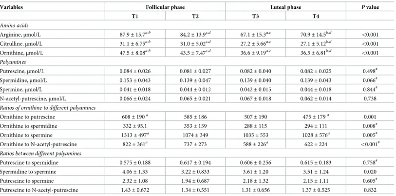
Table 1. Changes of variables across the menstrual cycle. Results are presented as mean± SD of 28 women at 4 time points (T1-T4).
Variables Follicular phase Luteal phase P value
T1 T2 T3 T4
Amino acids
Arginine,μmol/L 87.9± 15.7a.b 84.2± 13.9c.d 67.1± 15.3a.c 70.9± 14.5b.d <0.001
Citrulline,μmol/L 31.1± 6.75a.b 31.0± 5.02c.d 27.2± 5.66a.c 27.1± 5.12b.d <0.001
Ornithine,μmol/L 47.5± 8.08a.b 43.5± 7.47c.d 36.6± 9.19a.c 36.5± 6.81b.d <0.001
Polyamines
Putrescine,μmol/L 0.084± 0.026 0.081± 0.027 0.082± 0.040 0.082± 0.025 0.498#
Spermidine,μmol/L 0.153± 0.043 0.139± 0.047 0.139± 0.040 0.139± 0.043 0.066#
Spermine,μmol/L 0.041± 0.018 0.044± 0.012 0.042± 0.015 0.044± 0.018 0.844#
N-acetyl-putrescine,μmol/L 0.066± 0.024 0.065± 0.021 0.067± 0.018 0.062± 0.014 0.738
Ratios of ornithine to different polyamines
Ornithine to putrescine 608± 190a 585± 186 507± 190 475± 179a 0.001
Ornithine to spermidine 332± 95.1 353± 139 288± 115 294± 111 0.008#
Ornithine to spermine 1313± 497a 1074± 349 1035± 553 1028± 576a 0.005#
Ornithine to N-acetyl-putrescine 822± 361a 737± 273 588± 226a 622± 224 <0.001#
Ratios between different polyamines
Putrescine to spermidine 0.575± 0.188 0.617± 0.194 0.606± 0.256 0.615± 0.183 0.758#
Spermidine to spermine 4.06± 1.33 3.22± 0.833 3.61± 1.20 3.51± 1.24 0.020
Putrescine to spermine 2.32± 1.08 1.94± 0.687 2.18± 1.32 2.15± 1.11 0.605#
Putrescine to N-acetyl-putrescine 1.43± 0.672 1.34± 0.551 1.31± 0.656 1.37± 0.525 0.832
a,b,c,d
Same superscript letters at different time points indicate significant differences (P<0.01); one-way repeated measures ANOVA and Holm-Sidak multiple comparison procedures
#
repeated measures ANOVA on ranks and Tukey multiple comparison procedure
between T2 and T3 (ΔT3-T2) in NF-κB p65 activation were not related to changes in arginine concentrations, nor were changes in NF-κB p65 activation related to changes in progesterone concentrations.
Changes in polyamines and in ratios thereof across the menstrual cycle
(longitudinal approach)
Plasma concentrations are presented inTable 1. They were highest for spermidine, followed by putrescine, N-acetyl-putrescine and spermine (only about 30% of spermidine). Longitudi-nal changes in plasma putrescine, spermidine, spermine, and N-acetyl-putrescine across the menstrual cycle did not reach statistical significance in the entire study group (Table 1). When looking at individual women across a given menstrual cycle, substantial fluctuations, which differed between women and did not follow a specific pattern, became evident (Fig 6). Ratios of ornithine to putrescine, spermidine, spermine as well as to N-acetyl-putrescine, showed
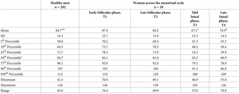
Table 2. Comparison of plasma arginine concentrations (μmol/L) of women across the menstrual cycle with reference ranges of 292 healthy men. Healthy men
n = 292
Women across the menstrual cycle
n = 28
Early follicular phase, T1
Late follicular phase, T2 Mid luteal phase, T3 Late luteal phase, T4 Mean 84.7a,b 87.9 84.2 67.1a 70.9b SD 16.3 15.7 13.9 15.3 14.5 5thPercentile 58.8 70.2 69.3 47.3 55.7 10thPercentile 64.5 73.7 70.5 48.2 58.4 25thPercentile 72.7 78.3 73.9 54.5 59.9 50thPercentilec 84.7 84.1 81.0 63.2 68.9 75thPercentile 96.1 95.0 92.0 79.1 78.9 90thPercentile 107 103 100 91.5 86.8 95PthPercentile 112 134 120 100 109 Minimum 41.5 70.0 69.1 46.9 55.0 Maximum 126 146 138 102 126 Range 85.6 76.4 69.0 55.0 70.8
Differences between phases of the menstrual cycle and healthy men were analyzed using
t-tests (healthy men vs. mid luteal phase) or Mann-Whitney Rank Sum Test (healthy men vs. early follicular, late follicular and late luteal phase). a,b
Same superscript letters indicate significant differences (P<0.01). cMedian
https://doi.org/10.1371/journal.pone.0200489.t002
Table 3. Regressions of amino acids (dependent variables) on progesterone (independent variables) at the 4 time points and regressions of changes (Δ) thereof between time points onΔ progesterone. Regression coefficients are presented for 28 women.
T1 T2 T3 T4 Δ T2-T1 Δ T3-T2 Δ T4-T3 Progesterone Δ Progesterone Arginine -0.150 0.004 -0.735a -0.125 Δ Arginine -0.206 -0.735a -0.677a Ornithine 0.033 0.011 -0.756a -0.306 Δ Ornithine -0.340 -0.693a -0.705a Citrulline 0.184 0.453 -0.636a -0.414 Δ Citrulline -0.353 -0.542b -0.501b a P < 0.001 b P < 0.01 https://doi.org/10.1371/journal.pone.0200489.t003
significant overall changes across the menstrual cycle (allP < 0.01), but post-hoc tests only reached significance (P < 0.01) for ratios of ornithine to putrescine, spermine as well as to N-acetyl-putrescine (Fig 7,Table 1). Ratios between different polyamines did not change across the menstrual cycle (Table 1).
Regressions of polyamines and of ratios thereof on progesterone
Ratios of ornithine to putrescine, spermidine, spermine as well as to N-acetyl-putrescine showed significant inverse relations (P < 0.01) with progesterone concentrations at T3 (Fig 8,Table 5, panel on the left hand side). Changes in ratios of ornithine to putrescine from T1 to T2 (ΔT2-T1) also showed a significant inverse relation with changes in progesterone concen-trations from T1 to T2 (ΔT2-T1) (Table 5, panel on the right hand side). In contrast, changes in ratios of putrescine to spermidine were positively related to changes in progesterone con-centrations (Table 5, panel on the right hand side).
Correlations between ornithine and polyamines and among polyamines
Ornithine concentrations were not significantly related to putrescine, spermidine, spermine and N-acetyl-putrescine concentrations (Table 6, panel on the left hand side), nor were changes in ornithine concentrations related to changes in putrescine, spermidine, spermine and N-acetyl-putrescine (Table 6, panel on the right hand side). In contrast, spermidine con-centrations were significantly related to putrescine concon-centrations at T2 (P < 0.01) and T3 (P < 0.001) (Table 6, panel on the left hand side), and spermine concentrations were signifi-cantly related to spermidine concentrations at T2 (P < 0.001) and T4 (P < 0.01) (Table 6, panel on the left hand side). Changes in putrescine concentrations between T3 and T4(ΔT4-T3) were significantly related to changes in spermidine (P < 0.01) and spermine concen-trations (P < 0.01), and changes in spermidine concenconcen-trations were significantly related to changes in spermine concentrations (P < 0.001) (Fig 9,Table 6, panel on the right hand side).
Discussion
This study for the first time provides data obtained at the plasma and PBMC level at luteal phase of the menstrual cycle, supporting the proposed regulatory node, comprising arginine and related amino acids, progesterone and NF-κB p65, aimed at successful preparation of pregnancy by demonstrating, as the first important finding of the present study, progesterone-related effects on plasma concentrations of arginine, ornithine and citrulline of a given men-strual cycle, including (i) longitudinal changes with lower concentrations of the three amino acids along with higher progesterone concentrations at luteal phase, a finding that was con-firmed by (ii) strong inverse relations of the three amino acids with progesterone at mid luteal phase, and strengthened by (iii) significant relations of changes in the three amino acid with changes in progesterone between late follicular and mid luteal as well as between mid and late luteal phase, and further highlighted by comparable slopes of the regression equations of (ii) and (iii).
Quantitatively, a 1 nmol/L increase in progesterone concentrations was related to a decrease in arginine concentrations of roughly 0.3μmol/L, which was regarded as a strong indicator of a progesterone-controlled amino acid decline at luteal phase. Compared to late
Fig 3. Linear regressions of plasma concentrations of arginine, ornithine and citrulline on serum progesterone concentrations at mid luteal phase (T3) of a given menstrual cycle in 28 women. Abbreviations: Arg, arginine; Cit,
citrulline; Orn, ornithine; Prog, progesterone.
Fig 4. Linear regressions of changes in plasma concentrations of arginine, ornithine and citrulline on changes in serum progesterone concentrations between late follicular and mid luteal phase (ΔT3-T2) (panels on the left hand side) and between mid-luteal and late luteal (ΔT4-T3) phase (panels on the right hand side) of a given menstrual cycle in 28 women. Abbreviations: Arg, arginine; Cit, citrulline;
Orn, ornithine; Prog, progesterone.
follicular phase, arginine at mid luteal phase was decreased by 19.8%, which is in line with pre-vious small-scale studies showing changes of about 10–15% [4,5]. Compared to reference ranges of healthy males, using the same method in the same laboratory, plasma arginine con-centrations were comparable at follicular phase, but significantly lower at luteal phase, reach-ing values below the 25thpercentile of healthy men in 86% (in 21 women at mid and 3 women
Table 4. Spearman rank correlations between arginine, ornithine and citrulline, at the 4 time points (T1-T4) as well as of changes (Δ) between time points.
Correla-tion coefficients are presented for 28 women.
T1 T2 T3 T4 Δ T2-T1 Δ T3-T2 Δ T4-T3 Arginine Δ Arginine Ornithine 0.436 0.519b 0.898a 0.675a Δ Ornithine 0.363 0.816a 0.775a Citrulline 0.035 0.345 0.587b 0.228 Δ Citrulline 0.211 0.702a 0.521b Ornithine Δ Ornithine Citrulline 0.154 0.422 0.729a 0.417 Δ Citrulline 0.228 0.645a 0.603a a P < 0.001 b P < 0.01 https://doi.org/10.1371/journal.pone.0200489.t004
Fig 5. Linear regressions of activation of the NF-κB p65 subunit in PBMC on plasma arginine concentrations at late follicular phase (T2) of a given menstrual cycle in 28 women. Abbreviations: Arg, arginine, NF-κB, nuclear factor kappa B.
at late luteal phase) and even below the 5thpercentile in 35.7% (in 10 women, all at mid luteal phase), suggesting that the luteal phase-specific arginine decline in the context of programmed preparation for successful pregnancy does not necessarily reach levels of deficiency, defined as being below the 5thpercentile of reference ranges.
Similar longitudinal changes in ornithine and citrulline concentrations suggest timed effects of the sharp increase in progesterone concentrations between late follicular and mid luteal phase on all three amino acids. For the first time, longitudinal changes were confirmed by strong inverse relations of all three amino acids with progesterone at mid luteal phase as well as between changes in the amino acids and progesterone between late follicular and mid luteal phase as well as between mid and late luteal phase. In contrast, at late luteal phase none of the three amino acids were significantly related to progesterone, probably because of absence of continuing physiological requirements in the absence of conception during the given menstrual cycle.
Regarding a mechanistic explanation of the findings, a number of luteal phase-specific mor-phological and functional changes shown in different studies may have contributed to arginine utilization, including increased expression of arginase [11] and eNOS [12] in endometrium as well as high expression of eNOS [13,14], NO-stimulated angiogenesis [15] and increased blood flow in corpus luteum [44]. Furthermore, a direct time- and dose-response effect of pro-gesterone on eNOS expression was shown in endometrial cells [45] as was a relation between increased blood flow in corpus luteum and serum progesterone concentrations [44].
Differences in the magnitudes of declines in amino acid concentrations from late follicular to mid luteal phase, which were, in decreasing order, 19.8% for arginine, the main substrate for NO-synthase [46] and arginase [47]; 16.0% for ornithine, a precursor of polyamines [9]; 12.1% for citrulline, a substrate forde novo synthesis of arginine [8]; could be explained by dif-ferent utilization and target levels at luteal phase according to their functional roles. Using sta-ble-isotope labelling in healthy young men, 15% of plasma arginine was shown to be used by arginase for synthesis of ornithine and urea, while total arginine utilization for NO synthesis did not exceed 1.2% [48]. However, such data are not available in the present study, because arginase and NO synthase activity have not been determined.
Lower ornithine and citrulline concentrations at luteal phase may have occurred as a conse-quence of reduced arginine availability and/or further utilization of ornithine for polyamine synthesis on one hand and of citrulline forde novo synthesis of arginine to compensate for high demands at mid luteal phase on the other hand.
As the second important finding for the proposed role of arginine in modulating immune functions for successful pregnancy, plasma arginine concentrations for the first time were identified, by using regression analysis, as predictors of the activation of the NF-κB p65 sub-unit in PBMC at late follicular phase, linking arginine to the role of NF-κB p65 in the immune response shift required for materno-fetal immune tolerance. So far, NF-κB p65 activation in PBMC was shown to be decreased in pregnancy [20], as well as to decline from follicular to luteal phase of the menstrual cycle, while associations with arginine or progesterone concen-trations have not been reported [29].
Regarding the underlying mechanisms of the findings of the present study, the arginine-mediated effect on NF-κB p65 activation could have been exerted through modulation of the T cell receptor CD3z expression [26], but CD3z expression was not determined in the present study. In trying to answer the question why similar relations were not observed at luteal phase,
Fig 6. Plasma putrescine, spermidine, spermine and N-acetyl-putrescine shown as individual concentrations across a given menstrual cycle in 28 women.
the strong effects of elevated progesterone on arginine concentrations, resulting in substantial utilization of arginine at luteal but not at follicular phase, could have jeopardized the regression of NF-κB p65 activation on arginine concentrations at luteal phase by selectively reducing argi-nine concentrations as the independent variable.
In contrast to arginine, changes in NF-κB p65 activation were not related to changes in pro-gesterone concentrations in the present study. Reduced nuclear translocation of NF-κB p65 upon exposure of LPS-stimulated macrophages to 100 nmol/L progesterone suggested direct inhibitory effects of progesterone on NF-κB activation [49]; however, comparably high pro-gesterone concentrations in plasma were only reached in three women of the present study. Interestingly, a study in healthy women showed higher NF-κB p65-DNA binding in the prolif-erative (at follicular phase) compared to the secretory (at luteal phase) endometrium [50], link-ing our findlink-ings in circulatory PBMC to evidence obtained in the endometrium.
Polyamine concentrations of the present study were comparable to those in 3 other studies including males and females [35,36,38], while the only 2 menstrual cycle studies reported 50-[37] and 2000-fold [39] higher spermidine and spermine concentrations. The analytical meth-ods yielding the high concentrations were ion-exchange amino acid analyzer [37] and HPLC UV detection [39], while the other studies used solid phase extraction (SPE)-LC/MS/MS [35], LC-MS [36] and radioimmunoassay [38].
Even though changes in polyamine concentrations due to increased demands at luteal phase could have been expected, plasma putrescine, spermidine, spermine, and N-acetyl-putrescine concentrations did not vary significantly across the menstrual cycle in the present study. When looking at individual women, substantial changes in all 4 polyamines were observed, which is in line with another study reporting individually different fluctuations in spermidine and spermine concentrations [37]. Changes in all 4 polyamines between late follic-ular and mid luteal phase were not related to changes in progesterone.
In contrast, significant longitudinal changes in ratios of ornithine, the principal precursor of polyamines, to each of the 4 polyamines—with differences in ratios of ornithine to putres-cine and ornithine to spermine between early follicular and late luteal phase and ratios of orni-thine to N-acetyl-putrescine between early follicular and mid luteal phase -, as well as
significant relations with progesterone at mid luteal phase could be attributed to the significant changes in and relations of ornithine with progesterone as the common numerator of the ratios.
The ratios of ornithine to putrescine and putrescine to spermidine could be regarded as proxies for the conversion of ornithine to putrescine and putrescine to spermidine, respec-tively. The inverse relation of changes between early and late follicular phase in progesterone concentrations with changes in ratios of ornithine to putrescine (i.e., with putrescine being the denominator) on one hand and positive relations of changes in progesterone with ratios of putrescine to spermidine (i.e., with putrescine being the numerator) on the other hand would suggest that progesterone exerts a putrescine-enhancing effect. However, progesterone levels at early and late follicular phase were very low and changes between early and late follicular phase were not significant. Moreover, such relations were not found in the presence of the pro-nounced increase in progesterone between late follicular and mid luteal phase, nor was an
Fig 7. Ratios of ornithine to putrescine, spermidine, spermine and N-acetyl-putrescine shown as box-and-whisker blots at early (T1) and late (T2) follicular and mid (T3) and late (T4) luteal phase across a given menstrual cycle in 28 women. Results were obtained by repeated measures ANOVA and repeated measures ANOVA on ranks for
normally and non-normally distributed data, respectively, along with all pairwise multiple comparison procedures (Holm-Sidak and Tukey tests, respectively); same superscripts indicate significant differences (P < 0.01) between time points.
increase in putrescine in the luteal phase observed, when a pronounced increase in progester-one occured. Given that all 4 polyamines did not change significantly across the menstrual cycle and were neither related to progesterone concentrations at mid luteal phase, nor were changes in all 4 polyamines between late follicular and mid luteal phase related to changes in
Fig 8. Linear regressions of plasma ratios of ornithine to putrescine, spermidine, spermine and N-acetyl-putrescine on serum progesterone concentrations at mid luteal phase (T3) of a given menstrual cycle in 28 women. Abbreviations: A-Putr, acetyl-putrescine; Orn, ornithine; Prog, progesterone; Put, putrescine; Spd,
spermidine; Spm, spermine.
https://doi.org/10.1371/journal.pone.0200489.g008
Table 5. Regressions of polyamines (dependent variables) on progesterone (independent variable) at the 4 time points and regressions of changes (Δ) thereof between time points onΔ progesterone. Regression coefficients are presented for 28 women.
T1 T2 T3 T4 Δ T2-T1 Δ T3-T2 Δ T4-T3 Progesterone Δ Progesterone Putrescine 0.135 0.024 0.142 0.080 Δ Putrescine 0.378 0.102 0.144 N-acetyl-putrescine 0.260 0.043 -0.013 0.097 Δ N-acetyl-putrescine 0.017 -0.172 -0.069 Spermidine 0.301 -0.075 0.063 0.022 Δ Spermidine -0.151 0.153 0.186 Spermine 0.049 -0.058 0.203 0.339 Δ Spermine 0.155 0.225 0.351
Ornithine to putrescine -0.115 -0.018 -0.557b -0.141 Δ Ornithine to putrescine -0.502b -0.455 -0.345
Ornithine to spermidine -0.365 0.210 -0.528b -0.097 Δ Ornithine to spermidine 0.094 -0.450 -0.448
Ornithine to spermine -0.159 0.119 -0.426b -0.283 Δ Ornithine to spermine -0.323 -0.416 -0.542b
Ornithine to N-acetyl-putrescine -0.368 -0.050 -0.485b -0.222 Δ Ornithine to N-acetyl-putrescine -0.164 -0.210 -0.245
Putrescine to spermidine -0.162 0.250 0.089 0.026 Δ Putrescine to spermidine 0.589a -0.025 -0.085
Putrescine to spermine -0.025 0.109 0.002 -0.236 Δ Putrescine to spermine 0.131 0.130 -0.319
Putrescine to N-acetyl-putrescine -0.239 -0.022 0.209 -0.025 Δ Putrescine to N-acetyl-putrescine 0.131 0.218 0.225
Spermidine to spermine 0.172 -0.067 -0.111 -0.366 Δ Spermidine to spermine -0.334 -0.106 -0.332
a P < 0.001 b
P < 0.01
https://doi.org/10.1371/journal.pone.0200489.t005
Table 6. Spearman rank correlations between ornithine and polyamines and among polyamines at the 4 time points (T1-T4) as well as of changes (Δ) between time points. Correlation coefficients are presented for 28 women.
T1 T2 T3 T4 Δ T2-T1 Δ T3-T2 Δ T4-T3 Ornithine Δ Ornithine Putrescine 0.332 0.221 -0.007 0.078 Δ Putrescine -0.088 0.053 -0.164 Spermidine 0.033 0.026 -0.087 -0.149 Δ Spermidine 0.311 -0.176 -0.218 Spermine 0.159 -0.137 -0.234 -0.274 Δ Spermine -0.160 -0.032 -0.228 N-acetyl-putrescine -0.157 -0.014 -0.011 -0.015 Δ N-acetyl-putrescine -0.011 0.256 0.103 Putrescine Δ Putrescine Spermidine 0.243 0.558b 0.648a 0.378 Δ Spermidine 0.426 0.202 0.535b Spermine 0.094 0.387 0.385 0.386 Δ Spermine 0.339 0.412 0.542b N-acetyl-putrescine 0.158 0.322 0.002 0.175 Δ N-acetyl-putrescine 0.436 0.109 -0.412 Spermidine Δ Spermidine Spermine 0.435 0.642a 0.383 0.589b Δ Spermine 0.409 0.259 0.712a a P < 0.001 b P < 0.01 https://doi.org/10.1371/journal.pone.0200489.t006
progesterone, these data suggest that generation of putrescine from ornithine and further con-versions to spermidine and spermine as well as N-acetyl-putrescine are not controlled by progesterone.
In conclusion, the present study provides data, at the plasma and PBMC level, supporting a physiological regulatory node aimed at preparation of successful pregnancy, including robust data on progesterone-associated luteal phase-specific declines in circulating arginine and related amino acid concentrations, based on (i) longitudinal changes, (ii) strong inverse associ-ations with progesterone concentrassoci-ations, and (iii) significant associassoci-ations between changes in arginine as well as arginine-related amino acids and changes in progesterone concentrations, and (iv) strong associations of declining arginine concentrations with declining NF-κB p65 activation, known to play a role in shifting the immune response towards materno-fetal toler-ance, while generation of putrescine from ornithine and further to spermidine and spermine as well as N-acetyl-putrescine does not seem to be controlled by progesterone.
Based on the fact that blood continuously supplies tissues and organs, plasma and PBMC were used as a systemic resource for studying amino acids, progesterone, and NF-κB p65 acti-vation in the present study. Interestingly, PBMC derived from non-pregnant women at luteal phase promoted progesterone production by human luteal cell cultures [51] and have thus been implicated in a systemic embryo-maternal cross-talk [52] and successfully used inin vitro fertilization [53], underpinning the physiological relevance of the proposed node at the circula-tory level for preparation of successful pregnancy.
There are also limitations of this study, given that (i) associations do not establish cause-and-effect relationships, (ii) additional factors may also be important, as evidenced, for instance, by lack of association between NF-κB p65 activation and progesterone despite its relation to arginine, and (iii) absence of additional measurements in PBMC, for instance, of arginase and eNOS, that could have allowed to quantify arginine consumption and the pro-posed conversions between amino acids. Furthermore, relations between the circulatory and organ levels, including information on physiological responses, such as NF-kB activation in relation to changes in amino acids, in the corpus luteum and endometrium would be of inter-est. To fully characterize the regulatory node and further address the physiological significance of the data presented in female fertility, future investigations are needed.
Acknowledgments
The authors thank Peter Pu¨rstner for helpful suggestions for the study design, and Theopisti Maimari, Verena Schaberl, Agnes Schriebl, Manfred Truber, and Gabriele Zechner for excel-lent technical assistance.
Author Contributions
Conceptualization: Gernot Faustmann, Johannes M. Roob, Brigitte M. Winklhofer-Roob. Data curation: Gernot Faustmann, Andreas Meinitzer, Christoph Magnes, Beate Tiran,
Bar-bara Obermayer-Pietsch, Hans-Ju¨rgen Gruber, Josep Ribalta, Edmond Rock, Johannes M. Roob, Brigitte M. Winklhofer-Roob.
Formal analysis: Gernot Faustmann, Andreas Meinitzer, Christoph Magnes, Beate Tiran,
Bar-bara Obermayer-Pietsch, Hans-Ju¨rgen Gruber, Brigitte M. Winklhofer-Roob.
Fig 9. Spearman rank correlations between changes in plasma concentrations of putrescine and spermidine, putrescine and spermine as well as spermidine and spermine between mid and late luteal phase (ΔT4-T3) of a given menstrual cycle in 28 women.
Funding acquisition: Josep Ribalta, Edmond Rock, Johannes M. Roob, Brigitte M.
Winklho-fer-Roob.
Investigation: Gernot Faustmann, Josep Ribalta, Edmond Rock, Johannes M. Roob, Brigitte
M. Winklhofer-Roob.
Methodology: Gernot Faustmann, Andreas Meinitzer, Christoph Magnes, Beate Tiran,
Bar-bara Obermayer-Pietsch, Hans-Ju¨rgen Gruber, Josep Ribalta, Edmond Rock, Johannes M. Roob, Brigitte M. Winklhofer-Roob.
Project administration: Josep Ribalta, Edmond Rock, Johannes M. Roob, Brigitte M.
Winkl-hofer-Roob.
Resources: Andreas Meinitzer, Christoph Magnes, Beate Tiran, Barbara Obermayer-Pietsch,
Josep Ribalta, Edmond Rock, Johannes M. Roob, Brigitte M. Winklhofer-Roob.
Supervision: Hans-Ju¨rgen Gruber, Josep Ribalta, Edmond Rock, Johannes M. Roob, Brigitte M. Winklhofer-Roob.
Validation: Gernot Faustmann, Andreas Meinitzer, Christoph Magnes, Beate Tiran, Barbara
Obermayer-Pietsch, Hans-Ju¨rgen Gruber, Josep Ribalta, Edmond Rock, Johannes M. Roob, Brigitte M. Winklhofer-Roob.
Visualization: Gernot Faustmann, Brigitte M. Winklhofer-Roob.
Writing – original draft: Gernot Faustmann, Brigitte M. Winklhofer-Roob.
Writing – review & editing: Gernot Faustmann, Andreas Meinitzer, Christoph Magnes, Beate
Tiran, Barbara Obermayer-Pietsch, Hans-Ju¨rgen Gruber, Josep Ribalta, Edmond Rock, Johannes M. Roob, Brigitte M. Winklhofer-Roob.
References
1. Solomon SJ, Kurzer MS, Howes Calloway D. Menstrual cycle and basal metabolic rate in women. Am J Clin Nutr. 1982; 36: 611–616.https://doi.org/10.1093/ajcn/36.4.611PMID:7124662
2. Lariviere F, Moussalli R, Garrel DR. Increased leucine flux and leucine oxidation during the luteal phase of the menstrual cycle in women. Am J Physiol. 1994; 267: E422–8.https://doi.org/10.1152/ajpendo. 1994.267.3.E422PMID:7943222
3. Calloway DH, Kurzer MS. Menstrual cycle and protein requirements of women. J Nutr. 1982; 112: 356– 366.https://doi.org/10.1093/jn/112.2.356PMID:7057271
4. Cox BD, Calame DP. Changes in plasma amino acid levels during the human menstrual cycle and in early pregnancy. A preliminary report. Horm Metab Res. 1978; 10: 428–433. https://doi.org/10.1055/s-0028-1093407PMID:711138
5. Moller SE, Moller BM, Olesen M, Fjalland B. Effects of oral contraceptives on plasma neutral amino acids and cholesterol during a menstrual cycle. Eur J Clin Pharmacol. 1996; 50: 179–84. PMID: 8737756
6. Yeung EH, Zhang C, Albert PS, Mumford SL, Ye A, Perkins NJ, et al. Adiposity and sex hormones across the menstrual cycle: the BioCycle Study. Int J Obes. 2013; 37: 237–243.
7. Morris SM. Arginine metabolism: boundaries of our knowledge. J Nutr. 2007; 137: 1602S–1609S. https://doi.org/10.1093/jn/137.6.1602SPMID:17513435
8. Dhanakoti SN, Brosnan JT, Herzberg GR, Brosnan ME. Renal arginine synthesis: studies in vitro and in vivo. Am J Physiol. 1990; 259: E437–42.https://doi.org/10.1152/ajpendo.1990.259.3.E437PMID: 1975989
9. Jenkinson CP, Grody WW, Cederbaum SD. Comparative properties of arginases. Comp Biochem Phy-siol. 1996; 114: 107–132.
10. Palmer RM, Ashton DS, Moncada S. Vascular endothelial cells synthesize nitric oxide from L-arginine. Nature. 1988; 333: 664–666.https://doi.org/10.1038/333664a0PMID:3131684
11. Tajima M, Harada T, Ishikawa T, Iwahara Y, Kubota T. Augmentation of arginase II expression in the human endometrial epithelium in the secretory phase. J Med Dent Sci. 2012; 59: 75–82. PMID: 23897115
12. Khorram O, Garthwaite M, Magness RR. Endometrial and myometrial expression of nitric oxide synthase isoforms in pre-and postmenopausal women. J Clin Endocrinol Metab. 1999; 84: 2226–2232. https://doi.org/10.1210/jcem.84.6.5759PMID:10372735
13. Friden BE, Runesson E, Hahlin M, Brannstrom M. Evidence for nitric oxide acting as a luteolytic factor in the human corpus luteum. Mol Hum Reprod. 2000; 6: 397–403. PMID:10775642
14. Vega M, Johnson MC, Diaz HA, Urrutia LR, Troncoso JL, Devoto L. Regulation of human luteal ste-roidogenesis in vitro by nitric oxide. Endocrine. 1998; 8: 185–191.https://doi.org/10.1385/ ENDO:8:2:185PMID:9704576
15. Reynolds LP, Grazul-Bilska AT, Redmer DA. Angiogenesis in the corpus luteum. Endocrine. 2000; 12: 1–9.https://doi.org/10.1385/ENDO:12:1:1PMID:10855683
16. Dulak J, Jozkowicz A, Dembinska-Kiec A, Guevara I, Zdzienicka A, Zmudzinska-Grochot D, et al. Nitric oxide induces the synthesis of vascular endothelial growth factor by rat vascular smooth muscle cells. Arterioscler Thromb Vasc Biol. 2000; 20: 659–666. PMID:10712388
17. Mitsube K, Zackrisson U, Bra¨nnstro¨m M. Nitric oxide regulates ovarian blood flow in the rat during the periovulatory period. Hum Reprod. 2002; 17: 2509–16. PMID:12351520
18. Chwalisz K, Garfield RE. Role of nitric oxide in implantation and menstruation. Hum Reprod. 2000; 15: 96–111. PMID:11041226
19. McCracken SA, Gallery E, Morris JM. Pregnancy-specific down-regulation of NF-kB expression in T cells in humans is essential for the maintenance of the cytokine profile required for pregnancy success. J Immunol. 2004; 172: 4583–4591. PMID:15034076
20. McCracken SA, Drury CL, Lee H-S, Morris JM. Pregnancy is associated with suppression of the nuclear factor kappa B/I kappa B activation pathway in peripheral blood mononuclear cells. J Reprod Immunol. 2003; 58: 27–47. PMID:12609523
21. Faas M, Bouman A, Moesa H, Heineman MJ, de Leij L, Schuiling G. The immune response during the luteal phase of the ovarian cycle: a Th2-type response? Fertil Steril. 2000; 74: 1008–1013. PMID: 11056250
22. Bronte V, Zanovello P. Regulation of immune responses by L-arginine metabolism. Nat Rev Immunol. 2005; 5: 641–654.https://doi.org/10.1038/nri1668PMID:16056256
23. Baniyash M. TCR zeta-chain downregulation: curtailing an excessive inflammatory immune response. Nat Rev Immunol. 2004; 4: 675–687.https://doi.org/10.1038/nri1434PMID:15343367
24. Rodriguez PC, Zea AH, DeSalvo J, Culotta KS, Zabaleta J, Quiceno DG, et al. L-arginine consumption by macrophages modulates the expression of CD3 zeta chain in T lymphocytes. J Immunol. 2003; 171: 1232–1239. PMID:12874210
25. Kropf P, Baud D, Marshall SE, Munder M, Mosley A, Fuentes JM, et al. Arginase activity mediates reversible T cell hyporesponsiveness in human pregnancy. Eur J Immunol. 2007; 37: 935–945.https:// doi.org/10.1002/eji.200636542PMID:17330821
26. Paul S, Schaefer BC. A new look at T cell receptor signaling to nuclear factor-kB. Trends Immunol. 2013; 34: 269–281.https://doi.org/10.1016/j.it.2013.02.002PMID:23474202
27. Lee K-Y, D’Acquisto F, Hayden MS, Shim J-H, Ghosh S. PDK1 nucleates T cell receptor-induced sig-naling complex for NF-kB activation. Science (80- ). 2005; 308: 114–118.
28. Vallabhapurapu S, Karin M. Regulation and function of NF-kappaB transcription factors in the immune system. Annu Rev Immunol. 2009; 27: 693–733.https://doi.org/10.1146/annurev.immunol.021908. 132641PMID:19302050
29. Faustmann G, Tiran B, Maimari T, Kieslinger P, Obermayer-Pietsch B, Gruber HJ, et al. Circulating lep-tin and NF-kB activation in peripheral blood mononuclear cells across the menstrual cycle. BioFactors. 2016; 42: 376–387.https://doi.org/10.1002/biof.1281PMID:27093900
30. van Nieuwenhoven ALV, Heineman MJ, Faas MM. The immunology of successful pregnancy. Hum Reprod Update. 2003; 9: 347–357. PMID:12926528
31. Mor G, Aldo P, Alvero AB. The unique immunological and microbial aspects of pregnancy. Nat Rev Immunol. 2017; 17: 469–482.https://doi.org/10.1038/nri.2017.64PMID:28627518
32. Greene JM, Feugang JM, Pfeiffer KE, Stokes J V, Bowers SD, Ryan PL. L-arginine enhances cell prolif-eration and reduces apoptosis in human endometrial RL95-2 cells. Reprod Biol Endocrinol. 2013; 11: 15.https://doi.org/10.1186/1477-7827-11-15PMID:23442442
33. Lefevre PLC, Palin MF, Murphy BD. Polyamines on the reproductive landscape. Endocr Rev. 2011; 32: 694–712.https://doi.org/10.1210/er.2011-0012PMID:21791568
34. KEGG: Kyoto Encyclopedia of Genes and Genomes. Arginine and proline metabolism. Available:http:// www.genome.jp/kegg-bin/show_pathway?rn00330+R01154
35. Magnes C, Fauland A, Gander E, Narath S, Ratzer M, Eisenberg T, et al. Polyamines in biological sam-ples: Rapid and robust quantification by solid-phase extraction online-coupled to liquid chromatogra-phy-tandem mass spectrometry. J Chromatogr A. 2014; 1331: 44–51.https://doi.org/10.1016/j.chroma. 2013.12.061PMID:24485539
36. Liu R, Bi K, Jia Y, Wang Q, Yin R, Li Q. Determination of polyamines in human plasma by high-perfor-mance liquid chromatography coupled with Q-TOF mass spectrometry. J Mass Spectrom. 2012; 47: 1341–6.https://doi.org/10.1002/jms.3084PMID:23019166
37. Lundgren DW, Farrell PM, Cohen LF, Hankins J. Fluctuations of unbound whole blood polyamine levels during the menstrual cycle. Proc Soc Exp Biol Med. 1976; 152: 81–85. PMID:1265084
38. Chaisiri P, Harper ME, Blamey RW, Peeling WB, Griffiths K. Plasma spermidine concentrations in patients with tumours of the breast or prostate or testis. Clin Chim Acta. 1980; 104: 367–375. PMID: 6156039
39. Gilad VH, Halperin R, Chen-Levy Z, Gilad GM. Cyclic changes of plasma spermine concentrations in women. Life Sci. 2002; 72: 135–141. PMID:12417247
40. Meinitzer A, Puchinger M, Winklhofer-Roob BM, Rock E, Ribalta J, Roob JM, et al. Reference values for plasma concentrations of asymmetrical dimethylarginine (ADMA) and other arginine metabolites in men after validation of a chromatographic method. Clin Chim Acta. 2007; 384: 141–148.https://doi.org/ 10.1016/j.cca.2007.07.006PMID:17689511
41. Davis JS, Darcy CJ, Piera K, McNeil YR, Woodberry T, Anstey NM. Ex-vivo changes in amino acid con-centrations from blood stored at room temperature or on ice: implications for arginine and taurine mea-surements. BMC Clin Pathol. 2009; 9: 10.https://doi.org/10.1186/1472-6890-9-10PMID:19941666 42. Roth M. Fluorescence reaction for amino acids. Anal Chem. 1971; 43: 880–882. PMID:5576608 43. Schwarz EL, Roberts WL, Pasquali M. Analysis of plasma amino acids by HPLC with photodiode array
and fluorescence detection. Clin Chim Acta. 2005; 354: 83–90.https://doi.org/10.1016/j.cccn.2004.11. 016PMID:15748603
44. Tamura H, Takasaki A, Taniguchi K, Matsuoka A, Shimamura K, Sugino N. Changes in blood-flow impedance of the human corpus luteum throughout the luteal phase and during early pregnancy. Fertil Steril. 2008; 90: 2334–2339.https://doi.org/10.1016/j.fertnstert.2007.10.056PMID:18249380 45. Khorram O, Han G. The influence of progesterone on endometrial nitric oxide synthase expression.
Fer-til Steril. 2009; 91: 2157–2162.https://doi.org/10.1016/j.fertnstert.2008.05.019PMID:18710710 46. Moali C, Boucher JL, Sari MA, Stuehr DJ, Mansuy D. Substrate specificity of NO synthases: detailed
com-parison of L-arginine, homo-L-arginine, their N omega-hydroxy derivatives, and N omega-hydroxynor-L-arginine. Biochemistry. 1998; 37: 10453–10460.https://doi.org/10.1021/bi980742tPMID:9671515 47. Hrabak A, Bajor T, Temesi A. Comparison of substrate and inhibitor specificity of arginase and nitric
oxide (NO) synthase for arginine analogues and related compounds in murine and rat macrophages. Biochem Biophys Res Commun. 1994; 198: 206–212. PMID:7507318
48. Castillo L, Beaumier L, Ajamit AM, Young VR. Whole body nitric oxide synthesis in healthy men deter-mined from [15N] arginine-to-[15N] citrulline labeling. Proc Natl Acad Sci. 1996; 93: 11460–11465. PMID:8876157
49. Su L, Sun Y, Ma F, Lu¨ P, Huang H, Zhou J. Progesterone inhibits Toll-like receptor 4-mediated innate immune response in macrophages by suppressing NF-κB activation and enhancing SOCS1 expres-sion. Immunol Lett. 2009; 125: 151–155.https://doi.org/10.1016/j.imlet.2009.07.003PMID:19607861 50. Gonza´lez-Ramos R, Rocco J, Rojas C, Sovino H, Poch A, Kohen P, et al. Physiologic activation of
nuclear factor kappa B in the endometrium during the menstrual cycle is altered in endometriosis patients. Fertil Steril. 2012; 97: 645–651.https://doi.org/10.1016/j.fertnstert.2011.12.006PMID: 22196717
51. Hashii K, Fujiwara H, Yoshioka S, Kataoka N, Yamada S, Hirano T, et al. Peripheral blood mononuclear cells stimulate progesterone production by luteal cells derived from pregnant and non-pregnant women: possible involvement of interleukin-4 and interleukin-10 in corpus luteum function and differentiation. Hum Reprod. 1998; 13: 2738–2744. PMID:9804222
52. Fujiwara H. Do circulating blood cells contribute to maternal tissue remodeling and embryo—maternal cross-talk around the implantation period? Mol Hum Reprod. 2009; 15: 335–343.https://doi.org/10. 1093/molehr/gap027PMID:19346239
53. Yoshioka S, Fujiwara H, Nakayama T, Kosaka K, Mori T, Fujii S. Intrauterine administration of autolo-gous peripheral blood mononuclear cells promotes implantation rates in patients with repeated failure of IVF–embryo transfer. Hum Reprod. 2006; 21: 3290–3294.https://doi.org/10.1093/humrep/del312 PMID:17021188


![Structures of [M(Ura-H)(Ura)]+ and [M(Ura-H)(H2O)n]+ (M = Cu, Zn, Pb; n = 1–3) complexes in the gas phase by IRMPD spectroscopy in the fingerprint region and theoretical studies](data:image/gif;base64,R0lGODlhAQABAIAAAP///wAAACH5BAEAAAAALAAAAAABAAEAAAICRAEAOw==)