HAL Id: tel-01252520
https://tel.archives-ouvertes.fr/tel-01252520
Submitted on 7 Jan 2016
HAL is a multi-disciplinary open access archive for the deposit and dissemination of sci-entific research documents, whether they are pub-lished or not. The documents may come from teaching and research institutions in France or abroad, or from public or private research centers.
L’archive ouverte pluridisciplinaire HAL, est destinée au dépôt et à la diffusion de documents scientifiques de niveau recherche, publiés ou non, émanant des établissements d’enseignement et de recherche français ou étrangers, des laboratoires publics ou privés.
Histone H3 variants and chaperones in Arabidopsis
thaliana heterochromatin dynamics
Matthias Benoit
To cite this version:
Matthias Benoit. Histone H3 variants and chaperones in Arabidopsis thaliana heterochromatin dy-namics. Agricultural sciences. Université Blaise Pascal - Clermont-Ferrand II, 2014. English. �NNT : 2014CLF22497�. �tel-01252520�
ÉCOLE DOCTORALE
DES SCIENCES DE LA VIE, SANTÉ, AGRONOMIE, ENVIRONNEMENT
Thèse
Présentée à l'Université Blaise Pascalpour l'obtention du grade de
DOCTEUR D'UNIVERSITÉ
Spécialité : Physiologie et Génétique Moléculaires
Soutenue le 17 Octobre 2014
Matthias BENOIT
HISTONE H3 VARIANTS AND CHAPERONES
IN ARABIDOPSIS THALIANA HETEROCHROMATIN DYNAMICS
Présidente : Dr. Chantal VAURY, GReD, Clermont-Ferrand Rapporteurs : Dr. Cristel CARLES, LPCV, Grenoble
Dr. Elaine DUNLEAVY, CCB, Galway
Dr. Ortrun MITTELSTEN SCHEID, GMI, Vienne Directrice de thèse : Dr. Aline PROBST, Université Blaise Pascal, Aubière
Génétique, Reproduction & Développement UMR CNRS 6293 - Clermont Université - INSERM U1103
! ! ! ! !
A Mesdames Cristel Carles, Elaine Dunleavy et Ortrun Mittelsten Scheid d’être venues des quatre coins de France et d’Europe pour évaluer ce travail, et à Chantal Vaury d’avoir presidé ce jury de thèse exclusivement féminin,
A Aline, la Chef, pour tout ce que tu m’as apporté durant ces quatre années. Merci de m’avoir donné ma chance et de m’avoir laissé t’accompagner dans cette aventure qui a commencé en terrasse d’un café Place de Jaude. Merci pour ton investissement, tes conseils et tes corrections sur la thèse même après deux heures du matin. Merci de ton optimisme quand le mien baissait. Merci de ta patience quand je laissais trainer mes plantes en serre. Tu as été et tu continueras à être un exemple pour moi,
A Sylvette, pour m’avoir accueilli à BIOMOVE et lancé dans le grand bain, pour ton aide et ton regard bienveillant sur le petit thésard que j’étais. Et pour Buda !
A Marie-Claude, ma maman du labo, d’avoir pris soin de moi pendant ces 4 années, à peigner des girafes (un peu), éplucher des cotylédons (beaucoup), papoter au café (beaucoup beaucoup) et à se raconter les hauts et les bas de nos vies. Les derniers mois sans toi au labo étaient déjà bien trop longs, tu vas me manquer,
A Chantal, d’avoir partagé bien plus que la moitié de ton bureau et d’avoir supporté mon coté bordélique (tiens où est ma clé USB ?!). J’espère que mon futur voisin de bureau sera aussi cool que toi !
A Céline, pour m’avoir énormément aidé et conseillé sur la fin de thèse. Merci de m’avoir transmis (ne serait-ce qu’un peu !) de ta rigueur et de ton ouverture d’esprit. Je ne mangerai plus de lentilles de la même façon,
A Sam pour ta joie et bonne humeur, ton aide sans faille, le style prof romantique et les soirées au Nirvana,
A Manu pour les enseignements partagés ensemble, les anecdotes sur Toulouse et tes chemisettes,
A Christophe pour ton pragmatisme et ton optimisme, ainsi que de m’avoir soutenu aussi bien pour la thèse que pour le monitorat,
A Benjamin et Ortrun de m’avoir fait profiter de leur expertise et gentillesse lors des comités de thèse. Merci à Ortrun et Jasmin pour l’accueil chaleureux reçu à Vienne,
A Marie-No pour le ménage. A Thierry pour le debrief rugby du week-end,
A Simon et ODI pour la triplette squash / picon bière / Baraka,
A Maryse et Marie-Jo, pour mes commandes et mes envois TOUJOURS hyper-urgents et hyper-importants,
A Leti, Angeles et Sébich pour leur bonne humeur et la bière de fin de journée (et ta chemise Sébich !),
A Isa pour l’enseignement et les (nombreuses !) discussions plus ou moins sérieuses sur les post-docs et l’avenir en général,
Aux copains de PCE et du PIAF pour les repas pizzas, bagels, food trucks, apéros …
A Margaux et Pierre, pour la relève,
A Elodie, dit Mich’, d’avoir été la vieille thésarde et de m’avoir fait l’historique du labo à mon arrivée. Merci pour les discussions venant de la paillasse de derrière et des répliques de la Cité de la Peur,
soutenance !
Aux copains thésards de l’équipe : A Axel, mon jumeau maléfique,
A Lauriane, ma jeune padawan chocolatphile, A Mélanie, ma Mad’moizelle,
Merci d’avoir été les compagnons du quotidien dans les bons moments et ceux plus difficiles. Un joyeux mélange de licornes et de petits poneys, de pauses thé/café/Rugbynistère, de débrief ciné, de petites bouffes sympas, de croustilles, de listes de ménage arrangées, de repas de midi salvateurs … !
A Lili et Valen pour leur soutien et amour sans faille,
A mes oncles, tantes, cousins, cousines, à travers toute la France,
A mon filleul que j’aime de tout mon cœur,
A mes grands-parents,
A mes parents et mon frère, pour leur soutien et amour indéfectible, ainsi que pour m’avoir encouragé si tôt à ouvrir mon esprit et découvrir le monde,
A Emilie de partager tout cela avec moi, de me pousser à aller toujours plus loin et de m’accompagner sur ce chemin pas toujours facile. Et puis au pire on s’aime.
Abstract
To understand how histones H3 are handled and how histone dynamics impact higher-order chromatin organization such as chromocenter formation in Arabidopsis, a comprehensive analysis of the different histone chaperone complexes is required. We identified and characterized the different subunits of the Arabidopsis HIR complex. AtHIRA is the central subunit and its loss affects non-nucleosomal histone levels, reduces nucleosomal occupancy not only at euchromatic but also at heterochromatic targets and alleviates transcriptional gene silencing. While the HIR complex-mediated histone deposition is dispensable for higher-order organization of Arabidopsis heterochromatin, I show that CAF-1 plays a central role in chromocenter formation. During post-germination development in cotyledons when centromeric and pericentromeric repeats cluster progressively into chromocenter structures, these repetitive elements but not euchromatic loci become enriched in H3.1 in a CAF-1-dependent manner. This enrichment, together with the appropriate setting of repressive histone post-translational marks, contributes to chromocenter formation, identifying chromatin assembly by CAF-1 as driving force in formation and maintenance of genome structure. Finally, while absence of HIR or CAF-1 complexes sustains viability, only the simultaneous loss of both severely impairs nucleosomal occupancy and plant development, suggesting a limited functional compensation between the different histone chaperone complexes and plasticity in histone variant interaction and deposition in plants.
Keywords: Heterochromatin dynamics, chromocenter, histone H3, histone chaperone, Arabidopsis thaliana
Afin d’étudier la prise en charge des histones H3 jusqu’à l’ADN et pour comprendre l’influence de leur dynamique dans l’organisation d’ordre supérieur de la chromatine, une analyse des chaperonnes d’histones a été menée. Nous avons identifié et caractérisé les sous-unités du complexe HIR, impliqué dans l’assemblage de la chromatine réplication-indépendante chez Arabidopsis. La perte d’AtHIRA, la sous-unité centrale du complexe, affecte le niveau d’histone soluble, l’occupation nucléosomale des régions euchromatiniennes et héterochromatiniennes ainsi que la mise sous silence transcriptionnel des séquences d’ADN répétées. Alors que le complexe HIR ne participe pas à l’organisation d’ordre supérieur de la chromatine, j’ai montré que CAF-1, impliqué dans l’assemblage de la chromatine au cours de la réplication, joue un rôle central dans la formation des chromocentres. Lors du développement post-germinatif des cotylédons, les séquences d’ADN répétées centromériques et péricentromériques se concentrent dans les chromocentres et s’enrichissent en histone H3.1 de manière CAF-1 dépendante. Cet enrichissement, associé à des modifications post-traductionnelles d’histones associées à un état répressif de la transcription, participe à la formation des chromocentres et met en évidence l’importance de l’assemblage de la chromatine par CAF-1 dans la structure et le maintien du génome. Alors que la perte individuelle de HIR ou de CAF-1 n’affecte pas la viabilité, l’absence des deux complexes altère fortement l’occupation nucléosomale et le développement des plantes. Ceci suggère que la compensation fonctionnelle entre ces complexes de chaperonnes ainsi que la plasticité des voies de dépôt des histones restent limitées.
Mots-clés: Dynamique de l’heterochromatine, chromocentre, histone H3, chaperonne d’histone, Arabidopsis
Table of contents
ABBREVIATIONS 3
LIST OF ILLUSTRATIONS 5
STATE OF THE ART 8
1. Organization of eukaryotic genomes in chromatin 9
1.1. Functional significance of chromatin 10
1.2. Heterochromatin is a specialized chromatin subdomain 12
1.2.1. Structure of heterochromatin 12
1.2.2. Heterochromatin is actively defined by epigenetic mechanisms 16
1.2.2.1. DNA methylation 16
1.2.2.1.1. Maintenance 17
1.2.2.1.2. Demethylation 19
1.2.2.2. Histone post-translational marks associated with heterochromatin 20
1.2.2.2.1. H3K9me2/3 21
1.2.2.2.2. H3K27me1 22
1.2.2.3. Chromatin remodeling factors 23
1.2.2.3.1. DDM1 23
1.2.2.3.2. MOM1 24
1.2.2.3.3. MORC 24
1.3. Dynamics of heterochromatin during developmental phase transitions in Arabidopsis 25
1.3.1. Heterochromatin remodeling during floral transition and dedifferentiation 25
1.3.1.1. Heterochromatin dynamics in cotyledons 27
1.3.1.1.1. Heterochromatin dynamics during seed development 27
1.3.1.1.2. Heterochromatin dynamics during germination and seedling growth 28
1.3.1.2. Environment-induced heterochromatin dynamics 30
2. Biology of histone H3 32
2.1. Canonical H3.1 33
2.1.1. Gene organization and expression 33
2.1.2. Protein structure 34
2.1.3. Genomic localization 35
2.1.4. Histone post-translational modifications 35
2.2. Replacement variant H3.3 37
2.2.1. Gene organization and expression 37
2.2.2. Protein structure 37
2.2.3. Genomic localization 38
2.2.4. Histone post-translational modifications 41
2.3. H3.3-like 42
2.4. CENH3 43
2.5. Histone dynamics in Arabidopsis development 44
3. Biological significance of histone chaperone networks 45
3.1. Chromatin Assembly Factor-1 (CAF-1) 45
3.1.1. Composition 45
3.1.2. Functions 47
3.2. Histone Regulator (HIR) complex 50
3.2.1. Composition 50
3.2.2. Functions 51
3.3. Other histone H3.3 chaperones 53
! ! ! ! !
OBJECTIVES 58
RESULTS 60
Chapter I: Characterization and functional analysis of H3.1, H3.3 and associated histone
chaperones in Arabidopsis 61
1.1. Characterization of Arabidopsis histone H3 chaperones 61
1.1.1. Study of expression of H3 chaperone-encoding genes 61
1.1.2. Analysis of the role of the H3 chaperones in vivo 62
1.1.3. Functional analysis of histone H3 chaperones and impact on chromatin organization 63
1.2. Characterization of Arabidopsis histone H3 variants 65
1.2.1. Study of expression of H3 variant-encoding genes 65
1.2.2. Generation of plants with reduced levels of canonical and H3 variants 66
1.2.3. Generation of histone tagged lines 67
1.2.4. Study of the role of post-translational modifications of the canonical H3.1 69
1.2.5. Importance of amino acids at position 87 and 90 for H3 deposition 70
Chapter II: The histone chaperone complex HIR controls nucleosome occupancy and
transcriptional silencing in plants 72
Chapter III: Role of CAF-1 mediated H3.1 deposition in chromocenter formation 122
DISCUSSION AND PERSPECTIVES 156
1. The HIR complex as a major player in maintenance of genome integrity 157
2. CAF-1 links chromatin assembly and epigenetic landscapes 159
3. Functional independence and interplay between CAF-1 and HIR 162
4. Functional diversification of canonical H3.1 and H3.3 variants 164
5. ASF1, one to rule them all? 167
6. Perspectives 168
MATERIAL AND METHODS 170
REFERENCES 183
APPENDICES 213
! ! ! ! ! ! ! ! ! ! ! ! ! ! ! ! ! ! ! ! ! ! ! ! ! ! ! ! !
Abbreviations
!
AGO4: Argonaute 4
ASF1: Anti-Silencing Factor 1 ATP: Adenosine Triphosphate
ATRX: Alpha Thalassemia/mental Retardation
X-linked syndrome
ATXR: Arabidopsis Trithorax-Related protein BAC: Bacterial Artificial Chromosome BLAST: Basic Local Alignment Search Tool Bp: Base pair
CAC: Chromatin Assembly Complex CAF-1: Chromatin Assembly Factor-1 CBN: Cabin
cDNA: Complementary DNA CENH3: Centromeric Histone H3 CENP-A: Centromere Protein A ChIP: Chromatin Immunoprecipitation ChIP-Seq: Chromatin
Immunoprecipitation-Sequencing
CLF: Curly Leaf cM: Centimorgan CMT: Chromomethylase Dag: Days after germination
DAPI: 4’,6’-diamidino-2-phenylindole DAXX: Death domain-associated protein DCL3: Dicer-Like 3
DDM1: Decrease in DNA Methylation 1 DLP: DAXX-Like Protein
DME: Demeter DML: Demeter-Like
DNA: Deoxyribonucleic Acid!
DNMT: DNA (cytosine-5)-Methyltransferase
DRM2: Domains Rearranged Methyltransferase 2 DRS: Direct RNA Sequencing
DSB: Double-Strand Break
DUO1: Duo Pollen 1!
E2F: Elongation Factor 2
FAS: Fasciata!
FISH: Fluorescent In Situ Hybridization FITC: Fluorescein Isothiocyanate
GABI: Genomanalyse im Biologischen System
Pflanze
gDNA: Genomic DNA GUS: β-Glucoronidase HIR: Histone Regulator HIRA: Histone Regulator A HP1: Heterochromatin Protein 1 HPT: Hygromycin Phosphotransferase HTR: Histone Three Related
HXK1: Hexokinase 1 H3K9: Histone 3 lysine 9 H3K27: Histone 3 lysine 27 IDN2: Involved in De Novo 2 Kb: Kilobase
kDa: Kilodalton KYP: Kryptonite LHP1: Like-HP1
lncRNA : Long non-coding RNA LSH: Lymphoid-Specific Helicase LTR: Long Terminal Repeat Mb: Megabase
! ! ! ! ! ! ! ! ! ! ! ! ! ! ! ! ! ! ! ! ! ! ! ! ! ! ! ! !
MEF2: Myocyte Enhancer Factor 2 MET1: Methyltransferase 1
miRNA: microRNA
M-MLV: Moloney Murine Leukemia Virus MOM1: Morpheus’ Molecule 1
MORC: Microrchidia
MSI1: Multicopy Suppressor of Ira 1
NASC: Nottingham Arabidopsis Stock Centre NCN: Nuclein
NOR: Nucleolus Organizer Region NRPE1: Nuclear RNA Polymerase E1 Nt: Nucleotide
OCS: Octopine Synthase
PCNA: Proliferating Cell Nuclear Antigen PCR: Polymerase Chain Reaction PHD: Plant Homeodomain Pol IV: RNA Polymerase IV
PRC: Polycomb Repressive Complex RdDM: RNA-directed DNA Methylation RDR2: RNA-Dependent RNA polymerase 2 RHF: Relative Heterochromatin Fraction ROS1: Repressor Of Silencing 1
rDNA: Ribosomal DNA
RNA: Ribonucleic Acid!
RNAi: Ribonucleic Acid interference RNAPII: RNA Polymerase II
rRNA: Ribosomal RNA!
RT-qPCR: Reverse Transcription-quantitative
Polymerase Chain Reaction
SAHF: Senescence-Associated Heterochromatin
Foci
SAIL: Syngenta Arabidopsis Insertion Library SD: Standard Deviation
SEM: Standard Error of the Mean SDS-PAGE: Sodium Dodecyl
Sulfate-Polyacrylamide Gel Electrophoresis!
SHH1: Sawadee Homeodomain Homolog 1
SIN3: SWI-independent 3!
siRNA: Small interfering RNA!
SPT5L: Suppressor of Ty insertion 5-Like sRNA: small RNA
SUMO: Small Ubiquitin-like Modifier
SU(VAR)3-9: Suppressor of Variegation 3-9 SUVH: Su(var)3-9 Homologue
SWI/SNF2: Switch/Sucrose Non Fermentable 2
TAIR: The Arabidopsis Information Resource!
T-DNA: Transfer-DNA
TPR: Tetratricopeptide Repeat
TSI: Transcriptionally Silent Information TSS: Transcription Start Site
TTS: Transcription Termination Site UBC28: Ubiquitin-conjugating Enzyme 28 UBN1: Ubinuclein1
UEV1C: Ubiquitin-conjugating Enzyme E2 Variant
1C
UHRF1: Ubiquitin-like containing PHD and RING
Finger domains 1
UTR: Untranslated Region UV: Ultraviolet
VIM1: Variant In Methylation 1 WISC: Wisconsin
WMD: Web MicroRNA Designer WT: Wild Type
XNP: ATRX homolog X-linked Nuclear Protein 4C: Circularized Chromosome Conformation
Capture
! ! ! ! ! ! ! ! ! ! ! ! ! ! ! ! ! ! ! ! ! ! ! ! ! ! ! ! !
List of illustrations
!
Figure 1: Epigenetic information is chromatin-based. 9
Figure 2: Different levels of chromatin compaction. 10
Figure 3: Chromatin arrangement in interphase nuclei. 11
Figure 4: Euchromatin and heterochromatin are two distinct chromatin states
in eukaryotes. 12
Figure 5: Heterochromatin is organized in chromocenters in Arabidopsis thaliana. 14 Figure 6: Chromocenters are enriched in repressive chromatin marks. 16
Figure 7: Model of CG and CHG methylation maintenance. 17
Figure 8: RNA-directed DNA methylation pathway. 18
Figure 9: At least four different chromatin states can be defined in Arabidopsis
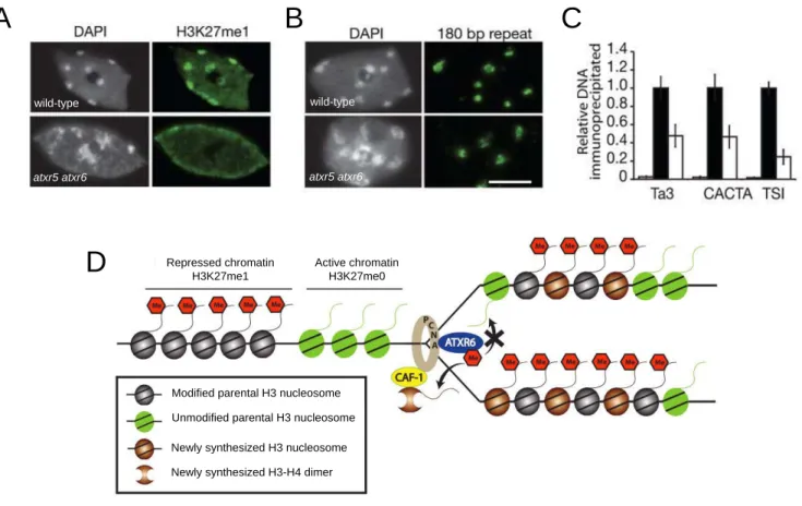
based on their enrichment in histone post-translational marks. 20 Figure 10: Arabidopsis atxr5 atxr6 mutants display defects in heterochromatin
organization and transcriptional silencing. 22
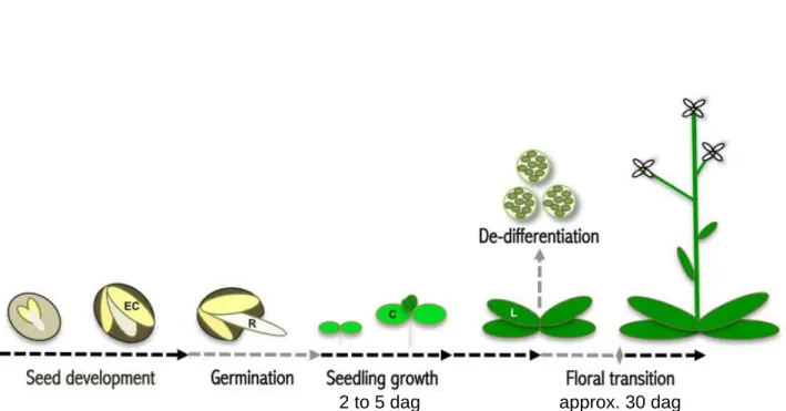
Figure 11: Overview of developmental phase transitions involving important
chromatin dynamics in Arabidopsis thaliana. 25
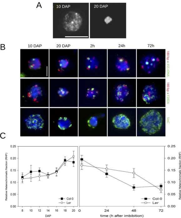
Figure 12: Heterochromatin dynamics in embryonic cotyledons during seed
development and germination. 27
Figure 13: Repressive chromatin marks localize to pre-chromocenters. 29 Figure 14: 5S rDNA chromatin organization is dynamic during early
post-germination development in cotyledons. 30
Figure 15: Prolonged high temperature stress results in heterochromatin
disorganization and release of transcriptional silencing. 31 Figure 16: Human canonical histone H3.1/H3.2 compared to replacement variant
H3.3. 33
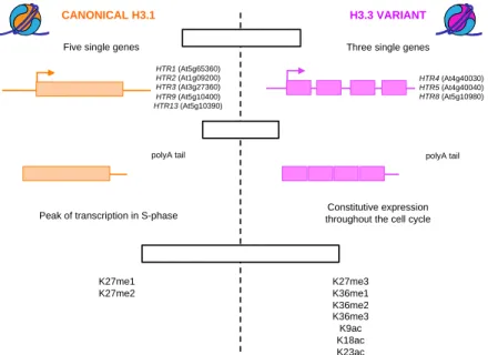
Figure 17: Summary of distinct features discriminating the canonical histone H3.1
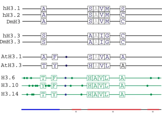
from the replacement variant H3.3 in Arabidopsis thaliana. 33 Figure 18: Schematic representation of amino acid sequence of canonical
and variant histone H3. 34
Figure 19: Arabidopsis histone H3.1 shows specific localization in the genome. 35 Figure 20: H3.1 is preferentially monomethylated at K27 by ATXR5 and ATXR6. 36 Figure 21: Arabidopsis histone H3.3 variant shows specific localization in the
genome. 39
Figure 22: Histone H3 variant dynamics at fertilization in Arabidopsis. 44 Figure 23: Histone chaperones are critical players in histone dynamics and
! ! ! ! ! ! ! ! ! ! ! ! ! ! ! ! ! ! ! ! ! ! ! ! ! ! ! ! !
Figure 25: Interaction between CAF-1 and the replication machinery tightly
links DNA replication and maintenance of chromatin states. 47 Figure 26: Effects of loss of CAF-1 on plant development and
maintenance of heterochromatin silencing. 49
Figure 27: CAF-1 is required for heterochromatin compaction. 50
Figure 28: Histone Regulator chaperone complex. 51
Figure 29: ASF1 is actively involved in maintenance of chromatin states
during DNA replication. 54
Figure 30: Schematic representation of the different histone H3-H4 assembly
networks as known in mammals. 57
Figure 31: Schematic representation of the different histone H3-H4 assembly
networks as known in Arabidopsis. 57
Figure 32: H3 chaperone-encoding genes are ubiquitously expressed. 61
Figure 33: Validation of H3 chaperone mutants. 62
Figure 34: Artificial miRNA efficiently interfere with AtHIRA, AtASF1A and
AtASF1B transcripts. 63
Figure 35: Loss of H3 chaperones does not lead to changes in transcription
of other chaperone genes. 63
Figure 36: Chromocenter formation is impaired in absence of CAF-1,
AtASF1A and AtASF1B while loss of HIR members has no effect. 64 Figure 37: Plants lacking CAF-1 display precocious switch to endoreplication
and increased polyploid nuclei content. 64
Figure 38: Transcriptional gene silencing at the multicopy GUS locus is maintained in all H3 chaperone mutants tested except fas1-4 mutants. 65 Figure 39: H3.1- and H3.3-encoding genes are ubiquitously expressed. 65 Figure 40: H3.1- and H3.3-encoding genes remain unaffected by loss of H3
chaperones, while H3.3-like genes are overexpressed in CAF-1 mutants. 66 Figure 41: Validation of T-DNA insertion mutants for H3.1- and H3.3-encoding genes. 66 Figure 42: Triple H3.3 mutants were obtained using a combination of T-DNA
insertions and artificial miRNA. 67
Figure 43: Transgenes encoding H3.1 and H3.3 proteins fused to a tag were
created to follow experimentally H3 dynamics. 67
Figure 44: e-H3.1 and e-H3.3 assembly into chromatin defines distinct genomic
regions. 68
Figure 45: Plants expressing mutated versions of e-H3.1. 70 Figure 46: Model for the interplay between CAF-1, SUVH, ATXR5 and ATXR6
! ! ! ! !
Figure 47: Expansion of cotyledons between 2 and 5 dag. 171
Table 1: H3-encoding genes in Arabidopsis. 44
Table 2: H3 chaperone-encoding genes characterized in Arabidopsis. 56
Table 3: Arabidopsis mutant lines used in this work. 69
Table 4: Primers used in this work. 172
Table 5: Plasmids used for cloning and expression of e-H3. 176
Table 6: PCR mix and program used for genotyping. 177
Table 7: Mix and program used for RT-PCR. 178
Table 8: Mix used for RT-qPCR, ChIP-qPCR and qPCR program. 179
Appendix 1: Restriction map of the pBIN-Hyg-TX plasmid. 214
Appendix 2: Restriction map of the pRS300 plasmid. 214
Appendix 3: Restriction map of the pDONR221 plasmid. 215
Appendix 4: Restriction map of the pMDC32 plasmid. 215
Appendix 5: Restriction map of the multi cloning site of pTP1. 216 Appendix 6: Restriction map of the multi cloning site of pTP9. 216 Appendix 7: Restrictition map of the pOZ-FH-C plasmid and cloning sites of
pOZ-FH-C and pOZ-FH-N. 217
Appendix 8: Restriction map of the pUB plasmid. 218
Appendix 9: Heterochromatin dynamics during developmental transitions in
! ! ! ! ! ! ! ! ! ! ! ! ! ! ! ! ! ! ! ! ! ! ! ! ! ! ! ! !
Figure 1: Epigenetic information is chromatin-based.
In the eukaryotic nucleus, DNA is organized together with histones forming the basic subunit of chromatin, the nucleosome, in which 146 bp of DNA wrap around a histone tetramere of H3-H4 and two H2A-H2B dimers. Candidates for key players in epigenetic inheritance are situated at different levels of chromatin and include DNA and histone modifications, histone variants, non-histone chromatin proteins that bind directly to DNA or histone modifications, and higher-order organization as well as spatial organization of a given locus within the nucleus. Histones can be post-translationally modified e.g. by acetylation, phosphorylation, methylation and each mark constitutes a signal red alone or in combination with other modifications on the same or neighbouring histones as the ‘histone code’. The presence of histone variants, in particular H3 and H2A variants adds further complexity to the epigenetic information (adapted from Probst et al., 2009).
Heterochromatin Euchromatin Histone modifications DNA methylation Histone variant DNA Nucleosome Chromatin fiber Nucleus Chromatin reader
1. Organization of eukaryotic genomes in chromatin
Individual cells of an organism comprise identical DNA content, but development and differentiation require specific gene expression patterns regulated in a time- and tissue-specific manner. How an organism controls the transcription of a given locus during development is therefore a key question. Besides specific sets of transcription factors and signaling cascades responding to environmental cues, epigenetic information, defined as mitotically heritable changes in gene expression that occur without alterations in DNA sequence (Riggs et al., 1996), is integrated at the chromatin level and is of critical importance regarding these questions.
Indeed, in eukaryotic organisms the genetic information encoded by the bulky linear DNA is tightly organized in a nucleoprotein structure called chromatin. Chromatin packages DNA to fit the small compartment of the cell nucleus, but also regulates DNA accessibility. The nucleosome is the basic subunit of chromatin and consists in DNA wrapped around histone proteins (Luger et al., 1997). Accessibility of the transcriptional machinery to DNA is determined by the levels of packaging based on electrostatic interactions between DNA and histones. Nucleosome features therefore directly impact accessibility of the nuclear machinery to the underlying DNA sequence, thus affecting fundamental DNA-based processes such as gene transcription, DNA replication and repair (Probst et al., 2009). Following this idea, chromatin appears as a potential framework for gene expression program maintenance and inheritance. Epigenetic information is essentially chromatin-based and includes covalent modifications at the level of DNA, histones, non-histone chromatin binding proteins, non-coding RNA and histone variants (Probst et al., 2009). Moreover, local properties of chromatin also have consequences on the higher-order nuclear organization and thus can influence the positioning of genes and entire chromosomes within the nucleus (Lieberman-Aiden et al., 2009; Saez-Vasquez and Gadal, 2010) (Figure 1). Therefore, epigenetic information carried by chromatin provides a form of memory critical for the maintenance of genome function, including both developmental gene expression patterns and the propagation of essential architectural features of the genome, such as chromosome positioning and definition of telomeres and centromeres (Probst et al., 2009).
DNA 10-nm fiber 30-nm fiber? Nucleus 2 nm 10 nm 30 nm Mitotic chromosome 700 nm 10 µm
Irregular folding of the 10-nm fiber? A
A
Figure 2: Different levels of chromatin compaction.
A. The 2 nm-long DNA molecule is wrapped around a core histone octamer and forms a 10-nm ‘‘beads-on-a-string’’ fiber. The 10-nm fiber has long been assumed to fold into a 30-nm fiber that could be organized in a one-start helical solenoid model or in a two-start helix conformation (not shown). Recent data argue instead for the presence in vivo of an irregularly folded 10-nm fiber. These intermediate chromatin structures subsequently participate in the higher-order chromatin organization of interphase nuclei or mitotic chromosomes (adapted from Maeshima et al., 2010 and Volle and Dalal., 2014).
B. Higher levels of chromatin folding exemplified by the distinct chromosomal territories occupied by human chromosomes 18 (green) and 19 (red) in the nuclear volume during interphase. Chromosomes 18 and 19 are revealed by fluorescent in situ hybridization in human primary lymphocytes (from Croft et al., 1999).
C. The nucleosome core particle consists of 146 bp of DNA wrapped in a ¾ superhelical turn around a histone octamer. The histone octamer consists of two molecules each of histone H2A, H2B, H3 and H4 with their N-terminal tails protruding from the globular domains (from Morales et al., 2001). First a H3-H4 dimer, then a tetramer is formed that associates with DNA to form a tetrameric particle and two H2A-H2B dimers complete the nucleosome core particle.
C
B
1.1.
Functional significance of chromatin
We distinguish four major levels of chromatin organization (Figure 2A): (i) the nucleosome, where DNA is tightly wrapped around the histone core; (ii) the 10-nm beads-on-a-string nucleosomal array (iii) the 30-nm fiber, with an estimated linear compaction of 100-200 kb/µm; (iv) higher folding levels, which position the chromatin fiber within a chromosome territory and allow the elevated compaction of metaphase chromosomes (Figure 2B). The general picture of chromatin is a polymer of nucleosomes, each of which is formed by an octamer of the core histones H2A, H2B, H3 and H4, around which a 146-bp DNA helix is wrapped 1.7 times in a left-handed superhelical fashion (Luger et al., 1997) (Figure 2C). The core histones are structurally defined by two distinct conserved motifs: the histone fold domain and the unstructured N-terminal and C-terminal histone tails that protrude from the globular part. The histone fold domain participates in interactions between core histones through three α-helices (α1, α2, and α3) connected by short loops (Arents and Moudrianakis, 1995). Histones are highly basic proteins, since their amino acid sequence is lysine- and argenine-rich, which tightly interact with the acidic phosphate groups of the DNA forming fourteen non-covalent histone-DNA contact points within the nucleosome (Luger et al., 1997). The nucleosome core assembly is achieved by the initial heterodimerization of H3/H4 followed by dimerization to form a (H3–H4)2 tetramer (Eickbush and Moudrianakis, 1978), initiating first contacts with DNA (Luger, 2001). Dimers of H2A/H2B then bind to the (H3–H4)2 tetramer creating new histone/DNA contact points and facilitate DNA wrapping around the histone octamer to form the nucleosome core complex (Hayes et al., 1990, 1991; Luger, 2001).
The beads-on-a-string model consists of a linear array of nucleosomes stabilized by the binding of linker histone H1 to linker DNA (Thomas, 1999; Maeshima et al., 2014) (Figure 2A). The 20-60 bp of linker DNA between nucleosomes is critical for the spatial orientation of the nucleosomal array and fixes the distance between adjacent nucleosomes (Grigoryev et al., 2009; Arya et al., 2010). This tightly controlled architecture establishes the 10-nm beads-on-a-string structure, which is a basis for further compaction of the chromatin fiber (Horn and Peterson, 2002; Mohd-Sarip and Verrijzer, 2004; Yelagandula et al., 2014). Existence of chromatin in form of a 30-nm fiber remains controversial due to
A
B
C
Figure 3: Chromatin arrangement in interphase nuclei.
A. Chromosome painting on a chicken fibroblast nucleus reveals chromosomes organized within exclusive chromosome territories (from Cremer and Cremer, 2001).
B. Interphase chromosomes of wheat meristematic root nuclei display Rabl configuration. Telomeres (in red) and centromeres (in green) are found at opposite poles of the interphase nucleus. Scale bars: 10 µm (from Santos and Shaw, 2004).
C. Chromosomes organize in distinct territories in Arabidopsis thaliana. DAPI-stained 4C leaf nucleus (left) and multi-color chromosome painting of individual Arabidopsis chromosome arms (right). Nucleoli (nu) and repetitive DNA sequences are unstained. Scale bar: 5 µm (from Pecinka et al., 2004).
A
B
structural complexity (Robinson and Rhodes, 2006; Maeshima et al., 2010; Joti et al., 2012). The 30-nm fiber is currently hypothesized following two models (Wu et al., 2007; Fransz and de Jong, 2011). The one-start helix (solenoid model) displays a linear arrangement and interactions between neighboring nucleosomes, while the two-start helix (zigzag model) exhibits repetitive units of nucleosomes that alternate into an irregular 3D zigzag architecture. However, some recent evidences suggest that in vivo, the 30-nm chromatin fiber could be preferentially replaced by irregularly folded 10-nm fibers participating in further organization of particular high-order chromatin domains within the nuclear volume (Maeshima et al., 2014; Volle and Dalal, 2014; Yelagandula et al., 2014).
Higher levels of folding locate the chromatin fiber within the 3D volume of the nucleus. The spatial organization of the chromosomes is not random and is mediated by many factors such as chromosome length, DNA sequence, changing gene activity during cell growth and differentiation, local or global chromatin context, nuclear volume or external stimuli. Within the nucleus, each chromosome preferentially occupies a proper subdomain called chromosome territory (Figure 3). Interestingly, gene-rich chromosomes are preferentially found in a central position (Croft et al., 1999; Kozubek et al., 2002; Chambeyron and Bickmore, 2004; Scheuermann et al., 2004), while gene-poor chromosomes tend to localize at the nuclear periphery, which is thought to form a transcriptionally repressive microenvironment (Croft et al., 1999; Boyle et al., 2001; Cremer and Cremer, 2001, 2010). However this organization is not universal since particularly highly-expressed genes have been described close to the nuclear periphery (Brown et al., 2006; Küpper et al., 2007). In mammals and plants, transcription leads to chromatin decondensation and looping out from the chromosome territory. Whether the distribution of chromosome territories is conserved through mitosis remains controversial (Gerlich et al., 2003; Kalmárová et al., 2008; Cremer and Cremer, 2010; Ishii and Houben, 2014). In Arabidopsis, chromosome territories location within the nucleus can be transmitted in a transient mirror-symmetrical pattern through mitosis (Berr and Schubert, 2007). Inside each chromosome territory, the centromere and the telomeres are arranged in a specific manner. Centromeric chromatin is mostly located at the periphery, near the nuclear membrane, whereas disposition of telomeric regions varies between species. In human cells, centromeres are partly localized to the nuclear periphery depending the cell cycle phase (Solovei et al., 2004). Plants with large genomes such as wheat and
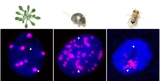
Figure 4: Euchromatin and heterochromatin are two distinct chromatin states in eukaryotes.
Two distinct forms of chromatin exist in eukaryotic genomes: on the one hand euchromatin, stained here in blue by DAPI in nuclei from Arabidopsis leaf tissue, mouse fibroblasts and Drosophila salivary glands, is relatively decondensed and includes chromatin domains with transcription-permissive features. On the other hand, heterochromatin remains condensed throughout the whole cell cycle and forms at repetitive sequences and transposable elements – here visible in pink by DNA fluorescence in situ hybridization of repetitive sequences (adapted from Probst et al., 2003. Pictures of mouse and Drosophila courtesy of A. Probst and E. Brasset).
Euchromatin
close relatives, as well as the budding yeast Saccharomyces cerevisiae exhibit a Rabl organization, where centromeres cluster at the spindle pole body, facing telomeres found in perinuclear foci on the opposite side of the nucleus. In Arabidopsis thaliana, the centromeres are located at the nuclear periphery (Fang and Spector, 2005), while telomeres cluster at the periphery of the nucleolus (Fransz et al., 2002; Probst et al., 2003).
1.2.
Heterochromatin is a specialized chromatin subdomain
1.2.1.
Structure of heterochromatin
Local modifications of the nucleosome structure and chromatin fiber organization translate in the formation of two major chromatin states in eukaryotes, visible in cytological preparations after coloration by DNA stains (Figure 4). The less compact euchromatin includes mainly genes and promotes a transcriptionally permissive environment. Densely packaged heterochromatin instead forms mostly at repetitive sequences and transposable elements, rendering them largely inaccessible to RNA polymerases by adopting a “closed” chromatin conformation. Heterochromatin thereby contributes to the control of potentially deleterious DNA elements and organizes repetitive DNA sequences at both centromeres and telomeres. Historically, heterochromatin has been cytologically defined in moss as chromosomal regions or chromosomes that remain intensely stained during the whole cell cycle, in contrast to the lightly stained euchromatin that decondenses at interphase (Heitz, 1928). This distinction was initially established by light microscopy using chromatin-staining dyes such as acetocarmine and then validated by other cytological techniques, such as Fluorescent In Situ Hybridization (FISH). The definition of heterochromatin has been enriched afterwards by molecular, biochemical and structural features together with the finding that heterochromatin contains abundant tandem-repeated DNA, wrapped around densely arranged nucleosomes relatively insensitive to DNase I (Chodavarapu et al., 2010; Shu et al., 2012, 2013), enriched in heavily methylated DNA and histones H3 methylated at lysine in position 9 (H3K9) and 27 (H3K27), thus promoting transcriptional repression, and subjected to late-replication (Karpen and Allshire, 1997; Hennig,
! ! ! ! ! ! ! ! ! ! ! ! ! ! ! ! ! ! ! ! ! ! ! ! ! ! ! ! !
1999; Henikoff, 2000; Chodavarapu et al., 2010; Almouzni and Probst, 2011; Shu et al., 2012). In most eukaryotic species, centromeric, pericentromeric and telomeric regions contain a high density of repetitive DNA sequences such as clusters of satellite sequences and transposons, and are principal candidates for heterochromatin formation (Martens et al., 2005; Schueler and Sullivan, 2006; Blasco, 2007; Slotkin and Martienssen, 2007; Schoeftner and Blasco, 2009). Centromeric and pericentromeric heterochromatin are required for correct chromosome segregation (Peters et al., 2001; Lippman and Martienssen, 2004; Dunleavy et al., 2005; Kanellopoulou et al., 2005; Pidoux and Allshire, 2005; Folco et al., 2008), and intra- and inter-chromosomal association of heterochromatin domains has been shown to be an important factor in the higher-order organization of chromosomes since it promotes the formation of specialized heterochromatin domains such as chromocenters in mouse (Guenatri et al., 2004) and Arabidopsis (Fransz et al., 2002). Chromocenters are conspicuous heterochromatin clusters containing most of the repetitive DNA content of the genome (Fransz et al., 2002; Guenatri et al., 2004).
In mouse Mus musculus domesticus, pericentromeres and centromeres consist of two types of repetitive DNA sequences spanning around 3.5% of the genome: the AT-rich major satellite repeats (6 Mb of 234 bp units) and the minor satellite repeats (600 kb of 123 bp units) respectively, localizing to the tip of the acrocentric chromosomes (Vissel and Choo, 1989; Choo, 1997; Lehnertz et al., 2003). In situ hybridization on metaphase chromosomes has shown that minor satellite sequences constitute the centromeric part, while major satellite repeats locate pericentromerically (Wong and Rattner, 1988; Joseph et al., 1989; Kuznetsova et al., 2006). Pericentromeres cluster to form chromocenters in interphase (Hsu et al., 1971; Guenatri et al., 2004). Chromocenter organization is found in most of the somatic mouse cells, but the levels of clustering and the spatial location of the chromocenters in the nucleus differ between cell types (Guenatri et al., 2004; Terranova et al., 2005; Solovei et al., 2009). At the molecular level, chromocenters are characterized by repressive epigenetic marks including DNA methylation, repressive post-translational modifications and strong enrichment in Heterochromatin Protein 1 (HP1) (Jeppesen et al., 1992; Peters et al., 2001; Lehnertz et al., 2003). The centromeric domain promotes kinetochore formation and the pericentromeric domain has been proposed to play a role in sister chromatid cohesion and proper
B
A
cc no noC
Figure 5: Heterochromatin is organized in chromocenters in Arabidopsis thaliana.
A. Schematic representation of the five Arabidopsis chromosomes (2n = 10) in the Columbia accession. Chromosome II and IV carry the 45S rDNA loci (Nucleolus Organizer Regions, NOR, blue). The 5S rDNA loci (red) are present on chromosomes III, IV and V, in close proximity to centromeric repeats (180 bp repeats, gray) and inside the pericentromeric domains (green) (from Benoit et al., 2013).
B. Left: Spread of an Arabidopsis leaf mesophyll nucleus stained with DAPI. Note the euchromatin in light gray and the nine brightly stained chromocenters (cc). The nucleolus (no) appears as a DAPI-unstained region. Scale bar: 5 µm. Right: Model of an Arabidopsis chromocenter of chromosome IV adjacent to the nucleolus (no). The chromocenter of chromosome IV comprises the 45S (blue), 5S (red), centromeric (light gray) and pericentromeric (green) repeats from which euchromatic loops (white) emanate (adapted from Fransz et al., 2002). Parts of the ribosomal DNA repeats which are actively transcribed are represented as 45S rDNA sequences (blue) that loop out from the chromocenter into the nucleolus (Probst et al., 2004) and 5S rDNA (red) loops within the euchromatin compartment (Mathieu et al., 2003) respectively.
C. Clustering of repetitive sequences in chromocenters revealed by FISH. Different repetitive sequences (5S, 45S, 180 bp, and Transcriptionally Silent Information (TSI) repeats) are revealed by DNA FISH (adapted from Douet et al., 2008 and Probst et al., 2003).
A
B
chromosome segregation (Guenatri et al., 2004). Indeed, mice lacking both the methylation at H3 lysine 9 (H3K9me) and HP1 at major satellites show chromosome missegregation (Peters et al., 2001; Lippman and Martienssen, 2004; Dunleavy et al., 2005; Kanellopoulou et al., 2005). The centromere region of the fruit fly Drosophila melanogaster displays a tripartite organization (Miklos and Cotsell, 1990), with the core domain consisting in gene-poor highly repetitive DNA and the flanking segments, observable only in polytene chromosomes, forms at middle repetitive DNA, interspersed with domains containing high density of genes (Eberl et al., 1993; Lohe et al., 1993). These heterochromatin domains are enriched in H3K9me2/3 and HP1 (Roy et al., 2010; Kharchenko et al., 2011; Riddle et al., 2011).
In Arabidopsis, heterochromatin is essentially composed of repetitive sequences and transposable elements, including the centromeric 180 bp repeats and interspersed pericentromeric repeats, as well as silenced 45S and 5S rRNA genes (Kumekawa et al., 2000; Nagaki et al., 2003; Benoit et al., 2013) (Figure 5A). The centromeres of Arabidopsis thaliana have been genetically mapped (Copenhaver et al., 1999; Heslop-Harrison et al., 1999). The centromere core is depleted from genes while silent and low-density genes (<1 per 100 kb compared to euchromatin with 1 gene per 5 kb) are found in pericentromeric regions (Kumekawa et al., 2000; The Arabidopsis Genome Initiative, 2000; Haupt et al., 2001; Hosouchi et al., 2002; Hall et al., 2003), together with various types of repetitive DNA elements, including transposons, retrotransposons and telomere-like repeats, were identified in the pericentromeric region (Richards, 1991; The Arabidopsis Genome Initiative, 2000). The organization of the Arabidopsis centromeric region translates in two domains: the centromere core, containing the functional centromere/kinetochore complex and consisting mainly in 180 bp repeats, and the flanking pericentromeric domains. Each Arabidopsis centromere core contains several megabases (near 20,000 copies spanning about 1.3 to 2.1 Mb) (Haupt et al., 2001) of the 180 bp repeat (Copenhaver et al., 1999; Kumekawa et al., 2000; Hosouchi et al., 2002; Hall et al., 2003), organized into long tandem arrays (The Arabidopsis Genome Initiative, 2000). In addition, a 398 bp fragment of the Athila2 Long Terminal Repeat (LTR) called 106B was discovered as associated with the 180 bp centromeric repeats (Thompson et al., 1996; Slotkin, 2010). The 106B repeat is interspersed as single copies within long stretches of 180 bp repeats. The centromere core region is flanked by pericentromeric heterochromatin domains that contain many transposons,
! ! ! ! ! ! ! ! ! ! ! ! ! ! ! ! ! ! ! ! ! ! ! ! ! ! ! ! !
retrotransposons and telomere-like repeats (Richards, 1991; The Arabidopsis Genome Initiative, 2000). The Athila family of LTR retrotransposons (approximately 500 copies) (Pélissier et al., 1996) and TSI (Transcriptionally Silent Information) repeats constitute most of the genomic content of pericentromeric heterochromatin regions of Arabidopsis chromosomes. TSI repeats exhibit sequence homology with the 3’ terminal part of the Athila retrotransposon (Steimer et al., 2000; Slotkin, 2010). Contrary to Drosophila, chromosome-specific repeats have not yet been identified in Arabidopsis except for the ribosomal gene clusters (Cloix et al., 2000). The genome of Arabidopsis thaliana encodes approximately 1000 copies of 5S rRNA genes, which are arranged as tandem arrays in several loci located in the pericentromeric heterochromatin of chromosomes 3, 4 and 5 in the Columbia accession (Campell et al., 1992; Layat et al., 2012b; Benoit et al., 2013) (Figure 5A). 5S rRNA gene transcription has been found only at the array located on chromosome 4 and at the large locus on chromosome 5, while 5S rRNA genes from chromosome 3 and from the small locus on chromosome 5 are transcriptionally silent (Cloix et al., 2002, 2003; Layat et al., 2012b). Interestingly, at active 5S rDNA loci only the most distal copies are expressed while units close from to centromere remain largely silenced and display increasing levels of mutations (The Arabidopsis Genome Initiative, 2000; Cloix et al., 2002; Vaillant et al., 2008). Upon developmental cues, some of the low-mutated silent units can revert and become transcribed to ensure appropriate amount of 5S rRNA. In Arabidopsis, the 45S rRNA genes are located at the tips of the short arms of chromosomes 2 and 4, in domains called Nucleolar Organizer Regions (NOR). The 570 to 750 copies of 45S rRNA genes are organized in tandem arrays (Copenhaver et al., 1995; Copenhaver and Pikaard, 1996; Layat et al., 2012b). Similar to the 5S rRNA genes, only a fraction of the 45S rRNA gene units are transcribed but the extent of this fraction can be dynamically modified during development (Pontvianne et al., 2010).
Centromeric and pericentromeric heterochromatin clusters with silenced 45S and 5S rDNA arrays of the same chromosome in a chromocenter (Fransz et al., 2002; Fransz and de Jong, 2011) (Figure 5B). In 4′,6′-diamidino-2-phenylindole (DAPI)-stained interphase nuclei, these chromocenters appear as 6-10 intensely stained chromatin foci (Fransz et al., 2002; Dittmer et al., 2007). Immunolocalization studies confirmed the enrichment in repressive chromatin marks, such as high levels of DNA methylation, H3K9me2 and H3K27me1 (Soppe et al., 2002; Probst et al.,
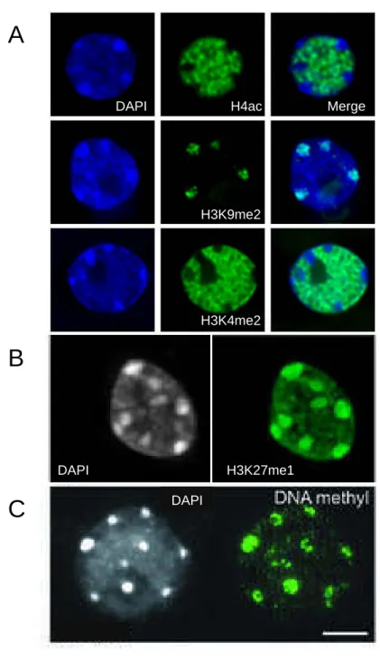
Figure 6: Chromocenters are enriched in repressive chromatin marks.
A. Immunolocalization of H4ac (top), H3K9me2 (middle) and H3K4me2 (bottom) in interphase nuclei isolated from young rosette leaves counterstained with DAPI. The repressive H3K9me2 mark colocalizes with heterochromatic chromocenters, while active marks H4ac and H3K4me2 are excluded (from Probst et al., 2003).
B. Immunodetection of H3K27me1 in interphase nuclei from young rosette leaves counterstained with DAPI. As a repressive histone post-translational modification, the H3K27me1 immunosignal is found colocalizing with chromocenters but is also present in euchromatin (from Mathieu et al., 2005).
C. Immunolocalization of DNA methylation with a 5mC specific antibody on interphase nuclear spreads from young rosette leaves counterstained with DAPI. The DNA methylation immunosignal concentrates in chromocenters. Scale bar: 5 µm (from Soppe et al., 2002).
B
A
C
H4ac H3K9me2 H3K4me2 DAPI Merge DAPI H3K27me1 DAPI2003; Mathieu et al., 2005) (Figure 6). The model emitted by Fransz and collaborators, based on FISH analysis in interphase nuclei, suggests that gene-rich euchromatin loops visualized using gene-rich Bacterial Artificial Chromosomes (BAC) as probes emanate from the chromocenters, thus defining chromosome territories (Fransz et al., 2002; Pecinka et al., 2004) (Figure 5B). Such an organization is important for proper compartmentalization and transcriptional silencing of repetitive DNA, meanwhile allowing efficient transcription of genes located in the euchromatic loops. Cytological observations were further confirmed by testing intra- and inter-chromosomal interactions using a 4C assay, revealing that heterochromatic repetitive sequences, including the ones dispersed along the chromosome arms, interact preferentially with each other thus generating specific and well-defined chromatin landscapes (Grob et al., 2013).
1.2.2.
Heterochromatin is actively defined by epigenetic
mechanisms
A defining feature of heterochromatin is that its transcriptionally repressed state and highly condensed structure self-perpetuates during the cell cycle in a region-specific manner (Probst et al., 2009). During S phase, epigenetic marks are diluted as a consequence of DNA replication. Therefore, sophisticated mechanisms that exploit the mutual reinforcement of DNA and histone modifications are required to ensure inheritance of chromatin marks and maintenance of heterochromatin in its specific higher-order structure (Probst et al., 2009).
1.2.2.1. DNA methylation
!
Cytosine residues are methylated in a wide diversity of organisms, including plants, mammals and Neurospora, while very low DNA methylation levels are present in yeast, Drosophila or Caenorhabditis elegans (Simpson et al., 1986; Lyko et al., 1999; Tang et al., 2012). DNA methylation has been shown since long to be critical for development in plants and mammals, involved in transcriptional gene silencing, imprinting and X-chromosome inactivation. Methylation acts in many ways and is
B A
Figure 7: Model of CG and CHG methylation maintenance.
A. Model of CG methylation maintenance during DNA replication. DNMT1 is targeted to the replication fork via interaction with UHRF1, specifically binding hemimethylated DNA, and PCNA. Following recruitment, DNMT1 fully reestablishes methylation patterns. In Arabidopsis thaliana, DNMT1 and UHRF1 orthologs, respectively MET1 and VIM, are involved in a similar manner in CG methylation maintenance (from Law and Jacobsen, 2010).
B. Model of CHG methylation maintenance in plants by a self-reinforcing loop between DNA and histone methylation. The DNA methyltransferase CMT3 establishes CHG methylation, which is recognized by SUVH4 histone methyltransferase. SUVH4 in turn dimethylates H3K9. This modification is then recognized by the chromodomain of CMT3 allowing maintenance of CHG methylation (from Law and Jacobsen, 2010).
susceptible to block binding of transcription factors, thus preventing transcription, or it can interact with chromatin modifiers that modify the neighbouring histones and enhancing transcriptional silencing. DNA methylation can occur in three different nucleotide sequence contexts: CG, CHG and CHH (H = C, T or A). CG methylation is observed widely in plants and mammals, while CHG as well as CHH are preferentially found in Arabidopsis. Cytosine methylation (5mC) is predominantly present at highly repetitive DNA, including centromeric and pericentromeric repeats, rDNA arrays and transposable elements (Figure 6C), where it often coexists with H3K9me2 and H3K27me1 (Figure 6AB), but also in the body of 30% of genes, many of which are characterized by moderate expression levels (Zhang et al., 2006; Zilberman et al., 2007; Vaughn et al., 2007; Bernatavichute et al., 2008; Cokus et al., 2008; Lister et al., 2008). Interestingly, while gene bodies contain mainly CG methylation (Cokus et al., 2008), CHG methylation is linked to centromeric and pericentromeric regions enriched in H3K9me2 (Bernatavichute et al., 2008), while CHH is a hallmark of long transposable elements (Zemach et al., 2013; Cavrak et al., 2014). CHH methylation establishment and maintenance rely on the production of small interfering RNA (siRNA) and the RNA-directed DNA methylation (RdDM) pathway.
1.2.2.1.1. Maintenance
!
In both mammals and plants, maintenance of CG methylation is achieved by a specific DNA methyltransferase associated to a co-factor that senses hemi-methylated DNA at the replication fork and by interaction with PCNA (Proliferating Cell Nuclear Antigen). In Arabidopsis, this is achieved by MET1 (DNA METHYLTRANSFERASE 1) and VIM1 (VARIATION IN METHYLATION 1) activity and in mammals, by DNMT1 (DNA cytosine-5-Methyltransferase 1) and UHRF1 (Ubiquitin-like containing PHD and RING Finger domains 1) (Law and Jacobsen, 2010) (Figure 7A). Moreover, the de novo DNA methyltransferases DNMT3A and DNMT3B are also involved in the maintenance of CG methylation patterns at some loci (Chen et al., 2003). Mutant alleles of MET1 display an important decrease in CG methylation levels, but also CHG and CHH, at centromeric repeats (Saze et al., 2003). Concomitantly, H3K9me2 is redistributed from the chromocenters, suggesting
Figure 8: RNA-directed DNA methylation pathway.
RNA-directed DNA Methylation (RdDM) necessitates the synthesis of single-stranded RNA (ssRNA), produced from the repetitive and transposon-rich DNA domains to be silenced by the plant-specific DNA-dependent RNA polymerase Pol IV. These transcripts are then processed by RDR2 for synthesis of double-strand RNA (dsRNA). After fragmentation in 24-nt long sequences by DCL3 the siRNA are loaded in AGO4 for transport to the sequence of origin where they are thought to recognize scaffold RNA originating from Pol V activity. Interaction between 24-nt siRNA and lncRNA is the basis for further recruitment of DRM2, the main DNA methyltransferase involved in RdDM, for methylation of the DNA domain (figure courtesy of M. Thomas).
RDR2 DCL3 Pol IV dsRNA AGO4 DRM2 ssRNA Pol V NRPE1 SPT5L AGO4 lncRNA
a fundamental role of CG methylation for H3K9me2 setting at chromocenters (Tariq et al., 2003; Mathieu et al., 2005).
DNA methylation in the CHG context is found widely in Arabidopsis and other plant genomes. Histone methylation by the KYP (KRYPTONITE) / SUVH4 (SUPPRESSOR OF VARIEGATION 3-9 HOMOLOGUE 4) methyltransferase together with the plant-specific DNA methyltransferase CMT3 (CHROMOMETHYLASE 3) sustain a feed-forward loop that maintains CHG methylation (Lindroth et al., 2001; Jackson et al., 2002; Johnson et al., 2007; Feng et al., 2010) (Figure 7B). Depletion of CMT3 leads to genome-wide decrease in CHG context methylation (Lindroth et al., 2004). CMT3 recognizes H3K9me2, which colocalizes with H3K27me1/2 at CMT3-controlled loci, suggesting that CMT3 recruitment relies on the combination of both marks (Lindroth et al., 2004; Du et al., 2012; Law et al., 2013).
CHH methylation is driven by two distinct pathways: one is the RdDM pathway that involves siRNA originating from repeats and transposon-rich heterochromatin domains and participates in the control of transcriptional gene silencing by targeting DNA methylation to the sequences to be suppressed (Lippman et al., 2004; Lippman and Martienssen, 2004) (Figure 8). 24-nt-long siRNA are produced from heterochromatic repeats and transposable elements. The siRNA synthesis is achieved by the plant-specific DNA-dependent RNA polymerase Pol IV, which produces single-stranded RNA from repetitive regions (Herr et al., 2005; Onodera et al., 2005; Sidorenko et al., 2009; Havecker et al., 2010). Pol IV transcripts are then converted into double-strand RNA by RNA-Dependent RNA polymerase 2 (RDR2) and subsequently cut in 24-nt-long sequences by DICER-LIKE3 (DCL3) (Xie et al., 2004). These 24-nt siRNA are then loaded in AGO4 (Argonaute 4) complexes which target the siRNA to the locus of origin by sequence homology (Chan et al., 2004; Qi et al., 2006; Ye et al., 2012) where they hybridize to genomic DNA or scaffold RNA (Wierzbicki et al., 2009). Scaffold RNA molecules are long non-coding RNA (lncRNA) produced independently from siRNA by Pol V (Wierzbicki et al., 2008, 2012). Complementarity between 24-nt siRNA and scaffold RNA is thought to help the recruitment of DNA methyltransferases, notably DRM2 (DOMAINS REARRANGED METHYLTRANSFERASE 2 – homolog of DNMT3) (Naumann et al., 2011), but the precise mechanism remains poorly characterized. siRNA-mediated heterochromatin
! ! ! ! ! ! ! ! ! ! ! ! ! ! ! ! ! ! ! ! ! ! ! ! ! ! ! ! !
formation is found in many other eukaryotes, including fission yeast, Drosophila and mammals (Hall et al., 2002; Volpe et al., 2002; Verdel et al., 2004).
The second pathway involves CHROMOMETHYLASE 2 (CMT2), methyltransferase maintaining both CHG and CHH. Indeed, drm1 drm2 cmt2 triple mutants loose all methylation in the CHH context (Stroud et al., 2014). Interestingly, DRM2 activity is thought to be promoted through the recruitment of Pol IV by the binding of methylated histone binding protein SHH1 to H3K9me1, H3K9me2 and H3K9me3, as observed for CMT3. While CMT3 exhibits no preference in binding affinity towards the number of methyl groups at H3K9, CMT2 interacts preferentially with H3K9me2 (Stroud et al., 2014). Together, these data suggest that DRM2, CMT2 and CMT3 control non-CG methylation in plants, and establish a self-reinforcing loop together with H3K9 methylation. Both CHG and CHH methylation also exist at low levels in mammalian embryonic stem cells where they are probably mediated by DNMT3A and DNMT3B (Ramsahoye et al., 2000; Chen et al., 2003; Lister et al., 2009).
The combination of genome-wide DNA methylation patterns with global nucleosome positioning maps revealed that DNA methylation deposition is influenced by nucleosome positioning in Arabidopsis (Chodavarapu et al., 2010). Consistently, DNA methyltransferases preferentially target nucleosome-bound DNA. The same observation has been described for human nucleosomal DNA, suggesting that DNA methylation and nucleosomal occupancy are tightly linked (Chodavarapu et al., 2010).
1.2.2.1.2. Demethylation
!
Removal of methylated cytosines occurs during epigenome reprogramming. In plants, four bifunctional helix-hairpin-helix DNA glycosylases and AP lysases are found. DME (DEMETER), DML2 (DEMETER-LIKE 2), DML3 (DEMETER-LIKE 3) and ROS1 (REPRESSOR OF SILENCING 1) excise methylated cytosines. The remaining 1-nt gap is then filled by a new unmethylated cytosine by a still unknown DNA ligase (Zhu, 2009). DML2, DML3 and ROS1 activities are mainly found in vegetative tissues and target hundreds of genes throughout the genome (Penterman et al., 2007; Yu et al., 2013). In the mammalian embryo, de novo DNA methylation of
Figure 9: At least four different chromatin states can be defined in Arabidopsis based on their
enrichment in histone post-translational marks.
A. The genome-wide distribution of the twelve chromatin marks analyzed defines four main chromatin states based upon heat map values ranging from 25% (light purple) to 100% (dark purple) (from Roudier et al., 2011).
B. The four chromatin states defined display different relative proportion of genomic features. While active marks-associated chromatin state 1 (CS1) and CS2 contain mostly genes, CS3 covers most of the transposable elements, which is coherent with its strong bias towards repressive mark enrichment. CS4 shows no particular enrichment in any of the studied chromatin marks and contains mainly genomic regions outside of genes and transposable elements (modified from Roudier et al., 2011).
A
the inner cell mass following massive demethylation is critical to drive the identity and stability of embryonic lineages, whereas the placenta remains mainly unmethylated (Law and Jacobsen, 2010; Seisenberger et al., 2013). In plants, genome-wide demethylation occurs during plant reproduction in the companion cells of the sperm and egg cells (Ibarra et al., 2012) and then during post-fertilization seed development (Gehring et al., 2009; Hsieh et al., 2009). At this stage the nutrient tissue, the endosperm, undergoes genome-wide demethylation in contrast to the embryo (Hsieh et al., 2009). This phenomenon is of particular importance for early embryogenesis by the establishment of maternal imprinted gene expression patterns (Pillot et al., 2010; Autran et al., 2011; Raissig et al., 2011). As a conclusion, DNA methylation reprogramming is important to establish epigenetic patterns of embryonic and extra-embryonic lineages.
1.2.2.2. Histone post-translational marks associated with heterochromatin
!
Histone tail domains are preferential targets for many and diverse post-translational modifications such as acetylation, methylation, and phosphorylation (Kouzarides, 2007). Such modifications can influence the biochemical properties of the nucleosome as well as its stability and interactions with remodeling factors (Zheng and Hayes, 2003), thus participating in the regulation of genome activity (Davie and Chadee, 1998) and higher-order chromatin architecture (Dorigo et al., 2003). Genome-wide studies argue for the role of epigenetic marks such as DNA methylation, histone post-translational modifications and chromatin binding proteins in establishing functional chromatin landscapes and defining euchromatin and heterochromatin (Filion et al., 2010; Roudier et al., 2011; Sequeira-Mendes et al., 2014) (Figure 9). Consequently, while transcribed regions of the genome bear marks permissive for transcription such as H3K4me2/3 and H4 acetylation (Figure 6A), repetitive sequences contained in chromocenters are enriched in histone marks involved in transcriptional repression such as H3K9 and H3K27 methylation (Soppe et al., 2002; Jasencakova et al., 2003; Probst et al., 2003; Lindroth et al., 2004; Mathieu et al., 2005; Naumann et al., 2005) (Figure 6AB).
! ! ! ! ! ! ! ! ! ! ! ! ! ! ! ! ! ! ! ! ! ! ! ! ! ! ! ! !