HAL Id: hal-02650775
https://hal.inrae.fr/hal-02650775
Submitted on 29 May 2020
HAL is a multi-disciplinary open access archive for the deposit and dissemination of sci-entific research documents, whether they are pub-lished or not. The documents may come from teaching and research institutions in France or abroad, or from public or private research centers.
L’archive ouverte pluridisciplinaire HAL, est destinée au dépôt et à la diffusion de documents scientifiques de niveau recherche, publiés ou non, émanant des établissements d’enseignement et de recherche français ou étrangers, des laboratoires publics ou privés.
inflammation and cancer
Jennifer Raisch, Arlette Darfeuille-Michaud, - Hang Thi Thu Nguyen
To cite this version:
Jennifer Raisch, Arlette Darfeuille-Michaud, - Hang Thi Thu Nguyen. Role of microRNAs in the immune system, inflammation and cancer. World Journal of Gastroenterology, Baishideng Publishing Group Co. Limited, 2013, 19 (20), pp.2985 - 2996. �10.3748/wjg.v19.i20.2985�. �hal-02650775�
Role of microRNAs in the immune system, inflammation
and cancer
Jennifer Raisch, Arlette Darfeuille-Michaud, Hang Thi Thu Nguyen
Jennifer Raisch, Arlette Darfeuille-Michaud, Hang Thi Thu Nguyen, M2iSH, UMR 1071 Inserm, University of Auvergne, 63000 Clermont-Ferrand, France
Jennifer Raisch, Arlette Darfeuille-Michaud, Hang Thi Thu Nguyen, INRA USC 2018, 63000 Clermont-Ferrand, France Arlette Darfeuille-Michaud, Centre Hospitalier Universitaire, 63000 Clermont-Ferrand, France
Author contributions: Raisch J and Nguyen HTT wrote the pa-per; Darfeuille-Michaud A supervised the manuscript.
Supported by The Ministère de la Recherche et de la Tech-nologie (JE2526, France), Inserm and University of Auvergne (UMR1071), INRA (USC-2018); Grants from the Association François Aupetit (AFA to Arlette Darfeuille-Michaud), and the European Union FP7 People Marie Curie International Incoming Fellowship (IIF to Hang Nguyen)
Correspondence to: Hang Thi Thu Nguyen, PhD, Assistant Professor, M2iSH, UMR 1071 Inserm, University of Auvergne, 28 place Henri Dunant, 63000 Clermont-Ferrand,
France. hang.nguyen@udamail.fr
Telephone: +33-4-73178372 Fax: +33-4-73178371 Received: January 24, 2013 Revised: March 29, 2013 Accepted: April 10, 2013
Published online: May 28, 2013
Abstract
MicroRNAs, a key class of gene expression regulators, have emerged as crucial players in various biological processes such as cellular proliferation and differentia-tion, development and apoptosis. In addidifferentia-tion, microR-NAs are coming to light as crucial regulators of innate and adaptive immune responses, and their abnormal expression and/or function in the immune system have been linked to multiple human diseases including inflammatory disorders, such as inflammatory bowel disease, and cancers. In this review, we discuss our current understanding of microRNAs with a focus on their role and mode of action in regulating the immune system during inflammation and carcinogenesis.
© 2013 Baishideng. All rights reserved.
Key words: MicroRNAs; Immune response; Inflamma-tion; Inflammatory bowel disease; Colorectal cancer Core tip: MicroRNAs (miRNAs), a key class of gene ex-pression regulators, have emerged as crucial players in various biological processes such as cellular prolifera-tion and differentiaprolifera-tion, development and apoptosis. A better understanding of the function of miRNAs is pro-viding new insights into the molecular basis of human pathologies, and new biomarkers for disease diagnosis and therapy.
Raisch J, Darfeuille-Michaud A, Nguyen HTT. Role of microR-NAs in the immune system, inflammation and cancer. World J
Gastroenterol 2013; 19(20): 2985-2996 Available from: URL:
http://www.wjgnet.com/1007-9327/full/v19/i20/2985.htm DOI: http://dx.doi.org/10.3748/wjg.v19.i20.2985
INTRODUCTION
MicroRNAs (miRNAs, miR) are small (approximately 20-22 nucleotides), non-coding RNAs that post-transcriptionally regulate gene expression by binding to the 3′-untranslated region of target mRNAs, leading to
mRNA degradation or translational inhibition[1].
Since the identification of the first miRNA, lin-4,
in Caenorabditis elegans in 1993[2,3], thousands of miRNA
genes have been identified in animal and plant genomes[4].
As a class, miRNAs account for about 1%-2% of genes
in worms, flies, and mammals[5]. Each miRNA can target
hundreds of mRNAs within a given cell type, and a single mRNA is often the target of multiple miRNAs, and thus over half of the human transcriptome is predicted to be under miRNA regulation, embedding this post-transcrip-tional control pathway within nearly every biological pro-cess[5].
Given its fundamental biological roles, it is not
sur-REVIEW
prising that miRNA expression is tightly controlled and that its deregulation can lead to various diseases. In this review, we summarize our current knowledge about the physiological role of miRNAs in mammalian biology and the manner in which miRNA activities contribute to dis-eases including inflammatory disorders and cancer.
MIRNA BIOSYNTHESIS AND
REGULATION
Biosynthesis
Our knowledge of miRNA biogenesis and regulation has
been greatly expanded in recent years[1]. The canonical
miRNA biogenesis takes place in a multi-step process
and involves two RNAse Ⅲ endonucleases, Dicer and
Drosha. MiRNAs are encoded by genomic DNA and
are most commonly transcribed by RNA polymerase Ⅱ,
which generates a primary miRNA (pri-miRNA) tran-script. Within the primary transcripts, miRNAs form stem-loop structures, which contain the mature miRNA as part of an imperfectly paired double-stranded stem connected by a short terminal loop. Pri-miRNAs are then processed by a microprocessor complex, a multiprotein complex with the two core components, Drosha and Di
George Syndrome critical region 8 (DGCR8)[6-8]. This
re-sults in the formation of a hairpin-shaped RNA molecule of 70-100 bp called miRNA precursor or pre-miRNA, which is then exported into the cytoplasm in a process involving the nucleocytoplasmic shuttle Exportin-5 and
in a Ran-GTP-dependent manner[9-11]. In cytoplasm,
the pre-miRNA hairpin is cleaved by the endonuclease DICER into an imperfect miRNA:miRNA* duplex of
21-23 nucleotides in length[12]. After separation of the
two strands of the duplex, one of the strands (the mature miRNA) is transferred into an Argonote (Ago) protein located in the RNA-induced silencing complex (RISC or miRISC), which is involved in the repression of gene expression by leading miRNAs to specific target mRNAs, whereas the other strand (the star-strand) is degraded. It has been shown that strand selection and RISC assembly in mammals are accomplished by a complex that contains Dicer, Ago and the double-stranded RNA binding
pro-tein TRBP[13-15]. MiRNAs target mRNAs by interacting
with sites of imperfect complementarity. Short “seed” sequences at the 5′-ends of miRNAs (nucleotides 2-8) are critical, and in some cases fully sufficient, for target selec-tion[16,17].
Regulation
Although there have been recent advances in our knowl-edge of the biogenesis of the miRNA pathway, relatively little is known about the mechanisms regulating the activ-ity of the pathway’s components. Several recent studies indicate that the regulation of miRNA expression and function occurs at three levels: transcription, processing and subcellular localization[17,18].
The first, and one of the most important, mechanisms controlling miRNA abundance is the regulation of
pri-miRNA transcription, which could be positively or nega-tively regulated by different factors such as transcription factors, enhancers, silencers and epigenetic modification
in miRNA promoters[16]. For example, the oncogene
c-myc can bind to the promoter of the miR-17-5p cluster, thereby up-regulating expression of the miRNAs encoded
by the cluster[19,20]. Similarly, the tumour suppressor p53
has been shown to upregulate the transcription of miR-34 family members, inhibiting important factors of cell
pro-liferation and survival, such as Bcl2 and Cdk4 and 6[21-24].
A region of miRNA genes is located within CpG islands involving the epigenetic control of miRNA transcription. It is estimated from recent works that 5%-10% of mam-malian miRNAs are epigenetically regulated[19,25-27].
Several post-transcriptional regulatory mechanisms that affect miRNA processing at different stages, from the pri-miRNA transcripts to the delivery of mature miR-NAs to their target mRmiR-NAs, have recently been
investi-gated[18]. For example, p53 can form a complex with
Dro-sha, which increases the processing of pri-miRNAs to
pre-miRNAs[28]. Histone deacetylase Ⅰ can enhance
pri-miRNA processing by deacetylating the protein DGCR8
of the microprocessor complex[29]. Cytokines such as
interferons have been shown to inhibit Dicer expression,
decreasing the processing of pre-miRNAs[30].
MICRORNAS AND IMMUNE SYSTEM
The immune system has evolved to maintain self-toler-ance and to recognize efficiently specific pathogens. The innate immune system acts as a first protector providing an immediate response to pathogens, and propagation of the innate response activates the adaptive immune system. Both innate and adaptive immune responses are highly regulated, and recent studies have shed light on the
role of miRNAs in this intricate system[31,32]. The role of
miRNAs in immune responses will be discussed in this section.
MicroRNAs and innate immune response
The innate immune system is activated via recognition of
pathogen-associated molecular patterns by toll-like
recep-tors (TLRs)[33], which will recruit adaptor proteins to the
receptor, followed by activation of downstream signalling pathways such as the nuclear factor
kappa-light-chain-enhancer of activated B cells (NF-κB) pathway[34]. This
signal transduction ultimately leads to induction of im-mune gene expression.
The first study examining the effect of lipopolysac-charide (LPS)-mediated activation of TLR signalling on miRNA production identified miR-155, miR-146a and miR-132, which are induced in human macrophages
by LPS[35]. Further analysis showed that miR-155 is
in-duced by LPS, cytokine IFN-β and various TLR ligands
in murine macrophages[36,37]. MiR-155, once induced, is
involved in the activation of tumor necrosis factor-α (TNF-α) and interleukin-6 (IL-6), enzyme linked
death domain protein, I B kinase ε, and receptor (TNF receptor superfamily)-interacting serine-threonine kinase
1[37]. MiR-155 plays a role in the innate immune response
by regulating suppressor of cytokine signalling (SOCS)-1, a negative regulator of dendritic cell antigen-presenting
capacity[37-39]. Likewise, miR-155-deficient dendritic cells
exhibit impaired antigen presentation and therefore are
unable to activate T cells to promote inflammation[39].
One study demonstrated that in human myeloid-derived DCs, knockdown of miR-155 expression significantly increased protein expression of the pro-inflammatory
cy-tokine IL-1[40]. The same study also showed that miR-155
directly inhibited expression of the pro-inflammatory signalling protein TAK1-binding protein 2 (TAB2, also known as MAP3K7IP2), which could be a mechanism
underlying its anti-inflammatory property[40]. In contrast,
other studies have shown that miR-155 can enhance in-flammatory responses. Overexpression of miR-155 in mouse bone marrow leads to a myeloproliferative pheno-type that is similar to that observed transiently after LPS
stimulation[41]. MiR-155 can negatively regulate SHIP1,
an important negative regulator of phosphoinositide
3-kinase (PI3K) and the downstream AKT pathway[42,43].
SHIP1, which is similar to SOCS1, is a negative regulator
of TLR4 signaling[44], and hence repression of SHIP1 by
miR-155 may counter this negative regulation and in-crease downstream AKT signalling.
Like miR-155, miR-146a is induced by LPS, TNF-α and IL-1β in a NF-κB-dependent manner. MiR-146a in turn inhibits expression of two components of the TLR4 signaling pathway, IL-1 receptor associated kinase
and TNF receptor-associated factor-6[35]. Thus, miR-146a
functions as a negative feedback regulator of the TLR/ NF-κB pathway. MiR-155 and miR-146 expression is in-creased in macrophages in response to LPS stimulation, while miR-125b expression is decreased. MiR-125b can target TNF-α mRNA, and a decrease in its expression leads to elevated TNF-α production and consequently
increased inflammatory response[37].
Macrophage inflammatory response to infection involves the upregulation of several miRNAs, such as
miR-21, miR-9 and miR-147[45-47]. These miRNAs can
also be induced by TLR signaling, and can negatively regulate activation of inflammatory pathways in myeloid cells. MiR-9 represses NF-κB subunit 1 (NFKB1/p50 unit) and helps to maintain a constant level of NF-κB1 protein expression during TLR4-mediated activation
of monocytes and neutrophils[46]. MiR-147 has been
shown to attenuate TLR2, TLR3 and TLR4-mediated production of inflammatory proteins such as TNF-α
and IL-6[47]. Induction of miR-21 inhibits PDCD4, an
IL-10 inhibitor, thereby derepressing IL-10. IL-10 in turn inhibits miR-155, allowing SHIP1 to be derepressed and
inhibit TLR signaling[45,48]. Hence, immune responses are
highly regulated by TLRs-mediated upregulation of dif-ferent miRNAs.
In addition to miRNA induction by TLR signaling, recent studies have also reported inflammatory
repres-sion, such as miR-155 represrepres-sion, in response to
anti-inflammatory cytokine IL-10[49].
MicroRNAs and adaptive immune response
In addition to their role in regulating the innate immune system, miRNAs have been implicated in adaptive immu-nity by controlling the development and activation of T and B cells.
T cells
Specific miRNA expression profiles have been reported
in different T cell subsets and stages of development[50-52],
suggesting that miRNA-mediated regulation of signal-ing networks in T cells, and probably other immune cells, is dynamic and highly regulated. Interestingly, miRNA
profiling in naive, effector and memory CD8+ T cells
has revealed that a few highly expressed miRNAs are dynamically regulated during antigen-specific T-cell
dif-ferentiation[52]. Mice exhibiting T-cell specific deletion
of Dicer had lower numbers of mature T cells with ab-normally developed T-cell subsets than wild-type mice, indicating that miRNAs are required for T cell develop-ment[53,54]. Two specific miRNAs have been implicated in
T cell development, and probably account for some of
the phenotype of Dicer-deficient T cells. The miR-17-92
cluster suppresses expression of pro-apoptotic proteins, including BCL-2-interacting mediators of cell death (BIM or BCL2L11) and phosphatase and tensin homologue. This miRNA cluster is thought to increase T cell survival during development and is expressed during the double
negative 2 stage of thymopoiesis[55].
The role of miRNAs in the differentiation of T cells into distinct effector T helper cell subsets has been recently reported. It was demonstrated that miR-326
regulates differentiation of TH17 cells both in vitro and
in vivo[56]. MiR-155 is implicated in regulatory T (T reg) cell
formation and function, since forkhead box P3 (FOXP3), a transcription factor that is required for the
develop-ment and function of Treg cells, may directly regulate the
expression of this miRNA[57]. Furthermore,
miRNA-155-deficient mice are immunodeficient, indicating the implication of miR-155 in homeostasis and the immune
system[39]. Similarly, using genetic deletion and transgenic
approaches, Thai and colleagues showed the important role of miR-155 in the mammalian immune system, specifically in regulating T helper cell differentiation and the germinal center reaction to produce an optimal T
cell-dependent antibody response[58]. Certain miRNAs,
such as the miR-17-92 cluster, might be involved in the development and function of T follicular helper cells (specialized T cells that provide selective signals to sup-porting geminal center B cells), which are essential for
long-lived antibody responses[59,60]. In addition, miR-181a,
which is increased during early T cell development and down-regulated in mature CD4 T cells such as Th1 and Th2 effector cells, can enhance TCR signaling strength by inhibiting multiple phosphatases that negatively regulate
ated with several human disorders such as inflammatory bowel disease (IBD) (Table 1), which is a chronic inflam-matory gastrointestinal disorder. Although the etiology of IBD remains largely unknown, extensive studies in the last decades have suggested that it involves environmental and genetic factors that lead to dysfunction of the epithe-lial barrier with consequent deregulation of the mucosal
immune system and responses to gut microbiota[76].
Distinguished miRNA expression profiles have been recently described in tissues of patients with active and inactive UC, CD, irritable bowel syndrome (IBS),
infec-tious colitis (IC), and microscopic colitis (MC)[77]. Wu and
colleagues demonstrated that active UC was associated of Dicer or Drosha in Treg cells led to lethal autoimmune
inflammatory disease, accompanied by impaired
develop-ment or function of Treg cells, indicating the role of
miR-NAs in Treg cell biology[62-64].
B cells
Distinct miRNA profiles in naive, germinal central and
post-germinal central B cells have been reported[65-67],
suggesting the implication of miRNAs in B cell devel-opment and maturation. A pioneer study showed that miR-181 is highly expressed in B cells of mouse bone marrow, and its ectopic expression in hematopoietic stem and progenitor cells resulted in an increase in the per-centage of B-lineage cells but not in T cells or myeloid
cells[68], indicating the role of lineage-specific miRNAs in
regulating lymphocyte development. Conditional deletion of Dicer in B cells completely arrested B cell
develop-ment in mice, which is thought to be due to dysregulated expression of the pro-apoptotic protein BIM, probably
during the selection of effective antigen receptors[69].
No-tably, B cells lacking miR-17-92 family and Dicer-deficient
B cells exhibited similar gene expression profiles[70],
sug-gesting that this miRNA cluster could play a determining role in the regulation of B cell development.
Recent studies have explored the role of miR-150, a miRNA specifically expressed by mature lymphocytes, in B cell differentiation[51,71,72]. MiR-150 expression increases
during B-cell maturation in bone marrow, and its consti-tutive expression blocked B cell development at the tran-sition from the pro-B-cell to pre-B-cell developmental stage, leading to severe defects in the production of
ma-ture B cells[71]. MiR-150-deficient mice exhibited a 2-fold
increase in splenic B-1 cell numbers, with a relative de-crease in those of B-2 cells, but had no apparent defect in the development of other lymphoid-derived T- and B-cell
types[72]. Mice expressing a miR-150 transgene early in life
also had dramatically impaired B cell development with normal T cell levels. These defects in miR-150 gain- and loss-of-function were further shown to be due to dysreg-ulation of c-Myb, a target of miR-150 and a transcription factor that controls multiple steps of lymphocyte
devel-opment[72]. MiR-155-deficient B cells showed defects in
antibody class switching and differentiation into plasma cells, resulting in an impaired humoral response to T
cell-dependent antigenic stimulation[39,58,73]. The constitutive
expression of miR-34a blocked B cell development at the pro-B to pre-B cell transition, leading to a reduction in
mature B cells[74]. This block appeared to be mediated by
miR-34a-inhibited expression of the transcription factor
Foxp1[74], which is an essential regulator of B cell
devel-opment[75]. Together, these studies show the important
role of miRNAs in normal B cell development.
MICRORNAS AND INFLAMMATORY
BOWEL DISEASE
As miRNAs play a critical role in the regulation of the immune system, failure of miRNA regulation is
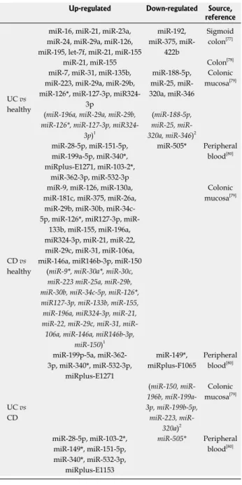
associ-Table 1 MicroRNAs dysregulated in ulcerative colitis and/or Crohn’s disease
Up-regulated Down-regulated Source, reference
UC vs healthy
miR-16, miR-21, miR-23a, miR-24, miR-29a, miR-126, miR-195, let-7f, miR-21, miR-155
miR-192, 375,
miR-422b
Sigmoid colon[77] miR-21, miR-155 Colon[78] miR-7, miR-31, miR-135b,
miR-223, miR-29a, miR-29b, miR-126*, miR-127-3p,
miR324-3p miR-188-5p, 25, miR-320a, miR-346 Colonic mucosa[79] (miR-196a, miR-29a, miR-29b,
miR-126*, miR-127-3p, miR324-3p)1 (miR-188-5p, 25, miR-320a, miR-346)2 miR-28-5p, miR-151-5p, miR-199a-5p, miR-340*, miRplus-E1271, miR-103-2*, miR-362-3p, miR-532-3p miR-505* Peripheral blood[80] CD vs healthy
miR-9, miR-126, miR-130a, miR-181c, miR-375, miR-26a,
miR-29b, miR-30b, 34c-5p, 126*, miR127-3p,
miR-133b, miR-155, miR-196a, miR324-3p, miR-21, miR-22, miR-29c, miR-31, miR-106a, miR-146a, miR146b-3p, miR-150
Colonic mucosa[79]
(miR-9*, miR-30a*, miR-30c, miR-223 miR-25a, miR-29b, miR-30b, miR-34c-5p, miR-126*, miR127-3p, miR-133b, miR-155, miR-196a, miR324-3p, miR-21, 22, 29c, 31, miR-106a, miR-146a, miR146b-3p,
miR-150)1 UC vs CD miR-199p-5a, miR-362-3p, miR-340*, miR-532-miR-362-3p, miRplus-E1271 miR-149*, miRplus-F1065 Peripheral blood[80] (150, miR-196b, miR-199a-3p, miR-199b-5p, 223, miR-320a)2 Colonic mucosa[79] miR-28-5p, miR-103-2*, miR-149*, miR-151-5p, miR-340*, miR-532-3p, miRplus-E1153 miR-505* Peripheral blood[80]
1miRNAs upregulated specifically in non-inflamed colonic mucosa; 2 miR-NAs downregulated specifically in non-inflamed tissue colonic mucosa. UC: Ulcerative colitis; CD: Crohn’s disease.
with the differential expression of 11 miRNAs (3 signifi-cantly decreased and 8 signifisignifi-cantly increased in UC tis-sues). MiR-192, the expression of which is decreased in active UC, was predominantly localized to colonic epithe-lial cells, and targeted macrophage inflammatory peptide
(MIP)-2α, a chemokine expressed by epithelial cells[77].
In colonic epithelial cells, TNF-α-induced MIP-2α ex-pression was inhibited by a miR-192 mimic. In contrast, miR-21 is significantly increased in patients with active UC compared to healthy subjects. In inactive UC patients, miR-375 and miR-422 expression was increased, while that of miR-192 was unaltered compared to healthy
sub-jects[77]. Inactive UC showed similar expression levels of
miR-375, miR-422b, and miR-23a to IBS and IC tissues. The miRNAs differently expressed in active UC were not dysregulated in MC and CD. This study highlights the specific miRNA expression patterns in active and inactive IBD tissues, and suggests that miRNAs could regulate expression of proteins implicated in the pathogenesis.
Another study showed the upregulated expression of several miRNAs in active UC compared to healthy co-lonic biopsies, suggesting that upregulation of miRNAs may be responsible for the development of intestinal
inflammation in UC[78]. MiR-21 was found among the
up-regulated miRNAs, which is consistent with the findings of Takagi et al[78].
Of interest, Fasseu and colleagues identified restricted subsets of miRNAs abnormally expressed in inactive
colonic mucosa of IBD patients[79]. This elegant study
identified 14 (in UC) and 23 (in CD) miRNAs with significantly altered expression (> 5-fold increase or < 0.05-fold decrease) in quiescent colonic mucosa com-pared to healthy control tissues. Eight of the miRNAs (miR-26a, -29a, -29b, -30c, -126*, -127-3p, -196a, -324-3p) were commonly dysregulated in non-inflamed UC and CD. Six miRNAs (miR-196b, -199a-3p, -199b-5p, -320a, -150, -223) displayed significantly distinct dysregulation of expression between non-inflamed UC and CD colonic biopsies. Interestingly, several miRNA genes with dys-regulated expression mapped within acknowledged IBD-susceptibility loci. In addition, significant dysregulated expression of four and five miRNAs specific to inflamed UC or CD tissues, respectively, compared to healthy
controls was observed[79]. This study sheds light on the
role of miRNAs as contributors to IBD susceptibility, in particular their implication in the onset and/or relapse of inflammation from quiescent mucosa of IBD patients.
There have been recent reports of differential miR-NA expression profiles in the peripheral blood of IBD
patients[80]. Four miRNAs (miR-199a5p, -362-3p, -532-3p
and miRplus-E1271) were upregulated and one miRNA (miRplus-F1065) was downregulated in the peripheral blood of patients with active CD, but not inactive CD,
compared to healthy controls[80]. Both active and inactive
CD patients had increased expression of miR-340 and decreased expression of miR-149 in the blood. Expres-sion of three miRNAs (miR-103-2, 262-3p, 532-3p) was increased in the blood of both active and inactive UC
patients. In addition, a subset of 11 miRNAs can
distin-guish active CD from active UC[80]. This study
important-ly supports the evidence that distinct peripheral blood miRNA profiles in different circulating immune cell types are associated with IBD.
Efforts have been made to understand the mecha-nisms underlying the implication of miRNAs in the pathogenesis of IBD. The potential association between single nucleotide polymorphisms (SNPs) in pre-miRNA coding regions and IBD susceptibility has been analyzed. A study in a Japanese cohort of 170 UC patients and 403 healthy controls revealed the association of three SNPs (rs11614913, rs2910164, and rs3746444) in coding regions
of pre-miR-196a2, pre-miR-146a and pre-miR-499[81].
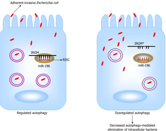
Of particular interest, the CD-associated SNP C313T in
immunity-related GTPase family, M (IRGM) gene caused
a loss in binding of miR-196[82] (Figure 1). IRGM plays
an important role in the immune system by its involve-ment in the autophagy process. In addition, miR-196 is overexpressed in the inflamed epithelium of CD patients and downregulates the IRGM protective variant (c.313C) but not the risk-associated allele (c.313T)[82]. Loss of tight
regulation of IRGM expression by miR-196 resulted in defects in autophagy-mediated control of intracellular
replication of adherent-invasive Escherichia coli (AIEC),
leading to abnormal persistence of AIEC in host cells (Figure 1). This suggests that the association of IRGM with CD could arise from abnormal miRNA-mediated IRGM regulation, which affects the efficacy of autopha-gy, thereby contributing a synonymous polymorphism as a likely causal variant.
Intestinal microbiota is increasingly recognized as a risk for, and a causal factor of, IBD. Our recent studies showed that miRNAs are involved in the regulation of
host gene expression by gut microbiota[83]. In another
study, we showed that miRNAs play a role in determining the unique physiological characteristics of intestinal epi-thelial cells, such as their differentiation during migration
along the crypt/villus axis[84]. In particular, expression of
CD98, a transmembrane glycoprotein that regulates inte-grin signalling, cellular homeostasis and innate immune
response in the gut[85], and its function are directly under
the control of miRNAs during the differentiation of
in-testinal epithelial cells[86]. MiRNAs could also be involved
in the upregulation of CD98 during intestinal
inflamma-tion and IBD[86]. The biological importance of miRNAs
in the pathogenesis of IBD is becoming clearer, and targeting miRNAs in the gastrointestinal tract may be a promising approach for future therapeutic opportunities.
MICRORNAS AND COLORECTAL
CANCERS
The transformation of a normal epithelium into a can-cerous state involves modifications in several genes that are involved in different stages of carcinogenesis such as apoptosis, proliferation, limitless replicative potential
Colorectal cancer (CRC) is one of the most common cancers worldwide. Its incidence is greater in industrial
countries than in developing countries[88]. MiRNAs have
been shown to play an important role in oncogenesis by regulating the expression of genes involved in cancer
initiation, promotion and development[89]. Hundreds of
miRNAs mapped to the human genome regions that are known to be altered in cancer, and a similar number of miRNAs are aberrantly expressed in cancerous tis-sues[90,91]. By analyzing miRNA expression profile
(miR-Nome) of prostate, stomach, pancreas, lung, breast and colon tumors, Volinia and colleagues identified a solid cancer miRNA signature including those with well-char-acterized cancer association, such as miR-17-5p, miR-20a,
miR-21, miR-92, miR-106a, and miR-155[92]. In particular,
21 miRNAs are up-regulated and 1 is down-regulated in
colon tumors compared to normal tissue[92]. MiRNA
pro-files can identify different tissue and tumor types better than mRNA expression patterns, making them attractive
targets for development as cancer biomarkers[93].
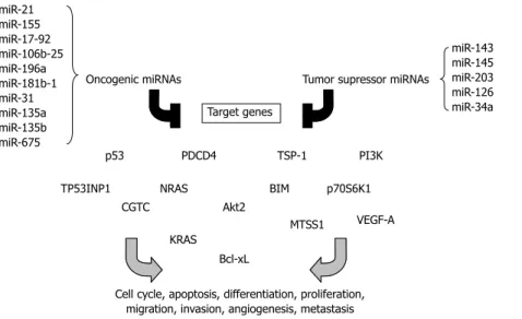
Distin-guished miRNA profiles can even be found in the serum of patients with cancers. The functions of such circulat-ing miRNAs have not been identified, but profilcirculat-ing of serum miRNAs might be a powerful approach for early cancer diagnosis. The cancer-associated miRNAs may function as oncogenes or tumor suppressors depending on their role in carcinogenesis. Some of the best ex-amples of such miRNAs will be discussed in this section
(Figure 2).
Oncogenic miRNAs
MiR-21 is one of the most up-regulated miRNAs in
vari-ous cancers, including CRC[92,94], and was identified as an
independent predictor of overall survival in the valida-tion set containing tumor samples from 113 patients with
CRC[95]. It has been shown that miR-21 is involved in
invasion, intravasation and metastasis processes by
target-ing the tumor suppressor PDCD4[96], and in CRC tissues
expression of miR-21 is inversely correlated with that of
PDCD4 compared to normal tissue[97]. Shibuya and
col-leagues suggested that miR-21 expression may predict
poor prognosis in CRC[98]. Likewise, these authors also
examined the prognostic value of miR-155 in CRC since its expression is up-regulated in tumor tissues compared
to normal adjacent tissues from CRC patients[98]. MiR-155
was previously shown to target the tumor protein 53-in-duced nuclear protein 1 (TP53INP1), a pro-apoptotic stress-induced p53 target, and significant reduction or loss of TP53INP1 expression was detected during
ad-enocarcinoma progression[99].
MiR-17-92 and miR-106b-25 clusters are known to be up-regulated in CRC stromal tissues compared with
normal stroma[100]. They include, respectively, multiple
mature miRNAs, miR-17, miR-18a, miR-19a, miR-20a,
miR-19b1, miR-92-1[101], and miR-106b, miR-93 and
miR-25[102]. These miRNA clusters play an important role
Adherent-invasive Escherichia coli
IRGM RISC miR-196 Regulated autophagy IRGM* miR-196 Dysregulated autophagy Decreased autophagy-mediated elimination of intracellular bacteria
Figure 1 Hypothetical model for the involvement of miR-196 in the pathogenesis of Crohn’s disease. MicroRNAs (miRNAs, miR) 196 normally targets
immu-nity-related GTPase family, M (IRGM) mRNA within RNA-induced silencing complex (RISC) for a negative regulation (left panel). The IRGM risk allele (IRGM*) mRNA lacks the binding site for miR-196 and therefore is not regulated by this miRNA (right panel). During Crohn’s disease, loss of tight regulation of IRGM* expression by miR-196 may lead to defects in autophagy with most intracellular bacteria replication occurring in dysfunctional vacuoles[82] (dotted cycle). This consequently results in
during carcinogenesis[92,100,103,104]. An anti-apoptotic
ef-fect of miR-17-92 appears to be one of the mechanisms underlying its procarcinogenic role in CRC
develop-ment and progression[105]. Abrogation of miR-92a leads
to cell apoptosis, and there is a correlation between the miR-17-92 overexpression in tumors of CRC patients and the downregulated expression of BIM, a member of
the Bcl-2 family that promotes apoptosis[105]. Some works
have reported that there is an interconnection between the expression of miR-17-92 cluster and angiogenesis, which occurs later in tumor development and is one of the most important stages in carcinogenesis. Dews and colleagues demonstrated that the anti-angiogenic fac-tors thrombospondin-1 (tsp-1) and connective tissue growth factor (CTGC) are down-regulated by this cluster in intestinal epithelial cells expressing constitutively the
oncogene c-myc[106], which was shown to be involved in
regulation of miR-17-92 expression[20]. MiR-18 targets
tsp-1 and miR-19 modulates the expression of CTGF[107].
Other miRNAs have also been identified as causal factors in colon carcinogenesis. For example, miR-196a had higher expression level in CRC tissues than in normal
epithelial tissues[108]. MiR-196a exerts a pro-oncogenic
in-fluence in CRC as a high level of its expression promotes the oncogenic phenotype of colorectal cancer cells such
as increased cell detachment, migration and invasion[109].
MiR-31 is often up-regulated in CRC and its high expres-sion associated with advanced tumor stage but the clinical
significance is unclear[110]. MiR-181b-1, 135a,
miR-135b, miR-675 are also known to be up-regulated in CRC
tumors[111]. MiR-135a is able to promote the growth and
invasion of CRC cells by targeting the metastasis sup-pressor 1[112].
Tumor suppressor miRNAs
Mir-143 and miR-145 are among the best examples of tumor suppressor miRNAs. The expression of these miRNAs is down-regulated in CRC tumors, and in other
cancers such as breast, prostate, cervical and lymphoid
cancer[113-115]. Many studies have reported that
down-regulation of miR-143 and miR-145 correlates with poor
prognosis[110,115,116]. The expression and
post-transcrip-tional maturation of these miRNAs were recently shown to be enhanced by the tumor suppressor p53 in response
to DNA damages in CRC cell lines[28,117]. In particular,
miR-143 is involved in inhibition of oncogene KRAS
ex-pression[118]. MiR-145 is reported to inhibit tumor growth
and angiogenesis by directly targeting p70S6K1[117], which
is activated by mTOR, and its overexpression in cancer
cells induces tumor angiogenesis[119-121]. Another study
reported that this miRNA is able to inhibit tumor growth and angiogenesis in breast cancer by targeting N-RAS
and VEGF-A, which are key players in carcinogenesis[122].
It was recently demonstrated that miR-34a is down-regulated in colon tumors and also in circulating
blood[123]. Furthermore, ectopic expression of miR-34a
in CRC cell line reduces cell proliferation, demonstrating that this miRNA has a tumor suppressive function in
co-lon carcinogenesis[124]. Several studies conducted in 2007
revealed that miR-34a can target p53, leading to apoptosis
and cell cycle arrest[21-24,125]. MiR-203 is identified as
an-other tumor suppressor miRNA. Its low expression was
found in vitro in CRC cell lines and was correlated with
tumor size in CRC. MiR-203 can inhibit proliferation of cancer cell lines[126]. Li et al[127] showed that miR-203
over-expression significantly decreased cell proliferation and survival and induced cell apoptosis in the p53-mutated CRC cells. The tumor suppressive role of miR-203 was
mediated by negatively regulating Akt2 expression via
mRNA degradation. In addition, overexpression of miR-203 decreased expression of the anti-apoptotic gene
Bcl-xL, leading to a resistance to apoptosis[127]. MiR-126
is specifically expressed in endothelial cells and is known to be down-regulated in CRC compared to normal tissue
via an unknown mechanism[128]. In vitro studies suggested
that a loss in negative regulation of p85 subunit of PI3K
miR-21 miR-155 miR-17-92 miR-106b-25 miR-196a miR-181b-1 miR-31 miR-135a miR-135b miR-675
Oncogenic miRNAs Tumor supressor miRNAs Target genes miR-143 miR-145 miR-203 miR-126 miR-34a p53 PDCD4 TSP-1 PI3K TP53INP1 NRAS BIM p70S6K1
Akt2 CGTC
KRAS
MTSS1 VEGF-A Bcl-xL
Cell cycle, apoptosis, differentiation, proliferation, migration, invasion, angiogenesis, metastasis
Figure 2 Overview of “oncogenic” and “tumor suppressor” microRNAs related to colorectal cancer described in this review, their targets and different carcinogenesis pathways in which they have been implicated.
by miR-126 could lead to a selective growth advantage during colon carcinogenesis[129].
CONCLUSION
MiRNAs are a class of gene regulators that have recently emerged as key players in the innate and adaptive immune system. Changes in miRNA expression are observed in many human diseases such as inflammatory bowel disease and cancers. Dysregulated miRNA expression profiles in IBD have been reported and could be used as diagnostic biomarkers but further studies are needed to examine the mechanism of their action in the etiopathogenesis of this disease and their clinical utility. Emerging evidence suggests that miRNAs play important roles in the patho-genesis of a limited range of human cancers. Some miR-NAs may be directly involved in cancer development by controlling cell differentiation and apoptosis, while others may be involved in cancers by targeting cancer oncogenes and/or tumor suppressors. Given the critical role of miRNAs, current studies are focusing on their association with CRC incidence and prognosis and on the possibility of using circulating miRNAs or fecal miRNA expression as noninvasive early detection biomarkers. These data suggest that miRNAs may be potential molecular classi-fiers, early detection biomarkers, and therapeutic targets for CRC. Finally, miRNA-based cancer therapy has been
limited to targeting a single miRNA[130,131]. However, it has
been recently shown that the small molecule enoxacin, a fluoroquinolone used as an antibacterial compound, enhances the miRNA-processing machinery by binding
to TRBP[132]. Thus, if most cancers are characterized by
a dysregulation of global mature miRNA expression, restoration of the global miRNome may be an attractive approach in cancer therapy. In conclusion, a better un-derstanding of the function of miRNAs is providing new insights into the molecular basis of human pathologies, and new biomarkers for disease diagnosis and therapy.
REFERENCES
1 Winter J, Jung S, Keller S, Gregory RI, Diederichs S. Many
roads to maturity: microRNA biogenesis pathways and their regulation. Nat Cell Biol 2009; 11: 228-234 [PMID: 19255566 DOI: 10.1038/ncb0309-228]
2 Wightman B, Ha I, Ruvkun G. Posttranscriptional regulation
of the heterochronic gene lin-14 by lin-4 mediates temporal pattern formation in C. elegans. Cell 1993; 75: 855-862 [PMID: 8252622 DOI: 10.1016/0092-8674(93)90530-4]
3 Lee RC, Feinbaum RL, Ambros V. The C. elegans
heteroch-ronic gene lin-4 encodes small RNAs with antisense comple-mentarity to lin-14. Cell 1993; 75: 843-854 [PMID: 8252621 DOI: 10.1016/0092-8674(93)90529-Y]
4 Kozomara A, Griffiths-Jones S. miRBase: integrating
mi-croRNA annotation and deep-sequencing data. Nucleic Acids Res 2011; 39: D152-D157 [PMID: 21037258 DOI: 10.1093/nar/ gkq1027]
5 Bartel DP. MicroRNAs: target recognition and regulatory
functions. Cell 2009; 136: 215-233 [PMID: 19167326 DOI: 10.1016/j.cell.2009.01.002]
6 Lee Y, Ahn C, Han J, Choi H, Kim J, Yim J, Lee J, Provost P,
Rådmark O, Kim S, Kim VN. The nuclear RNase III Drosha
initiates microRNA processing. Nature 2003; 425: 415-419 [PMID: 14508493 DOI: 10.1038/nature01957]
7 Gregory RI, Yan KP, Amuthan G, Chendrimada T, Doratotaj
B, Cooch N, Shiekhattar R. The Microprocessor complex me-diates the genesis of microRNAs. Nature 2004; 432: 235-240 [PMID: 15531877 DOI: 10.1038/nature03120]
8 Denli AM, Tops BB, Plasterk RH, Ketting RF, Hannon GJ.
Processing of primary microRNAs by the Microprocessor complex. Nature 2004; 432: 231-235 [PMID: 15531879 DOI: 10.1038/nature03049]
9 Lund E, Güttinger S, Calado A, Dahlberg JE, Kutay U.
Nu-clear export of microRNA precursors. Science 2004; 303: 95-98 [PMID: 14631048 DOI: 10.1126/science.1090599]
10 Yi R, Qin Y, Macara IG, Cullen BR. Exportin-5 mediates the nuclear export of pre-microRNAs and short hairpin RNAs. Genes Dev 2003; 17: 3011-3016 [PMID: 14681208 DOI: 10.1101/gad.1158803]
11 Bohnsack MT, Czaplinski K, Gorlich D. Exportin 5 is a RanGTP-dependent dsRNA-binding protein that mediates nuclear export of pre-miRNAs. RNA 2004; 10: 185-191 [PMID: 14730017]
12 Grishok A, Pasquinelli AE, Conte D, Li N, Parrish S, Ha I, Baillie DL, Fire A, Ruvkun G, Mello CC. Genes and mecha-nisms related to RNA interference regulate expression of the small temporal RNAs that control C. elegans developmental timing. Cell 2001; 106: 23-34 [PMID: 11461699 DOI: 10.1016/ S0092-8674(01)00431-7]
13 Chendrimada TP, Gregory RI, Kumaraswamy E, Norman J, Cooch N, Nishikura K, Shiekhattar R. TRBP recruits the Dicer complex to Ago2 for microRNA processing and gene silencing. Nature 2005; 436: 740-744 [PMID: 15973356 DOI: 10.1038/nature03868]
14 Haase AD, Jaskiewicz L, Zhang H, Lainé S, Sack R, Gatignol A, Filipowicz W. TRBP, a regulator of cellular PKR and HIV-1 virus expression, interacts with Dicer and functions in RNA silencing. EMBO Rep 2005; 6: 961-967 [PMID: 16142218 DOI: 10.1038/sj.embor.7400509]
15 Gregory RI, Chendrimada TP, Cooch N, Shiekhattar R. Hu-man RISC couples microRNA biogenesis and posttranscrip-tional gene silencing. Cell 2005; 123: 631-640 [PMID: 16271387 DOI: 10.1016/j.cell.2005.10.022]
16 Rüegger S, Großhans H. MicroRNA turnover: when, how, and why. Trends Biochem Sci 2012; 37: 436-446 [PMID: 22921610 DOI: 10.1016/j.tibs.2012.07.002]
17 Carthew RW, Sontheimer EJ. Origins and Mechanisms of miRNAs and siRNAs. Cell 2009; 136: 642-655 [PMID: 19239886]
18 Siomi H, Siomi MC. Posttranscriptional regulation of mi-croRNA biogenesis in animals. Mol Cell 2010; 38: 323-332 [PMID: 20471939 DOI: 10.1016/j.molcel.2010.03.013] 19 Breving K, Esquela-Kerscher A. The complexities of
microR-NA regulation: miRandering around the rules. Int J Biochem Cell Biol 2010; 42: 1316-1329 [PMID: 19800023 DOI: 10.1016/ j.biocel.2009.09.016]
20 O’Donnell KA, Wentzel EA, Zeller KI, Dang CV, Mendell JT. c-Myc-regulated microRNAs modulate E2F1 expression. Nature 2005; 435: 839-843 [PMID: 15944709 DOI: 10.1038/na-ture03677]
21 Raver-Shapira N, Marciano E, Meiri E, Spector Y, Rosenfeld N, Moskovits N, Bentwich Z, Oren M. Transcriptional ac-tivation of miR-34a contributes to p53-mediated apoptosis. Mol Cell 2007; 26: 731-743 [PMID: 17540598 DOI: 10.1016/ j.molcel.2007.05.017]
22 He L, He X, Lim LP, de Stanchina E, Xuan Z, Liang Y, Xue W, Zender L, Magnus J, Ridzon D, Jackson AL, Linsley PS, Chen C, Lowe SW, Cleary MA, Hannon GJ. A microRNA compo-nent of the p53 tumour suppressor network. Nature 2007;
447: 1130-1134 [PMID: 17554337 DOI: 10.1038/nature05939]
23 Tarasov V, Jung P, Verdoodt B, Lodygin D, Epanchintsev A, Menssen A, Meister G, Hermeking H. Differential regulation
of microRNAs by p53 revealed by massively parallel se-quencing: miR-34a is a p53 target that induces apoptosis and G1-arrest. Cell Cycle 2007; 6: 1586-1593 [PMID: 17554199 DOI: 10.4161/cc.6.13.4436]
24 Chang TC, Wentzel EA, Kent OA, Ramachandran K, Mul-lendore M, Lee KH, Feldmann G, Yamakuchi M, Ferlito M, Lowenstein CJ, Arking DE, Beer MA, Maitra A, Mendell JT. Transactivation of miR-34a by p53 broadly influences gene expression and promotes apoptosis. Mol Cell 2007; 26: 745-752 [PMID: 17540599 DOI: 10.1016/j.molcel.2007.05.010] 25 Brueckner B, Stresemann C, Kuner R, Mund C, Musch T,
Meister M, Sültmann H, Lyko F. The human let-7a-3 locus contains an epigenetically regulated microRNA gene with oncogenic function. Cancer Res 2007; 67: 1419-1423 [PMID: 17308078 DOI: 10.1158/0008-5472.CAN-06-4074]
26 Han L, Witmer PD, Casey E, Valle D, Sukumar S. DNA methylation regulates MicroRNA expression. Cancer Biol Ther 2007; 6: 1284-1288 [PMID: 17660710 DOI: 10.4161/ cbt.6.8.4486]
27 Toyota M, Suzuki H, Sasaki Y, Maruyama R, Imai K, Shi-nomura Y, Tokino T. Epigenetic silencing of microRNA-34b/c and B-cell translocation gene 4 is associated with CpG island methylation in colorectal cancer. Cancer Res 2008;
68: 4123-4132 [PMID: 18519671 DOI: 10.1158/0008-5472.
CAN-08-0325]
28 Suzuki HI, Yamagata K, Sugimoto K, Iwamoto T, Kato S, Miyazono K. Modulation of microRNA processing by p53. Nature 2009; 460: 529-533 [PMID: 19626115 DOI: 10.1038/na-ture08199]
29 Wada T, Kikuchi J, Furukawa Y. Histone deacetylase 1 en-hances microRNA processing via deacetylation of DGCR8. EMBO Rep 2012; 13: 142-149 [PMID: 22222205 DOI: 10.1038/ embor.2011.247]
30 Wiesen JL, Tomasi TB. Dicer is regulated by cellular stresses and interferons. Mol Immunol 2009; 46: 1222-1228 [PMID: 19118902 DOI: 10.1016/j.molimm.2008.11.012]
31 Dalal SR, Kwon JH. The Role of MicroRNA in Inflammatory Bowel Disease. Gastroenterol Hepatol (N Y) 2010; 6: 714-722 [PMID: 21437020]
32 Lu LF, Liston A. MicroRNA in the immune system, microR-NA as an immune system. Immunology 2009; 127: 291-298 [PMID: 19538248]
33 Takeda K, Kaisho T, Akira S. Toll-like receptors. Annu Rev Immunol 2003; 21: 335-376 [PMID: 12524386 DOI: 10.1146/an-nurev.immunol.21.120601.141126]
34 Dunne A, O’Neill LA. Adaptor usage and Toll-like receptor signaling specificity. FEBS Lett 2005; 579: 3330-3335 [PMID: 15876435 DOI: 10.1016/j.febslet.2005.04.024]
35 Taganov KD, Boldin MP, Chang KJ, Baltimore D. NF-kappaB-dependent induction of microRNA miR-146, an inhibitor targeted to signaling proteins of innate immune responses. Proc Natl Acad Sci USA 2006; 103: 12481-12486 [PMID: 16885212 DOI: 10.1073/pnas.0605298103]
36 O’Connell RM, Taganov KD, Boldin MP, Cheng G, Balti-more D. MicroRNA-155 is induced during the macrophage inflammatory response. Proc Natl Acad Sci USA 2007; 104: 1604-1609 [PMID: 17242365 DOI: 10.1073/pnas.0610731104] 37 Tili E, Michaille JJ, Cimino A, Costinean S, Dumitru CD,
Adair B, Fabbri M, Alder H, Liu CG, Calin GA, Croce CM. Modulation of miR-155 and miR-125b levels following lipo-polysaccharide/TNF-alpha stimulation and their possible roles in regulating the response to endotoxin shock. J Immu-nol 2007; 179: 5082-5089 [PMID: 17911593]
38 Lu LF, Thai TH, Calado DP, Chaudhry A, Kubo M, Tanaka K, Loeb GB, Lee H, Yoshimura A, Rajewsky K, Rudensky AY. Foxp3-dependent microRNA155 confers competitive fitness to regulatory T cells by targeting SOCS1 protein. Immunity 2009; 30: 80-91 [PMID: 19144316 DOI: 10.1016/ j.immuni.2008.11.010]
39 Rodriguez A, Vigorito E, Clare S, Warren MV, Couttet P,
Soond DR, van Dongen S, Grocock RJ, Das PP, Miska EA, Vetrie D, Okkenhaug K, Enright AJ, Dougan G, Turner M, Bradley A. Requirement of bic/microRNA-155 for normal immune function. Science 2007; 316: 608-611 [PMID: 17463290 DOI: 10.1126/science.1139253]
40 Ceppi M, Pereira PM, Dunand-Sauthier I, Barras E, Reith W, Santos MA, Pierre P. MicroRNA-155 modulates the inter-leukin-1 signaling pathway in activated human monocyte-derived dendritic cells. Proc Natl Acad Sci USA 2009; 106: 2735-2740 [PMID: 19193853 DOI: 10.1073/pnas.0811073106] 41 O’Connell RM, Rao DS, Chaudhuri AA, Boldin MP,
Tagan-ov KD, Nicoll J, Paquette RL, Baltimore D. Sustained expres-sion of microRNA-155 in hematopoietic stem cells causes a myeloproliferative disorder. J Exp Med 2008; 205: 585-594 [PMID: 18299402 DOI: 10.1084/jem.20072108]
42 Costinean S, Sandhu SK, Pedersen IM, Tili E, Trotta R, Per-rotti D, Ciarlariello D, Neviani P, Harb J, Kauffman LR, Shidham A, Croce CM. Src homology 2 domain-containing inositol-5-phosphatase and CCAAT enhancer-binding protein beta are targeted by miR-155 in B cells of Emicro-MiR-155 transgenic mice. Blood 2009; 114: 1374-1382 [PMID: 19520806 DOI: 10.1182/blood-2009-05-220814]
43 O’Connell RM, Chaudhuri AA, Rao DS, Baltimore D. Ino-sitol phosphatase SHIP1 is a primary target of miR-155. Proc Natl Acad Sci USA 2009; 106: 7113-7118 [PMID: 19359473 DOI: 10.1073/pnas.0902636106]
44 Sly LM, Rauh MJ, Kalesnikoff J, Song CH, Krystal G. LPS-induced upregulation of SHIP is essential for endotoxin tolerance. Immunity 2004; 21: 227-239 [PMID: 15308103 DOI: 10.1016/j.immuni.2004.07.010]
45 Sheedy FJ, Palsson-McDermott E, Hennessy EJ, Martin C, O’Leary JJ, Ruan Q, Johnson DS, Chen Y, O’Neill LA. Nega-tive regulation of TLR4 via targeting of the proinflammatory tumor suppressor PDCD4 by the microRNA miR-21. Nat Immunol 2010; 11: 141-147 [PMID: 19946272 DOI: 10.1073/ pnas.0810909106]
46 Bazzoni F, Rossato M, Fabbri M, Gaudiosi D, Mirolo M, Mori L, Tamassia N, Mantovani A, Cassatella MA, Locati M. Induction and regulatory function of miR-9 in human monocytes and neutrophils exposed to proinflammatory signals. Proc Natl Acad Sci USA 2009; 106: 5282-5287 [PMID: 19289835]
47 Liu G, Friggeri A, Yang Y, Park YJ, Tsuruta Y, Abraham E. miR-147, a microRNA that is induced upon Toll-like recep-tor stimulation, regulates murine macrophage inflammarecep-tory responses. Proc Natl Acad Sci USA 2009; 106: 15819-15824 [PMID: 19721002 DOI: 10.1073/pnas.0901216106]
48 O’Neill LA, Sheedy FJ, McCoy CE. MicroRNAs: the fine-tuners of Toll-like receptor signalling. Nat Rev Immunol 2011;
11: 163-175 [PMID: 21331081]
49 McCoy CE, Sheedy FJ, Qualls JE, Doyle SL, Quinn SR, Mur-ray PJ, O’Neill LA. IL-10 inhibits miR-155 induction by toll-like receptors. J Biol Chem 2010; 285: 20492-20498 [PMID: 20435894 DOI: 10.1074/jbc.M110.102111]
50 Merkerova M, Belickova M, Bruchova H. Differential ex-pression of microRNAs in hematopoietic cell lineages. Eur J Haematol 2008; 81: 304-310 [PMID: 18573170 DOI: 10.1111/ j.1600-0609.2008.01111.x]
51 Monticelli S, Ansel KM, Xiao C, Socci ND, Krichevsky AM, Thai TH, Rajewsky N, Marks DS, Sander C, Rajewsky K, Rao A, Kosik KS. MicroRNA profiling of the murine hematopoi-etic system. Genome Biol 2005; 6: R71 [PMID: 16086853 DOI: 10.1186/gb-2005-6-8-r71]
52 Wu H, Neilson JR, Kumar P, Manocha M, Shankar P, Sharp PA, Manjunath N. miRNA profiling of naive, effector and memory CD8 T cells. PLoS One 2007; 2: e1020 [PMID: 17925868 DOI: 10.1371/journal.pone.0001020]
53 Cobb BS, Nesterova TB, Thompson E, Hertweck A, O’Con-nor E, Godwin J, Wilson CB, Brockdorff N, Fisher AG, Smale ST, Merkenschlager M. T cell lineage choice and
differen-tiation in the absence of the RNase III enzyme Dicer. J Exp Med 2005; 201: 1367-1373 [PMID: 15867090 DOI: 10.1084/ jem.20050572]
54 Muljo SA, Ansel KM, Kanellopoulou C, Livingston DM, Rao A, Rajewsky K. Aberrant T cell differentiation in the absence of Dicer. J Exp Med 2005; 202: 261-269 [PMID: 16009718 DOI: 10.1084/jem.20050678]
55 Xiao C, Srinivasan L, Calado DP, Patterson HC, Zhang B, Wang J, Henderson JM, Kutok JL, Rajewsky K. Lymphopro-liferative disease and autoimmunity in mice with increased miR-17-92 expression in lymphocytes. Nat Immunol 2008; 9: 405-414 [PMID: 18327259 DOI: 10.1038/ni1575]
56 Du C, Liu C, Kang J, Zhao G, Ye Z, Huang S, Li Z, Wu Z, Pei G. MicroRNA miR-326 regulates TH-17 differentiation and is associated with the pathogenesis of multiple sclerosis. Nat Immunol 2009; 10: 1252-1259 [PMID: 19838199 DOI: 10.1038/ ni.1798]
57 Zheng Y, Josefowicz SZ, Kas A, Chu TT, Gavin MA, Ruden-sky AY. Genome-wide analysis of Foxp3 target genes in developing and mature regulatory T cells. Nature 2007; 445: 936-940 [PMID: 17237761 DOI: 10.1038/nature05563] 58 Thai TH, Calado DP, Casola S, Ansel KM, Xiao C, Xue Y,
Murphy A, Frendewey D, Valenzuela D, Kutok JL, Schmidt-Supprian M, Rajewsky N, Yancopoulos G, Rao A, Rajewsky K. Regulation of the germinal center response by microR-NA-155. Science 2007; 316: 604-608 [PMID: 17463289 DOI: 10.1126/science.1141229]
59 Johnston RJ, Poholek AC, DiToro D, Yusuf I, Eto D, Barnett B, Dent AL, Craft J, Crotty S. Bcl6 and Blimp-1 are reciprocal and antagonistic regulators of T follicular helper cell differ-entiation. Science 2009; 325: 1006-1010 [PMID: 19608860 DOI: 10.1126/science.1175870]
60 Yu D, Rao S, Tsai LM, Lee SK, He Y, Sutcliffe EL, Srivastava M, Linterman M, Zheng L, Simpson N, Ellyard JI, Parish IA, Ma CS, Li QJ, Parish CR, Mackay CR, Vinuesa CG. The transcriptional repressor Bcl-6 directs T follicular helper cell lineage commitment. Immunity 2009; 31: 457-468 [PMID: 19631565 DOI: 10.1016/j.immuni.2009.07.002]
61 Li QJ, Chau J, Ebert PJ, Sylvester G, Min H, Liu G, Braich R, Manoharan M, Soutschek J, Skare P, Klein LO, Davis MM, Chen CZ. miR-181a is an intrinsic modulator of T cell sensi-tivity and selection. Cell 2007; 129: 147-161 [PMID: 17382377 DOI: 10.1016/j.cell.2007.03.008]
62 Chong MM, Rasmussen JP, Rudensky AY, Littman DR. The RNAseIII enzyme Drosha is critical in T cells for preventing lethal inflammatory disease. J Exp Med 2008; 205: 2005-2017 [PMID: 18725527 DOI: 10.1084/jem.20081219]
63 Liston A, Lu LF, O’Carroll D, Tarakhovsky A, Rudensky AY. Dicer-dependent microRNA pathway safeguards regu-latory T cell function. J Exp Med 2008; 205: 1993-2004 [PMID: 18725526]
64 Zhou X, Jeker LT, Fife BT, Zhu S, Anderson MS, McManus MT, Bluestone JA. Selective miRNA disruption in T reg cells leads to uncontrolled autoimmunity. J Exp Med 2008; 205: 1983-1991 [PMID: 18725525]
65 Tan LP, Wang M, Robertus JL, Schakel RN, Gibcus JH, Diep-stra A, Harms G, Peh SC, Reijmers RM, Pals ST, Kroesen BJ, Kluin PM, Poppema S, van den Berg A. miRNA profiling of B-cell subsets: specific miRNA profile for germinal center B cells with variation between centroblasts and centrocytes. Lab Invest 2009; 89: 708-716 [PMID: 19349957 DOI: 10.1038/ labinvest.2009.26]
66 Basso K, Sumazin P, Morozov P, Schneider C, Maute RL, Kitagawa Y, Mandelbaum J, Haddad J, Chen CZ, Califano A, Dalla-Favera R. Identification of the human mature B cell miRNome. Immunity 2009; 30: 744-752 [PMID: 19446474] 67 Xiao C, Rajewsky K. MicroRNA control in the immune
sys-tem: basic principles. Cell 2009; 136: 26-36 [PMID: 19135886] 68 Chen CZ, Li L, Lodish HF, Bartel DP. MicroRNAs modulate
hematopoietic lineage differentiation. Science 2004; 303: 83-86
[PMID: 14657504 DOI: 10.1126/science.1091903]
69 Koralov SB, Muljo SA, Galler GR, Krek A, Chakraborty T, Kanellopoulou C, Jensen K, Cobb BS, Merkenschlager M, Rajewsky N, Rajewsky K. Dicer ablation affects antibody diversity and cell survival in the B lymphocyte lineage. Cell 2008; 132: 860-874 [PMID: 18329371]
70 Ventura A, Young AG, Winslow MM, Lintault L, Meissner A, Erkeland SJ, Newman J, Bronson RT, Crowley D, Stone JR, Jaenisch R, Sharp PA, Jacks T. Targeted deletion reveals essential and overlapping functions of the miR-17 through 92 family of miRNA clusters. Cell 2008; 132: 875-886 [PMID: 18329372 DOI: 10.1016/j.cell.2008.02.019]
71 Zhou B, Wang S, Mayr C, Bartel DP, Lodish HF. miR-150, a microRNA expressed in mature B and T cells, blocks early B cell development when expressed prematurely. Proc Natl Acad Sci USA 2007; 104: 7080-7085 [PMID: 17438277] 72 Xiao C, Calado DP, Galler G, Thai TH, Patterson HC, Wang J,
Rajewsky N, Bender TP, Rajewsky K. MiR-150 controls B cell differentiation by targeting the transcription factor c-Myb. Cell 2007; 131: 146-159 [PMID: 17923094]
73 Vigorito E, Perks KL, Abreu-Goodger C, Bunting S, Xiang Z, Kohlhaas S, Das PP, Miska EA, Rodriguez A, Bradley A, Smith KG, Rada C, Enright AJ, Toellner KM, Maclennan IC, Turner M. microRNA-155 regulates the generation of immu-noglobulin class-switched plasma cells. Immunity 2007; 27: 847-859 [PMID: 18055230]
74 Rao DS, O’Connell RM, Chaudhuri AA, Garcia-Flores Y, Geiger TL, Baltimore D. MicroRNA-34a perturbs B lympho-cyte development by repressing the forkhead box transcrip-tion factor Foxp1. Immunity 2010; 33: 48-59 [PMID: 20598588 DOI: 10.1016/j.immuni.2010.06.013]
75 Hu H, Wang B, Borde M, Nardone J, Maika S, Allred L, Tucker PW, Rao A. Foxp1 is an essential transcriptional reg-ulator of B cell development. Nat Immunol 2006; 7: 819-826 [PMID: 16819554]
76 Blumberg R, Cho J, Lewis J, Wu G. Inflammatory bowel disease: an update on the fundamental biology and clinical management. Gastroenterology 2011; 140: 1701-1703 [PMID: 21530735 DOI: 10.1053/j.gastro.2011.03.013]
77 Wu F, Zikusoka M, Trindade A, Dassopoulos T, Harris ML, Bayless TM, Brant SR, Chakravarti S, Kwon JH. MicroRNAs are differentially expressed in ulcerative colitis and alter expression of macrophage inflammatory peptide-2 alpha. Gastroenterology 2008; 135: 1624-1635.e24 [PMID: 18835392] 78 Takagi T, Naito Y, Mizushima K, Hirata I, Yagi N,
Tomat-suri N, Ando T, Oyamada Y, Isozaki Y, Hongo H, Uchiyama K, Handa O, Kokura S, Ichikawa H, Yoshikawa T. Increased expression of microRNA in the inflamed colonic mucosa of patients with active ulcerative colitis. J Gastroenterol Hepatol 2010; 25 Suppl 1: S129-S133 [PMID: 20586854]
79 Fasseu M, Tréton X, Guichard C, Pedruzzi E, Cazals-Hatem D, Richard C, Aparicio T, Daniel F, Soulé JC, Moreau R, Bouhnik Y, Laburthe M, Groyer A, Ogier-Denis E. Identification of re-stricted subsets of mature microRNA abnormally expressed in inactive colonic mucosa of patients with inflammatory bowel disease. PLoS One 2010; 5: e13160 [PMID: 20957151] 80 Wu F, Guo NJ, Tian H, Marohn M, Gearhart S, Bayless TM,
Brant SR, Kwon JH. Peripheral blood microRNAs distin-guish active ulcerative colitis and Crohn’s disease. Inflamm Bowel Dis 2011; 17: 241-250 [PMID: 20812331 DOI: 10.1002/ ibd.21450]
81 Okubo M, Tahara T, Shibata T, Yamashita H, Nakamura M, Yoshioka D, Yonemura J, Kamiya Y, Ishizuka T, Nakagawa Y, Nagasaka M, Iwata M, Yamada H, Hirata I, Arisawa T. Asso-ciation study of common genetic variants in pre-microRNAs in patients with ulcerative colitis. J Clin Immunol 2011; 31: 69-73 [PMID: 20848167 DOI: 10.1007/s10875-010-9461-y] 82 Brest P, Lapaquette P, Souidi M, Lebrigand K, Cesaro A,
Vouret-Craviari V, Mari B, Barbry P, Mosnier JF, Hébuterne X, Harel-Bellan A, Mograbi B, Darfeuille-Michaud A,
Hof-man P. A synonymous variant in IRGM alters a binding site for miR-196 and causes deregulation of IRGM-dependent xenophagy in Crohn’s disease. Nat Genet 2011; 43: 242-245 [PMID: 21278745]
83 Dalmasso G, Nguyen HT, Yan Y, Laroui H, Charania MA, Ayyadurai S, Sitaraman SV, Merlin D. Microbiota modulate host gene expression via microRNAs. PLoS One 2011; 6: e19293 [PMID: 21559394 DOI: 10.1371/journal.pone.0019293] 84 Dalmasso G, Nguyen HT, Yan Y, Laroui H, Srinivasan S, Sitaraman SV, Merlin D. MicroRNAs determine human intestinal epithelial cell fate. Differentiation 2010; 80: 147-154 [PMID: 20638171]
85 Nguyen HT, Merlin D. Homeostatic and innate immune responses: role of the transmembrane glycoprotein CD98. Cell Mol Life Sci 2012; 69: 3015-3026 [PMID: 22460579 DOI: 10.1007/s00018-012-0963-z]
86 Nguyen HT, Dalmasso G, Yan Y, Laroui H, Dahan S, Mayer L, Sitaraman SV, Merlin D. MicroRNA-7 modulates CD98 expression during intestinal epithelial cell differentiation. J Biol Chem 2010; 285: 1479-1489 [PMID: 19892711]
87 Hanahan D, Weinberg RA. Hallmarks of cancer: the next generation. Cell 2011; 144: 646-674 [PMID: 21376230] 88 Weitz J, Koch M, Debus J, Höhler T, Galle PR, Büchler MW.
Colorectal cancer. Lancet 2005; 365: 153-165 [PMID: 15639298] 89 Dalmay T, Edwards DR. MicroRNAs and the hallmarks of
cancer. Oncogene 2006; 25: 6170-6175 [PMID: 17028596 DOI: 10.1038/sj.onc.1209911]
90 Kasinski AL, Slack FJ. Epigenetics and genetics. MicroRNAs en route to the clinic: progress in validating and targeting microRNAs for cancer therapy. Nat Rev Cancer 2011; 11: 849-864 [PMID: 22113163 DOI: 10.1038/nrc3166]
91 Calin GA, Sevignani C, Dumitru CD, Hyslop T, Noch E, Yendamuri S, Shimizu M, Rattan S, Bullrich F, Negrini M, Croce CM. Human microRNA genes are frequently located at fragile sites and genomic regions involved in cancers. Proc Natl Acad Sci USA 2004; 101: 2999-3004 [PMID: 14973191 DOI: 10.1073/pnas.0307323101]
92 Volinia S, Calin GA, Liu CG, Ambs S, Cimmino A, Petrocca F, Visone R, Iorio M, Roldo C, Ferracin M, Prueitt RL, Yanai-hara N, Lanza G, Scarpa A, Vecchione A, Negrini M, Harris CC, Croce CM. A microRNA expression signature of human solid tumors defines cancer gene targets. Proc Natl Acad Sci USA 2006; 103: 2257-2261 [PMID: 16461460]
93 Lu J, Getz G, Miska EA, Alvarez-Saavedra E, Lamb J, Peck D, Sweet-Cordero A, Ebert BL, Mak RH, Ferrando AA, Downing JR, Jacks T, Horvitz HR, Golub TR. MicroRNA expression profiles classify human cancers. Nature 2005; 435: 834-838 [PMID: 15944708 DOI: 10.1038/nature03702] 94 Meng F, Henson R, Wehbe-Janek H, Ghoshal K, Jacob ST,
Patel T. MicroRNA-21 regulates expression of the PTEN tumor suppressor gene in human hepatocellular cancer. Gas-troenterology 2007; 133: 647-658 [PMID: 17681183]
95 Schetter AJ, Leung SY, Sohn JJ, Zanetti KA, Bowman ED, Yanaihara N, Yuen ST, Chan TL, Kwong DL, Au GK, Liu CG, Calin GA, Croce CM, Harris CC. MicroRNA expression profiles associated with prognosis and therapeutic outcome in colon adenocarcinoma. JAMA 2008; 299: 425-436 [PMID: 18230780]
96 Asangani IA, Rasheed SA, Nikolova DA, Leupold JH, Col-burn NH, Post S, Allgayer H. MicroRNA-21 (miR-21) post-transcriptionally downregulates tumor suppressor Pdcd4 and stimulates invasion, intravasation and metastasis in colorectal cancer. Oncogene 2008; 27: 2128-2136 [PMID: 17968323]
97 Chang KH, Miller N, Kheirelseid EA, Ingoldsby H, Hen-nessy E, Curran CE, Curran S, Smith MJ, Regan M, McAn-ena OJ, Kerin MJ. MicroRNA-21 and PDCD4 expression in colorectal cancer. Eur J Surg Oncol 2011; 37: 597-603 [PMID: 21546206]
98 Shibuya H, Iinuma H, Shimada R, Horiuchi A, Watanabe T.
Clinicopathological and prognostic value of microRNA-21 and microRNA-155 in colorectal cancer. Oncology 2010; 79: 313-320 [PMID: 21412018]
99 Gironella M, Seux M, Xie MJ, Cano C, Tomasini R, Gom-meaux J, Garcia S, Nowak J, Yeung ML, Jeang KT, Chaix A, Fazli L, Motoo Y, Wang Q, Rocchi P, Russo A, Gleave M, Dagorn JC, Iovanna JL, Carrier A, Pébusque MJ, Dusetti NJ. Tumor protein 53-induced nuclear protein 1 expression is repressed by miR-155, and its restoration inhibits pancre-atic tumor development. Proc Natl Acad Sci USA 2007; 104: 16170-16175 [PMID: 17911264]
100 Nishida N, Nagahara M, Sato T, Mimori K, Sudo T, Tanaka F, Shibata K, Ishii H, Sugihara K, Doki Y, Mori M. Microarray analysis of colorectal cancer stromal tissue reveals upregula-tion of two oncogenic miRNA clusters. Clin Cancer Res 2012;
18: 3054-3070 [PMID: 22452939 DOI: 10.1158/1078-0432.
CCR-11-1078]
101 Stefani G, Slack FJ. Small non-coding RNAs in animal de-velopment. Nat Rev Mol Cell Biol 2008; 9: 219-230 [PMID: 18270516]
102 Hudson RS, Yi M, Esposito D, Glynn SA, Starks AM, Yang Y, Schetter AJ, Watkins SK, Hurwitz AA, Dorsey TH, Stephens RM, Croce CM, Ambs S. MicroRNA-106b-25 cluster expres-sion is associated with early disease recurrence and targets caspase-7 and focal adhesion in human prostate cancer. Oncogene 2012; Epub ahead of print [PMID: 22986525 DOI: 10.1038/onc.2012.424]
103 Petrocca F, Vecchione A, Croce CM. Emerging role of miR-106b-25/miR-17-92 clusters in the control of transforming growth factor beta signaling. Cancer Res 2008; 68: 8191-8194 [PMID: 18922889]
104 Mendell JT. miRiad roles for the miR-17-92 cluster in development and disease. Cell 2008; 133: 217-222 [PMID: 18423194]
105 Tsuchida A, Ohno S, Wu W, Borjigin N, Fujita K, Aoki T, Ueda S, Takanashi M, Kuroda M. miR-92 is a key oncogenic component of the miR-17-92 cluster in colon cancer. Cancer Sci 2011; 102: 2264-2271 [PMID: 21883694 DOI: 10.1111/ j.1349-7006.2011.02081.x]
106 Dews M, Homayouni A, Yu D, Murphy D, Sevignani C, Wentzel E, Furth EE, Lee WM, Enders GH, Mendell JT, Thomas-Tikhonenko A. Augmentation of tumor angiogen-esis by a Myc-activated microRNA cluster. Nat Genet 2006;
38: 1060-1065 [PMID: 16878133]
107 Urbich C, Kuehbacher A, Dimmeler S. Role of microRNAs in vascular diseases, inflammation, and angiogenesis. Cardio-vasc Res 2008; 79: 581-588 [PMID: 18550634]
108 Motoyama K, Inoue H, Takatsuno Y, Tanaka F, Mimori K, Uetake H, Sugihara K, Mori M. Over- and under-expressed microRNAs in human colorectal cancer. Int J Oncol 2009; 34: 1069-1075 [PMID: 19287964]
109 Schimanski CC, Frerichs K, Rahman F, Berger M, Lang H, Galle PR, Moehler M, Gockel I. High miR-196a levels pro-mote the oncogenic phenotype of colorectal cancer cells. World J Gastroenterol 2009; 15: 2089-2096 [PMID: 19418581] 110 Slaby O, Svoboda M, Fabian P, Smerdova T, Knoflickova D,
Bednarikova M, Nenutil R, Vyzula R. Altered expression of miR-21, miR-31, miR-143 and miR-145 is related to clinico-pathologic features of colorectal cancer. Oncology 2007; 72: 397-402 [PMID: 18196926]
111 Schee K, Fodstad Ø, Flatmark K. MicroRNAs as biomarkers in colorectal cancer. Am J Pathol 2010; 177: 1592-1599 [PMID: 20829435]
112 Zhou W, Li X, Liu F, Xiao Z, He M, Shen S, Liu S. MiR-135a promotes growth and invasion of colorectal cancer via metastasis suppressor 1 in vitro. Acta Biochim Biophys Sin (Shanghai) 2012; 44: 838-846 [PMID: 23017832]
113 Esquela-Kerscher A, Slack FJ. OncomiRs - microRNAs with a role in cancer. Nat Rev Cancer 2006; 6: 259-269 [PMID: 16557279]