HAL Id: inserm-02080160
https://www.hal.inserm.fr/inserm-02080160
Submitted on 26 Mar 2019
HAL is a multi-disciplinary open access
archive for the deposit and dissemination of
sci-entific research documents, whether they are
pub-lished or not. The documents may come from
teaching and research institutions in France or
abroad, or from public or private research centers.
L’archive ouverte pluridisciplinaire HAL, est
destinée au dépôt et à la diffusion de documents
scientifiques de niveau recherche, publiés ou non,
émanant des établissements d’enseignement et de
recherche français ou étrangers, des laboratoires
publics ou privés.
ultrasensitive rapid diagnostic test for detection of
asymptomatic infections in Eastern Myanmar
Jordi Landier, Warat Haohankhunnatham, Smita Das, Kamonchanok
Konghahong, Peter Christensen, Jathee Raksuansak, Pase
Phattharakokoedbun, Ladda Kajeechiwa, May Thwin, Ihn Jang, et al.
To cite this version:
Jordi Landier, Warat Haohankhunnatham, Smita Das, Kamonchanok Konghahong, Peter Christensen,
et al.. Operational performance of a Plasmodium falciparum ultrasensitive rapid diagnostic test for
detection of asymptomatic infections in Eastern Myanmar. Journal of Clinical Microbiology, American
Society for Microbiology, 2018, 56 (8), pp.565 - 583. �10.1128/JCM.00565-18�. �inserm-02080160�
Operational Performance of a Plasmodium falciparum
Ultrasensitive Rapid Diagnostic Test for Detection of
Asymptomatic Infections in Eastern Myanmar
Jordi Landier,a,bWarat Haohankhunnatham,aSmita Das,cKamonchanok Konghahong,aPeter Christensen,a
Jathee Raksuansak,aPase Phattharakokoedbun,aLadda Kajeechiwa,aMay Myo Thwin,aIhn Kyung Jang,c Mallika Imwong,d
Jacher Wiladphaingern,aKhin Maung Lwin,aClare Ling,aStephane Proux,aGonzalo J. Domingo,cGilles Delmas,a,e
François H. Nostena,e
aShoklo Malaria Research Unit, Mahidol-Oxford Tropical Medicine Research Unit, Faculty of Tropical Medicine,
Mahidol University, Mae Sot, Thailand
bInstitut de Recherches pour le Développement (IRD), Aix Marseille Université, INSERM, SESSTIM, Marseille,
France
cPATH, Diagnostics Program, Seattle, Washington, USA
dMahidol-Oxford Tropical Medicine Research Unit, Faculty of Tropical Medicine, Mahidol University, Bangkok,
Thailand
eCentre for Tropical Medicine and Global Health, Nuffield Department of Medicine, University of Oxford,
Oxford, United Kingdom
ABSTRACT In the Greater Mekong Subregion in Southeast Asia, malaria elimina-tion strategies need to target all Plasmodium falciparum parasites, including those carried asymptomatically. More than 70% of asymptomatic carriers are not detected by current rapid diagnostic tests (RDTs) or microscopy. An HRP2-based ultrasensitive RDT (uRDT) developed to improve the detection of low-density in-fections was evaluated during prevalence surveys within a malaria elimination program in a low-transmission area of eastern Myanmar. Surveys were conducted to identify high-prevalence villages. Two-milliliter venous blood samples were collected from asymptomatic adult volunteers and transported to the laboratory.
Plasmodium parasites were detected by RDT, uRDT, microscopy, ultrasensitive
qPCR (uPCR), and multiplex enzyme-linked immunosorbent assay (ELISA). The sensitivity, specificity, and predictive positive and negative values of RDT and uRDT were calculated compared to uPCR and ELISA. Parasite and antigen con-centrations detected by each test were defined using uPCR and ELISA, respec-tively. A total of 1,509 samples, including 208 P. falciparum-positive samples were analyzed with all tests. The sensitivity of the uRDT was twofold higher than that of RDT, 51.4% versus 25.2%, with minor specificity loss, 99.5% versus 99.9%, against the combined reference (uPCR plus ELISA). The geometric mean para-sitemia detected by uRDT in P. falciparum monospecific infections was 3,019 par-asites per ml (95% confidence interval [95% CI], 1,790 to 5,094; n ⫽ 79) com-pared to 11,352 parasites per ml (95% CI, 5,643 to 22,837; n ⫽ 38) by RDT. The sensitivities of uRDT and RDT dropped to 34.6% and 15.1%, respectively, for the matched tests performed in the field. The uRDT performed consistently better than RDT and microscopy at low parasitemias. It shows promising characteristics for the identification of high-prevalence communities and warrants further evalu-ation in mass screening and treatment interventions.
KEYWORDS Plasmodium falciparum, asymptomatic infection, low density
parasitemia, low transmission setting, malaria, rapid tests, ultrasensitive RDT
Received 4 April 2018 Returned for modification 29 April 2018 Accepted 5 June 2018
Accepted manuscript posted online 13 June 2018
Citation Landier J, Haohankhunnatham W, Das S, Konghahong K, Christensen P, Raksuansak J, Phattharakokoedbun P, Kajeechiwa L, Thwin MM, Jang IK, Imwong M, Wiladphaingern J, Lwin KM, Ling C, Proux S, Domingo GJ, Delmas G, Nosten FH. 2018. Operational performance of a Plasmodium falciparum ultrasensitive rapid diagnostic test for detection of asymptomatic infections in Eastern Myanmar. J Clin Microbiol
56:e00565-18.https://doi.org/10.1128/JCM
.00565-18.
Editor Geoffrey A. Land, Carter BloodCare & Baylor University Medical Center Copyright © 2018 Landier et al. This is an open-access article distributed under the terms
of theCreative Commons Attribution 4.0
International license.
Address correspondence to Jordi Landier, jordi.landier@ird.fr.
crossm
on March 26, 2019 by guest
http://jcm.asm.org/
E
limination of Plasmodium falciparum malaria is a major goal for all countries in Southeast Asia, and specifically in the Greater Mekong Subregion (GMS), where antimalarial resistance threatens the progress made over the last 15 years (1). An increasing number of countries outside Southeast Asia are also committing to malaria elimination by 2030 (2).In the GMS, reports of large numbers of asymptomatic malaria infections not detected by standard rapid diagnostic test (RDT) or microscopy (3) and insufficient protection provided by long-lasting insecticide-treated nets (LLINs) (4, 5) led to the development of P. falciparum elimination interventions which mainly focus on the prevention of human transmission (6–8): prompt treatment of clinical episodes with effective artemisinin combination therapy (ACT) and a single low dose of primaquine as a gametocytocidal agent (9) and identification and treatment of asymptomatic carriers (7). In low-transmission settings, these individuals can successfully transmit parasites to mosquitos even when they are infected at low-density parasitemias (10, 11). Recent evidence from the GMS also suggests that 20% of asymptomatically infected individ-uals may carry their infection forⱖ4 months (12).
When asymptomatic carriers are tested by point-of-care methods such as micros-copy and RDT in low-transmission settings, the infection is often not detected because these individuals generally harbor low parasite densities and P. falciparum antigen concentrations, respectively (3, 13, 14). In the GMS, a series of prevalence surveys were conducted using high-volume ultrasensitive quantitative PCR (uPCR) detection. Accord-ing to the observed distribution of P. falciparum parasitemias, more than 80% of all P.
falciparum-infected carriers harbor infections with parasite densities less than 100,000
parasites/milliliter (p/ml) (3). Consequently, surveys relying on standard RDT, which reliably detects parasitemias above 100,000 p/ml identify only 20% of the distribution and are unable to estimate community prevalence with sufficient precision (3, 15). Likewise, screening and treatment or focal mass drug administration (MDA) interven-tions relying on RDT conducted in low-incidence settings have missed significant numbers of cases and yielded disappointing results (14, 16, 17). Highly sensitive laboratory methods such as uPCR require central laboratories and skilled staff and are costly (3, 18). The delay between sample collection and results excludes their use for screening and treatment interventions. Even less constrained molecular methods with intermediate sensitivity (i.e., loop amplification-mediated PCR [LAMP]) often remain too complex or demanding to be deployed routinely in the remote areas where malaria persists (19).
The identification of low-density parasite carriers is therefore an important bottle-neck to current interventions. The need for tools to bridge the gap in sensitivity between point-of-care tests and laboratory methods resulted in a new generation of tests, including the Alere Malaria Ag P.f Ultra Sensitive HRP2-based ultrasensitive RDT (uRDT) (20, 21). It can be anticipated that this is the first of a new generation of more sensitive malaria RDTs that will become available to malaria programs. Previously the uRDT was evaluated only under laboratory conditions with frozen or cultured samples (20, 21).
The objective of this study was to confirm the sensitivity, specificity, and detection properties of the uRDT for the detection of low-density P. falciparum infections in asymptomatic individuals in eastern Myanmar. This region of low and seasonal malaria incidence is currently targeted for malaria elimination and could benefit from rapid deployment of the uRDT if the performance characteristics are adequate (22).
MATERIALS AND METHODS
Context. From 11 June 2016 to 17 January 2017, the evaluation of the uRDT was included
prospectively in routine baseline surveys conducted in 39 villages in Hpapun Township, Kayin State, Myanmar, to identify high-prevalence villages as part of the Malaria Elimination Task Force (METF) program (22). The surveys included two steps: (i) an exhaustive RDT and uRDT screening of the village population and (ii) collection of venous blood samples from a random subsample of adult village inhabitants. The data presented here correspond to field and laboratory results obtained for the subsample participating in the two steps.
on March 26, 2019 by guest
http://jcm.asm.org/
Participants. Participants were the inhabitants of the villages selected randomly or targeted for a
prevalence survey within the METF program (22). Survey teams took samples from adults who agreed to participate, attempting to balance samples across sex and broad age groups, until reaching the sample size needed based on the full village population (see below). This sample size represented a significant proportion of the village population. Assuming that 50% of inhabitants were older than 18, which was verified in a complete census obtained during MDA interventions, the sample size often comprised between 30 and 50% of adult village population. Blood samples (2-ml samples of venous blood) were taken from adults who provided informed consent for laboratory analyses. Individuals with an axillary
temperature ofⱖ37.5°C or reporting a history of fever in the previous 2 days were excluded from the
analysis.
Ethics statement. First, community engagement teams sought community approval ahead of the
survey date. Then, survey participants received individual information in their language, and informed consent was obtained from each individual before they provided a venous blood sample. Appropriate treatment for Plasmodium falciparum or Plasmodium vivax was available for all RDT-positive individuals. The METF project has ethical approval from the Lower Myanmar Department of Medical Research Ethics’ committee (reference 73/ETHICS2014, dated 25 November 2014, and renewed in November 2015 and 2016 under the same reference). The specific use of a 40-l whole-blood aliquot for supplementary control for HRP2 detection was approved by the Oxford Tropical Research Ethics Committee (OxTREC 516-17). A favorable opinion was also expressed by representatives of the communities in the Thailand-Myanmar border region within the Tak Community Advisory Board (TCAB-09/REV/2016).
Blood collection and handling. A 2-ml venous blood sample was collected from each participant
in an EDTA Vacutainer. After homogenization, venous samples were stored at 4°C in a cooler box and transported to the Shoklo Malaria Research Unit (SMRU) laboratory in Mae Sot, Thailand, within 24 h from sample collection.
In the laboratory, 5l whole blood was used to perform one normal RDT, 5 l whole blood was used
to perform one uRDT, and approximately 10l was used to perform a thick blood smear of standardized
thickness (6l spread on a 12-mm circle) and a thin blood smear. A 40-l aliquot was kept at ⫺80°C for
HRP2 detection using the Quansys ELISA reference assay. The remaining whole blood was centrifuged,
500l packed red blood cells (RBC) were used for the ultrasensitive qPCR detection, and the remaining
aliquot of packed RBC was stored at⫺80°C.
RDT and uRDT testing. All whole-blood specimens were tested in singlet according to the
manufacturer’s instructions, using the SD BIOLINE Malaria Ag P.f. P.v. RDT (Standard Diagnostics, Inc./Alere, Republic of Korea) referred to hereafter as “RDT” and using the Alere Malaria Ag P.f. Ultra Sensitive uRDT (manufactured by Standard Diagnostics, Inc., Republic of Korea) referred to hereafter as “uRDT.”
In the field, survey team members performed the uRDT and RDT at the sample collection site for each participant providing a venous blood sample. The results were interpreted by a single reader in a nonblind manner as invalid (no control line or no proper clearance of the test), positive (control line and test line present), or negative (control line present, no test line) without reporting signal intensity.
In the laboratory, experienced laboratory technicians blind to field test results performed the RDT and uRDT. The uRDT was performed before the RDT and read by two independent readers who were blind to each other’s reading results and to the results of all other tests conducted in the laboratory. The results were interpreted as invalid (no control line or no proper clearance of the test), positive (control line and test line present), or negative (control line present, no test line). Furthermore, each laboratory reader graded the strength of the signal in tests interpreted as positive in four categories: inconclusive (or
“doubtful”, intensity of 1 or⫹/⫺⫺), faint positive (intensity of 2 or ⫹/⫺), positive (intensity of 3 or ⫹), and
strong positive (intensity of 4 or⫹⫹). Intensities 2 to 4 corresponded to unambiguous results and were
defined as “conclusive” in the analysis. A third reader interpreted the test result if the two prior readers disagreed on the test result.
Microscopy. Thick smears were declared negative if no parasites were seen in 200 high-power fields
(HPF) (total magnification,⫻1,000), equivalent to 0.27l of blood examined.
Reference assays. Three references were used to estimate the performance of the uRDT, RDT, and
microscopy (Table 1).
Quansys human malaria 4-plex ELISA (reference A). Reference antigen detection and
quantifica-tion by Quansys ELISA (Quansys Biosciences, Logan, UT) were conducted at the PATH laboratory (Seattle, TABLE 1 Definitions of reference methods
uPCR resulta
Quansys ELISA resultb
PfHRP2 detected PfHRP2 not detected
P. falciparum DNA P. vivax DNA Plasmodium DNA
Plasmodium DNA not
detected (negative) P. falciparum DNA P. vivax DNA Plasmodium DNA
Plasmodium DNA not
detected (negative)
Reference A ⫹ ⫹ ⫹ ⫹ ⫺ ⫺ ⫺ ⫺
Reference B ⫹ ⫺ ⫺ ⫺ ⫹ ⫺ ⫺ ⫺
Reference C ⫹ ⫹ ⫹ ⫺ ⫹ ⫺ ⫺ ⫺
aReference A corresponds to Quansys ELISA result only (positive if and only if P. falciparum HRP2 [PfHRP2] antigen present). Reference B corresponds to uPCR result
only (positive if and only if P. falciparum DNA present). Reference C combines uPCR and Quansys ELISA (positive if P. falciparum DNA present or if PfHRP2 and Plasmodium DNA [Plasmodium spp. or P. vivax] present).
bA sample is considered positive (⫹) (or negative [⫺]) according to the reference.
on March 26, 2019 by guest
http://jcm.asm.org/
WA) using the 40-l whole-blood aliquot stored at ⫺80°C following methods described previously (20). Three Plasmodium antigens in a sample were detected by the Quansys human malaria 4-plex ELISA: P. falciparum histidine-rich protein 2 (PfHRP2), P. vivax lactate dehydrogenase (PvLDH), and Plasmodium LDH (PanLDH). All assays are performed in the same well for each clinical specimen (unpublished results). Samples were defined as PfHRP2 antigen positive if the PfHRP2 concentration was measured above 2.3 pg/ml by Quansys ELISA.
High-volume ultrasensitive qPCR approach (reference B). The method for high-volume
ultrasen-sitive qPCR (uPCR) detection of malaria parasites with a lower limit of detection of 0.02 parasites/l was
performed as described previously (18, 22). Briefly, the detection was conducted in two steps. First, a high-volume ultrasensitive qPCR detecting and quantifying Plasmodium genus parasites was performed in duplicate to quantify the parasite concentration. Samples exhibiting a discrepant result or a difference
of⬎2 cycle threshold (CT) between the two duplicates were reanalyzed. Samples exhibiting a second
discrepant result between duplicates upon reanalysis were interpreted as undetermined and excluded from the analysis. Second, two species identification single-target real-time PCR (RT-PCR) targeting species-specific sequences of P. falciparum and P. vivax were performed on positive samples.
Combined reference (reference C). The combined results of Quansys ELISA and uPCR were used at
the third reference assay to evaluate the performance of the uRDT. This combined reference improved the species determination of each assay. Samples were defined as positive for P. falciparum according to reference C if P. falciparum-specific DNA was detected by uPCR or if Plasmodium or P. vivax DNA was detected by uPCR together with PfHRP2 by Quansys ELISA.
Data management. Demographic data (age, sex, and fever status) on surveyed participants were
recorded in the field on paper forms. Each participant providing a venous blood sample was associated with a preprinted label sticker that included a unique identifier (consisting of a sample order number and a village code) printed clearly and in a barcode. A preprinted sticker was added to each participant’s demographic data form and sample tube. All laboratory tests (uRDT, RDT, microscopy, and uPCR) were conducted using the unique identifier or barcode. The paper data were entered twice into a Microsoft Access database, and all discrepant values were checked against the original forms.
Statistical analyses. Statistical analysis was conducted using STATA 14.1 (Stata Corp., College
Station, TX, USA). For each reference method, the sensitivity, specificity, positive predictive value, and negative predictive value were calculated. Binomial 95% confidence intervals (95% CI) were calculated. Quantitative variables (parasitemia or PfHRP2 concentration) were described by their mean and inter-quartile range (IQR) and were transformed by logarithm base 10 for graphic representation. All parasite densities were expressed in parasites per milliliter (p/ml).
The proportion of positive tests per category of parasite density (measured by uPCR) was calculated for samples with a single P. falciparum infection. The continuous probability of obtaining a positive test according to P. falciparum parasitemia was estimated by logistic regression, using the decimal logarithm of parasitemia (in parasites per milliliter) to predict the test result for each test (uRDT, RDT, field uRDT, or field RDT). Samples with Plasmodium DNA and PfHRP2 (positive by reference C but not reference B) or with mixed P. falciparum and P. vivax DNA detected by uPCR were excluded from these analyses: the quantification of parasitemia conducted during the genus-specific step of the uPCR did not distinguish P. falciparum-specific parasitemia from P. vivax or other potentially coinfecting species.
Sample size. The sample size for the small-sample venous blood survey was calculated in order to
identify a malaria prevalence of 40% by uPCR with a 90% confidence interval of 10%. The number of samples required was between 30 and 65 samples per village (22).
The number of samples required to evaluate the performance of uRDT compared to uPCR was
calculated based on its specifications (detection of parasitemia above 10 parasites/l) and previous
evaluation (20). The uRDT was expected to identify about 50% of uPCR-positive samples. To estimate a 50% sensitivity compared to uPCR, with a 95% confidence interval of 5%, the sample size required was 250 P. falciparum uPCR-positive samples, which required collecting 2,500 samples in Hpapun township
(P. falciparum uPCR prevalence of⬃10% across 143 surveys [23]).
RESULTS
Overview. Across 39 villages surveyed, 1,656/3,847 (43%) adult participants were
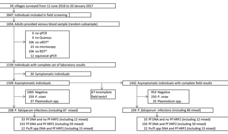
randomly selected to provide venous blood. Of these 1,656 samples, 1,509 samples from asymptomatic participants from 36 villages had a result with each of the five laboratory malaria detection tests and were included in the analysis (Fig. 1). The median age of participants was 36 years (IQR, 27 to 48 years; n⫽ 1,507), and the proportion of female participants was 52.9% (798/1,509). A median of 45 samples per village (IQR, 33 to 50 samples) was collected.
Combined results of uPCR analysis and Quansys ELISA (reference C) identified 1,005 negative samples, 259 P. vivax infections, 121 P. falciparum infections, 87 mixed P.
falciparum and P. vivax infections, and 37 Plasmodium infections of undetermined
species (Fig. 1).
uRDT detection of asymptomatic P. falciparum infections in the laboratory. For
the detection of P. falciparum infections (reference C), the sensitivity of the uRDT was 51.4% (95% CI, 44.4 to 58.4%) with a specificity of 99.5% (95% CI, 98.9 to 99.8%)
on March 26, 2019 by guest
http://jcm.asm.org/
(Table 2). The sensitivity and specificity of the uRDT were similar compared to the other references (Table 2): 52.0% (95% CI, 44.9 to 59.0%) and 99.3% (95% CI, 98.7 to 99.7%) for PfHRP2 detection (reference A), respectively, and 53.8% (95% CI, 46.3 to 61.1%) and 98.9% (95% CI, 98.2 to 99.4%) for P. falciparum DNA-positive samples (reference B), respectively. In comparison, the sensitivities of RDT and microscopy were similar and low (ranging from 23.6 to 27.4% [Table 2]). As a result, uRDT consistently exhibited a twofold increase in sensitivity compared to the sensitivity of RDT and microscopy.
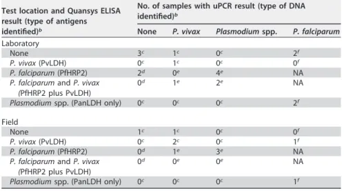
In total, 18 samples were positive for P. falciparum by uRDT in the laboratory and negative by at least one reference (references A to C) (Table 3). Only 5/18 (28%) uRDT-positive samples were negative by all references and corresponded to “true” false-positive results: 3 samples contained neither detected Plasmodium DNA nor antigens, 1 sample was P. vivax positive by uPCR and malaria negative by Quansys ELISA, and 1 sample was P. vivax positive by uPCR and Quansys ELISA. Among the remaining samples, 4/18 (22%) were positive by reference B and C, 7/18 (39%) were positive by reference C only, and 2/18 (11%) were positive by reference A only.
Direct comparison of uRDT with standard RDT and microscopy in the labora-tory. uRDT detected 100% (52/52) of RDT-positive P. falciparum infections (reference C),
while RDT detected only 49% (52/107) of uRDT-positive infections. Likewise, uRDT detected 98% (48/49) of microscopy-positive infections, while microscopy detected only 45% (48/107) of uRDT-positive infections.
uRDT detection of asymptomatic P. falciparum infections in the field. Overall,
1,442 asymptomatic adults with results for all five laboratory tests had also been tested in the field by uRDT and RDT (Fig. 1). The sensitivity of field testing was lower than in the laboratory for both uRDT and RDT, with respective sensitivities of 34.8% (95% CI, 28.3 to 41.8%) and 15.2% (95% CI, 10.6 to 20.9%) compared to reference C (Table 2). The uRDT in the field remained twofold more sensitive than the RDT compared to all three
FIG 1 Flowchart of study participants and results. Species results obtained by the combination of Plasmodium DNA detection by uPCR and Plasmodium antigens
by Quansys ELISA (reference C) are displayed.
on March 26, 2019 by guest
http://jcm.asm.org/
references (Table 2). The specificity of the tests conducted in the field was not significantly different from the tests conducted in the laboratory, 99.7% (95% CI, 99.2 to 99.9%) for uRDT and 99.9% (95% CI, 99.6 to 100%) for RDT.
In the field, there were 10 positive uRDT results corresponding to at least one negative reference test (Table 3). Four samples were negative by all reference methods, while two samples were positive according to reference B and C and four samples were positive according to reference C.
Direct comparison of field uRDT with laboratory uRDT. Out of 1,442 samples
with complete field and laboratory results, 75 were positive by uRDT in the field, and 113 were positive by uRDT in the laboratory. Field and laboratory results agreed in 67 positive samples and 1,321 negative samples. Eight samples were positive only by field uRDT, of which four were also positive by at least one reference method and four were negative by all reference methods. Conversely, 46 samples were positive in the labo-ratory but not in the field, of which 41 were also positive by at least one reference method and five were negative by all references.
The sensitivity of field testing compared to the same test in the laboratory was 59.2% (95% CI, 49.6 to 68.4%) for uRDT and 60.8% (95% CI, 46.1 to 74.2%) for RDT. The
TABLE 2 Specificity, sensitivity, and positive and negative predictive values for uRDT, standard RDT, and microscopy compared to
reference methodsa
Reference and test Test result
No. of negative samples
No. of samples with
P. falciparum
Specificity/sensitivity
(95% CI)b PPV/NPV (95% CI)c
Ref A (Quansys ELISA)
Lab uRDT Negative 1,298 97 Sp⫽ 99.3 (98.7–99.7) PPV⫽ 92.1 (85.5–96.3)
P. falciparum 9d 105 Se⫽ 52.0 (44.9–59.0) NPV⫽ 93.0 (91.6–94.3)
Lab RDT Negative 1,306 151 Sp⫽ 99.9 (99.6–100) PPV⫽ 98.1 (89.7–100)
P. falciparum 1 51 Se⫽ 25.2 (19.4–31.8) NPV⫽ 89.6 (88–91.2)
Microscopy Negative 1,306 153 Sp⫽ 99.9 (99.6–100) PPV⫽ 98.0 (89.4–99.9)
P. falciparum 1 49 Se⫽ 24.3 (18.5–)30.8 NPV⫽ 89.5 (87.8–91.0)
Field uRDT Negative 1,238 129 Sp⫽ 99.5 (99–99.8) PPV⫽ 92.0 (83.4–97.0)
P. falciparum 6d 69 Se⫽ 34.8 (28.2–41.9) NPV⫽ 90.6 (88.9–92.1)
Field RDT Negative 1,242 168 Sp⫽ 99.8 (99.4–100) PPV⫽ 93.8 (79.2–99.2)
P. falciparum 2 30 Se⫽ 15.2 (10.5–20.9) NPV⫽ 88.1 (86.3–89.7)
Ref B (uPCR)
Lab uRDT Negative 1,309 86 Sp⫽ 98.9 (98.2–99.4) PPV⫽ 87.7 (80.3–93.1)
P. falciparum 14d 100 Se⫽ 53.8 (46.3–61.1) NPV⫽ 93.8 (92.4–95.0)
Lab RDT Negative 1,322 135 Sp⫽ 99.9 (99.6–100) PPV⫽ 98.1 (89.7–100)
P. falciparum 1 51 Se⫽ 27.4 (21.1–34.4) NPV⫽ 90.7 (89.1–92.2)
Microscopy Negative 1,321 138 Sp⫽ 99.8 (99.5–100) PPV⫽ 96.0 (86.3–99.5)
P. falciparum 2 48 Se⫽ 25.8 (19.7–32.7) NPV⫽ 90.5 (88.9–92)
Field uRDT Negative 1,251 116 Sp⫽ 99.4 (98.8–99.7) PPV⫽ 89.3 (80.1–95.3)
P. falciparum 8d 67 Se⫽ 36.6 (29.6–44) NPV⫽ 91.5 (89.9–92.9)
Field RDT Negative 1,258 152 Sp⫽ 99.9 (99.6–100) PPV⫽ 96.9 (83.8–99.9)
P. falciparum 1 31 Se⫽ 16.9 (11.8–23.2) NPV⫽ 89.2 (87.5–90.8)
Ref C (combined uPCR and Quansys ELISA)
Lab uRDT Negative 1,294 101 Sp⫽ 99.5 (98.9–99.8) PPV⫽ 93.9 (87.8–97.5)
P. falciparum 7d 107 Se⫽ 51.4 (44.4–58.4) NPV⫽ 92.8 (91.3–94.1)
Lab RDT Negative 1,301 156 Sp⫽ 100 (99.7–100) PPV⫽ 100 (93.2–100)
P. falciparum 0 52 Se⫽ 25 (19.3–31.5) NPV⫽ 89.3 (87.6–90.8)
Microscopy Negative 1,300 159 Sp⫽ 99.9 (99.6–)100 PPV⫽ 98.0 (89.4–99.9)
P. falciparum 1 49 Se⫽ 23.6 (18.0–29.9) NPV⫽ 89.1 (87.4–90.7)
Field uRDT Negative 1,234 133 Sp⫽ 99.7 (99.2–99.9) PPV⫽ 94.7 (86.9–98.5)
P. falciparum 4d 71 Se⫽ 34.8 (28.3–41.8) NPV⫽ 90.3 (88.6–91.8)
Field RDT Negative 1,237 173 Sp⫽ 99.9 (99.6–100) PPV⫽ 96.9 (83.8–99.9)
P. falciparum 1 31 Se⫽ 15.2 (10.6–20.9) NPV⫽ 87.7 (85.9–89.4)
aThe references (Ref) are combined reference uPCR plus Quansys (PfDNA or Plasmodium DNA plus PfHRP2), reference P. falciparum DNA detection by uPCR, and
reference PfHRP2 detection by Quansys.
bThe specificity (Sp) and sensitivity (Se) and 95% confidence interval are shown.
cThe positive predictive value (PPV) and negative predictive value (NPV) and 95% confidence interval are shown.
dSee Table 3 for details on uRDT false-positive samples.
on March 26, 2019 by guest
http://jcm.asm.org/
corresponding specificity was 99.4% (95% CI, 98.8 to 99.7%) for uRDT and 99.9% (95% CI, 99.6 to 100%) for RDT.
Quantitative detection of parasites and PfHRP2. The qualitative increase in
sensitivity of the uRDT compared to standard RDT and microscopy was verified in terms of quantitative PfHRP2 detection by Quansys ELISA and parasitemia by uPCR. Quanti-tative analysis was performed on samples with a monospecific P. falciparum infection by uPCR. PfHRP2 was also quantified in these samples by Quansys ELISA (Fig. 2). In this population of asymptomatic carriers, the P. falciparum infections detected by uPCR had low densities ranging from 10 to 544,666 parasites per ml (p/ml) (geometric mean, 734 p/ml; 95% CI, 481 to 1,117 p/ml).
In the laboratory, the geometric mean parasitemia detected by uRDT was 3,019 p/ml (95% CI, 1,790 to 5,094 p/ml; n⫽ 79), compared to 11,352 (95% CI, 5,643 to 22,837 p/ml;
n ⫽ 38) by RDT (Table 4). The uRDT performed better than the RDT to detect
parasitemia between 1,000 and 10,000 p/ml (27/31 [87%] by uRDT compared to 15/31 [48%] by RDT) and between 100 and 1,000 p/ml (24/50 [48%] by uRDT compared to 3/50 [6%] by RDT). The performance of microscopy was similar to that of RDT. All three methods performed well above 100,000 p/ml (7/7 [100%]) and poorly below 100 p/ml (Table 4).
The median PfHRP2 concentration detected was 1,991 pg/ml (IQR, 588 to 8,377 pg/ml; n⫽ 75) by uRDT compared to 8,377 pg/ml (IQR, 3,910 to 16,500 pg/ml; n ⫽ 37) by RDT (Table 5). The uRDT detected 100% (37/37) of samples above 2,000 pg/ml, 88% (29/33) of samples between 100 pg/ml and 2,000 pg/ml, and 19% (9/47) of samples with detectable HRP2 below 100 pg/ml. In contrast, the RDT detected 95% (35/37), 6% (2/33), and 0% (0/47), respectively (Table 5).
TABLE 3 Number of samples per reference result for all samples with a P.
falciparum-positive uRDT and a P. falciparum-negative result for at least one reference (of references A to C)a
Test location and Quansys ELISA result (type of antigens identified)b
No. of samples with uPCR result (type of DNA identified)b
None P. vivax Plasmodium spp. P. falciparum
Laboratory
None 3c 1c 0c 2f
P. vivax (PvLDH) 0c 1c 0c 0f
P. falciparum (PfHRP2) 2d 0e 4e NA
P. falciparum and P. vivax (PfHRP2 plus PvLDH)
0d 1e 2e NA
Plasmodium spp. (PanLDH only) 0c 0c 0c 2f
Field
None 1c 1c 0c 0f
P. vivax (PvLDH) 0c 2c 0c 1f
P. falciparum (PfHRP2) 0d 1e 3e NA
P. falciparum and P. vivax (PfHRP2 plus PvLDH)
0d 0e 0e NA
Plasmodium spp. (PanLDH only) 0c 0c 0c 1f
aA color-coded version of Table 3 is available in the supplemental material (Table S1).
bSee footnotes c to f below. Values with footnotes c and d are false-positive results by reference C (n⫽ 7 for
laboratory and n⫽ 4 for field). Values with footnotes c and f are false-positive results by reference A (n ⫽ 9
for laboratory and n⫽ 6 for field). Values with footnotes c, d, and e are false-positive results by reference B
(n⫽ 14 for laboratory and n ⫽ 8 for field). NA, not applicable (corresponds to PfHRP2-positive and
PfDNA-positive samples [true PfDNA-positive according to all references]).
cP. falciparum-negative samples by all three reference methods. These are most likely false-positive uRDT results.
dPfHRP2-positive, PfDNA-negative, and Plasmodium DNA-negative samples (positive by reference A
only). These two samples could correspond to recently cleared infections with persisting HRP2 antigenemia.
ePfHRP2-positive, PfDNA-negative, and Plasmodium DNA-positive samples (P. falciparum positive by reference
A and C).
fPfHRP2 negative and PfDNA positive (P. falciparum positive by reference B and C).
on March 26, 2019 by guest
http://jcm.asm.org/
In the field, the geometric mean parasitemia and median HRP2 concentration detected were higher: for uRDT, 4,960 p/ml and 3,910 pg/ml; for RDT, 14,237 p/ml and 2,937 pg/ml (Table 4).
The probability of a monospecific P. falciparum infection to be given a positive rapid test result increased with parasitemia. In the laboratory, the probability of a positive test was significantly higher for uRDT than for RDT for infections between 50 and 300,000 p/ml (Fig. 3A). In the field, the probability of a positive test was significantly higher for uRDT than for RDT for infections between 200 and 100,000 p/ml (Fig. 3B). At any
FIG 2 Increased range of PfHRP2 detection in uRDT compared to RDT and corresponding increase in detection of
lower parasitemias for rapid tests performed in the laboratory (A) or in the field (B). Parasitemia measured by uPCR and the corresponding PfHRP2 concentration by Quansys ELISA are presented for samples positive for P. falciparum only by uPCR (monospecific infection) and by type of detection method. Vertical lines indicate PfHRP2 concen-trations of 100 and 2,000 pg/ml, while horizontal lines correspond to 1,000 and 100,000 parasites/ml (see the text). Mixed P. falciparum plus P. vivax uPCR-positive samples and Plasmodium DNA-positive samples with PfHRP2 are presented in Fig. S1 in the supplemental material.
on March 26, 2019 by guest
http://jcm.asm.org/
TABLE 4 Detection properties of the different tests by parasitemia for monospecific P. falciparum uPCR-positive infections (P. falciparum by reference B) a Test Parasitemia (no. of parasites/ml) b No. of infections detected by parasitemia category (% of uPCR reference) c Geometric mean (95% CI) Median (IQR) > 100,000 p/ml 10,000–100,000 p/ml 1,000–10,000 p/ml 100–1,000 p/ml < 100 p/ml Total Lab uRDT 3,019 (1,790–5094) 2,939 (484–13,748) 7 (100) 17 (94) 27 (87) 24 (48) 4 (11) 79 (55) Lab RDT 11,352 (5,643–22,837) 11,144 (4,377–52,429) 7 (100) 12 (67) 15 (48) 3 (6) 1 (3) 38 (27) Microscopy 19,132 (10,701–34,205) 15,151 (6,919–54,225) 7 (100) 16 (89) 12 (39) 1 (2) 0 (0) 36 (25) uPCR reference 734 (481– 1,117) 497 (90–5,506) 7 (100) 18 (100) 31 (100) 50 (100) 37 (100) 143 (100) Field uRDT 4,960 (2,657–9,260) 6,764 (632–18,924) 7 (100) 15 (83) 17 (57) 14 (29) 2 (6) 55 (39) Field RDT 14,237 (4,659–43,509) 15,229 (2,410–170,102) 7 (100) 7 (39) 5 (17) 3 (6) 1 (3) 23 (16) uPCR reference 742 (485–1135) 493 (95–4,941) 7 (100) 18 (100) 30 (100) 49 (100) 36 (100) 140 (100) aCorresponding graphs are presented in Fig. S2 in the supplemental material. b95% CI, 95% confidence interval; IQR, interquartile range. cParasitemia is given in the number of parasites per milliliter (p/ml).
on March 26, 2019 by guest
http://jcm.asm.org/
Downloaded from
parasitemia, the probability of a positive test was lower in the field than in the laboratory (Fig. 3A and B).
Readability. In the laboratory, the two independent readers interpreting uRDT
agreed on the results of 1,489 out of 1,509 samples (98.7%), of which 1,385 samples were interpreted negative and 104 positive. Of the 104 uRDT-positive readings, there were 102 P. falciparum infections and 2 false-positive results compared to reference C. Of the 20 discordant results between the first and second reader, 6 corresponded to P.
falciparum infections, and 14 were negative. For these discordant results, the third
reader interpeted 5/6 as P. falciparum infections and 5/14 as negative (Fig. 4). The positive signals were conclusive according to both readers in 77/104 (74.0%) concordant uRDT readings, corresponding to 76 P. falciparum infections and 1 false-positive result compared to reference C. Among the remaining 37 false-positive results by uRDT (27 concordant and 10 discordant results interpreted positive by reader 3), 13 were graded “inconclusive” by one reader only and 24 were graded “inconclusive” by both readers.
In total, 31 (29%) of 107 true P. falciparum infections identified by uRDT in the laboratory had very low intensity signals (12 graded “inconclusive” once and 19 twice). Misinterpretation of negative tests as very low intensity positive results also resulted in 6/7 false-positive results. The field tests corresponding to samples with low intensity in the laboratory were much less likely to be interpreted as positive: only 30% (8/26) of tests where at least one reader gave an inconclusive result compared to 75% (57/76) for samples with conclusive interpretation in the laboratory (Fig. 4).
DISCUSSION
Summary. In this study,⬎1,500 asymptomatic individuals were screened using the
uRDT in a low-incidence setting of eastern Myanmar. The performance of the uRDT against combined uPCR and Quansys ELISA (reference C) showed a twofold increase in sensitivity compared to that of the RDT, 51.4% versus 25.2%, respectively. The speci-ficities of the two rapid tests were similar, 99 to 100%. This twofold increase in sensitivity was also observed for performance against Quansys ELISA (reference A) and uPCR (reference B). It resulted from an improved detection of low-density infections between 100 and 10,000 p/ml. The sensitivity of uRDT was still higher than the sensitivity of RDT in the field, but both were lower than the same test performed in the laboratory.
Limitations and biases. The study was conducted as part of prevalence surveys
routinely aimed at “hotspot” identification within an elimination program (22). The surveys were conducted in different seasons in 35 villages of different malaria intensity profiles, ranging from 0 to ⬎40% P. falciparum prevalence based on uPCR detection (23). At the individual level, the venous blood surveys involved a random fraction of the adult population. In the smaller villages (e.g., 20 households,⬃100 inhabitants), the sample may have been similar to a continuous series of villagers when nearly 50% of the adult population was sampled. However, since only asymptomatic participants TABLE 5 Detection properties of the different tests by concentration for monospecific P.
falciparum uPCR-positive infections (P. falciparum by reference B)
Test
Median HRP2 concn (pg/ml) (IQR)
No. of infections detected by category of HRP2 concn (% of uPCR reference) >2,000 pg/ml 100–2,000 pg/ml <100 pg/ml Total Lab uRDT 1,991 (588–8,377) 37 (100) 29 (88) 9 (19) 75 (64) Lab RDT 8,377 (3,910–16,500a) 35 (95) 2 (6) 0 (0) 37 (32) Microscopy 4,780 (3,187–14,216) 27 (73) 6 (18) 2 (4) 35 (30) uPCR reference 517 (19–3,558) 37 (100) 33 (100) 47 (100) 117 (100) Field uRDT 3,910 (882–10,378) 34 (94) 16 (48) 3 (7) 53 (47) Field RDT 12,937 (7,811–16,500a) 22 (61) 0 (0) 0 (0) 22 (19) uPCR reference 553 (19–3,648) 36 (100) 33 (100) 45 (100) 114 (100)
aThe upper limit of quantification of the reference test was 16,500, which was reached in a total of 17
samples.
on March 26, 2019 by guest
http://jcm.asm.org/
were included in this analysis, it is unlikely that sampling could introduce a bias related to individual response to Plasmodium infections, which could potentially affect the test result.
The evaluations reported here correspond to two very different conditions: in the laboratory, two independent expert laboratory technicians evaluated the test under optimal conditions. The field survey team members received a 2-day training on uRDT before the start of their activities. A small team of six persons shared a heavy workload to obtain participant consent, collect demographic data, test all participants using RDT and uRDT, and collect venous blood from the subsample of participants. No dedicated workspace was available when surveys were conducted door to door. The availability of adequate light sources may also have been an issue where active rural populations were often most available in the early morning or in the evening, outside their daily activities.
As a result, it is evident that additional attention is needed to identify very faint signals as positive results: approximately 30% (37/114, of which 31 were also uPCR positive) of uRDTs interpreted as positive by two laboratory readers involved at least
FIG 3 Probability of a P. falciparum-positive test for uRDT compared to RDT according to the parasitemia
in uPCR-positive single P. falciparum infections. (A) Lab test results (solid line) and 95% CI (shaded area); field test results (dashed line). (B) Field test results (solid line) and 95% CI (shaded area); lab test results (dashed line).
on March 26, 2019 by guest
http://jcm.asm.org/
FIG 4 Readability of the uRDT in the laboratory. Agreement between readers, semiquantitative assessment of signal intensity, corresponding results acc ording to reference C, and matching results of field uRDT testing.
on March 26, 2019 by guest
http://jcm.asm.org/
Downloaded from
one of them grading the signal as “inconclusive” or doubtful, and only 30% (10/31) of these were identified in field conditions. Better training materials may be required to ensure that field users of the tests can more confidently interpret weak signals.
Using capillary blood instead of venous blood might have led to a lower sensitivity of the uRDT in the field than in the laboratory. However, a similar decrease in sensitivity was measured for the RDT, with field testing also detecting close to 60% of laboratory-positive samples. No difference was previously shown between results for capillary and venous blood testing for microscopically detectable parasitemia (24). This seems to confirm that the lower sensitivity might be related, rather, to readability issues associ-ated with faint signals in the field.
Generalizability. In a pilot study in the same setting, the sensitivity and specificity
of the uRDT compared to uPCR were estimated to be 44% and 99.8%, respectively, based on only nine P. falciparum infections detected by uPCR, of which four were PfHRP2 positive, and zero were positive by RDT (20). The present study estimated the sensitivity and specificity more precisely and demonstrated a twofold increase in detection by uRDT compared to RDT or microscopy. The performance for both uRDT and RDT under field conditions was approximately 40% lower than in the laboratory. It will be important to understand what interventions such as proficiency testing or better job aids could improve the operational performance of RDTs in the context of surveys. Both uRDT and RDT detected a significant number of infections below their lower limit of detection (LLOD), which leads to the geometric mean parasitemia detected being lower than the LLOD. This is potentially driven by the HRP2 antigen dynamics in the blood, which results in a nonstoichiometric relationship between HRP2 and para-sitemia.
Indeed, HRP2 persists in the bloodstream following parasite clearance for at least 7 days and up to several weeks (25, 26). A high-sensitivity HRP2-based assay raised concerns that it would detect residual HRP2 in the absence of parasites and thus increase the diagnosis of resolved infections. Out of 114 samples positive by uRDT in this study, only 2 had confirmed PfHRP2 antigens by Quansys ELISA but no Plasmodium DNA by uPCR. In contrast, Quansys ELISA detected HRP2 associated with P. falciparum DNA in 96/114 samples and with P. vivax DNA or undetermined Plasmodium DNA in 7/114 samples. HRP2 persistence was therefore not a major source of misdiagnosis among asymptomatic adults in this context.
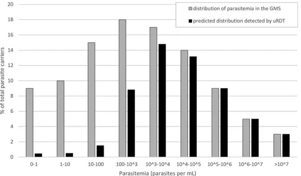
Given the distribution of parasitemia proposed by Imwong et al. (3) and the results obtained here, we extrapolate that the uRDT will be able to detect approximately 50% of all P. falciparum infections in GMS settings (Fig. 5). Standard RDT and microscopy already estimate reliably the number of carriers harboring parasitemias of ⱖ10,000 parasites/ml (Table 4). The improvement from uRDT comes from detecting infections with densities between 100 and 10,000 parasites/ml, a range of parasitemia which includes 35% of all infected carriers (Fig. 5).
Implications. Quality evidence regarding the size of the low-density infectious
reservoir acquired directly in the field could simplify the targeting of interventions for the rapid elimination of P. falciparum malaria and bring the decision-making closer to the field level (27). However, strategies targeting asymptomatic carriers will be widely adopted by national malaria control programs only if they rely on a simple, field-robust, and inexpensive diagnostic test to measure malaria prevalence and/or identify individ-uals harboring low-density P. falciparum infections. The uRDT evaluated here shows promising qualities in terms of cost (approximately 1 U.S. dollar/test), of testing instructions compared to the standard RDT, and of the range of low-density infections detected.
The uRDT will improve the accuracy of reservoir size measurement over RDT differently between contexts, since the distribution of low- versus high-density infec-tions is determined by the epidemiology of malaria. In this study, the detection of ⬎50% of uPCR-positive samples suggests that uRDT could be an adequate survey tool
on March 26, 2019 by guest
http://jcm.asm.org/
to estimate accurately the size of the low-density asymptomatic reservoir in the GMS (Fig. 5).
Given its increased performance compared to RDT, uRDT is also expected to improve effectiveness of mass screening and treatment (MSAT) interventions in low-incidence settings. In eastern Myanmar, MDA with a median 90% participation resulted in a median 90% decrease of prevalence in villages also accessing malaria early diagnosis and treatment at the community level (7, 23). With a similar 90% participation in an MSAT intervention, approximately 45% of P. falciparum-infected carriers in the village would be detected, resulting at best in a 50% decrease in prevalence. However, given the range of parasitemia detected by uRDT, ⬎95% of P. falciparum parasite biomass at the time of MSAT would be destroyed. Predicting the impact of an uRDT-based MSAT addressing only 50% of carriers is out of the scope of this study. It involves local transmission dynamics, which could be targeted by additional vector control interventions, and complex within-host interactions (duration of carriage, spon-taneous clearance, parasite density oscillations), which have recently begun to be investigated using highly sensitive methods (12).
Finally, as recently confirmed, a significant limitation of the uRDT is that it cannot detect P. falciparum infections with hrp2 hrp3 double deletion mutations (21). While
hrp2 hrp3 double deletion mutations are not frequent in the GMS and seem to be fairly
localized, this limitation could be addressed by including a second equally sensitive marker of P. falciparum infection on the same test (28, 29).
Conclusion. This study tested the performance of a new uRDT in the laboratory and
in the field using a large set of well-characterized surveys conducted within a P.
falciparum elimination program in the GMS. The uRDT detected close to 50% of
estimated P. falciparum infections, a twofold improvement compared to the numbers previously detected by standard RDT or microscopy. Field and in silico evaluations of uRDT in specific use cases, such as prevalence estimation, reactive case detection, or MSAT, and in different settings, are now necessary to define its future role in elimina-tion strategies.
FIG 5 Theoretical distribution of P. falciparum parasitemia among carriers in the Greater Mekong Subregion (reference data
from Imwong et al. [3] shown by the gray bars) and the corresponding fraction of each class that would be detected by uRDT (Table 4, laboratory uRDT results shown by the black bars). If used in optimal conditions, uRDT is predicted to detect up to 56% of the carriers in the GMS setting.
on March 26, 2019 by guest
http://jcm.asm.org/
SUPPLEMENTAL MATERIAL
Supplemental material for this article may be found athttps://doi.org/10.1128/JCM .00565-18.
SUPPLEMENTAL FILE 1, PDF file, 0.4 MB.
ACKNOWLEDGMENTS
We thank the communities of Karen/Kayin State, Myanmar, who participated in the surveys, the members of the survey teams, and particularly Saw B’Let, Keerati Kitti-tawee, Armon Santirad, Chode Wanachaloemlep, Saw Mar Nay, METF Area 1 coordina-tor Saw Nyunt Win, and the field team members, as well as SMRU laboracoordina-tory and logistics staff.
This work was supported by the Wellcome Trust (041843), the Bill and Melinda Gates Foundation (OPP1117507 and OPP1135840), the Regional Artemisinin Initiative (Global Fund against AIDS, Tuberculosis and Malaria). SMRU is part of the Mahidol Oxford University Research Unit, supported by the Wellcome Trust of Great Britain.
REFERENCES
1. Imwong M, Suwannasin K, Kunasol C, Sutawong K, Mayxay M, Rekol H, Smithuis FM, Hlaing M, Tun KM, Van Der Pluijm RW, Tripura R, Miotto O, Menard D, Dhorda M, Day NPJ, White NJ, Dondorp AM. 2017. The spread of artemisinin-resistant Plasmodium falciparum in the Greater Mekong Subregion: a molecular epidemiology observational study. Lancet Infect
Dis 17:491– 497.https://doi.org/10.1016/S1473-3099(17)30048-8.
2. Rabinovich RN, Drakeley C, Djimde AA, Hall BF, Hay SI, Hemingway J, Kaslow DC, Noor A, Okumu F, Steketee R, Tanner M, Wells TNC, Whittaker MA, Winzeler EA, Wirth DF, Whitfield K, Alonso PL. 2017. malERA: an updated research agenda for malaria elimination and eradication. PLoS
Med 14:1–17.https://doi.org/10.1371/journal.pmed.1002456.
3. Imwong M, Stepniewska K, Tripura R, Peto TJ, Lwin KM, Vihokhern B, Wongsaen K, von Seidlein L, Dhorda M, Snounou G, Keereecharoen L, Singhasivanon P, Sirithiranont P, Chalk J, Nguon C, Day NPJ, Nosten F, Dondorp A, White NJ. 2016. Numerical distributions of parasite densities
during asymptomatic malaria. J Infect Dis 213:1322–1329.https://doi
.org/10.1093/infdis/jiv596.
4. Parker DM, Carrara VI, Pukrittayakamee S, McGready R, Nosten FH. 2015. Malaria ecology along the Thailand-Myanmar border. Malar J 14:388. https://doi.org/10.1186/s12936-015-0921-y.
5. Smithuis FM, Kyaw M, Phe U, van der Broek I, Katterman N, Rogers C, Almeida P, Kager PA, Stepniewska K, Lubell Y, Simpson JA, White NJ. 2013. The effect of insecticide-treated bed nets on the incidence and prevalence of malaria in children in an area of unstable seasonal
trans-mission in western Myanmar. Malar J 12:363.https://doi.org/10.1186/
1475-2875-12-363.
6. Maude RJ, Socheat D, Nguon C, Saroth P, Dara P, Li G, Song J, Yeung S, Dondorp AM, Day NP, White NJ, White LJ. 2012. Optimising strategies for Plasmodium falciparum malaria elimination in Cambodia: primaquine, mass drug administration and artemisinin resistance. PLoS One
7:e37166.https://doi.org/10.1371/journal.pone.0037166.
7. Landier J, Kajeechiwa L, Thwin MM, Parker DM, Chaumeau V, Wilad-phaingern J, Imwong M, Miotto O, Patumrat K, Duanguppama J, Cer-queira D, Malleret B, Renia L, Nosten S, Von Seidlein L, Ling C, Proux S, Corbel V, Simpson JA, Dondorp AM, White NJ, Nosten FH. 2017. Safety and effectiveness of mass drug administration to accelerate elimination of artemisinin-resistant falciparum malaria: a pilot trial in four villages of
Eastern Myanmar. Wellcome Open Res 2:81.https://doi.org/10.12688/
wellcomeopenres.12240.1.
8. Thu AM, Phyo AP, Landier J, Parker DM, Nosten FH. 2017. Combating multidrug-resistant Plasmodium falciparum malaria. FEBS J 284:
2569 –2578.https://doi.org/10.1111/febs.14127.
9. Landier J, Parker DM, Thu AM, Carrara VI, Lwin KM, Bonnington CA, Pukrittayakamee S, Delmas G, Nosten FH. 2016. The role of early
detec-tion and treatment in malaria eliminadetec-tion. Malar J 15:363.https://doi
.org/10.1186/s12936-016-1399-y.
10. Gonçalves BP, Kapulu MC, Sawa P, Guelbéogo WM, Tiono AB, Grignard L, Stone W, Hellewell J, Lanke K, Bastiaens GJH, Bradley J, Nébié I, Ngoi JM, Oriango R, Mkabili D, Nyaurah M, Midega J, Wirth DF, Marsh K, Churcher TS, Bejon P, Sirima SB, Drakeley C, Bousema T. 2017. Examining
the human infectious reservoir for Plasmodium falciparum malaria in
areas of differing transmission intensity. Nat Commun 8:1133.https://
doi.org/10.1038/s41467-017-01270-4.
11. Tadesse FG, Slater HC, Chali W, Teelen K, Lanke K, Belachew M, Menberu T, Shumie G, Shitaye G, Okell LC, Graumans W, van Gemert G-J, Kedir S, Tesfaye A, Belachew F, Abebe W, Mamo H, Sauerwein R, Balcha T, Aseffa A, Yewhalaw D, Gadisa E, Drakeley C, Bousema T. 2018. The relative contribution of symptomatic and asymptomatic Plasmodium vivax and Plasmodium falciparum infections to the infectious reservoir in a
low-endemic setting in Ethiopia. Clin Infect Dis 66:1883–1891.https://doi
.org/10.1093/cid/cix1123.
12. Nguyen TN, von Seidlein L, Nguyen TV, Truong PN, Hung SD, Pham HT, Nguyen TU, Le TD, Dao VH, Mukaka M, Day NP, White NJ, Dondorp AM, Thwaites GE, Hien TT. 2018. The persistence and oscillations of submi-croscopic Plasmodium falciparum and Plasmodium vivax infections over time in Vietnam: an open cohort study. Lancet Infect Dis 18:565–572. https://doi.org/10.1016/S1473-3099(18)30046-X.
13. Tadesse FG, Van Den Hoogen L, Lanke K, Schildkraut J, Tetteh K, Aseffa A, Mamo H, Sauerwein R, Felger I, Drakeley C, Gadissa E, Bousema T. 2017. The shape of the iceberg: quantification of submicroscopic Plas-modium falciparum and PlasPlas-modium vivax parasitaemia and
gameto-cytaemia in five low endemic settings in Ethiopia. Malar J 16:99.https://
doi.org/10.1186/s12936-017-1749-4.
14. Cook J, Xu W, Msellem M, Vonk M, Bergström B, Gosling R, Al-Mafazy A-W, McElroy P, Molteni F, Abass AK, Garimo I, Ramsan M, Ali A, Mårtens-son A, Björkman A. 2015. Mass screening and treatment on the basis of results of a Plasmodium falciparum-specific rapid diagnostic test did not reduce malaria incidence in Zanzibar. J Infect Dis 211:1476 –1483. https://doi.org/10.1093/infdis/jiu655.
15. Mogeni P, Williams TN, Omedo I, Kimani D, Ngoi JM, Mwacharo J, Morter R, Nyundo C, Wambua J, Nyangweso G, Kapulu M, Fegan G, Bejon P. 2017. Detecting malaria hotspots: a comparison of rapid diagnostic test, microscopy, and polymerase chain reaction. J Infect Dis 216:1091–1098. https://doi.org/10.1093/infdis/jix321.
16. Eisele TP, Bennett A, Silumbe K, Finn TP, Chalwe V, Kamuliwo M, Ha-mainza B, Moonga H, Kooma E, Chizema Kawesha E, Yukich J, Keating J, Porter T, Conner RO, Earle D, Steketee RW, Miller JM. 2016. Short-term impact of mass drug administration with dihydroartemisinin plus pip-eraquine on malaria in southern province Zambia: a cluster-randomized
controlled trial. J Infect Dis 214:1831–1839. https://doi.org/10.1093/
infdis/jiw416.
17. van Eijk AM, Ramanathapuram L, Sutton PL, Kanagaraj D, Sri Lakshmi Priya G, Ravishankaran S, Asokan A, Tandel N, Patel A, Desai N, Singh R, Sullivan SA, Carlton JM, Srivastava HC, Eapen A. 2016. What is the value of reactive case detection in malaria control? A case-study in India and
a systematic review. Malar J 15:67.https://doi.org/10.1186/s12936-016
-1120-1.
18. Imwong M, Hanchana S, Malleret B, Rénia L, Day NPJ, Dondorp A, Nosten F, Snounou G, White NJ. 2014. High-throughput ultrasensitive molecular
on March 26, 2019 by guest
http://jcm.asm.org/
techniques for quantifying low-density malaria parasitemias. J Clin
Mi-crobiol 52:3303–3309.https://doi.org/10.1128/JCM.01057-14.
19. Britton S, Cheng Q, McCarthy JS. 2016. Novel molecular diagnostic tools for malaria elimination: a review of options from the point of view of high-throughput and applicability in resource limited settings. Malar J
15:88.https://doi.org/10.1186/s12936-016-1158-0.
20. Das S, Jang IK, Barney B, Peck R, Rek JC, Arinaitwe E, Adrama H, Murphy M, Imwong M, Haohankhunnatham W, Ling CL, Proux S, Rist M, Seilie AM, Hanron A, Daza G, Chang M, Nakamura T, Kalnoky M, Labarre P, Murphy SC, McCarthy JS, Nosten F, Greenhouse B, Allauzen S, Domingo GJ. 2017. Performance of a high-sensitivity rapid diagnostic test for Plasmodium falciparum malaria in asymptomatic individuals from Uganda and Myanmar and naive human challenge infections. Am J Trop
Med Hyg 97:1540 –1550.https://doi.org/10.4269/ajtmh.17-0245.
21. Das S, Peck RB, Barney R, Jang IK, Kahn M, Zhu M, Domingo GJ. 2018. Performance of an ultra-sensitive Plasmodium falciparum HRP2-based rapid diagnostic test with recombinant HRP2, culture parasites, and
archived whole blood samples. Malar J 17:118.https://doi.org/10.1186/
s12936-018-2268-7.
22. Parker DM, Landier J, Thu AM, Lwin KM, Delmas G, Nosten FH, Malaria Elimination Task Force Group. 2017. Scale up of a Plasmodium fal-ciparum elimination program and surveillance system in Kayin
State, Myanmar. Wellcome Open Res 2:98.https://doi.org/10.12688/
wellcomeopenres.12741.2.
23. Landier J, Parker DM, Thu AM, Lwin KM, Delmas G, Nosten FH, the Malaria Elimination Task Force Group. 2018. Effect of generalised access to early diagnosis and treatment and targeted mass drug administration on Plasmodium falciparum malaria in Eastern Myanmar: an observa-tional study of a regional elimination programme. Lancet 391:
1916 –1926.https://doi.org/10.1016/S0140-6736(18)30792-X.
24. Sandeu MM, Bayibéki AN, Tchioffo MT, Abate L, Gimonneau G, Awono-Ambéné PH, Nsango SE, Diallo D, Berry A, Texier G, Morlais I. 2017. Do the venous blood samples replicate malaria parasite densities found in capillary blood? A field study performed in naturally-infected
asymp-tomatic children in Cameroon. Malar J 16:345.https://doi.org/10.1186/
s12936-017-1978-6.
25. Marquart L, Butterworth A, McCarthy JS, Gatton ML. 2012. Modelling the dynamics of Plasmodium falciparum histidine-rich protein 2 in human malaria to better understand malaria rapid diagnostic test performance.
Malar J 11:74.https://doi.org/10.1186/1475-2875-11-74.
26. Mayxay M, Pukrittayakamee S, Chotivanich K, Looareesuwan S, White NJ. 2001. Persistence of Plasmodium falciparum HRP-2 in successfully treated acute falciparum malaria. Trans R Soc Trop Med Hyg 95:179 –182. 27. Cohen JM, Le Menach A, Pothin E, Eisele TP, Gething PW, Eckhoff PA, Moonen B, Schapira A, Smith DL. 2017. Mapping multiple components of malaria risk for improved targeting of elimination interventions. Malar
J 16:459.https://doi.org/10.1186/s12936-017-2106-3.
28. Cheng Q, Gatton ML, Barnwell J, Chiodini P, McCarthy J, Bell D, Cun-ningham J. 2014. Plasmodium falciparum parasites lacking histidine-rich protein 2 and 3: a review and recommendations for accurate reporting.
Malar J 13:283.https://doi.org/10.1186/1475-2875-13-283.
29. Baker J, Ho M-F, Pelecanos A, Gatton M, Chen N, Abdullah S, Albertini A, Ariey F, Barnwell J, Bell D, Cunningham J, Djalle D, Echeverry DF, Gamboa D, Hii J, Kyaw MP, Luchavez J, Membi C, Menard D, Murillo C, Nhem S, Ogutu B, Onyor P, Oyibo W, Wang SQ, McCarthy J, Cheng Q. 2010. Global sequence variation in the histidine-rich proteins 2 and 3 of Plasmodium falciparum: implications for the performance of malaria
rapid diagnostic tests. Malar J 9:129.https://doi.org/10.1186/1475-2875
-9-129.