HAL Id: hal-02373125
https://hal.archives-ouvertes.fr/hal-02373125
Submitted on 20 Nov 2020
HAL is a multi-disciplinary open access
archive for the deposit and dissemination of
sci-entific research documents, whether they are
pub-lished or not. The documents may come from
teaching and research institutions in France or
abroad, or from public or private research centers.
L’archive ouverte pluridisciplinaire HAL, est
destinée au dépôt et à la diffusion de documents
scientifiques de niveau recherche, publiés ou non,
émanant des établissements d’enseignement et de
recherche français ou étrangers, des laboratoires
publics ou privés.
fluorescent probes for cellular imaging
Doina Sirbu, Julien Diharce, Ivana Martinić, Nicolas Chopin, Svetlana
Eliseeva, Gerald Guillaumet, Stephane Petoud, Pascal Bonnet, Franck Suzenet
To cite this version:
Doina Sirbu, Julien Diharce, Ivana Martinić, Nicolas Chopin, Svetlana Eliseeva, et al.. An original class
of small sized molecules as versatile fluorescent probes for cellular imaging. Chemical Communications,
Royal Society of Chemistry, 2019, 55 (54), pp.7776-7779. �10.1039/C9CC03765A�. �hal-02373125�
COMMUNICATION
Received 00th January 20xx, Accepted 00th January 20xx DOI: 10.1039/x0xx00000x
An Original Class of Small Sized Molecules as Versatile Fluorescent
Probes for Cellular Imaging
Doina Sirbu,
aJulien Diharce,
aIvana Martinić,
bNicolas Chopin,
aSvetlana V. Eliseeva,
bGérald
Guillaumet,
aStéphane Petoud,
bPascal Bonnet
aand Franck Suzenet*
aAn unusual class, compact in sizes, of fluorescent probes based on pyridazino-1,3a,6a-triazapentalene scaffolds exhibits highly fluorescent properties (quantum yield values up to 73%, large Stokes shifts, emission wavelengths located in the green-yellow range, excellent solubility) with very good photostability suitable for optical imaging applications.
Fluorescent organic molecules are essential compounds for the detection, quantification and understanding of biological processes applied in chemical biology, biochemistry, biomedical research and diagnostics.1 The main advantages of fluorescent
organic molecules include high versatility, moderate molecular size and weight, chemical stability, and ability to exhibit switchable or activatable spectroscopic properties. This fluorogenic behavior can be initiated by a chemical or biochemical process according to four main different reaction mechanisms: pH sensitivity, complexation/decomplexation, formation or cleavage of a covalent bond and redox reactions.2-5
Although numerous fluorophores have already been described in the literature, the diversity of the molecular frameworks of those commonly used for cell imaging probes is often limited to coumarin, xanthene (fluorescein, rhodamine, Texas), BODIPY and cyanine cores.6,7 These dyes cannot be considered as ideal
probes for optical imaging since none of them combine high fluorescence with optimal absorption and emission wavelengths, good chemical and photostability, sufficient water solubility and small molecular weight/size.
Therefore, the discovery of novel fluorophore-based scaffolds remains an important challenge to offer new highly versatile platforms providing alternative and/or complementary molecular tools for novel bioimaging applications.8-12 In this
context, focusing on very low molecular weight/size fluorophores is of major interest for minimizing pharmacokinetic alterations of the targeting biomolecules to which they are linked.13 Small dyes may also find specific
interest in ligase fusion proteins.14 Obviously, those
fluorophores have to rely on (i) optimized spectroscopic properties, including high quantum yield values, large Stokes shifts and good photostability,15,16 and (ii) biological media
compatibility i.e. good chemical stability, low cytotoxicity, cell permeability and solubility, in order to be considered as suitable probes for cellular imaging.
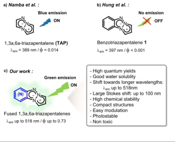
a)Namba et al. : Our work : N N N (N) Fused 1,3a,6a-triazapentalenes N N N b)Hung et al. : c) em= 389 nm /= 0.014 Benzotriazapentalene 1 OFF ON 1,3a,6a-triazapentalene (TAP) ON
- High quantum yields - Good water solublity
- Shift towards longer wavelengths:
emup to 518nm - Large Stokes shift: up to 100 nm - High chemical stability - Compact structures - Easy modulation - Photostable - Non toxic N N N em= 397 nm /< 0.001 emup to 518 nm /up to 0.73 Blue emission Green emission No emission
Fig 1 Summary of the state of the art in the field of triazapentalene derivatives.
Photophysical properties of 1a,3a,6a-triazapentalene (a) benzotriazapentalene (b), and novel nitrogen-containing fused ring TAP derivatives (c).
A few years ago, Namba’s team pioneered the fluorescent properties of the 1,3a,6a-triazapentalene (TAP) derivatives (Figure 1a)17 and showed that this highly condensed dipolar
structure offers very promising spectroscopic properties and suitability for biological applications.18,19 To study the impact of
the TAP substituents on spectroscopic properties, the development of specific synthetic pathways was required.20-26
a.Institut de Chimie Organique et Analytique - ICOA UMR7311, rue de Chartres, 45100 Orléans, France. E-mail: franck.suzenet@univ-orleans.fr
b.Centre de Biophysique Moléculaire CNRS UPR 4301, rue Charles Sadron, 45071 Orléans Cedex 2, France.
Electronic Supplementary Information (ESI) available: Experimental procedures, characterization data, 1H and 13C NMR spectra, theoretical calculations,
spectroscopic analysis, cellular optical imaging, cytotoxicity tests. See DOI: 10.1039/x0xx00000x
COMMUNICATION
Journal Name
2 | J. Name., 2012, 00, 1-3 This journal is © The Royal Society of Chemistry 20xx
Although some improvements of this highly promising TAP scaffold were observed, further investigations towards fused ring chemical entities have not been considered so far as benzotriazapentalene 1 as a fused ring system were already reported as non-fluorescent (Figure 1b).17
Herein, we report the design, the synthesis, the fluorescent properties and the cellular imaging compatibility of a novel class of small nitrogen-containing tricyclic frameworks. We demonstrate the crucial impact of the additional fused heteroaromatic ring on the TAP system towards promising spectroscopic properties for such a low molecular weight structure with excellent chemical stability.
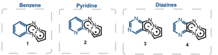
In the course of a research program on the design of energetic compounds, we identified pyrazolo-triazolo-pyridine27 and
pyrazolo-triazolo-pyrazine scaffolds28 (Figure 2, compounds 2
and 4, respectively). We postulated that the electron deficient character of the fused azine or diazine cycle could favor an intramolecular charge transfer (ICT) and promote the emission of this tricyclic scaffold (Figure 1c).
In order to study the effect induced by the presence of nitrogen atoms in the (di)azine rings on the fluorescent properties, we managed to synthesize and characterize the tricyclic scaffolds 1, 2, 3 and 4 (Figure 2). N N N N N N N N N N N N N N N N N
Benzene Pyridine Diazines
1 2 3 4
Fig. 2 Chemical structures of compounds 1, 2, 3 and 4.
Synthesis of compounds 1,29 227 and 428 was undertaken
following reported protocols and they were isolated in correct yields (see Supporting Information). Regarding compound 3, an original synthetic pathway was developed in four steps involving a key copper catalyzed C-N bond formation30 (see SI
for details). Having prepared the triazapentalene scaffolds 1-4, we then carried out the complete spectroscopic characterization (Table 1). The absence of fluorescence for compound 1 was confirmed.17 The azine scaffold (compound 2)
showed very weak fluorescence in contrast to its diazine isomers (compounds 3 and 4) which exhibited intense fluorescence bands with a very promising quantum yield values of 15% in DMSO.
Furthermore, the spectroscopic data clearly showed that the number and position of nitrogen atoms in the additional aromatic ring greatly affects the absorption and emission bands positions in agreement with the calculations (SI, Table S1). Interestingly, the presence of an extra nitrogen atom in compound 2 induced 17 nm and 49 nm red shifts for absorption and emission bands respectively compared to compound 1. This trend is confirmed with a pyrazine-containing fused aromatic ring (compound 4) with a remarkable bathochromic effect for both absorption and emission bands offering a very compact organic fluorescent scaffold with an emission maximum at 516 nm. It should be noted that the position of these two additional nitrogen atoms is particularly relevant as is illustrated with
compound 3 for which a blue shift of the absorption and emission bands was observed.
Table 1 Spectroscopic characterization of the tricyclic compounds 1, 2, 3 and 4 in DMSO. Dye abs[nm] [a] em[nm] [b] [nm] [c] max[d] [e] 1 370 397 27 12700 <0.001 2 387 446 59 18700 0.018 3 362 440 78 1200 0.15 4 418 516 98 15500 0.15
[a] Apparent maxima of absorption bands of the dyes. [b] Apparent maxima of emission bands of the dye. [c] Stokes Shift values calculated as the difference between the maxima of absorption and emission bands. [d] Units: L.mol-1.cm-1. [e] is the relative fluorescence quantum yield using Coumarin
153 (=0.38 in EtOH) as a reference standard.31
In addition, scaffold 4 showed a much higher molar extinction coefficient compared to compound 3, giving rise to an improved brightness. The pyrazino-[1a,3a,6a]-triazapentalene 4 stands out as our model scaffold which could be easily optimized by a simple and large scope of chemical modulation.
Interestingly, the fluorescent tricyclic scaffold 4 displayed a good solubility in water (>1 mg.mL-1) along with an apparent
position of the emission maximum larger than 530 nm, a Stokes shift of approximately 130 nm and a molar extinction coefficient of higher than 12000 M-1.cm-1. The excellent chemical stability
of dye 4 was also observed from pH4 to pH11 in water solutions. The solvatochromic behavior of 4 was obtained by recording absorption and emission spectra in various solvents. Whereas almost no change in the positions of the apparent absorption maxima was observed, a strong bathochromic effect was noticed from apolar to protic polar solvents characterized by a larger Stokes shift in water (128 nm) compared to chloroform (85 nm) (see SI for details).
In order to provide gain insights into the effect of nitrogen additions on the (het)aryl part of the tricyclic molecules (1 to 4), TD-DFT calculations were carried out10,32 (see SI for details) and
analysis of frontier orbitals for each compound was performed. The electronic density of the HOMO appeared, in general, to be weakly impacted by the substitution of a carbon atom by a nitrogen. The presence of nitrogen on the fused ring decreased the HOMO electronic density on adjacent atoms without any impact on the 1,3a,6a-triazapentalene moiety. In contrast, the pyridine ring in 2 displaced the LUMO electronic density from the 1,3a,6a-triazapentalene moiety to the 6-member ring, increasing the positive partial charges at the carbons and the π-deficient character of the fused ring (see Figure 3 and Table S1). These two complementary effects may explain the enhancement of the intramolecular charge transfer (ICT).
Figure 3. Major atomic contribution to the LUMO density for
The pyrazine ring in 4 slightly emphasized the effect on the LUMO atomic contribution compared to 1 while the pyrimidine ring in 3 did not induce the same behavior. Indeed, by comparing 3 and 4, the LUMO density is less localized on the pyrimidine part than on the pyrazine. Consequently, the enhanced π-electron deficient character of the pyrazine could Scheme 2 Synthesis of various diversified pyrazinyl-1a,3a,6a-triazapentalenes
from chloropyrazine 5 (pathway A) and from organometallic coupling processes with halogeno-triazapentalene 4i or 4j (pathway B).
Ar-B(OH)2 Pd(PPh3)4 K2CO3 N N N N N N N N N N N N N N N R N N Cl N3 N N H R (N) + 5 1) K2CO3,CH3CN, reflux, 1 to 6h 2) Thermolysis, 165 °C (N) Pathway A Pathway B 4i-j 4a-h 6a-g 1,4-dioxane/H2O or 1,4-dioxane/EtOH reflux or microwave 150 °C, 18min to 18h N N NN N OMe N N NN N N N N N N CF3 N N NN N CO2Et N N N N N CO2Et N N NN N N N N NN N N N N NN N CF3 4a, 26 % 4b, 48 % 4d, 40 % 4c, 41 % 4f, 73 % 4g, 77 % 4e, 57 % 4h, 85 % OMe N N NN N 4, 79 % N N N N N Br 4j, 97 % N N NN N I 4i, 93 % N N NN N 6a, 88 % N N NN N 6b, 70 % OMe N N N N N 6d, 82 % NMe2 N N NN N 6e, 85 % COOEt N N NN N 6c, 86 % S N N N N N 6g, 74 % N N N N NN N 6f, 45 % CF3 X Ar
explain the better fluorescence properties observed for compound 4.
With this information in hand, the potential of this new family of fluorophores was further investigated by considering the influence of an extra nitrogen in the azole ring as well as the effect of substituents on the reactivity and fluorescence properties. Various tetrazapentalenes and triazapentalenes bearing donor or acceptor groups on the azole moiety were synthesized in moderate to excellent yields in accordance with synthetic pathway A. In addition, aryl substituted
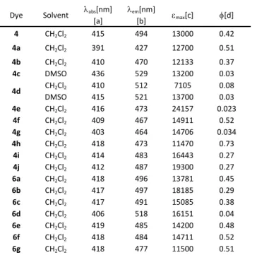
triazapentalenes were obtained through a Suzuki cross-coupling reaction following pathway B (Scheme 2, see SI for details). Table 2. Spectroscopic characterization of tricyclic analogs of
(pyrazinyl)-1a,3a,6a-triazapentalene and –tetrazapentalene in CH2Cl2 and DMSO.
Dye Solvent abs[a][nm] em[b][nm] max[c] [d]
4 CH2Cl2 415 494 13000 0.42 4a CH2Cl2 391 427 12700 0.51 4b CH2Cl2 410 470 12133 0.37 4c DMSO 436 529 13200 0.03 4d CH2Cl2 DMSO 410 415 512 521 7105 13700 0.08 0.03 4e CH2Cl2 416 473 24157 0.023 4f CH2Cl2 409 467 14911 0.52 4g CH2Cl2 403 464 14706 0.034 4h CH2Cl2 418 473 11470 0.73 4i CH2Cl2 414 483 16443 0.27 4j CH2Cl2 412 487 19300 0.27 6a CH2Cl2 418 496 13781 0.45 6b CH2Cl2 417 497 18185 0.29 6c CH2Cl2 417 491 15085 0.38 6d CH2Cl2 406 518 16151 0.04 6e CH2Cl2 419 485 14200 0.48 6f CH2Cl2 418 484 14711 0.52 6g CH2Cl2 418 477 11500 0.51
[a] Apparent maxima of absorption bands. [b] Apparent maxima of emission band. [c] Units: L.mol-1.cm-1. [d] is the relative fluorescence quantum yield
estimated by using Coumarin 153 (=0.38 in EtOH) as a reference standard.31
As shown in Table 2, the presence of an extra nitrogen on the azole ring (for compounds 4a and 4b) produced a strong hypochromic effect without affecting the other spectroscopic properties (ϕ and ). In contrast, an electron donating group wherever the position (compounds 4c, 4d and 6d) produced a bathochromic effect, while being detrimental for the quantum yield. However, an electron-withdrawing group (CF3, CO2Et,
ArCF3 and ArCO2Et for compounds 4f, 4h, 6f and 6e respectively)
was highly beneficial to the quantum yield (up to 73% in CH2Cl2
for compound 4h) when attached in position 3 of the azole. The molar extinction coefficient was greatly improved when the ester group was introduced in position 2. Finally, it is noteworthy that excitation and emission wavelengths were almost unaffected by the presence of an aryl group on the azole moiety (6a vs 4).
In order to demonstrate the ability of the novel triazapentalene compounds to be used as fluorescence probes for the imaging of living cells, epifluorescence microscopy experiments were performed on HeLa cells incubated with dye 6e. The resulting images suggest that this dye can be taken up by cells and the signal in the green region of the visible spectrum was recorded upon excitation using a 414 nm band pass 46 nm filter (see SI, Figure S9, Top) whereas, a minimal autofluorescence signal was observed for untreated cells (see SI, Figure S9, Bottom). Considering the very good quality of the signal in cells, the photostability of dye 6e was evaluated and compared to a commercially available bodipy fluorophore, LysoTracker Green DND-26. Interestingly, compound 6e shows a very good
COMMUNICATION
Journal Name
4 | J. Name., 2012, 00, 1-3 This journal is © The Royal Society of Chemistry 20xx
photostability (Figure 4) with a remaining signal up to 480 s (see SI for details). In this context, the cytotoxicity of probe 6e was evaluated with the Alamar Blue assay and more than 95% of cell viability was observed up to 170 µM concentration (see SI for details).
a (0s) b (30 s) c (100 s)
Fig 4 Photobleaching experiments (epifluorescence microscopy) performed on
HeLa cells incubated during 1h 30 min with a 95 µM solution of compound 6e (top) or with 50 nM LysoTracker Green DND-26 (bottom) after exposure to the continuous excitation light at 414 nm, band pass 46 nm filter during different times: (a) 0 s, (b) 30 s, (c) 100 s. 63× objective.
Conclusions
We created an original versatile tricyclic scaffold offering highly promising fluorescent properties. The fused diazine on the triazapentalene ring induced a strong bathochromic shift of the emission bands combined with improved quantum yield values due to a probable ICT process. This family appears to be non-toxic, highly photostable, chemically stable and some compounds are amphiphilic. With epifluorescence microscopy experiments we demonstrated that compound 6e can serve as a fluorescent probe for the optical imaging of living cells. A combination of small size/low molecular weight, good photostability, enhanced photophysical properties, large Stokes shifts, and versatility makes the reported fused triazapentalene family of fluorophores a promising tool for biological applications and is currently being explored for optical imaging.
Conflicts of interest
There are no conflicts to declare.
Acknowledgment
We thank the master students- Emeline Cristofaro and Dorian Guillot, for preliminary theoretical calculations of some compounds. This work was supported by the French ministry of research (D.S.), Labex SynOrg (ANR-11-LABX-0029), Labex IRON (ANR-11-LABX-0018-01), University of Orléans and Région Centre Val de Loire. S.P. acknowledges support from Institut National de la Santé et de la Recherche Médicale (INSERM). In addition, this work is partially supported by la Ligue Contre le Cancer, La Région Centre and the Réseau Canaux Ioniques du Cancéropôle Grand Ouest.
Notes and references
1 M. F. Juette, D. S. Terry, M. R. Wasserman, Z. Zhou, R. B. Altman, Q. Zheng and S.C Blanchard, Curr. Opin. Chem. Biol. 2014, 20, 103.
2 X. Li, X. Gao, W. Shi and H. Ma, Chem. Rev., 2014, 114, 590. 3 H. Zhu, J. Fan, J. Du and X. Peng, Acc. Chem. Res., 2016, 49,
2115.
4 Y. Niko, P. Didier, Y. Mely, G.-I Konishi and A. S. Klymchenko,
Scientific Reports, 2016, 6, 18870.
5 A. S. Klymchenko, Acc. Chem. Res., 2017, 50, 366. 6 L. D., Lavis and R. T. Raines, ACS Chem. Biol. 2008, 3, 142. 7 L. D.,Lavis and R. T. Raines, ACS Chem. Biol. 2014, 9, 855. 8 E. Kim, M. Koh, J. Ryu and S. B. Park, J. Am. Chem. Soc. 2008,
130, 12206.
9 E. J. Choi, E. Kim, Y. Lee, A. Jo and S. Bum Park, Angew.
Chem. Int. Ed., 2014, 53, 1346.
10 Y. Cheng, G. Li, Y. Liu, Y. Shi, G. Gao, D. Wu, J. Lan and J. You,
J. Am. Chem. Soc. 2016, 138, 4730.
11 E. Heyer, P. Lory, J. Leprince, M. Moreau, A. Romieu, M. Guardigli, A. Roda and R. Ziessel, Angew. Chem. Int. Ed., 2015, 54, 2995.
12 B. M. White, Y. Zhao, T. E. Kawashima, B. P. Branchaud, M. D. Pluth and R. Jasti, ACS Cent. Sci., 2018, 1173.
13 H. Kobayashi, M. Ogawa, R. Alford, P. L. Choyke and Y. Urano, Chem. Rev., 2010, 110, 2620.
14 Luke D. Lavis, Biochemistry, 2017, 56, 5165.
15 Q. Zheng, M. F. Juette, S. Jockusch, M. R. Wasserman, Z. Zhou, R. B. Altmana and S. C. Blanchard, Chem. Soc. Rev., 2014, 43,1044.
16 Z. Liu, L. D. Lavis and E. Betzig, Mol. Cell, 2015, 58, 644. 17 K. Namba, A. Osawa, S. Ishizaka, N. Kitamura and K. Tanino,
J. Am. Chem. Soc., 2011, 133, 11466.
18 R. Kamada, F. Tano, F. Kudoh, N. Kimura, Y. Chuman, A. Osawa, K. Namba, K. Tanino and K. Sakaguchi, PLoS ONE, 2016, 11(8): e016062.
19 J.-I. Sawada, A. Osawa, T. Takeuchi, M. Kaneda, S. Oishi, N. Fujii, A. Asai, K. Tanino and K. Namba, Bioorg. Med. Chem.
Lett., 2016, 26, 5765.
20 K. Namba, A. Mera, A. Osawa, E. Sakuda, N. Kitamura and K. Tanino, Org. Lett., 2012, 14, 5554.
21 R. Cai, D. Wang, Y. Chen, W. Yan, N. R. Geise, S. Sharma, H. Li, J. L. Petersen, M. Li and X. Shi, Chem. Commun., 2014, 50, 7303.
22 K. Namba, A. Osawa, A. Nakayama, A. Mera, F. Tano, Y. Chuman, E. Sakuda, T. Taketsugu, K. Sakaguchi, N. Kitamura and K. Tanino, Chem. Sci., 2015, 6, 1083.
23 A. Nakayama, S. Nishio, A Otani, A. Mera, A. Osawa, K. Tanino and K. Namba, Chem. Pharm. Bull., 2016, 64, 830 24 A. Mera, M. Ito, A. Nakayama and K. Namba, Chem. Lett.,
2017, 46, 539.
25 T. Hayashi, A. Osawa, T. Watanabe, Y. Murata, A. Nakayama and K. Namba, Tetrahedron Lett., 2017, 58, 1961.
26 B. Verbelen and W. Dehaen, Org.Lett. 2016, 18, 6412. 27 C. Nyffenegger, E. Pasquinet, F. Suzenet, D. Poullain, C.
Jarry,J.-M. Léger and G. Guillaumet, Tetrahedron, 2008, 64, 9567.
28 C. Nyffenegger, E. Pasquinet, F. Suzenet, D. Poullain and G. Guillaumet, Synlett, 2009, 1318.
29 B. M. Lynch and Y.-Y. Hung, J. Heterocycl. Chem., 1965, 2, 218.
30 H.-J. Cristau, P. P. Cellier, J.-F. Spindler and M. Taillefer, Eur.
J. Org. Chem., 2004, 695.
31 M. Brouwer and A. M. Brouwer, Pure Appl. Chem., 2011, 83, 2213.
32 D. Jacquemin, B. Mennucci and C. Adamo, Phys. Chem.