HAL Id: hal-02502377
https://hal.univ-lorraine.fr/hal-02502377
Submitted on 2 Dec 2020HAL is a multi-disciplinary open access archive for the deposit and dissemination of sci-entific research documents, whether they are pub-lished or not. The documents may come from teaching and research institutions in France or abroad, or from public or private research centers.
L’archive ouverte pluridisciplinaire HAL, est destinée au dépôt et à la diffusion de documents scientifiques de niveau recherche, publiés ou non, émanant des établissements d’enseignement et de recherche français ou étrangers, des laboratoires publics ou privés.
Self-Assembly of Porphyrin Nanostructures at the
Interface Between Two Immiscible Liquids
Andrés Molina-Osorio, David Cheung, Colm O’Dwyer, Andrew Stewart, M.
Dossot, Grégoire Herzog, Micheál Scanlon
To cite this version:
Andrés Molina-Osorio, David Cheung, Colm O’Dwyer, Andrew Stewart, M. Dossot, et al.. Self-Assembly of Porphyrin Nanostructures at the Interface Between Two Immiscible Liquids. Journal of Physical Chemistry C, American Chemical Society, 2020, �10.1021/acs.jpcc.0c00437�. �hal-02502377�
Self-assembly of Porphyrin Nanostructures at the
Interface between Two Immiscible Liquids
Andrés F. Molina-Osorio,1 David Cheung,2 Colm O’Dwyer,3,4 Andrew A. Stewart,5 Manuel
Dossot,6 Grégoire Herzog,6 and Micheál D. Scanlon.1,4,*
1The Bernal Institute and Department of Chemical Sciences, School of Natural Sciences,
University of Limerick (UL), Limerick V94 T9PX, Ireland.
2School of Chemistry, National University of Ireland, Galway, University Road, Galway, Ireland.
3School of Chemistry, and Tyndall National Institute, University College Cork, Cork, T12 YN60
Ireland.
4Advanced Materials and Bioengineering Research (AMBER) centre.
5The Bernal Institute and Department of Physics, School of Natural Sciences, University of
Limerick (UL), Limerick V94 T9PX, Ireland.
6CNRS-Université de Lorraine, LCPME UMR 7564, 405 Rue de Vandoeuvre, 54600
Villers-lès-Nancy, France.
This document is a postprint. Final version has been published in Journal of Physical Chemistry
ABSTRACT. One of the many evolved functions of photosynthetic organisms is to synthesize
light harvesting nanostructures from photoactive molecules such as porphyrins. Engineering
synthetic analogues with optimized molecular order necessary for the efficient capture and harvest
of light energy remains challenging. Here, we address this challenge by reporting the self-assembly
of zinc(II) meso-tetrakis(4-carboxyphenyl)porphyrins into films of highly ordered nanostructures.
The self-assembly process takes place selectively at the interface between two immiscible liquids
(water|organic solvent), with kinetically stable interfacial nanostructures formed only at pH values
close to the pKa of the carboxyphenyl groups. Molecular dynamics simulations suggest that the
assembly process is driven by an interplay between the hydrophobicity gradient at the interface
and hydrogen bonding in the formed nanostructure. Ex situ XRD analysis and in situ UV/vis and
steady state fluorescence indicates the formation of chlathrate type nanostructures that retain the
emission properties of their monomeric constituents. The self-assembly method presented here
avoids the use of acidic conditions, additives such as surfactants and external stimuli, offering an
alternative for the realization of light-harvesting antennas in artificial photosynthesis technologies.
1. INTRODUCTION
Photosynthetic organisms universally exploit antenna systems to capture high energy photons and
funnel this excitation energy toward a coupled reaction centre.1,2 Self-assembled molecular
antennae consisting of multi-layers of chromophores, such as porphyrins, can potentially function
as high efficiency light harvesters due to their exceptionally high molar absorption coefficients
(105 cm–1M–1).3 However, to mimic the evolved efficiency of these antennas and avoid
“concentration quenching” of the excited state at disordered trap sites,3 the supramolecular packing
of the individual chromophores must be precisely controlled and show long-range molecular
Molecular self-assemblies at “soft” liquid|air or immiscible liquid|liquid interfaces can exhibit
the required macroscale long-range order.6 These soft interfaces are exceptionally smooth, and
have no inherent defects leading to an unrivalled macroscale uniformity in molecule-interface
interactions.7,8 By contrast, the grain boundaries, step defects and edge sites always present at
solid|liquid interfaces can impede diffusion of adsorbed molecules, trapping them in local energy
minima as molecules stick to defect sites.9,10 The uniform templating of adsorbed molecules at
liquid|air or immiscible liquid|liquid interfaces has been exploited to create a variety of porphyrin
assemblies,11–16 yet a facile and robust route to create highly-ordered porphyrin nanostructures
remains challenging.
Here we describe the self-assembly of light harvesting nanostructures from readily available,
water-soluble and symmetrically substituted porphyrins at mild pH conditions. Thus, we avoid
acidic conditions (that would lead to the expulsion of the central metal ion),17 as well as the use of
synthetically challenging amphiphilic porphyrin molecules,18 and more complicated routes using
additives (e.g., divalent cations or surfactants)19–21 or external stimuli (e.g., electric fields).22–24 We
chose zinc(II) meso-tetrakis(4-carboxyphenyl)porphyrin (ZnTPPc) as a prototypical model system
to demonstrate this new means of self-assembly at an immiscible aqueous|organic interface.
Simply contacting aqueous ZnTPPc solutions prepared in citrate buffer at pH values between 5.1
and 6.0 with a neat, immiscible organic phase of -trifluorotoluene (TFT) lead to the
immediate formation of films of porphyrin nanostructures. We rationalise our findings using
molecular dynamics (MD) simulations that highlights the key role of the hydrophobicity gradient
at the immiscible aqueous|organic interface, and carboxylic acid-carboxylate hydrogen bonding
interactions in the formed nanostructure. The latter were found to be maximised at pH values close
stable interfacial nanostructures. The presence of multi-layers with strong visible light absorption,
and the crystalline macroscale long-range molecular order in the porphyrin nanostructure, suggests
these films as promising light-harvesting antennae in artificial photosynthetic technologies.
2. EXPERIMENTAL SECTION
2.1. Chemicals. All reagents were used as received without further purification.
Meso-tetrakis(4-carboxyphenyl)porphyrin (H2TPPc, ≥98%) and its zinc(II) derivative (ZnTPPc, ≥98%)
were obtained from Porphychem. Lithium hydroxide (LiOH, ≥98%), citric acid (H3Cit, ≥99.5%),
decamethylferrocene (97%), and 1,2-dichloroethane (DCE, ≥99.0%) were purchased from
Sigma-Aldrich, and α,α,α-trifluorotoluene (TFT, ≥99%) from Acros Organics. All aqueous solutions were
prepared using Milli-Q® deionized water (18.2 MΩ·cm). Aqueous solutions of ZnTPPc were
prepared by directly dissolving the solid in the lithium citrate buffer pre-adjusted to the desired
pH, followed by sonication of the solution for three minutes. Initially, H2TPPc was insoluble in
the buffer at neutral pH. Therefore, the solid was dissolved first in LiOH and the pH subsequently
adjusted with H3Cit. The ionic strength of each lithium citrate buffer solution containing either
porphyrin was maintained at 10 (±2) mM. For the photoelectrochemistry experiments,
bis(triphenylphosphoranylidene) ammonium chloride (BACl, 97%) and lithium
tetrakis(pentafluorophenyl)borate diethyletherate ([Li(OEt2)]TB) were obtained from
Sigma-Aldrich and Boulder Scientific Company, respectively.
Bis(triphenylphosphoranylidene)ammonium tetrakis(pentafluorophenyl)borate (BATB) was
prepared by metathesis of BACl and Li(OEt2)]TB, as described previously.26
2.2. Characterisation methodology. The films of Por-INs were gently transferred to a silicon
substrate for SEM, XRD and Raman characterisation, or an Agar Scientific TEM grid (Holey
interface. Prior to imaging, the samples were sequentially rinsed with water and TFT, and dried
under a stream of nitrogen gas. TEM images were acquired on a Thermo Fisher double aberration
corrected Titan Themis, spot size 6, on a Gatan Oneview detector. SEM images were obtained on
a FEI Quanta 650 FEG high resolution SEM. X-ray diffraction patterns (XRD) were acquired in
-2 geometry with a Phillips Xpert PW3719 diffractometer using Cu Kα radiation (λ = 0.15418 nm, operation voltage 40 kV, current 40 mA). Patterns were also acquired in -2 geometry using
a PANalytical X'pert PRO XRD. UV/vis absorbance spectra were collected in a Thermo Scientific
Evolution 60S UV/vis spectrophotometer with illumination provided by a Xenon light source
(accuracy ±0.8 nm). Steady-state fluorescence experiments were performed in a LS 55 Perkin
Elmer Fluorescence spectrometer. The experimental configuration implemented to obtain UV/vis
and steady-state fluorescence spectra of the Por-INs is illustrated in Figure S1. Raman
measurements were carried out using a Horiba Jobin Yvon T64000 spectrometer equipped with a
nitrogen cooled CCD detector. The laser wavelength was 532 nm with a power of 13 mW. Raman
spectra were obtained in 10 acquisitions of 30 seconds duration.
2.3. Adsorption isotherms. Vials containing biphasic systems of ZnTPPc or H2TPPc in lithium
citrate buffer (10 mM ionic strength, pH 5.8) at different initial bulk concentrations and TFT as
the organic phase were prepared and left to stand for 24 hours. After this time, the Por-INs formed
and all remaining unadsorbed porphyrin in the bulk aqueous phase was extracted by thoroughly
rinsing with porphyrin-free buffer solution. The solutions containing unadsorbed porphyrin were
collected and analysed by UV/vis absorbance spectroscopy to quantify the porphyrin concentration
therein (final bulk concentration). By subtracting the final from the initial bulk concentrations, the
surface concentration (number of moles adsorbed per geometric area of aqueous|organic interface)
2.4. Molecular dynamics simulations. Simulations of interfacial adsorption and assembly were
performed using a pre-equilibrated water|TFT interface, consisting water and TFT regions with
4000 water molecules and 1226 TFT molecules, respectively. Full details are provided in the
Supporting Information.
2.5. Photocurrent transient measurements at an electrified aqueous|organic interface.
Photocurrent measurements with a DC illumination were performed using the LED driver
provided by Metrohm Autolab in conjunction with a PGSTAT204 in a 4-electrode configuration
as presented in Figure S2. The overlap of the emission spectrum of the LED with the absorption
spectra of ZnTPPc, H2TPPc and their respective Por-INs is presented in Figure S3. The
determination of the photon flux at the electrified liquid|liquid interface as a function of the LED
driving current is outlined in Figure S4.
3. RESULTS AND DISCUSSION
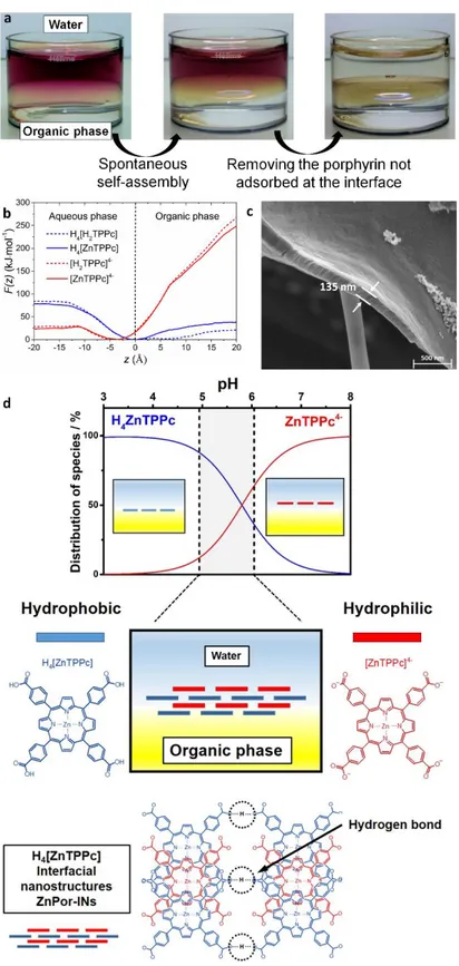
3.1. Triggering the formation of interfacial nanostructures. The selective formation of
ZnTPPc nanostructures at the interface between water and TFT was observed upon contacting a
ZnTPPc aqueous solution with neat TFT. A yellow/green colour was observed at the water|TFT
interface within minutes, easily distinguishable from the purple colour of the bulk ZnTPPc aqueous
solution, and associated with the formation of porphyrin interfacial nanostructures (Por-INs)
(Figure 1a). Self-assembly was observed only at pH values where the ratio between neutral,
protonated (H4[ZnTPPc]) and tetra-anionic, deprotonated ([ZnTPPc]4–) species was close to 1
(pH= pKaCOOH = 5.8), see Figure S5. Mono-, di- and triprotonated ZnTPPc species may also be
present. However, only one pKa was observed for the potentiometric titration of ZnTPPc with
within a very narrow interval of pH. Nevertheless, it must be noted that the interfacial protonation
equilibria is complex. Despite using a buffered aqueous electrolyte, an implicit assumption cannot
be made that the pH at the liquid|liquid boundary, where nanostructure formation is initiated, is
the same as in the bulk. Numerous studies have shown that local interfacial pH values can be
several units different from the bulk aqueous phase.27–29 Furthermore, the pKa of the
carboxyphenyl groups may differ from their bulk values, observed previously for other acid/base
equilibria at the water|air interface.30,31
MD simulations show that the hydrophilic, anionic [ZnTPPc]4– species prefer to sit in water at a
distance of ~3-5 Å above the interface due to their hydrophilic nature (Figure 1b), and suggest that
initially, hydrophobic neutral H4[ZnTPPc] species accumulate at the interface driven by the
hydrophobicity gradient at the immiscible liquid|liquid interface (Figure 1b and Figure S7). This
interfacial layer acts as a template structure for the [ZnTPPc]4– species to adsorb via carboxylic
acid-carboxylate hydrogen-bonding and π-π interactions. In this manner, a crystalline film of
ZnTPPc nanostructures builds up layer-by-layer at the interface. Akin to the clathrate crystals of
ZnTPPc developed by Goldberg and coworkers,32–36 the strength of each individual hydrogen bond
or π-π interaction may be insubstantial, but the cooperative effect allows the net enthalpies of these
multivalent interactions to cumulatively rival the strength of a covalent bond and stabilise the
Por-IN. Ex situ scanning electron microscopy (SEM) images reveal the thickness of the film of
Figure 1. Formation of porphyrin interfacial nanostructures. a) Optical images of the formation of
Por-INs at the interface between water and TFT. b) Computed potential of mean force or free energy profiles for translation of ZnTPPc molecules across the water|TFT interface, averaged over
10 ns of free molecular dynamics, with the carboxylate groups on the 4-carboxyphenyl-substitutents either fully deprotonated, [ZnTPPc]4– (solid red line) or fully protonated H
4[ZnTPPc]
(solid blue line). c) Scanning electron microscopy (SEM) image of a film of ZnPor-INs transferred from the water|TFT interface to a copper grid with a holey carbon substrate. d) Distribution percentage of neutral H4[ZnTPPc] (blue line) and anionic [ZnTPPc]4– (red line) porphyrin species
in the aqueous phase as a function of pH, with a schematic description of the layered structure of the ZnPor–INs, and chlathrate structure of ZnPor-INs obtained from ex situ XRD analysis (vide
infra). Note, for clarity, only a simple hydrogen bonding model is displayed for the chlathrate
structure of the ZnPor-INs. However, coordination of carboxyl groups to the central zinc atom is also important to form a well ordered 3D-structure, as described vide infra.
Control experiments and MD simulations demonstrated that ZnTPPc and free-base H2TPPc
molecules are kinetically stable in solution at pH 5.8, and do not undergo spontaneous bulk
aggregation in the concentration range studied (Figure S8). This indicates that the Por-INs form
only by self-assembly in situ at the water|TFT interface. Thus, to achieve selective Por-IN
formation, the pH of the aqueous solution must be controlled across a narrow pH range between
5.1 and 6.0 (Figure 1d). More alkaline conditions inhibit formation of the Por-INs due to
electrostatic repulsion between tetra-anionic porphyrins. More acidic conditions induce the
spontaneous formation of aggregates in the bulk aqueous phase (Figure S9). This pH dependency
of Por-IN formation indicates that cooperative H-bonding is key for self-assembly of the
nanostructure. This finding is in line with a detailed examination of the photoelectrochemistry of
ZnTPPc and decamethylferrocene at the water|1,2-dichloroethane (DCE) interface by Girault and
co-workers.37 Based on the photocurrent dependence on the angle of polarisation under total
internal reflection, they concluded that the spontaneous 2D coverage of ZnTPPc molecules at the
interface is pH dependent, sharply decreasing above pH 6. While no interfacial nanostructures
that spontaneous 2D assembly is the initial step in the generation of 3D interfacial nanostructures.
Furthermore, nanostructure self-assembly is not restricted to a single immiscible biphasic system,
with experiments showing that Por-INs also form selectively at the water|DCE interface (Figure
S10).
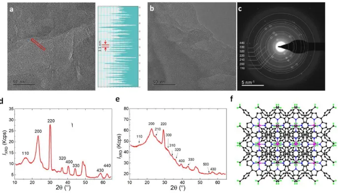
3.2. Molecular structure of the Por-INs. Transmission electron microscopy (TEM) and X-ray
diffraction (XRD) analysis confirm the crystalline and layered nature of the porphyrin interfacial
nanostructures. Ex situ TEM images with corresponding selected area electron diffraction (SAED)
analysis, and patterns from ZnPor-INs (Figure 2a-c) were acquired after immobilization of the film
on an amorphous hydrophilized glass substrate. The diffraction pattern for ZnPor-INs bears a
striking resemblance to NAFS (nanofilm on a solid surface) structural models for liquid phase
interfacial synthesis of highly ordered molecular nanosheets (Figure 2a).38 SAED and XRD
estimate a 0.55-0.57 nm interplanar spacing between {110} planes, with a ~1.1 nm periodicity
measured from HRTEM lattice fringes. The intense 220 reflection is consistent with a lack of
layer-on-layer stacking order (due to non-registered stacking), but a highly crystalline nanosheet
material is formed. The (200) reflections indicate strong axial coordination that is a distinct feature
of layering along the c-axis normal to the support. Clearly discernible (hk0) reflections are
consistent with a tetragonal unit cell with preferred growth orientation along the plane of the
liquid|liquid interface (Figure 2b,c). Experiments with nanostructures of the free base H2TPPc
(H2Por-INs) demonstrate the key role of the central metal ion in enhancing the crystallinity of the
film of porphyrin interfacial nanostructures. H2Por-INs grown in the same way also form a
crystalline 2D layered material, and show a similar XRD pattern but with a more pronounced
amorphous background and suppressed reflection intensity compared to the ZnPor-INs (Figure
structure of the ZnPor-INs is illustrated in Figure 2f. This structure assumes interdigitation to
account for the reduced unit cell spacing compared to a NAFS-1 or NAFS-2 tetragonal
metal-organic nanosheet crystalline structure.
Figure 2. Characterisation of ZnPor-INs and H2Por-INs ex situ by transmission electron
microscopy (TEM) and X-ray diffraction (XRD). a) TEM image of ZnPor-INs transferred from the water|TFT interface to a copper grid with a holey carbon substrate, taken at 185 k magnification. The profile of the lattice fringing shown on the right is taken from the area marked by the red box in the TEM image. b) TEM image of ZnPor-INs, taken at 185 k magnification at an accelerating voltage of 300 kV and c) the corresponding diffraction pattern with a 40 μm selected area aperture and camera length of 580 mm. d) XRD pattern of the ZnPor-INs transferred to a silicon substrate. Patterns were acquired in ω-2θ geometry and clearly discernible (hk0) reflections confirms a tetragonal unit cell and dominant c-axis layering. e) XRD of the H2Por-INs. f) Schematic representation of the crystalline and layered clathrate-type structure of the
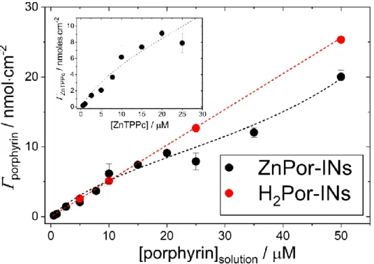
3.3. The interfacial nanostructures represent a kinetically trapped metastable state rather than a thermodynamically stable state. The interfacial concentration of Por-INs was measured
as a function of the solution concentration of ZnTPPc and H2TPPc, respectively. Over the
concentration range studied (0.5-100 µM), ZnTPPc adsorption followed a Brunauer-Emmet-Teller
(BET) isotherm behaviour, whereas H2TPPc adsorption followed a linear isotherm behaviour
(Figure 3). Using the BET model for liquid phase adsorption reported by Ebadi et al.,39 the
isotherm obtained for ZnTPPc adsorption was fit to Eqn. 1:
Г = Г𝑚 𝐾1𝐶𝑒𝑞.
(1−K2Ceq.)(1−𝐾2𝐶𝑒𝑞.+𝐾1𝐶𝑒𝑞.) (1)
in which 𝐾1 and 𝐾2 are the pseudo-equilibrium constants of adsorption to form the first and second
layers of the ZnPor-INs, Г and Г𝑚 are the equilibrium and monolayer porphyrin surface
concentrations, respectively, and 𝐶𝑒𝑞. is the equilibrium concentration of the porphyrin in solution.
Non-linear fitting determined Г𝑚 as 13.08 (±0.94) nmolcm–2, and 𝐾
1 and 𝐾2 as 4.55 x 10⁻² (±0.25)
and 8.79 x 10⁻³ (±2.21 x 10⁻⁴) Lµmol, respectively (95% confidence). Thus, the Gibbs free energy
of adsorption (ΔGads.) of ZnTPPc molecules to form the first and second layers of the ZnPor-INs
are 7.65 kJmol–1 and 11.72 kJmol–1, respectively. These values suggest that the interfacial
nanostructures represent a kinetically trapped metastable state rather than a thermodynamically
stable state. In other words, the interfacial molecular self-assembly process is under kinetic control,
and such out-of-equilibrium self-assembly systems are known to yield porphyrin nanostructures
that are inaccessible through the spontaneous thermodynamic process.40 The latter kinetic control
has also been observed in supramolecular polymerization of porphyrins,41 and the self-assembly
Figure 3 Adsorption isotherms obtained at 20 °C of ZnTPPc and H2TPPc at the water|TFT
interface. Inset: Adsorption isotherm data for ZnTPPc at the water|TFT interface, highlighting the Brunauer-Emmet-Teller (BET) behavior
The difference in the adsorption isotherms for ZnTPPc and H2TPPc further emphasises the
influence of the metal centre during porphyrin interfacial adsorption. Previous studies at an
aqueous|dodecane interface demonstrated that the adsorption of an oil-soluble metalloporphyrin
was highly dependent on the nature of the central metal ion due to the different water coordination
geometries around different metals.45 Also, the study by Girault and co-workers37 noted vide supra,
concluded that there is a direct link between the surface coverage and orientation of ZnTPPc
adsorbed at the interface. A dihedral angle between the porphyrin ring and the surface normal in
the range of 60 to 75° was observed, depending on the bulk ZnTPPc concentration and the
application of a Galvani potential difference at the liquid|liquid interface, and rationalized in terms
MD simulations to quantify the influence of the presence or absence of Zn2+, and the protonation
state, on the adsorption of ZnTPPc and H2TPPc at the water|TFT interface. For all model variants,
there is a significant degree of orientational freedom (Figure S11), with all molecules adopting a
range of orientations relative to the interface. This is reflected in the orientational angle probability
distributions which have peaks at a low (<15°) angle but remain non-zero for all angles. (Figure
S11). This suggests that the difference in the adsorption behaviour stems from different
supramolecular packing modes within the Por-INs and not their interaction with the liquid|liquid
interface.
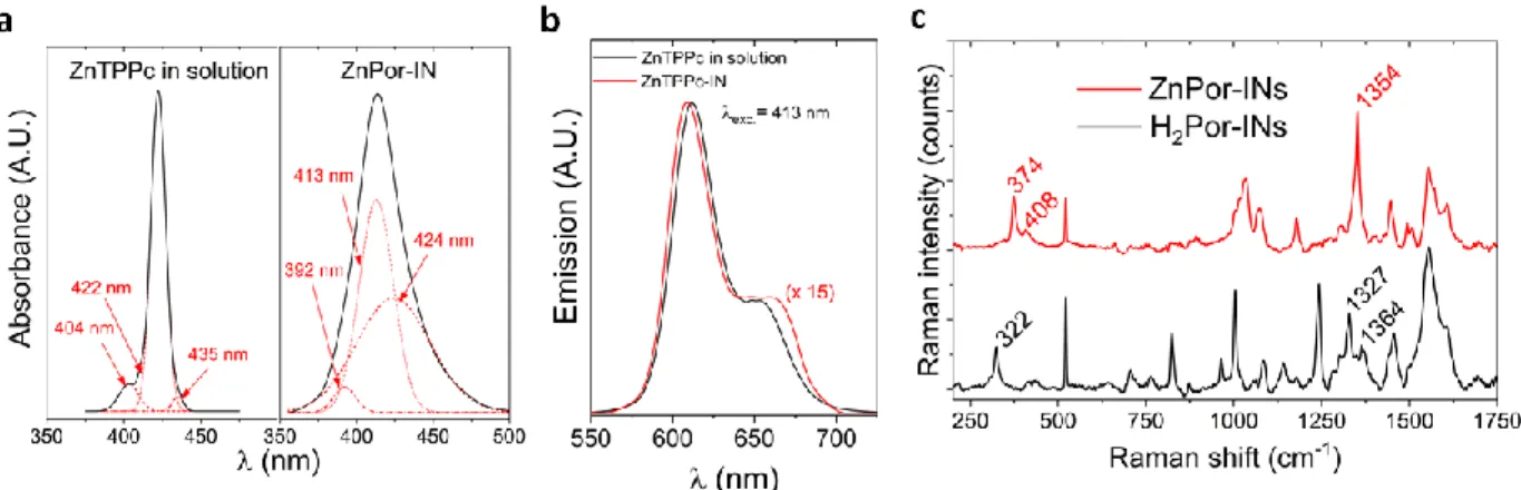
3.4. Influence of nanostructure formation on the photophysical properties of the assembly.
In situ UV/vis absorption spectra of ZnTPPc in solution and ZnPor-INs are shown in Figure 4a
and Figure S12. The λmax. value of the Soret band for the ZnPor-INs is blue-shifted (from 422 to
413 nm with respect to ZnTPPc in solution). Perturbation of the electronic absorption spectra in
terms of shifts and broadening of the Soret band, indicate the presence of multiple structural
domains within the ZnPor-INs. Analogous to ZnTPPc in solution, ZnPor-INs display two Q bands,
indicating that the porphyrin ring remains metallated with Zn2+ and thus retains the D
4h symmetry.
Additionally, in solution, ZnTPPc presented one main peak centred at 422 nm (Figure 4a and Table
S1). This peak further indicates the monomeric nature of ZnTPPc under the experimental
conditions used during this work. The latter is supported with the adherence of ZnTPPc in solution
to the Beer-Lambert law across an extensive concentration range (Figure S12), and with previous
Figure 4. Spectroscopic characterisation of the ZnPor-INs by in situ UV/vis absorbance and
steady-state fluorescence, and ex situ Raman spectroscopy. Deconvolution of the Soret absorbance bands for a) ZnTPPc in solution and ZnPor-INs. b) Comparison of the fluorescence emission spectra of ZnTPPc in solution and ZnPor-INs for an excitation wavelength (λexc.) of 418 nm. c)
Comparison of the Raman spectra of films of ZnPor- and H2Por-INs transferred from the
water|TFT interface to a silicon substrate (see Table S2 for peak assignments).
In situ steady-state fluorescence spectra was measured for an excitation wavelength (λexc.) of 413
nm (Figure 4b). Comparison of the emission profiles of ZnTPPc in solution and the ZnPor-INs
shows that the Q(0,0) transient is slightly blue-shifted and Q(0,1) transition slightly red-shifted
upon nanostructure formation. Following Kasha’s exciton model, the ZnPor-INs can be classified
as H-type structures (with a blue shifted max., see Figure 4a) where strong − overlap is expected.
− interactions are reported to lower exciton diffusion and fluorescence rates.47,48 However,
energy transfer within these types of molecular assemblies is also affected by other parameters
such as intermolecular distance, molecular packing orientation or molecular crystallinity.49 The
importance of the latter is highlighted in several works where high carrier mobility is observed in
structures with strong − stacking interactions.49,50 For example, Vohra et al.50 reported
efficient active layers in solar cells. Thus, the retention of fluorescence emission after
nanostructure formation demonstrates how the long-range molecular order in the nanostructure
leads to diminished concentration quenching, a key attribute when designing light-harvesting
antennae in artificial photosynthetic technologies. Additional dependence of the emission
properties of these nanostructures on the excitation wavelength is presented in Figures S13 and
S14.
Since ZnTPPc self-assembles in mild pH conditions, expulsion of Zn2+ was avoided, as
confirmed by analysis of the effect of nanostructure formation on the vibrational modes of the
ZnPor- and H2Por-INs by ex situ Raman spectroscopy (Figure 4c). Prominent differences between
the two are entirely consistent with previous comparisons of metallo- and free-base
4-carboxyphenyl-substituted porphyrin Raman spectra (Table S2).51,52 The retention of Zn2+ is a key
advantage of the interfacial self-assembly method described over common aggregation methods
in bulk solution at acidic conditions. The presence of the metal increases the inter-system crossing
(ISC) rate constant, kISC, due to the heavy atom effect, increasing the probability of the forbidden
S1→ T1 transition. From the T1 state, relaxation may occur via phosphorescence or charge transfer.
The long-lived (up to millisecond) excited triplet state lifetimes provides sufficient time for the
excited state to efficiently interact with ground state quencher molecules.53
As a proof-of-concept, the photoactivity of the ZnPor-INs was demonstrated by mediating
interfacial photo-induced electron transfer (PET) between redox species chemically confined to
different sides of a liquid|liquid interface using the methodology pioneered by Girault and
co-workers (Figure 5a).37,53–55 The multi-layers of porphyrin in the ZnPor-INs floating at the interface
function as light harvesters. Upon illumination, the generated triplet excited state was reductively
transfer from O2. The charge separation was accompanied by an electrical photocurrent through
an external circuit, and the latter increased on application of a more positive Galvani potential
difference (Figure 5b-c).
Control cyclic voltammograms (CVs) were carried out, in the dark and with chopped LED
illumination, in the presence of the ZnPor-INs at the interface and decamethylferrocene in the TFT
phase (Figure S15). Charge transfer peaks due to the adsorbed ZnPor-INs precluded the study of
applied Galvani potentials less than –0.1 V (Figure S15). Decamethylferrocene is a relatively
strong electron donor, capable of reducing dissolved O2, and consequently leading to a dark current
at positive Galvani potentials with an onset of ~+0.15 V. The mechanism underlying this dark
current has been identified previously as the reduction of O2 by first proton transfer to the organic
phase, followed by formation of a decamethylferrocene-hydride. The latter then reacts with
dissolved O2 in the TFT to generate a peroxyl radical species, HO2•.56 However, as seen in Figure
S15, the photocurrents obtained are superimposed upon this dark current and can be resolved by
background subtraction of the dark current (as is the case for the photocurrent transients shown in
Figures 5b and c).
A major future perspective outside the scope of this article is an in-depth exploration of PET at
electrified liquid|liquid interfaces using this photoactive film. In particular, a detailed analysis on
the potential dependence of the magnitude and line-shape of the photocurrent transient
measurements is required and will lead to an elegantly simple system to realise a new approach to
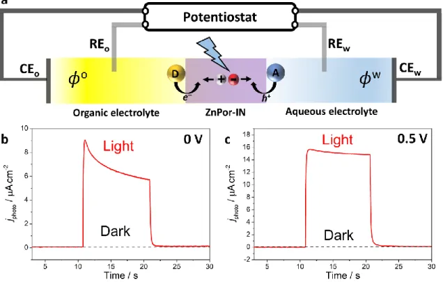
Figure 5. Photoconversion at the interface between two immiscible liquids. a) Schematic of
“soft-photoconversion”; converting light energy to chemical energy using dye-sensitised electrified
liquid|liquid interfaces. The donor species (D) is decamethylferrocene and the acceptor species (A)
is O2. The ZnTPPc triplet excited state in the crystalline, layered ZnPor-INs undergoes reductive
quenching by decamethylferrocene, with O2 regenerating the ground state. Thus, light energy is
converted to chemical energy in the form of the oxidised donor (D+) and reduced acceptor (A–)
spatially separated on either side of the water|TFT interface. Photocurrent transients were
measured b) at 0 V and c) at a positive polarisation of the interface (0.5 V), at pH 5.8 and 5 mM
decamethyferrocene in the TFT phase. The ZnPor-INs floating at the water|TFT interface were
illuminated with an LED (470 nm at 50 mW·cm–2) controlled by the potentiostat. The water|TFT
interface was electrified using a specialised 4-electrode electrochemical cell (CEw and CEo are the
electrodes in each phase). The supporting organic electrolyte was 5 mM bis(triphenylphosphoranylidene)ammonium tetrakis(pentafluorophenyl)borate (BATB).
4. CONCLUSIONS
The defect-free nature of the water|organic interface provides an ideal platform to self-assemble
interfacial nanostructures with unique structural arrangements. In this Article, we report the
self-assembly of interfacial nanostructures of zinc(II) meso-tetrakis(4-carboxyphenyl)porphyrin. The
nanostructures are stabilised by cooperative hydrogen bonding and, due to the templating
interaction of the interface with adsorbed porphyrin molecules, possess a highly ordered structure.
This approach uniquely harnesses the difference in hydrophobicity between the neutral protonated
and tetra-anionic non-protonated versions of the porphyrin at pKa conditions, combined with the
introduction of a hydrophobicity gradient to trigger interfacial self-assembly. We open a new
avenue to kinetically stable porphyrin nanostructure formation under mild experimental conditions
without the need for acidic pH, designer amphiphilic porphyrin molecules, aggregation-inducing
additives or external triggers. The feasibility of using such nanostructures for light collection and
harvesting was demonstrated in situ by measuring photocurrents associated with interfacial
photo-induced electron transfer across the water|TFT interface.
ASSOCIATED CONTENT Supporting Information
Supporting experimental methods (in situ microscopy, molecular dynamic simulations,
formation, potentiometric titration data, molecular dynamic studies of porphyrins in the bulk
(dimerisation free energy calculations) or at the liquid|liquid interface (mechanism of
layer-by-layer formation of the porphyrin interfacial nanostructures, determining probability distributions of the angle of orientation of adsorbed porphyrins), spectroscopic studies of the porphyrins in bulk
aqueous solutions (influence concentration and pH on dimerisation or aggregation, respectively),
in situ spectroscopic studies of porphyrin interfacial nanostructures (UV/vis, fluorescence and
Raman). AUTHOR INFORMATION Corresponding Author *micheal.scanlon@ul.ie (M.D. Scanlon) ORCID ID numbers Andrés F. Molina-Osorio: 0000-0001-8356-6381 David L. Cheung: 0000-0002-3994-2295 Colm O’Dwyer: 0000-0001-7429-015X Andrew Stewart: 0000-0002-3081-5644 Manuel Dossot: 0000-0003-0575-025X Grégoire Herzog: 0000-0003-1932-9300 Micheál D. Scanlon: 0000-0001-7951-7085 Author Contributions
The manuscript was written through contributions of all authors. All authors have given approval
ACKNOWLEDGMENTS
This publication has emanated from research by M.D.S. and A.F.M.-O. supported by the
European Research Council through a Starting Grant (agreement no. 716792) and in part by a
research grant from Science Foundation Ireland (SFI) (grant number 13/SIRG/2137). M.D.S. and
A.F.M.–O. acknowledge funding through Irish Research Council New Foundations Awards (2014
and 2015) to facilitate the research. A.M.O., M.D.S., G.H. and M.D. are grateful to the support of
the Irish Research Council and Campus France for travel support between the French and Irish
groups through their joint ULYSSES programme. G. H. is grateful to the French Programme
Investissement d’Avenir (PIA) “Lorraine Université d’Excellence” (Reference No.
ANR-15-IDEX-04-LUE) for the partial financial support of this work. C.O.D. acknowledges support from
Science Foundation Ireland (SFI) under Grant Numbers 13/TIDA/E2761, 14/IA/2581 and
15/TIDA/2893. Computational facilities and support for the molecular dynamics simulations were
provided by the Irish Centre for High-End Computing (ICHEC). Ivan Robayo-Molina (University
of Limerick) is acknowledged for assistance in carrying out the potentiometric titrations.
REFERENCES
(1) Scholes, G. D.; Fleming, G. R.; Olaya-Castro, A.; Van Grondelle, R. Lessons from Nature
about Solar Light Harvesting. Nat. Chem. 2011, 3 (10), 763–774.
(2) Mirkovic, T.; Ostroumov, E. E.; Anna, J. M.; Van Grondelle, R.; Govindjee; Scholes, G. D.
Light Absorption and Energy Transfer in the Antenna Complexes of Photosynthetic
Organisms. Chem. Rev. 2017, 117 (2), 249–293.
Porphyrins and Chlorophylls. J. Mater. Chem. A 2018, 6 (16), 6710–6753.
(4) Magna, G.; Monti, D.; Di Natale, C.; Paolesse, R.; Stefanelli, M. The Assembly of
Porphyrin Systems in Well-Defined Nanostructures: An Update. Molecules 2019, 24 (23),
4307.
(5) Medforth, C. J.; Wang, Z.; Martin, K. E.; Song, Y.; Jacobsen, J. L.; Shelnutt, J. A.
Self-Assembled Porphyrin Nanostructures. Chem. Commun. 2009, 7345 (47), 7261–7277.
(6) Bigioni, T. P.; Lin, X. M.; Nguyen, T. T.; Corwin, E. I.; Witten, T. A.; Jaeger, H. M.
Kinetically Driven Self Assembly of Highly Ordered Nanoparticle Monolayers. Nat. Mater.
2006, 5 (4), 265–270.
(7) Michael, D.; Benjamin, I. Molecular Dynamics Simulation of the Water|nitrobenzene
Interface. J. Electroanal. Chem. 1998, 450 (2), 335–345.
(8) Strutwolf, J.; Barker, A. L.; Gonsalves, M.; Caruana, D. J.; Unwin, P. R.; Williams, D. E.;
Webster, J. R. P. Probing Liquid|liquid Interfaces Using Neutron Reflection Measurements
and Scanning Electrochemical Microscopy. J. Electroanal. Chem. 2000, 483 (1), 163–173.
(9) Mendes, A. C.; Baran, E. T.; Reis, R. L.; Azevedo, H. S. Self-Assembly in Nature: Using
the Principles of Nature to Create Complex Nanobiomaterials. WIREs Nanomed
Nanobiotechnol 2013, 5 (6), 582–612.
(10) Grzybowski, B. A.; Wilmer, C. E.; Kim, J.; Browne, K. P.; Bishop, K. J. M. Self-Assembly:
From Crystals to Cells. Soft Matter 2009, 5 (6), 1110–1128.
(11) Rong, Y.; Chen, P.; Wang, D.; Liu, M. Porphyrin Assemblies through the Air/water
Supramolecular Chirality. Langmuir 2012, 28 (15), 6356–6363.
(12) Ponce, C. P.; Araghi, H. Y.; Joshi, N. K.; Steer, R. P.; Paige, M. F. Spectroscopic and
Structural Studies of a Surface Active Porphyrin in Solution and in Langmuir-Blodgett
Films. Langmuir 2015, 31 (50), 13590–13599.
(13) Babu, S. S.; Bonifazi, D. Self-Organization of Polar Porphyrinoids. Chempluschem 2014,
79 (7), 895–906.
(14) Numata, M.; Kinoshita, D.; Taniguchi, N.; Tamiaki, H.; Ohta, A. Self-Assembly of
Amphiphilic Molecules in Droplet Compartments: An Approach toward Discrete
Submicrometer-Sized One-Dimensional Structures. Angew. Chemie - Int. Ed. 2012, 51 (8),
1844–1848.
(15) Xie, F.; Zhuo, C.; Hu, C.; Liu, M. H. Evolution of Nanoflowers and Nanospheres of Zinc
Bisporphyrinate Tweezers at the Air/water Interface. Langmuir 2017, 33 (15), 3694–3701.
(16) Qiu, Y.; Chen, P.; Liu, M. Interfacial Assemblies of Atypical Amphiphilic Porphyrins:
Hydrophobicity/hydrophilicity of Substituents, Annealing Effects, and Supramolecular
Chirality. Langmuir 2010, 26 (19), 15272–15277.
(17) Nagatani, H.; Watarai, H. Direct Spectrophotometric Measurement of Demetalation
Kinetics of 5,10,15,20-tetraphenylporphyrinatozinc(II) at the Liquid−liquid Interface by a
Centrifugal Liquid Membrane Method. Anal. Chem. 1998, 70 (14), 2860–2865.
(18) Lin, L.; Wang, T.; Lu, Z.; Liu, M.; Guo, Y. In Situ Measurement of the Supramolecular
Chirality in the Langmuir Monolayers of Achiral Porphyrins at the Air/aqueous Interface
6733.
(19) Qian, D. J.; Nakamura, C.; Miyake, J. Multiporphyrin Array from Interfacial
Metal-Mediated Assembly and Its Langmuir-Blodgett Films. Langmuir 2000, 16 (24), 9615–9619.
(20) Cheung, D. L.; Carbone, P. How Stable Are Amphiphilic Dendrimers at the Liquid–liquid
Interface? Soft Matter 2013, 9 (29), 6841–6850.
(21) Guo, P.; Zhao, G.; Chen, P.; Lei, B.; Jiang, L.; Zhang, H.; Hu, W.; Liu, M. Porphyrin
Nanoassemblies via Surfactant-Assisted Assembly and Single Nanofiber Nanoelectronic
Sensors for High-Performance H2O2 Vapor Sensing. ACS Nano 2014, 8 (4), 3402–3411.
(22) Nagatani, H.; Samec, Z.; Brevet, P.-F.; Fermín, D. J.; Girault, H. H. Adsorption and
Aggregation of Meso-tetrakis(4-Carboxyphenyl)porphyrinato Zinc(II) at the Polarized
water|1,2-Dichloroethane Interface. J. Phys. Chem. B 2003, 107 (3), 786–790.
(23) Yamamoto, S.; Nagatani, H.; Imura, H. Potential-Induced Aggregation of Anionic
Porphyrins at Liquid|liquid Interfaces. Langmuir 2017, 33 (39), 10134–10142.
(24) Yamamoto, S.; Nagatani, H.; Morita, K.; Imura, H. Potential-Dependent Adsorption and
Orientation of Meso- Substituted Porphyrins at Liquid|liquid Interfaces Studied by
Polarization-Modulation Total Internal Reflection Fluorescence Spectroscopy. J. Phys.
Chem. C 2016, 120 (13), 7248–7255.
(25) Maiti, N. C.; Mazumdar, S.; Periasamy, N. J- and H-Aggregates of Porphyrin−Surfactant
Complexes: Time-Resolved Fluorescence and Other Spectroscopic Studies †. J. Phys.
Chem. B 1998, 102 (97), 1528–1538.
Liquid-Liquid Interfaces. Electrochim. Acta 2019, 328, 1–9.
(27) Zhao, X.; Ong, S.; Wang, H.; Eisenthal, K. B. New Method for Determination of Surface
pKa Using Second Harmonic Generation. Chem. Phys. Lett. 1993, 214 (2), 203–207.
(28) Xiao, X. D.; Vogel, V.; Shen, Y. R. Probing the Proton Excess at Interfaces by Second
Harmonic Generation. Chem. Phys. Lett. 1989, 163 (6), 555–559.
(29) Castro, A.; Bhattacharyya, K.; Eisenthal, K. B. Energetics of Adsorption of Neutral and
Charged Molecules at the Air/water Interface by Second Harmonic Generation:
Hydrophobic and Solvation Effects. J. Chem. Phys. 1991, 95 (2), 1310–1315.
(30) Eisenthal, K. B. Liquid Interfaces Probed by Second-Harmonic and Sum-Frequency
Spectroscopy. Chem. Rev. 1996, 96 (4), 1343–1360.
(31) Tamburello-Luca, A. A.; Hébert, P.; Antoine, R.; Brevet, P. F.; Girault, H. H. Optical
Surface Second Harmonic Generation Study of the Two Acid/base Equilibria of Eosin B at
the Air/water Interface. Langmuir 1997, 13 (16), 4428–4434.
(32) Diskin-Posner, Y.; Goldberg, I. From Porphyrin Sponges to Porphyrin Sieves: A Unique
Crystalline Lattice of Aquazinc tetra(4-Carboxyphenyl)porphyrin with Nanosized
Channels. Chem. Commun. 1999, 1961–1962.
(33) Diskin-Posner, Y.; Patra, G. K.; Goldberg, I. Crystal Engineering of 2-D and 3-D
Multiporphyrin Architectures − The Versatile Topologies of
Tetracarboxyphenylporphyrin-Based Materials. Eur. J. Inorg. Chem. 2001, 2001 (10), 2515–2523.
(34) Diskin-Posner, Y.; Dahal, S.; Goldberg, I. New Effective Synthons for Supramolecular
(35) George, S.; Goldberg, I. Self-Assembly of Supramolecular Porphyrin Arrays by Hydrogen
Bonding: New Structures and Reflections. Cryst. Growth Des. 2006, 6 (3), 755–762.
(36) Goldberg, I. Crystal Engineering of Metalloporphyrin Molecular Sieves and Zeolite
Analogues. Acta Crystallogr. Sect. A Found. Crystallogr. 2000, 56 (7), s122–s122.
(37) Jensen, H.; Kakkassery, J. J.; Nagatani, H.; Fermín, D. J.; Girault, H. H. Photoinduced
Electron Transfer at Liquid|liquid Interfaces. Part IV. Orientation and Reactivity of Zinc
tetra(4-Carboxyphenyl) Porphyrin Self-Assembled at the water|1,2-Dichloroethane
Junction. J. Am. Chem. Soc. 2000, 122 (44), 10943–10948.
(38) Motoyama, S.; Makiura, R.; Sakata, O.; Kitagawa, H. Highly Crystalline Nanofilm by
Layering of Porphyrin Metal-Organic Framework Sheets. J. Am. Chem. Soc. 2011, 133 (15),
5640–5643.
(39) Ebadi, A.; Soltan Mohammadzadeh, J. S.; Khudiev, A. What Is the Correct Form of BET
Isotherm for Modeling Liquid Phase Adsorption? Adsorption 2009, 15 (1), 65–73.
(40) Fukui, T.; Kawai, S.; Fujinuma, S.; Matsushita, Y.; Yasuda, T.; Sakurai, T.; Seki, S.;
Takeuchi, M.; Sugiyasu, K. Control over Differentiation of a Metastable Supramolecular
Assembly in One and Two Dimensions. Nat. Chem. 2017, 9 (5), 493–499.
(41) Ogi, S.; Sugiyasu, K.; Manna, S.; Samitsu, S.; Takeuchi, M. Living Supramolecular
Polymerization Realized through a Biomimetic Approach. Nat. Chem. 2014, 6 (3), 188–
195.
(42) Wu, H.; Ting, J. M.; Werba, O.; Meng, S.; Tirrell, M. V. Non-Equilibrium Phenomena and
(16).
(43) Tidhar, Y.; Weissman, H.; Wolf, S. G.; Gulino, A.; Rybtchinski, B. Pathway-Dependent
Self-Assembly of Perylene Diimide/peptide Conjugates in Aqueous Medium. Chem. - A
Eur. J. 2011, 17 (22), 6068–6075.
(44) Yan, Y.; Huang, J.; Tang, B. Z. Kinetic Trapping-a Strategy for Directing the
Self-Assembly of Unique Functional Nanostructures. Chem. Commun. 2016, 52 (80), 11870–
11884.
(45) Nagatani, H.; Watarai, H. Specific Adsorption of Metal Complexes of
Tetraphenylporphyrin at Dodecane-Water Interface. Chem. Lett. 1997, 26 (2), 167–168.
(46) Pasternack, R. F.; Francesconi, L.; Raff, D.; Spiro, E. Aggregation of nickel(II), copper(II),
and zinc(II) Derivatives of Water-Soluble Porphyrins. Inorg. Chem. 1973, 12 (11), 2606–
2611.
(47) Huijser, A.; Savenije, T. J.; Kroeze, J. E.; Siebbeles, L. D. A. Exciton Diffusion and
Interfacial Charge Separation in Meso- tetraphenylporphyrin/TiO2 Bilayers: Effect of Ethyl
Substituents. J. Phys. Chem. B 2005, 109 (43), 20166–20173.
(48) Verma, S.; Ghosh, H. N. Exciton Energy and Charge Transfer in Porphyrin
Aggregate/semiconductor (TiO2) Composites. J. Phys. Chem. Lett. 2012, 3 (14), 1877–
1884.
(49) Ya-Rui, S.; Hui-Ling, W.; Ya-Ting, S.; Yu-Fang, L. Theoretical Study of the Charge
Transport Mechanism in π-Stacked Systems of Organic Semiconductor Crystals.
(50) Vohra, V.; Kawashima, K.; Kakara, T.; Koganezawa, T.; Osaka, I.; Takimiya, K.; Murata,
H. Efficient Inverted Polymer Solar Cells Employing Favourable Molecular Orientation.
Nat. Photonics 2015, 9 (6), 403–408.
(51) Vlčková, B.; Matějka, P.; Šimonová, J.; Čermáková, K.; Pančoška, P.; Baumruk, V.
Surface-Enhanced Resonance Raman Spectra of Free Base
5,10,15,20-tetrakis(4-Carboxyphenyl)porphyrin and Its Silver Complex in Systems with Silver Colloid: Direct
Adsorption in Comparison to Adsorption via Molecular Spacer. J. Phys. Chem. 1993, 97
(38), 9719–9729.
(52) Cotton, T. M.; Schultz, S. G.; Van Duyne, R. P. Surface-Enhanced Resonance Raman
Scattering from Water-Soluble Porphyrins Adsorbed on a Silver Electrode. J. Am. Chem.
Soc. 1982, 104 (24), 6528–6532.
(53) Fermín, D. J.; Dung Duong, H.; Ding, Z.; Brevet, P.-F.; Girault, H. H. Photoinduced
Electron Transfer at Liquid/liquid Interfaces Part II. A Study of the Electron Transfer and
Recombination Dynamics by Intensity Modulated Photocurrent Spectroscopy (IMPS).
Phys. Chem. Chem. Phys. 1999, 1 (7), 1461–1467.
(54) Fermín, D. J.; Duong, H. D.; Ding, Z.; Brevet, P. F.; Girault, H. H. Photoinduced Electron
Transfer at Liquid/liquid Interfaces. Part III. Photoelectrochemical Responses Involving
Porphyrin Ion Pairs. J. Am. Chem. Soc. 1999, 121 (43), 10203–10210.
(55) Fermín, D. J.; Ding, Z.; Duong, H. D.; Brevet, P.-F.; Girault, H. H. Photoinduced Electron
Transfer at Liquid/Liquid Interfaces. 1. Photocurrent Measurements Associated with
Heterogeneous Quenching of Zinc Porphyrins. J. Phys. Chem. B 1998, 102 (50), 10334–
(56) Jane Stockmann, T.; Deng, H.; Peljo, P.; Kontturi, K.; Opallo, M.; Girault, H. H. Mechanism
of Oxygen Reduction by Metallocenes near Liquid|liquid Interfaces. J. Electroanal. Chem.
TOC graphic
Zn porphyrins self-assemble at the interface between two immiscible liquids driven by a