HAL Id: inserm-00114072
https://www.hal.inserm.fr/inserm-00114072
Submitted on 15 Nov 2006
HAL is a multi-disciplinary open access
archive for the deposit and dissemination of
sci-entific research documents, whether they are
pub-lished or not. The documents may come from
teaching and research institutions in France or
abroad, or from public or private research centers.
L’archive ouverte pluridisciplinaire HAL, est
destinée au dépôt et à la diffusion de documents
scientifiques de niveau recherche, publiés ou non,
émanant des établissements d’enseignement et de
recherche français ou étrangers, des laboratoires
publics ou privés.
Mitochondrial activities in human cultured skin
fibroblasts contaminated by Mycoplasma hyorhinis.
Niklas Darin, Norman Kadhom, Jean-Jacques Brière, Dominique Chretien,
Cécile Bébéar, Agnès Rötig, Arnold Munnich, Pierre Rustin
To cite this version:
Niklas Darin, Norman Kadhom, Jean-Jacques Brière, Dominique Chretien, Cécile Bébéar, et al..
Mitochondrial activities in human cultured skin fibroblasts contaminated by Mycoplasma hyorhinis..
BMC Biochemistry, BioMed Central, 2003, 4, pp.15. �10.1186/1471-2091-4-15�. �inserm-00114072�
Open Access
Research article
Mitochondrial activities in human cultured skin fibroblasts
contaminated by Mycoplasma hyorhinis
Niklas Darin
1, Norman Kadhom
1, Jean-Jacques Brière
1,
Dominique Chretien
1, Cécile M Bébéar
2, Agnès Rötig
1, Arnold Munnich
1and
Pierre Rustin*
1Address: 1Unité de Recherches sur les Handicaps Génétiques de l'Enfant, INSERM U393, Tour Lavoisier, Hôpital Necker Enfants-Malades, 149 Rue
de Sèvres, 75743 Paris Cedex 15, France and 2Laboratoire de Bactériologie, Université Victor Segalen Bordeaux 2, 33076, Bordeaux Cedex, France
Email: Niklas Darin - niklas.darin@vgregion.se; Norman Kadhom - benit@necker.fr; Jean-Jacques Brière - jejac.briere@free.fr; Dominique Chretien - chretien@necker.fr; Cécile M Bébéar - Cecile.Bebear@labbebear.u-bordeaux2.fr; Agnès Rötig - roetig@necker.fr; Arnold Munnich - munnich@necker.fr; Pierre Rustin* - rustin@necker.fr
* Corresponding author
Abstract
Background: Mycoplasma contaminations are a recurrent problem in the use of cultured cells,
including human cells, especially as it has been shown to impede cell cycle, triggering cell death under various conditions. More specific consequences on cell metabolism are poorly known.
Results: Here we report the lack of significant consequence of a heavy contamination by the
frequently encountered mycoplasma strain, M. hyorhinis, on the determination of respiratory chain activities, but the potential interference when assaying citrate synthase. Contamination by M.
hyorhinis was detected by fluorescent imaging and further quantified by the determination of the
mycoplasma-specific phosphate acetyltransferase activity. Noticeably, this latter activity was not found equally distributed in various mycoplasma types, being exceptionally high in M. hyorhinis.
Conclusion: While we observed a trend for respiration reduction in heavily contaminated cells,
no significant and specific targeting of any respiratory chain components could be identified. This suggested a potential interference with cell metabolism rather than direct interaction with respiratory chain components.
Background
The mollicutes (lat., molis, soft; cutis, skin) or mycoplasmas, with over 100 different species, are the smallest self-repli-cating organisms known at present and constitute a dis-tinct class within the prokaryotes characterized by their lack of a rigid cell wall. They can be classified into fermen-tative strains, which gain energy by fermentation of carbo-hydrates and non-fermentative strains that are unable to metabolize carbohydrates via glycolysis. The
mycoplas-external surface of cells, but can also penetrate these [1]. In humans, M. pneumoniae is a frequent cause of respira-tory infections, and is at the origin of approximately 20% of all community-acquired pneumonias [2]. The myco-plasmas may also lead to genitourinary and neonatal infections [3]. In addition, mycoplasmas have been impli-cated in the pathogenesis of AIDS [4] and rheumatoid arthritis [5], although their precise contribution is still under debate. Being 'minimal cells', mycoplasma have
Published: 04 November 2003
BMC Biochemistry 2003, 4:15
Received: 20 August 2003 Accepted: 04 November 2003 This article is available from: http://www.biomedcentral.com/1471-2091/4/15
© 2003 Darin et al; licensee BioMed Central Ltd. This is an Open Access article: verbatim copying and redistribution of this article are permitted in all media for any purpose, provided this notice is preserved along with the article's original URL.
BMC Biochemistry 2003, 4 http://www.biomedcentral.com/1471-2091/4/15
also been used to investigate the machinery of self-repli-cating organisms [6].
Beside health problems, mycoplasma contamination con-stitutes one frequent problem when studying cultured cells (estimated frequency varying from 5 to 35%). The strains M. hyorhinis, M. orale, M. arginini, M. fermetans, M.
hominis and Acholeplasma laidlawii represent 90–95% of
the contaminating isolates [7,8]. Contamination is ini-tially often difficult to detect because the contaminated culture grows well and appears normal by ordinary light microscopy. In addition, mycoplasma is highly conta-gious and can rapidly spread through the cell stocks. The possible consequences of mycoplasma infection for the host-cells are multiple and variable, ranging from no apparent effect to extensive changes with inhibition of cell proliferation, induction of apoptosis, induction of cytokines and oxidative radicals, and malignant transfor-mation [9–12]. There is also a possibility that mycoplas-mal biological activities may erroneously be interpreted as being of host origin [13].
The consequences of mycoplasma-contamination on the host cell metabolism are not well established. In myco-plasma-infected individuals, a decrease of cell respiration, dehydrogenase activities and ATP content have been described in tracheal cell explants and the cytochrome c oxidase (COX) activity has been claimed to be decreased in muscle tissue [14,15]. In cultured cells, the consump-tion of the nutrients in the medium may affect the cell metabolism by interfering with deoxyribonucleic acid, ribonucleic acid and protein synthesis [16]. The aim of this study was to evaluate the in vitro consequences of cul-tured skin fibroblasts contamination by one frequent mycoplasma strain, M. hyorhinis, on mitochondrial enzyme determination. In the course of this study, M.
hyorhinis was quantified by a new and convenient
approach using the assay of the phosphate acetyltrans-ferase activity, absent from human cultured skin fibroblasts.
Results
Mycoplasma detection
The fluorescent Hoechst 33258 stain test showed the occurrence of nucleic acid-rich particles in the cytosol of four fibroblast cell lines. These were no longer observed after Ciprofloxacin treatment in any of these cell lines, denoting the initial presence of contaminating myco-plasma (Fig. 1). The strain of contaminating mycomyco-plasma was subsequently identified to be M. hyorhinis by PCR, DNA staining and enzyme-linked immunosorbent assay (ELISA) (not shown).
Mitochondrial activities
After Ciprofloxacin treatment, intact fibroblast respiration tends to slightly increase (about 10% more than infected cells). A similar trend was observed for succinate, and for malate plus glutamate oxidation, by digitonin-permeabi-lized fibroblasts (not shown). A detailed spectrophoto-metric study of mitochondrial respiratory chain (RC) activities was next performed on both heavily contami-nated and mycoplasma-free fibroblasts (Table 1). None of the RC enzyme activities of the studied cells was found significantly changed by the Ciprofloxacin treatment. Sev-eral activities were found slightly decreased or increased upon antibiotic treatment of the cells, but the large varia-tions associated with these measurements make these apparent changes unreliable. We next analysed the activity ratios between the components of the RC previously shown to represent the most sensitive parameters for detecting changes in RC enzyme activities [17,18]. The dif-ferences between treated and untreated cells were not sta-tistically different for any of the ratios (Table 1). Similarly none of these ratios was found significantly different from
In situ fluorescent stain of Mycoplasma hyorhinis
Figure 1
In situ fluorescent stain of Mycoplasma hyorhinis. The
Hoechst 33258 fluorescent stain was used before (a) and after ciprofloxacin treatment of cultured skin fibroblasts (b). Positive (c) and negative control (d) were slides from Sigma Mycoplasma Stain Kit (MYC-1; 3T6-Swiss Albino;
ATCC*CCL96).
a b
the corresponding value obtained from mycoplasma-free, Ciprofloxacin-naïve, fibroblasts (Table 1).
Phosphate acetyltransferase activity
Interestingly enough, in the course of citrate synthase activity assay, we noticed that contaminated cell extracts readily reduced DTNB in phosphate buffer before the addition of oxaloacetate (Fig. 2A). This reducing activity was not detectable anymore in treated cells. Such a rapid reduction of DTNB in the mycoplasma-contaminated cells suggested the presence of a mycoplasmal enzyme capable of cleaving acetyl-CoA. Two such enzymes have been identified in mycoplasma, namely the acetyl-CoA acetyltransferase, which forms CoA-SH and acetoacetyl-CoA from 2 acetyl-acetoacetyl-CoA, and the phosphate-dependant phosphate acetyltransferase, which yields CoA-SH and acetate from one molecule of acetyl-CoA. In order to dis-tinguish between these two enzymes, the experiment was repeated in a buffered solution with or without phosphate (Fig. 2B). The strict dependency of the activity on the pres-ence of phosphate indicated that the phosphate acetyl-transferase activity was present in M. hyorhinis. The activity was next measured in titrated suspension of this myco-plasma. Owing to the high specific activity of the enzyme, the activity liberated from as low as 45,000 organisms could be spectrophotometrically detected (Fig. 3). For the
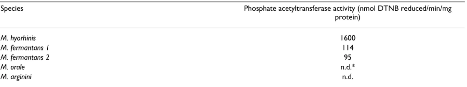
sake of comparison, a similar assay was performed on three other mycoplasma species, M. fermentans, M. orale,
M. arginini. A ten time lower specific activity was detected
in the former species, while no activity could be detected in the two latter species (Table 2).
Discussion
The contamination by mycoplasmas is a frequent prob-lem encountered when studying cultured cells. The myco-plasmas may have a myriad of different effects upon the infected host cells, which may moreover depends on the mycoplasma species. However, all the mollicutes exam-ined so far have truncated respiratory systems, lacking a complete tricarboxylic acid cycle and having no quinones or cytochromes, ruling out oxidative phosphorylation as an ATP-generating mechanism [19]. Our analysis of RC activity in four human skin fibroblast cell lines heavily contaminated by M. hyorhinis indicates a potential mild decrease of cell respiration and substrate oxidation in the mycoplasma contaminated cells, but no significant effect on any of the RC components. Slight decrease of cell respiration might result from a competition in the use of intermediate substrates resulting from proliferating myco-plasmas in the cell cytosol.
Table 1: Respiratory chain enzyme activities in mycoplasma contaminated fibroblasts before and after antibiotic treatment. Values are means ± 1 SD of values obtained independently on 4 different fibroblast cultures. Data between brackets represent values for uninfected, Ciprofloxacin-naïve, fibroblasts (n > 400). NQR, NADH quinone oxidoreductase; SCCR, Succinate cytochrome c reductase; GPCCR, glycerol-3-phosphate cytochrome c reductase; QCCR, quinol cytochrome c reductase; COX, cytochrome c oxidase; CS, citrate synthase; SD, Standard deviation.
Fibroblasts heavily contaminated with M. hyorhinis Fibroblasts after Ciprofloxacin treatment
Enzyme activity (nmol/min/mg protein) (n = 4) (n = 4)
NQR* 20.2 ± 3.8 15.8 ± 0.8 SCCR 22.0 ± 13.6 19.2 ± 3.0 GPCCR 7.8 ± 3.1 6.7 ± 1.8 QCCR 70.9 ± 25.3 66.6 ± 15.3 COX 71.1 ± 25.3 49.7 ± 8.0 ATPase* 31.1 ± 6.2 43.3 ± 7.7 CS 18.8 ± 7.3 15.2 ± 4.2
Enzyme activty ratios
COX/SCCR 3.7 ± 1.14 2.7 ± 0.84 (3.1 ± 0.2) COX/GPCCR 9.3 ± 0.84 8.0 ± 3.0 (7.5 ± 0.4) COX/QCCR 1.0 ± 0.21 0.8 ± 0.29 (0.7 ± 0.05) QCCR/SCCR 3.7 ± 1.15 3.5 ± 0.94 (4.0 ± 0.2) QCCR/GPCCR 9.3 ± 1.5 10.5 ± 3.9 (11.7 ± 0.5) SCCR/GPCCR 2.6 ± 0.61 2.9 ± 0.64 (2.2 ± 0.1) COX/CS 4.2 ± 2.0 3.6 ± 1.6 (2.8 ± 0.2) COX/ATP* 6.5 ± 2.0 2.8 ± 0.43 (3.4 ± 0.6) COX/NQR* 9.9 ± 2.4 7.6 ± 0.49 (10.1 ± 1.6) ATP/NQR* 1.6 ± 0.38 2.7 ± 0.61 (2.1 ± 0.4)
BMC Biochemistry 2003, 4 http://www.biomedcentral.com/1471-2091/4/15
The studied mycoplasma, M. hyorhinis, is a fermentative mollicute usually inhabiting the nasal cavity of swine and is one of the most common cell culture contaminants encountered, probably originating from the bovine sera [8]. The mechanisms of ATP-generation in the mollicutes are not fully understood, but among a number of poten-tial energy-yielding mechanisms in mollicutes is the
ace-tate kinase-ATP-generating pathway [20]. In this pathway acetyl-phosphate needs to be produced through the action of phosphate acetyltransferase on acetyl-CoA:
Phosphate acetyltransferase activity of Mycoplasma hyorhinis can be confused with citrate synthase activity of cultured skin fibroblasts contaminated by M. hyorhinis
Figure 2
Phosphate acetyltransferase activity of Mycoplasma hyorhinis can be confused with citrate synthase activity of cultured skin fibroblasts contaminated by M. hyorhinis. A: Measurement of the citrate synthase activity before (a) and
after (b) treatment showing, before oxaloacetate addition, the rapid reduction of DTNB in the mycoplasma-contaminated fibroblasts as compared to treated cells. The experiment was started with the addition of Triton X100-permeabilized fibrob-lasts and DTNB followed by the addition of acetylCoA and oxaloacetate. B: Measurement of the DTNB reduction in myco-plasma-contaminated cells in the presence (d) or absence of phosphate (c) in the medium. The experiment started with the addition of cells and DTNB, followed by the addition of acetyl-CoA. In b and c, 10 mM phosphate was later added to the cuvette. Numbers along the traces are nmol DTNB reduced/min/mg protein and represent mean values of experiments carried out independently on the 4 different cell lines. Rather large standard variations (between 20 to 30%) were calculated presuma-bly reflecting variable level of cell contamination by M. hyorhinis (not shown).
A
B
a
b
d
c
oxaloacetate
acetyl-CoA
PO
4
acetyl-CoA
1
6
5
1
5
2
308
0.05 A
2 min
2 min
0.05 A
1
7
2
1
7
5
302
A
B
a
b
d
c
oxaloacetate
acetyl-CoA
PO
4
acetyl-CoA
1
6
5
1
5
2
308
0.05 A
2 min
2 min
0.05 A
1
7
2
1
7
5
302
Although these enzymes have been reported to be com-monly found in both fermentative and non-fermentative mollicutes [21], we found phosphate acetyltransferase mostly active in the two fermentative species (M. hyorhinis and M. fermentans). The enzyme was indeed particularly active in M. hyorhinis with two major consequences. On one hand, the activity of this enzyme can be confused
is not carefully checked to be dependent on oxaloacetate. On the other hand, measurement of the phosphate acetyl-transferase activity represents a quite sensitive, conven-ient, and costless tool to detect the presence of M.
hyorhinis in biological samples, particularly in human
cul-tured cells where it represents a common contaminant. Its routine measurement along with citrate synthase assay may prove to be a valuable supplement to the diagnostic arsenal of mycoplasma detection.
Conclusion
The above data indicate that M. hyorhinis results in a potential mild decrease of cell respiration and substrate oxidation in the mycoplasma contaminated cells, but does not have any significant effect on the RC compo-nents. With the noticeable exception of citrate synthase, our study indicates that contamination by M. hyorhinis should not influence routine diagnostic procedures used to detect mitochondrial defects in cultured skin fibroblasts.
Methods
Mycoplasma detection and species identification
As a consequence of reduced cell proliferation and increased cell mortality, mycoplasma contamination was suspected in four human fibroblast cell lines and detec-tion initially performed with the fluorescent Hoechst 33258 stain using the Mycoplasma stain kit (Sigma-Aldrich Co. Ltd, Irvine, UK) according to the manufac-turer's description. The identification of M. hyorhinis as the contaminating species was carried out using molecu-lar typing by PCR-based methods [22,23].
Cell culture
Fibroblast cultures were established from skin biopsies obtained from 3-year to 77-year old individuals for diag-nostic purpose but no evidence of RC dysfunction. Cells were cultured in RPMI 1640 (Life technologies SARL, Cergy Pontoise, France) supplemented with glutamax (446 mg/l), 10% foetal calf serum, 100 µg/ml streptomy-cin, 100 IU/ml penicillin, 200 µM uridine and 2.5 mM
Table 2: Phosphate acetyltransferase activity in different mycoplasma species. Numbers are the mean values of triplicates. M. fermantans 1 and 2 represent two different batches. DTNB, dithionitrobenzoic acid.
Species Phosphate acetyltransferase activity (nmol DTNB reduced/min/mg
protein) M. hyorhinis 1600 M. fermantans 1 114 M. fermantans 2 95 M. orale n.d.* M. arginini n.d.
*n.d., no detectable DTNB reduction after 20 min incubation.
Phosphate acetyltransferase activity in titrated suspensions of
Mycoplasma hyorhinis
Figure 3
Phosphate acetyltransferase activity in titrated sus-pensions of Mycoplasma hyorhinis. Measurement of
DTNB reduction as a function of organisms/ml. Notice that 9 × 10⁶ organisms represents about 30 µg protein, one
color-changing units (CCU) corresponding roughly to 10 to 100 organisms [29].
0
5
10
15
20
25
30
35
0
1
2
3
4
n
m
o
l
D
T
N
B
r
e
d
u
ced
/m
in
0 9 18 27 36
10
6mycoplasmas/ml
35
30
25
20
15
10
5
0
BMC Biochemistry 2003, 4 http://www.biomedcentral.com/1471-2091/4/15
sodium pyruvate, at 37°C under standard conditions [24]. When indicated, cells were treated with 20 µg/ml Ciprofloxacin (Bayer, Leverkusen, Germany) for 14 days and culture medium changed every 3–4 day. Cipro-floxacin treatment is considered as a safe and effective method for elimination of cell culture mycoplasmas [25].
Cell respiration and enzyme activities
Polarographic measurements were performed on fresh cells before and after 1 week of the antibiotic treatment. Cell pellets frozen on the same occasion were thawed and used for spectrophotometric measurements, which were performed blindly. Intact fibroblast respiration and mito-chondrial substrate oxidation with malate and glutamate and with succinate (Figure 1) were polarographically stud-ied in cell suspensions of digitonin-permeabilized fibrob-lasts as previously described [26]. The activity of the RC complexes, NADH-ubiquinone reductase (NQR, complex I), succinate-cytochrome c reductase (SCCR, complex II-III), glycerol-3-phosphate cytochrome c reductase, (GPCCR, glycerol-3-Phosphate dehydrogenase + complex III), decylubiquinol-cytochrome c reductase (QCCR, com-plex III), cytochrome c oxidase (COX, comcom-plex IV), mito-chondrial ATP synthetase (ATPase, complex V) were spectrophotometrically measured on freeze-thaw permea-bilized fibroblasts in cell suspension as previously described [26,27]. The NQR, and ATPase activity were measured in 1 ml of 10 mM Tris-HCl buffer (pH 8.0) containing 0.8 mM NADH, 1 mM KCN, 50 µM decylubi-quinone for NQR assay, subsequently added with 3 µM rotenone, 5 mM MgCl2, 10 mM KCl, 2 mM PEP, 0.5 mM ATP, 4 IU lactic dehydrogenase and pyruvate kinase for ATPase assay. Cytochrome c reductase activities (SCCR, GCCR, QCCR) were measured in 1 ml of 10 mM KH2PO4 (pH 7.8), EDTA 2 mM in the presence of 1 mM KCN, 3 µM rotenone and 0.2 mM ATP. Succinate (10 mM), glyc-erol 3-phosphate (20 mM), decylubiquinol (50 µM) were used as substrates for detremining SCCR, GCCR and QCCR, respectively. COX activity was measured in 1 ml of 10 mM KH2PO4 (pH 6.5) in the presence of 2.5 mM lau-ryl-maltoside, and 10 µM reduced cytochrome c. Citrate synthase was spectrophotometrically measured at 412– 600 nm in the presence of 0.1% Triton X-100 by following the appearance of the free SH-group of the released CoA-SH upon the addition of 10 mM oxaloacetate to a cell sus-pension to which 100 µM acetyl-CoA and 2 mM DTNB (Dithionitrobenzoic acid; Ellman's reagent) have been added [28]. In order to discriminate between the phos-phate-independent acetyl-CoA acetyltransferase and the phosphate-dependant phosphate acetyltransferase, the analysis was performed either in 10 mM KH2PO4 or in 10 mM MOPS (3-N-morpholino-propane-sulfonic acid) (pH 7.8). All measurements were carried out at 37°C and assay media supplemented with 1 mg/ml bovine serum albu-min. All chemicals were analytical reagent grade from
Sigma-Aldrich Chimie (Saint Quentin Fallavier; France). The protein content was measured using the Bradford method.
Statistical analyses
Student's T-test was performed using the Statview 5.0 soft-ware (SAS Institute Inc. USA).
Authors' contributions
ND carried out most of the polarographic and spectro-photometric assay of the RC; NK took care of the cell cul-tures; JJB and DC worked on the enzyme assays carried out on mycoplasma strains which were grown and pro-vided by CB; PR conceived of the study, wrote the paper; AR and AM participated in its design and coordination. All authors read and approved the final manuscript.
Acknowledgements
This project was supported by Grants from the Wenner-Gren Founda-tions, the Swedish Association of Neurologically Disabled, the Swedish Medical Association and the Association Française contre l'Ataxie de Frie-dreich (AFAF)
References
1. Razin S, Yogev D and Naot Y: Molecular biology and
pathogenic-ity of mycoplasmas. Microbiol Mol Biol Rev 1998, 62:1094-1156.
2. Waites KB and Taylor-Robinson D: Mycoplasma and
Urea-plasma. Manual of clinical microbiology 7thth edition. Edited by: Murray
P B. Washington, D.C., ASM Press; 1999:782-794.
3. Blanchard A., and C. M. Bébéar: Mycoplasmas of humans.
Molecu-lar biology and pathogenicity of mycoplasmas Edited by: S Razin and R Herrmann. London, Kluwer Academic/Plenum Publishers; 2002:45-71.
4. Blanchard A and Montagnier L: AIDS-associated mycoplasmas.
Annu Rev Microbiol 1994, 48:687-712.
5. Gilroy CB, Keat A and Taylor-Robinson D: The prevalence of
Mycoplasma fermentans in patients with inflammatory arthritides. Rheumatology (Oxford) 2001, 40:1355-1358.
6. Razin S: Peculiar properties of mycoplasmas: the smallest
self-replicating prokaryotes. FEMS Microbiol Lett 1992, 79:423-431.
7. Hay RJ, Macy ML and Chen TR: Mycoplasma infection of cultured
cells. Nature 1989, 339:487-488.
8. Drexler HG and Uphoff CC: Contamination of cell culture,
mycoplasma. Encyclopedia of cell technology Volume 1. Edited by: Spier
R E. New York, John Wiley & Sons, Inc.; 2000:609-627.
9. Avron A and Gallily R: Mycoplasma stimulates the production
of oxidative radicals by murine peritoneal macrophages. J
Leukoc Biol 1995, 57:264-268.
10. Sasaki T, Shintani M, Yamada K, Okumura H and Kihara K: Behavior
of Mycoplasma hominis in a human diploid cell culture system. Microbiol Immunol 1981, 25:537-543.
11. Sokolova IA, Vaughan AT and Khodarev NN: Mycoplasma
infec-tion can sensitize host cells to apoptosis through contribu-tion of apoptotic-like endonuclease(s). Immunol Cell Biol 1998, 76:526-534.
12. Tsai S, Wear DJ, Shih JW and Lo SC: Mycoplasmas and
oncogen-esis: persistent infection and multistage malignant transformation. Proc Natl Acad Sci U S A 1995, 92:10197-10201.
13. Choi JW, Haigh WG and Lee SP: Caveat: mycoplasma arginine
deiminase masquerading as nitric oxide synthase in cell cultures. Biochim Biophys Acta 1998, 1404:314-320.
14. Gabridge MG: Metabolic consequences of Mycoplasma
pneu-moniae infection. Isr J Med Sci 1987, 23:574-579.
15. Astrom E, Friman G and Pilstrom L: Effects of viral and
myco-plasma infections on ultrastructure and enzyme activities in human skeletal muscle. Acta Pathol Microbiol Scand [A] 1976, 84:113-122.
Publish with BioMed Central and every scientist can read your work free of charge "BioMed Central will be the most significant development for disseminating the results of biomedical researc h in our lifetime."
Sir Paul Nurse, Cancer Research UK Your research papers will be:
available free of charge to the entire biomedical community peer reviewed and published immediately upon acceptance cited in PubMed and archived on PubMed Central yours — you keep the copyright
BioMedcentral
16. Doersen CJ and Stanbridge EJ: Effects of mycoplasma
contami-nation on phenotypic expression of mitochondrial mutants in human cells. Mol Cell Biol 1981, 1:321-329.
17. Chretien D, Gallego J, Barrientos A, Casademont J, Cardellach F, Munnich A, Rotig A and Rustin P: Biochemical parameters for
the diagnosis of mitochondrial respiratory chain deficiency in humans, and their lack of age-related changes. Biochem J 1998, 329:249-254.
18. Rustin P, Chretien D, Bourgeron T, Wucher A, Saudubray JM, Rotig A and Munnich A: Assessment of the mitochondrial
respira-tory chain. Lancet 1991, 338:60.
19. Pollack JD, Williams MV and McElhaney RN: The comparative
metabolism of the mollicutes (Mycoplasmas): the utility for taxonomic classification and the relationship of putative gene annotation and phylogeny to enzymatic function in the smallest free-living cells. Crit Rev Microbiol 1997, 23:269-354.
20. Kahane I, Razin S and Muhlrad A: Possible role of acetate kinase
in ATP generation in Mycoplasma hominis and acheoplasma laidlawii. FEMS Microbiol Lett 1978, 3:143-145.
21. Muhlrad A, Peleg I, Robertson JA, Robinson IM and Kahane I:
Ace-tate kinase activity in mycoplasmas. J Bacteriol 1981, 147:271-273.
22. Teyssou R, Poutiers F, Saillard C, Grau O, Laigret F, Bove JM and Bebear C: Detection of mollicute contamination in cell
cul-tures by 16S rDNA amplification. Mol Cell Probes 1993, 7:209-216.
23. Dussurget O., A. Henry, B. Lemercier, and D. Roulland-Dussoix:
Polymerase chain reaction-based diagnosis of mollicute infection of commercial animal sera. J. Microbiol. Methods 1994, 20:125-135.
24. Bourgeron T, Chretien D, Amati P, Rotig A, Munnich A and Rustin P:
Expression of respiratory chain deficiencies in human cul-tured cells. Neuromuscul Disord 1993, 3:605-608.
25. Schmitt K, Daubener W, Bitter-Suermann D and Hadding U: A safe
and efficient method for elimination of cell culture myco-plasmas using ciprofloxacin. J Immunol Methods 1988, 109:17-25.
26. Rustin P, Chretien D, Bourgeron T, Gerard B, Rotig A, Saudubray JM and Munnich A: Biochemical and molecular investigations in
respiratory chain deficiencies. Clin Chim Acta 1994, 228:35-51.
27. Chretien D, Benit P, Chol M, Lebon S, Rotig A, Munnich A and Rustin P: Assay of mitochondrial respiratory chain complex I in
human lymphocytes and cultured skin fibroblasts. Biochem
Bio-phys Res Commun 2003, 301:222-224.
28. Srere PA: Citrate synthase. Meth Enzymol 1969, 13:3-11. 29. Bernet C, Garret M, de Barbeyrac B, Bebear C and Bonnet J:
Detec-tion of Mycoplasma pneumoniae by using the polymerase chain reaction. J Clin Microbiol 1989, 27:2492-2496.