HAL Id: hal-02171187
https://hal.sorbonne-universite.fr/hal-02171187
Submitted on 29 Aug 2019HAL is a multi-disciplinary open access archive for the deposit and dissemination of sci-entific research documents, whether they are pub-lished or not. The documents may come from teaching and research institutions in France or abroad, or from public or private research centers.
L’archive ouverte pluridisciplinaire HAL, est destinée au dépôt et à la diffusion de documents scientifiques de niveau recherche, publiés ou non, émanant des établissements d’enseignement et de recherche français ou étrangers, des laboratoires publics ou privés.
Synergistic convergence of microbiota-specific systemic
IgG and secretory IgA
Karim Dorgham, Hela El Kafsi, Sébastien Andre, Eric Oksenhendler, Jehane
Fadlallah, Delphine Sterlin, Claire Fieschi, Christophe Parizot, Gaëlle Autaa,
Pascale Ghillani-Dalbin, et al.
To cite this version:
Karim Dorgham, Hela El Kafsi, Sébastien Andre, Eric Oksenhendler, Jehane Fadlallah, et al.. Syn-ergistic convergence of microbiota-specific systemic IgG and secretory IgA. Journal of Allergy and Clinical Immunology, Elsevier, 2019, 143 (4), pp.1575-1585.e4. �10.1016/j.jaci.2018.09.036�. �hal-02171187�
Synergistic convergence of microbiota-specific systemic IgG and secretory IgA
1
2 Jehane Fadlallah1,3†, Delphine Sterlin1†, Claire Fieschi3, Christophe Parizot1, Karim 3 Dorgham1,
Hela El Kafsi1, Gaëlle Autaa1, Pascale Ghillani-Dalbin1, Catherine Juste2, Patricia Lepage2, 4
Marion Malphettes3, Lionel Galicier3, David Boutboul3, Karine Clément4,5,6,7, Sébastien 5 André4,5,6, Florian Marquet4,5,6, Christophe Tresallet8, Alexis Mathian1, Makoto Miyara1, Eric 6 Oksenhendler3, Zahir Amoura1, Hans Yssel1, Martin Larsen1*, Guy Gorochov1*
7
Affiliations:
8 9
1Sorbonne Université, INSERM, Centre d’Immunologie et des Maladies Infectieuses
(CIMI-10
Paris), AP-HP Hôpital Pitié-Salpêtrière, F-75013 Paris, France 11
2UMR1319 Micalis, INRA, Jouy-en-Josas, France.
12
3Université Paris Diderot Paris 7, Department of Clinical Immunology, Hôpital Saint-Louis,
13
Assistance Publique Hôpitaux de Paris (APHP), EA3518, 75010, Paris, France 14
4INSERM, UMR_S 1166, NutriOmics Team, F-75013, Paris, France;
15
5Sorbonne Universités, UPMC University Paris 06, UMR_S 1166, F-75005, Paris, France;
16
6Institute of Cardiometabolism and Nutrition, ICAN, Pitié-Salpêtrière Hospital, Assistance
17
Publique Hôpitaux de Paris, F-75013, Paris, France;
18
7Assistance Publique Hôpitaux de Paris, Pitié-Salpêtrière Hospital, Nutrition, Endocrinology
19
Departments, F-75013, Paris, France 20
8Assistance Publique Hôpitaux de Paris, Pitié-Salpêtrière Hospital, Department of surgery,
F-21
75013, Paris, France 22
23 24
†These authors contributed equally to this work
25
*To whom correspondence should be addressed: guy.gorochov@upmc.fr or 26
martin.larsen@upmc.fr. 27
28
Conflict of interests: The authors declare no competing interests.
29
Funding: The study was financed by Institut national de la santé et de la recherche médicale
30
(Inserm) and Agence Nationale de la Recherche (MetAntibody ANR) 31
Abstract (<250 words)
33 34
Background: Besides intestinal barrier function, the host tolerates gut commensals through
35
both innate and adaptive immune mechanisms. It is now clear that gut commensals induce 36
local immunoglobulin A (IgA) responses, but it remains unclear whether anti-microbiota 37
responses remain confined to the gut. 38
Objective: The aim of this study was to investigate systemic and intestinal responses against
39
the whole microbiota under homeostatic conditions, and in the absence of IgA. 40
Methods: We analyzed blood and feces from healthy donors, patients with selective IgA
41
deficiency (SIgAd) and common variable immunodeficiency (CVID). Immunoglobulin-42
coated bacterial repertoires were analyzed by combined bacterial fluorescence-activated cell 43
sorting and 16S rRNA sequencing, and bacterial lysates were probed by western blot analysis 44
with healthy donors serums. 45
Results: Although absent from the healthy gut, serum anti-microbiota IgG are present in
46
healthy individuals, and increased in SIgAd patients. IgG converge with non-overlapping 47
secretory IgA repertoires to target the same bacteria. Each individual targets a diverse, 48
microbiota repertoire whose proportion inversely correlates with systemic inflammation. 49
Finally, Intravenous Immunoglobulin preparations (IVIG) target much less efficiently CVID 50
gut microbiota than healthy microbiota. 51
Conclusion: Secretory IgA is pivotal for induction of tolerance to gut microbiota.
SIgAd-52
associated inflammation is inversely correlated with systemic anti-commensal IgG responses, 53
which may thus serve as a second line of defense. We speculate that SIgAd patients could 54
benefit from oral IgA supplementation. Our data also suggest that IVIG preparations might be 55
supplemented with IgG from IgA deficient patients pools in order to offer a better protection 56
against gut bacterial translocations in CVID. 57
58 59
Key Messages:
60 61
- Systemic IgG and secretory IgA bind a common spectrum of commensals. 62
- Increased proportions of IgG+ microbiota and inflammatory markers in SIgAd. 63
- IVIG poorly target CVID and SIgAd gut microbiota. 64
Capsule summary:
65
Serum anti-microbiota IgG are present in healthy individuals, and increased in SIgAd. IVIG 66
only bind a small fraction of SIgAd gut microbiota. Oral IgA and IgA/IgG supplementation 67
should be considered in SIgAd and CVID, respectively. 68
Key words (<10): gut microbiota, anti-commensal IgG, secretory IgA, IgA deficiency,
69 CVID, IVIG. 70 71 Abbreviations: 72 Ig: Immunoglobulin 73
SIgAd : Selective IgA deficiency 74
CVID: Common Variable Immunodeficiency 75
IVIG: Intravenous Immunoglobulin 76
77
Acknowledgments: The authors wish to thank Emma Slack for advice, Jean-Michel Batto for
78
discussions, Joel Doré, Fabienne Beguet-Crespel and Emma Slack for providing bacterial 79
strains. 80
Funding: The study was financed by: Institut national de la santé et de la recherche médicale
81
(Inserm), Agence Nationale de la Recherche (MetAntibody, ANR-14-CE14-0013), Fondation 82
pour l’Aide a la Recherche sur la Sclérose En Plaques (ARSEP). 83
84 85 86
Introduction 87
88
Gut commensal bacteria contribute to several beneficial properties to the host. This complex 89
community provides metabolic functions, prevents pathogen colonization and enhances 90
immune development. A symbiotic relationship is maintained using host innate and adaptive 91
immune responses such as antimicrobial compounds and mucus secretion, as well as IgA 92
production 1,2. However, the gastrointestinal tract remains an important reservoir for potential 93
bloodstream infections that involve Enterobacteriaceae, Enterococcus species or other Gram-94
negative bacilli 3,4. The physical gut barrier, but also innate and adaptive immune 95
mechanisms, control host-microbiota mutualism, reducing the risk of bacterial translocation 96
and systemic immune activation. Murine models of innate immune deficiency indeed develop 97
high seric IgG levels against gut microbiota 2. Significant titers of IgG targeting E. coli were 98
also reported either in patients with inflammatory bowel diseases or in mice lacking secretory 99
IgA 5,6. Nevertheless, based on recent murine studies, the notion has emerged that induction 100
of systemic IgG responses against gut symbiotic bacteria is not necessarily a consequence of 101
mucosal immune dysfunction or epithelial barrier leakiness. Healthy mice actively generate 102
systemic IgG against a wide range of commensal bacteria under homeostatic conditions, 103
which are passively transferred to the neonates through the maternal milk 7. Serum IgG that
104
specifically recognize symbiotic Gram-negative bacteria confer protection against systemic 105
infections by these same bacteria. Because such IgG target a conserved antigen in commensal 106
and pathogens, they also enhance elimination of pathogens such as Salmonella 8. 107
IgG-expressing B cells are present in human gut lamina propria during steady state 108
conditions, and represent 3-4% of the total gut B cells. About two-third of IgG+ lamina 109
propria antibodies react with common intestinal microbes 9. Inflammatory bowel disease is 110
associated with a marked increase in gut IgG+ B cells that might contribute to the observed 111
elevated serum anti-E. coli IgG levels in these patients 9. However, to which extent gut IgG+ 112
B cells contribute to the serum IgG repertoire, remains elusive. Focusing on anti-113
transglutaminase 2 antibodies, it has been shown a low degree of clonal relationship between 114
serum and intestinal IgG 10. Altogether, it remains unknown whether secretory and serum 115
anti-bacteria antibodies have identical targets or whether digestive and systemic antibody 116
repertoires are shaped by distinct microbial consortia. 117
118
In this study, we report that human serum IgG bind a broad range of commensal bacteria. We 119
also demonstrate for the first time the convergence of intestinal IgA and serum IgG responses 120
toward the same microbial targets, under homeostatic conditions. Private anti-microbiota IgG 121
specificities are induced in IgA-deficient patients, but are not found in IgG pools from healthy 122
donors, partially explaining why substitutive IgG cannot regulate antibody deficiency-123
associated gut dysbiosis and intestinal translocation. Finally, in both controls and IgA-124
deficient patients, systemic anti-microbiota IgG responses correlate with reduced 125
inflammation suggesting that systemic IgG responses contribute to the gut microbiota 126 confinement. 127 128 129 130 131 132
Results
133 134
1/ Convergence of intestinal IgA and serum IgG toward the same bacterial cells
135
To determine the level of humoral systemic response against fecal microbiota, we have 136
elaborated a flow cytometric assay derived from a previously reported technology 11. This 137
protocol allows to probe concomitantly IgA and IgG microbiota coating. We found that 138
approximately 8% of the fecal microbiota is targeted by secretory IgA (median[min-max]%; 139
8[0.8-26.7]%; n=30) in healthy donors, in concordance with previous reports 12. As shown, 140
the proportion of bacteria in vivo bound by secretory IgA in human feces is highly variable 141
between healthy individuals (Figure 1B). IgG-bound bacteria are virtually absent from healthy 142
human feces (median [min-max]%; 0.03[0-0.16]%; n=30 ; Figure S1 and 1A), in agreement 143
with the lack of IgG transport to the intestinal lumen. In healthy donors, seric IgG bound a 144
median rate of 1.1% of fecal bacteria (median [min-max]%; 1.1[0.2-3.2]%; Figure 1B). 145
Surprisingly, seric IgG targeted exclusively secretory IgA bound bacteria (Figure 1A). 146
Conversely, all IgA-coated bacteria (IgA+ bacteria) were not targeted by seric IgG. Of note, 147
an irrelevant human monoclonal IgG (chimeric anti-human TNF containing a human Fc IgG 148
fraction) exhibits markedly reduced binding to IgA+ bacteria, compared to serum IgG (Figure 149
1A, S2), demonstrating that IgG binding to IgA-coated bacteria is mostly Fab-mediated. 150
To confirm that systemic IgG binding is directed against IgA-bound bacteria, we evaluated in 151
vitro serum IgG binding to cultivable bacterial strains. We selected four bacterial strains that 152
were not preferentially bound by IgA in human feces and four others that were previously 153
defined as classical IgA targets in vivo 12–14. As shown in Figure 2, IgG from healthy 154
individuals (n = 30) bind much more significantly Bifidobacterium longum, Bifidobacterium 155
adolescentis, Faecalibacterium prausnitzii and Escherichia coli, known to be particularly 156
enriched in the IgA-coated fraction of healthy individuals, than three different strains of 157
Bacteroides sp. and Parabacteroides distasonis, known to be particularly enriched in the IgA-158
uncoated fraction of the fecal microbiota (Figure 2A-B). The majority of anti-commensal IgG 159
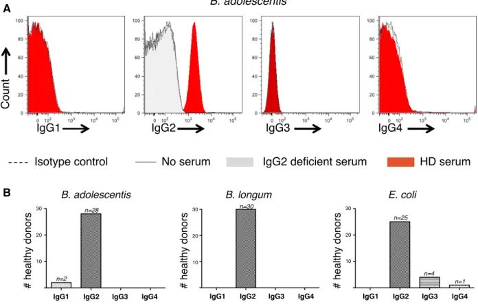
antibodies are of the IgG2b and IgG3 isotypes in mice. Using isotype-specific secondary 160
antibodies we detected minimal IgG1 binding, but high seric IgG2 reactivity, to 161
Bifidobacterium adolescentis, Bifidobacterium longum and Escherichia coli, suggesting that 162
IgG2 is involved in commensals targetting in humans (Figure S3). 163
Since anti-commensal IgG might possibly be triggered during mucosal immune responses, we 164
characterized lamina propria B cells and detected the presence of IgG2+ B cells throughout 165
the intestine (Figure S4). Of note, IgG transcripts are more abundant in LP tissue that in 166
PBMCs, as measured by qPCR (Figure S4). 167
These results demonstrate that human IgG recognize a wide range of commensal under 168
homeostatic conditions. Systemic humoral immunity (notably IgG2) converges with mucosal 169
immunity to bind the surface of commensals. 170
171
2/ Inter-individual variability and non overlapping anti-commensal IgA and IgG
172
molecular targets.
173 174
It was previously suggested that murine IgG would target a restricted number of bacterial 175
proteins and favored highly conserved outer membrane proteins 8. Reactivity of human serum 176
IgG against bacterial lysates from a Gram-negative strains was evaluated by immunoblotting. 177
We observed that IgG labeled several E. coli bands (Figure 2C), suggesting that multiple 178
bacterial products are involved in the induction of systemic antibodies. Interestingly, this 179
analysis reveals a great deal of inter-individual variability, as it is not always the same 180
bacterial products that react with the tested serums. We then compared the overlap between 181
bacterial products labeled by IgG and IgA and found distinct binding profiles (Figure 2C). 182
Finally, in the 5 individuals tested, although some bacterial products (notably a 15 Kd 183
antigen) are frequently targeted in most subjects and without isotype restriction, it clearly 184
appears that IgA and IgG never share exactly the same binding pattern at a molecular level. 185
Taken together, these results demonstrate although IgG converges with IgA to bind the 186
surface of commensals, it appears that IgA and IgG do not systematically target the same 187
bacterial antigens, even at the individual level. 188
189
3/ Private anti-microbiota IgG specificities are induced in IgA-deficient patients
190
The existence of seric IgG able to bind IgA-coated bacteria could equally suggest that some 191
gut bacteria (or bacterial antigens) might cross the intestinal barrier: (i) in spite of IgA, or (ii) 192
because of IgA. In order to explore these two putatively opposing roles for IgA, we studied 193
the systemic anti-commensal IgG response in SIgAd. These patients had undetectable seric 194
and digestive IgA levels while seric IgG were in the normal range15. Anti-microbiota IgG 195
levels were significantly higher in SIgAd compared to controls (median [min-max]%; 3.3[0.2-196
20.2]% versus 1.1%[0.2-3.2]%; Figure 3A). Using irrelevant human IgG, we confirmed that, 197
like in healthy donors, IgG interact with fecal bacteria in a Fab-dependent manner (Figure 198
S2B). These data support an enhanced triggering of systemic IgG immunity against fecal 199
microbiota when lacking secretory IgA, as shown in the murine model of polymeric 200
immunoglobulin receptor deficiency 6. 201
Considering this high level of anti-microbiota IgG in SIgAd, and the similarity of SIgAd and 202
healthy microbiota composition15, we investigated how anti-microbiota IgG repertoires from 203
healthy donors and IgA deficient patients were overlapping. Using polyclonal IgG from 204
pooled serum of healthy donors, we assessed IgG-bound microbiota using either healthy or 205
SIgAd purified microbiota. We showed that pooled polyclonal IgG and autologous healthy 206
sera recognized a similar percentage of fecal bacteria (median [min-max]%;1[0-3.7] % vs 207
1.1[0.2-3.2]%, respectively, figure 3B-C). In contrast, pooled polyclonal IgG bound a smaller 208
bacterial fraction of IgA deficient-microbiota compared to autologous patient serum (median 209
[min-max]%;0.4[0-3.6] % vs 3.3[0.2-20.2] %, figure 3B-C ). In order to test whether similar 210
specificities are induced in all or most IgA deficient individuals, we compared their IgG 211
reactivity to autologous or heterologous gut microbiota. In this experiment (Figure 3D), each 212
IgA-deficient microbiota was incubated either with autologous serum (i.e.: autologous 213
condition), or with serum from an unrelated IgA deficient individual (i.e.: heterologous 214
condition). As shown in Figure 3D, no significant difference was seen between autologous or 215
heterologous conditions (median autologous IgG+ microbiota 1.2% versus median 216
heterologous IgG+ microbiota 1.4%). Of note, heterologous seric IgG also predominantly 217
interact with fecal microbiota in a Fab-dependent manner (Figure S2C). 218
This set of data suggests that peculiar anti-microbiota IgG specificities are induced in IgA-219
deficient patients, but not in healthy individuals. 220
4/ IgG specifically recognize a broad spectrum of bacteria
221
To more deeply decipher anti-commensal IgG specificities in both healthy donors and IgA 222
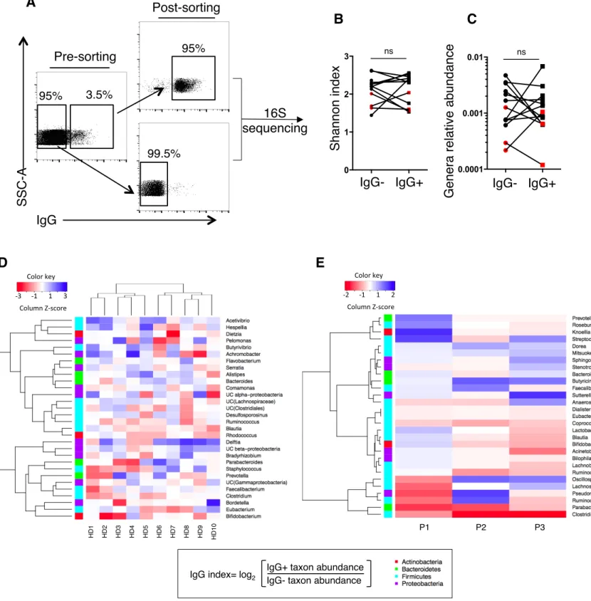
deficient patients, we next performed a stringent flow-sorting to isolate IgG-bound bacteria 223
and identified their taxonomy by 16S rRNA sequencing (Figure 4A). We observed extensive 224
inter-individual variability at genus level irrespective of immunological status (healthy donors 225
vs IgA deficient patients). Microbial diversity calculated by Shannon index varied between 226
donors, but on average bacterial diversity of IgG+ and IgG- bacteria was not significantly
227
different (Figure 4B). We postulated that IgG might preferentially interact with dominant 228
taxa, and therefore compared relative abundance of IgG-bound and IgG-unbound genera. 229
Both fractions exhibited equal distributions of rare and abundant genera (Figure 4C), thus IgG 230
target commensals irrespectively of their frequency. Interestingly, we found that individual 231
IgG+ and IgG- fecal bacterial profiles were remarkably different, supporting a strong IgG bias
232
against peculiar taxa that cannot be explained by an expansion of the latter. Besides, anti-233
commensals IgG were not restricted to pathobionts, but also targeted symbiotic genera such as 234
Faecalibacterium, whose the most common species (i.e.: F.prausnitzii) has been assigned 235
anti-inflammatory properties in both healthy donors and IgA deficient patients (Figure 4D-E) 236
16. From this part we conclude that anti-commensal IgG recognize a diverse array of both
237
pathobionts and commensal bacteria. Importantly, each individual harbored a private IgG 238
antimicrobial signature. 239
240
5/ High anti-microbiota IgG levels correlate with reduced systemic inflammation
241
Microbiota-specific serum IgG responses contribute to symbiotic bacteria clearance in 242
periphery and maintain mutualism in mice 2. We thus hypothesized that anti-commensals IgG
243
might influence the balance of systemic inflammatory versus regulatory responses in humans. 244
Hence, we measured plasma levels of sCD14 (a marker of monocyte activation, 17) and 245
observed that seric IgG-coated bacteria inversely correlated with soluble CD14 (r=-0.42, 246
p<0.005; Figure 5A) in both healthy donors and SIgAd patients. These results are in line with 247
the finding that IgG replacement therapy reduced endotoxemia 18. To further explore the 248
potential link between anti-microbiota IgG and systemic inflammation, we explored CVID 249
patients (characterized by both IgG and IgA defects). These patients benefit from IVIG 250
treatment. Yet, we show that IVIG do not efficiently bind CVID microbiota. As shown in 251
Figure 5B, IVIG bound a reduced fraction of CVID microbiota compared to control 252
microbiota (median [min-max]%; 0.37[0.00-1.14]% vs 1.06[0.00-3.7]%). We then determined 253
plasma levels of sCD14 and IL-6 (an inflammatory cytokine reflecting T-cell activation) and 254
evaluated the expression of PD-1 (a T-cell co-inhibitory molecule induced after activation) on 255
CD4+ T cells. IL-6 as well as sCD14 levels were consistently higher in CVID patients than in 256
healthy donors (IL-6, median [min-max]%, 1.8(0.7-60.1) pg/ml versus 0.6(0.33-2.4) pg/ml; 257
sCD14, median [min-max]%; 2063 (590-5493) pg/ml versus median 2696(1147-4283) pg/ml; 258
Figure 5C-D). Moreover, CD45RA-PD1+CD4+ T cells tended to increase in CVID patients, 259
as compared with healthy donors (median [min-max]%; 20.3(4.26-59.6)% versus 10(2.09-260
41.9)%, Figure 5E). 261
Altogether, in both controls and IgA-deficient patients, systemic anti-microbiota IgG 262
responses correlate with reduced inflammation. 263
264 265
Discussion
266
Anti-commensal IgG have been described in patients with inflammatory diseases 5,19,20. Here, 267
we characterize for the first time a broad anti-commensal IgG response under homeostatic 268
conditions in humans. Previous work demonstrated that symbiotic Gram-negative bacteria 269
disseminate spontaneously and drive systemic IgG responses 8. We show here that a diverse 270
array of commensal bacteria, including Gram-positive and Gram-negative species, can induce 271
systemic IgG. We show that a pathobiont like E. coli induce less systemic IgG responses than 272
a presumably beneficial symbiont like B. adolescentis (Fig. 2B). Therefore the systemic IgG 273
response in healthy humans does not appear preferentially driven by pathobionts, but also by 274
commensals. In mice it has been shown that commensal microbes induce serum IgA 275
responses that protect against sepsis21, illustrating the consequence of systemic anti-microbial 276
IgA binding to both pathogenic strains and commensals. We postulate that systemic anti-277
microbiota IgG, also mainly induced by commensals, could have the same protective role. 278
Strikingly, systemic IgG and secretory IgA converge towards the same autologous microbiota 279
subset. Yet, it seems unlikely that secretory IgA enhances systemic IgG responses, since IgA 280
deficiency is associated with high proportions of IgG+ microbiota, as detected using bacterial 281
flow cytometry on SIgAd microbiota labeled with autologous serum. In addition, induction of 282
anti-commensal IgG has been shown to be microbiota-dependent, but IgA-independent in 283
mice 2,6. Systemic IgG could reflect asymptomatic gut microbiota translocation episodes in
284
healthy individuals. Repeated bacterial translocations might occur more frequently in the 285
absence of secretory IgA, accounting for elevated anti-microbiota IgG levels in these patients. 286
IgA do not activate complement via the classical pathway 22. Interestingly, the anti-287
Bifidobacterium adolescentis IgG response is primarily restricted to the IgG2 isotype (Figure 288
S3), which less efficiently triggers the classical route of complement than IgG1 and IgG3 23.
289
Furthermore, IgG2 poorly interact with type I FcγRs, while IgG1 and IgG3 demonstrate 290
affinity for most FcγRs 24. These distinct binding patterns have functional consequences. IgG1 291
antibodies mediate phagocytosis and induce potent pro-inflammatory pathways while IgG2 292
are rather involved in dendritic cell or B cell activation 25,26. Besides its specific Fc domain 293
interaction, IgG2 is usually, but not exclusively, associated with anti-carbohydrate responses 294
27. IgA was also recently shown to bind multiple microbial glycans 28. Thus, IgA and IgG2
295
could be viewed as playing similar roles, but in different compartments. Much effort has been 296
recently expended to develop bacterial glycan or protein microarray. Glycomics could 297
represent a new option in order to better decipher anti-microbiota antibody targets 27,29. 298
Importantly, we show that IgA and IgG do not systematically target the same bacterial 299
antigens at an individual level (Figure 2C). Therefore IgG and IgA epitopes are not strictly 300
overlapping. This result could further illustrate antibacterial IgA/IgG synergy, and explain the 301
absence of isotype competition allowing the observed IgA/IgG co-staining of bacteria (Figure 302
1). 303
Recent studies suggested that murine secretory IgA are polyreactive and bind a broad but 304
defined subset of microbiota 30,31. Similarly, up to 25% of intestinal IgG+ plasmablasts could 305
produce polyreactive antibodies 9. We therefore hypothesized that the cross-reactive potential 306
of anti-commensal IgG may act as a first line of defense against potentially harmful bacteria. 307
In line with this idea, it can be noted that homeostatic anti-commensal IgG confer protection 308
against pathogens such as Salmonella 8. Conversely, IgG directed against Klebsiella
309
pneumoniae, an opportunistic pathogen, cross-react with commensal microbes32. Clonally
310
related memory B cells expressing cross-specific anti-K. pneumoniae antibodies were found 311
in both lamina propria and peripheral blood in humans suggesting that generation of anti-312
commensal antibodies might be triggered in the mucosal compartment. At the same time, 313
anti-commensal memory B cells might recirculate in periphery32. Altogether, it appears
314
possible that bacteria-specific IgG would arise from the gut, as all bacteria-specific IgG 315
isotypes we characterized in human sera are also present in the gut (Fig. S4), and also because 316
a large proportion of gut IgG+ B cells are expected to be commensal-specific9. However, it 317
remains presently unknown whether serum IgG responses mainly originate from the gut 318
and/or are induced the periphery following bacterial translocation. 319
We report that each individual harbors a private set of anti-commensal IgG in both healthy 320
donors and IgA deficient patients. Since our analysis was limited to 3 IgA deficient patients, 321
further study might precisely reveal how SIgAd anti-commensal IgG bind a distinct set of 322
commensals. While IVIG preparations contain an extended set of anti-commensal IgG, we 323
observe that IVIG less efficiently bind CVID microbiota. These observations are consistent 324
with reported alterations of gut microbiota in CVID patients 33. Microbiota perturbations are 325
also associated with selective IgA deficiency. The latter perturbations are less pronounced 326
than in CVID, since the presence of IgM appears to preserve SIgAd microbiota diversity15. 327
Nevertheless, IgA deficiency condition is also associated in severe cases with bacterial 328
translocation, colitis and dysbiosis. These complications are not accessible to substitutive Ig 329
replacement therapy 34. Indeed, IVIG do not appear to contain high-enough concentrations as 330
well as appropriate specificities of anti-commensal IgG. As shown in Figure 3, healthy 331
control serum usually less efficiently binds IgA deficient microbiota than autologous serum. 332
Similarly, IVIG poorly targets CVID gut microbiota (Figure 5B). In addition, local mucosal 333
antibody responses might be important in regulating microbiota composition in a way that 334
cannot be substituted by IVIG. These findings expand our understanding of how IVIG fail to 335
treat intestinal symptoms in CVID and IgA deficient patients. Dysbiosis and gastro-336
intestinal complications might not accessible to substitutive Ig replacement therapy, since, as 337
we show, healthy IgG repertoire does not contain adequate “dysbiotic-specific” antibodies. 338
It was recently shown in mice that maternally-derived anti-commensal IgG dampen aberrant 340
mucosal immune responses and strengthen epithelial barrier 7,35. The contribution of systemic 341
anti-commensal IgG to the regulation of microbiota/immune homeostasis was not explored in 342
the latter studies. Here, we show that anti-commensal IgG are negatively associated with 343
sCD14, suggesting they might quell inflammation. In support of this, we measured higher 344
levels of sCD14 and IL-6 in plasma of patients lacking both IgA and IgG compared to 345
controls (Figure 5). 346
347
Altogether, these data suggest that systemic IgG and intestinal IgA cooperate in different 348
body compartments to limit systemic pro-inflammatory pathways. While selective IgA 349
deficient patients harbour elevated seric anti commensal IgG levels, CVID patients can not 350
mount an appropriate IgG response. These findings suggest that : in selective IgA deficiency, 351
microbiota confinement is obtained at the price of a strong inflammatory response, and in 352
CVID, confinement is lost and Ig replacement therapy do not substitute for a specific 353
autologuous IgG response. We therefore propose that IgA supplementation might have 354
beneficial effects on gut dysbiosis and systemic inflammatory disorders associated with 355
antibody deficiencies. IgA might be orally delivered through a carrier system allowing colon 356
delivery. Polymers such as gellan gum or pectin, are degraded specifically by the colonic 357
microbiota and could thus release polymer-bound IgA locally 36.
358 359
In summary, we report for the first time a systemic anti-commensal IgG response that is 360
restricted to intestinal IgA-coated bacteria in humans. We demonstrate that in the absence of 361
IgA, anti-commensal IgG responses are amplified and associated with reduced systemic 362
inflammation. Finally, the present study provides new therapeutic perspectives based on IgA 363
supplementation in patients with CVID or SIgAd, while SIgAd -derived IgG supplementation 364
might be considered in CVID. 365
366
Materials and Methods
367
Human samples 368
Fresh stool and blood samples were simultaneously collected from n=30 healthy donors, n=15 369
selective IgA deficiency and n=10 common variable immunodeficiency patients. 370
Healthy donors were recruited among laboratory staff and relatives. Patients followed for 371
clinical manifestations associated with antibody deficiencies were recruited from two French 372
clinical immunology referral centers (Department of Clinical Immunology at Saint Louis 373
hospital and Department of Internal Medecine at Pitié-Salpêtrière hospital, Paris). Patient's 374
inclusion criteria were (i) undetectable seric IgA levels (<0,07 mg/mL) in at least three 375
previous samples in the past year (ii) either selective IgA deficiency (n=15 selective IgA 376
deficient patients), or associated with IgG and/or IgM deficiency integrating a global antibody 377
production defect (n=10 CVID patients). Clinical and biological data were collected at 378
inclusion time. 379
Surgical samples from histologically normal intestine were obtained from twelve donors 380
undergoing gastric bypass or tumorectomy at Pitié-Salpêtrière hospital, Paris. 381
382
Oral and written consent were obtained from patients and healthy donors before inclusion in 383 the study. 384 385 PBMC and plasma 386
30 mL of blood were collected in ACD tubes (BD Vacutainer®) and PBMC were isolated by 387
density gradient procedure (Ficoll 400, Eurobio, Les Ulis, France) and then stored in liquid 388
nitrogen after soft freezing in isopropanol. Supernatants were collected as plasma and 389
immediately stored at -80°C. 390
391
Stool collection and whole microbiota purification 392
Stool were collected immediately after emission in a container allowing anaerobic bacteria 393
preservation (Anaerocult band, Merck, Darmstadt, Germany), aliquoted in a CO2-rich 02-low 394
atmosphere and stored at -80°C. Fecal microbiota were extracted by gradient purification in 395
anaerobic conditions (Freter chamber) as previously described 37. Briefly, thawed feces were 396
diluted in 1x-PBS (Eurobio), 0,03% w/v sodium deoxycholate (NaDC), 60% w/v Nycodenz 397
(Sigma-aldrich, St Louis, USA) and loaded on a continuous density gradient obtained by a 398
freezing-thawing cycle of a Nycodenz solution. Fecal bacteria were obtained after 399
ultracentrifugation (14567 x g, 45 min, +4°C) (Beckman Coulter ultracentrifuge, swinging 400
rotor SW28) and washed three times in 1x-PBS (Eurobio), 0,03% w/v sodium NaDC. The 401
final pellet was diluted in 1xPBS-10%Glycerol, immediately frozen in liquid nitrogen and 402
then stored at -80°C. 403
404
Bacterial Flow Cytometry 405
Specific seric antibodies levels against purified microbiota or cultivable strains were assessed 406
by a flow cytometry assay as previously described 11. Briefly, 107 bacteria (purified
407
microbiota or cultivable strains) were fixed in a solution of 4% paraformaldehyde and 408
simultaneously stained with a cell proliferation dye (eFluor 450, eBiosciences, CA, USA). 409
After washing with 1mL of a 1x-PBS solution, cells were resuspended to a final concentration 410
of 4.108 bacteria/mL in a 1x-PBS, 2% w/v BSA, 0.02% w/v Sodium azide solution. Then 107 411
bacteria were incubated in a 96-V bottom well plate with a 10µg/mL IgG solution (from 412
either human serum or pooled human IgG Hizentra® - CSL Behring France or human anti- 413
TNF Remicade® - MSD France) per condition. Immune complexes were washed twice with a 414
1x-PBS, 2% w/v BSA, 0.02% w/v Sodium azide (200 µL/well, 4000 x g, 10 minutes, +4°C) 415
and then incubated with secondary conjugated antibodies, either isotype controls mix or goat 416
anti-human IgA-FITC and goat anti-human IgG-A647 (Jackson Immunoresearch 417
Laboratories, West Grove, USA). Acquisition of the cells events was performed on a FACS 418
CANTO II flow cytometer (Becton Dickinson) after washing and analysis was performed 419
with Flow-Jo software (Treestar, Ashland, USA). Medians of fluorescence were used to 420
measure the seric IgG response levels against the cultivable strains. Intestinal IgA binding 421
was quantified by the same assay without incubation with seric immunoglobulins. Results are 422
expressed as median, minimum and maximum percentages throughout the manuscript. 423
424
Cytokines quantification 425
IL-6 and IL-10 were measured in the serum using a 3-step digital assay relying on Single 426
Molecule Array (Simoa) technology HD-1 Analyzer (Quanterix Corporation, Lexington, 427
USA). Working dilutions were 1/4 for all sera in working volumes of 25µL. Lower limit of 428
quantification for IL-6 and IL-10 are respectively of 0.01, 0.021 pg/mL. 429
430
Soluble CD14 quantification 431
Soluble CD14 was quantified in plasma (400-fold dilution) by ELISA (Quantikine® ELISA 432
kit, R&D, Minneapolis, USA). Experimental procedure followed the manufacturer's 433
recommendations. Lower limit of quantification for soluble CD14 is of 6 pg/mL. 434
435
Peripheral blood mononuclear cell phenotyping 436
T cell phenotyping was performed using a combination of the following antibodies : CD3-437
H500, CCR7-PE-Cy7, CD4-APC-Cy7 (BD Biosciences), CD45RA-PercP Cy5.5 (e-438
Bioscience), CD8-A405 (Invitrogen), CD279-APC (BioLegend). Acquisition of cells events 439
was performed using a FACS CANTO II flow cytometer (Becton Dickinson) and analysis 440
was performed using the Flow-Jo software (Treestar). 441
442
Intestinal B cells phenotyping 443
Lamina propria was digested by collagenase A (Roche) in RPMI (Life Technologies) for 30 444
minutes at 37°C. Lymphocytes were purified by centrifugation over Ficoll 400 (Eurobio) and 445
stained with the following antibodies: anti-CD45 APC-H7, anti-CD19 BV421, anti-IgD FITC, 446
anti-CD27 PE-Cy7 (all purchased from BD Biosciences), and anti-IgA PE (Jackson 447
Immunoresearch), or anti-IgG1 PE, anti-IgG2 AF488, anti-IgG3 A647 (Southern Biotech). 448
Dead cells were excluded with LIVE/DEADTM Fixable Aqua Dead Cell Stain Kit 449
(Invitrogen). Acquisition of cells events was performed using a FACS CANTO II flow 450
cytometer (Becton Dickinson) and analysis was performed using the Flow-Jo software 451
(Treestar). 452
453
Analysis of IgG-coated bacteria 454
Purified microbiota (109/condition) was washed in 1x-PBS and stained with isotype control 455
(A647-conjugated Goat IgG, Jackson Immunoresearch Laboratories) as a negative control or 456
anti-human IgG-A647 (Jackson Immunoresearch Laboratories). Acquisition and sorting were 457
performed on a 2 lasers- 2 ways Fluorescent-activated cell sorter (S3 cell sorter, Bio-Rad 458
Laboratories, California, USA). 106 bacteria per fraction were collected and immediately 459
stored at -80°C as dry pellets. Purity for both fractions was systematically verified after 460
sorting with a minimum rate of 80%. Genomic DNA was extracted and the V3–V4 region of 461
the 16S rRNA gene was amplified by semi-nested PCR. Primers V3fwd (+357): 5’ 462
TACGGRAGGCAGCAG 3’ and V4rev (+857): 5’ ATCTTACCAGGGTATCTAATCCT 3’ 463
were used during the first round of PCR (10 cycles). Primers V3fwd and X926_Rev (+926) 5’ 464
CCGTCAATTCMTTTRAGT 3’ wre used in the second PCR round (40 cycles). Polymerase 465
chain reaction amplicon libraries were sequenced using a MiSeq Illumina platform (Genotoul, 466
Toulouse, France). The open source software package Quantitative Insights Into Microbial 467
Ecology (QIIME) 38 was used to analysed sequences with the following criteria: (i) minimum 468
and maximum read length of 250 bp and 500 bp respectively, (ii) no ambiguous base calls, 469
(iii) no homopolymeric runs longer than 8 bp and (iv) minimum average Phred score > 27 470
within a sliding window of 50 bp. Sequences were aligned with NAST against the 471
GreenGenes reference core alignment set (available in QIIME as
472
core_set_aligned.fasta.imputed) using the ‘align_seqs.py’ script in QIIME. Sequences that did 473
not cover this region at a percent identity > 75% were removed. Operational taxonomic units 474
were picked at a threshold of 97% similarity using cd-hit from ‘pick_otus.py’ script in 475
QUIIME. Picking workflow in QUIIME with the cd-hit clustering method currently involves 476
collapsing identical reads using the longest sequence-first list removal algorithm, picking 477
OTU and subsequently inflating the identical reads to recapture abundance information about 478
the initial sequences. Singletons were removed, as only OTU that were present at the level of 479
at least two reads in more than one sample were retained (9413 ± 5253 sequences per 480
sample). The most abundant member of each OTU was selected through the ‘pick_rep_set.py’ 481
script as the representative sequence. The resulting OTU representative sequences were 482
assigned to different taxonomic levels (from phylum to genus) using the GreenGenes database 483
(release August 2012), with consensus annotation from the Ribosomal Database Project naïve 484
Bayesian classifier [RDP 10 database, version 6 39. To confirm the annotation, OTU 485
representative sequences were then searched against the RDP database, using the online 486
program seqmatch (http://rdp.cme.msu.edu/seqmatch/seqmatch_intro.jsp) and a threshold 487
setting of 90% to assign a genus to each sequence. 488
489
Immunoblotting 490
108 CFU of wild type Escherichia coli were freezed (-80°C) and thawed (37°C) three times in 491
30µL of lysis buffer (50mM Tris-HCL, 8M urea). Lysis efficiency was verified by Gram 492
staining. Proteins were separated using 4%-20% polyacrylamide gel electrophoresis (Mini-493
PROTEAN TGX Stain-Free Precast Gels; Bio-Rad) in reducing conditions (dithiothreitol 494
DTT and sodium dodecyl sulfate SDS, Bio-Rad) and transferred to nitrocellulose. Membranes 495
were incubated with 10µg/ml of human seric IgG or IgA of different healthy donors. Human 496
IgG were detected with horseradish peroxidase-conjugated goat anti-human IgG used at 497
1:50,000 or goat anti-human IgG used at 1:20,000 followed by enhanced chemi-luminescence 498
revealing reaction (ClarityTM Western ECL, Bio-Rad). Human IgA were detected with 499
horseradish peroxidase-conjugated goat anti-human IgA used at 1:20 000 (Bethyl 500
Laboratories). All incubations were in PBS with 5% non fat milk and washing steps in 1x-501
PBS with 0.1% Tween. 502
503
IgG gene expression analysis 504
Total RNA of jejunal lamina propria fraction and PBMC were extracted with the RNeasy 505
Mini kit (QIAGEN). cDNAs were synthesized from and prepared with M-MLV reverse 506
transcriptase (Promega). SYBR green primers were designed by manufacturer (Roche) and 507
used for qRT-PCR using the 7300 real time PCR system (Applied Biosystem). Data were 508
normalized to ribosomal 18S RNA. 509
510 511 512
Figure legends:
513 514
Figure 1: Systemic IgG and secretory IgA recognize a common spectrum of commensals.
515
A. Representative flow cytometry dot plot showing from bottom to top isotype control, 516
endogenous secretory IgA (without serum), human IgG anti-TNF (10μg/ml ; irrelevant 517
IgG) and autologous systemic IgG (10μg/ml) to fecal microbiota in a healthy donor. 518
B. Flow cytometry analysis of the fraction of fecal microbiota bound by either secretory 519
IgA, seric IgG or both in healthy donors (n=30). Median values are indicated and 520
subgroups are compared with a non-parametric Mann-Whitney test. 521
522
Figure 2 : Systemic IgG bind a broad spectrum of commensals
523
A. Flow cytometry analysis of serum IgG binding to cultivated bacterial strains. Grey 524
histograms represent isotype controls and dark lines anti-IgG staining. 525
B. Flow cytometry analysis of serum IgG binding levels to 8 different bacterial strains in 526
healthy donors (n=30). Blue strains (left) are typically poorly coated by secretory IgA 527
from healthy individuals while pink strains (right) are representative of typical IgA 528
targets15
. Results are presented as Δ Median Fluorescence Intensity (MFI) i.e.: IgG = 529
MFI IgG serum – MFI IgG negative control. Red bars show medians. Kruskal-wallis 530
test was used to calculate p-value. 531
C. Representative immunoblotting of Escherichia coli lysates probed with five different 532
healthy human serums, with a normalized IgA and IgG levels. Ponceau staining 533
indicates total amounts of bacteria lysates loaded. IgA and IgG binding were assessed 534
by an HRP conjugated secondary antibody. 535
536
Figure 3: IgA deficient patients harbour private anti-commensal IgG responses.
A. Flow cytometry analysis of fecal microbiota bound by autologous seric IgG in healthy 538
donors (n=30) and IgA deficient patients (n=15). Red bars represent medians. P-value 539
was calculated by Mann-Whitney test. 540
B. Representative flow cytometry analysis of autologous seric IgG binding (left) or 541
polyclonal IgG derived from pooled serum of healthy donors binding (right) to fecal 542
microbiota. In a healthy donor (top) and in an IgA deficient patient (bottom). 543
C. Flow cytometry analysis of the IgG-bound fecal microbiota with IgG from autologous 544
serum or polyvalent IgG in healthy donors (n=30) and IgA deficient patients (n=15). 545
P-values were calculated by Wilcoxon-paired test. 546
D. Flow cytometry detection of IgG on IgA deficient microbiota (n=9), following 547
incubation with autologous serum or heterologous serum from another, randomly 548
picked, IgA deficient individual. P-value was calculated by Wilcoxon-paired test. 549
550
Figure 4: Private IgG anti-microbial signatures.
551
A. Sorting strategy of IgG-bound and IgG-unbound microbiota in 10 healthy donors and 552
3 IgA deficient patients. Composition of sorted subsets was next analysed by 16S 553
rRNA sequencing. 554
B. Genera diversity in IgG+ and IgG- sorted fractions calculated by Shannon index. Dark 555
symbols correspond to healthy donors, red symbols to IgA deficient patients. 556
C. Median relative abundance of genera in IgG+ and IgG- sorted fractions. Dark symbols 557
correspond to healthy donors, red symbols to IgA deficient patients. 558
D. IgG responses toward the 30 most frequent genera in 10 healthy donors. IgG response 559
to a given bacteria is expressed as a calculated IgG index (as defined in the box), 560
outlining genera more likely serum IgG-bound in red.. Genera and individuals are 561
grouped using a hierarchical clustering algorithm. 562
E. IgG responses (defined by IgG index) toward the 30 most frequent genera in 3 IgA 563
deficient patients. 564
565
Figure 5: Microbiota specific IgG and inflammation
566
A. Percentage of serum IgG-bound microbiota correlated with sCD14 levels in 567
autologous serum of healthy donors (triangles) and SIgAd patients (dark points). 568
Spearman coefficient (r) and p-value (p) are indicated. 569
B. Flow cytometry analysis of IgG-bound microbiota following IVIG exposure in healthy 570
donors and CVID patients. 571
C. sCD14 levels measured by ELISA in plasmas of healthy donors and CVID patients. 572
D. Seric IL-6 levels measured by Simoa technology in plasmas of healthy donors and 573
CVID patients. 574
E. Flow cytometry analysis of CD4+CD45RA-PD-1+ lymphocytes in peripheral blood 575
mononuclear cells of healthy donors and CVID patients. Percentage among CD4+ T 576
cells is presented. 577
For all dot plots, black lines represent medians. Mann-Whitney test was used to calculate p-578
values (*p<0.05, ***p<0.001) 579
580
Figure S1: In vivo intestinal IgG binding to gut microbiota
581
Flow cytometry analysis of the fraction of fecal microbiota bound by intestinal IgG in healthy 582
donors (HD; n=30) and selective IgA deficient patients (SIgAd; n=15). Pink bars represent 583
medians. 584
585
Figure S2: Anti-commensals IgG react mostly in a Fab-dependent manner
586
(A-B) Flow cytometry analysis of 30 healthy (A) and 15 IgA deficient (B) fecal microbiota 587
samples incubated with seric IgG or human IgG anti-TNF TNFα. 588
(C) Flow cytometry analysis of 10 IgA deficient fecal microbiota samples incubated with 589
heterologous seric IgG or human IgG anti-TNF TNFα. 590
Wilcoxon-paired test was used to calculate p-values. **p<0.01;***p<0.001; ****p<0.0001 591
592
Figure S3: Anti-commensals IgG are mostly of IgG2 isotype
593
A. Representative flow cytometry analysis of serum IgG1, IgG2, IgG3 and IgG4 binding 594
to Bifidobacterium adolescentis. Grey histograms represent serum from an IgG2 595
deficient patient that served as negative control, red histograms represent serum from 596
a healthy donor. This donor was scored IgG2+ and IgG1- against Bifidobacterium 597
adolescentis. 598
B. Flow cytometry analysis of IgG1, IgG2, IgG3 and IgG4 binding to Bifidobacterium 599
adolescentis, Bifidobacterium longum and Escherichia coli in 30 healthy donors. 600
601
Figure S4: IgG2+ B cells are present in human gut lamina propria.
602
A. Proportions of surface IgA+, IgG1+, IgG2+, or IgG3+ cells among lamina propria 603
CD19+CD27+IgD- switched B cells were detected by flow cytometry in jejunum (n = 604
4, pink symbols), ileum (n = 2, black symbols) or colon (n = 2, blue symbols) samples. 605
B. Cgamma transcripts were determined by RT-qPCR in lamina propria (LP) and 606
peripheral blood mononuclear cells (PBMC) from 4 severely obese patients. Results 607
are expressed as fold expression in LP over PBMC (mean ± SEM) 608
609 610 611
References
612 613
1. Honda K, Littman DR. The microbiota in adaptive immune homeostasis and disease. 614
Nature. 2016;535:75. 615
2. Slack E, Hapfelmeier S, Stecher B, Velykoredko Y, Stoel M, Lawson MAE, et al. 616
Innate and adaptive immunity cooperate flexibly to maintain host-microbiota mutualism. 617
Science. 2009;325:617–20. 618
3. Donskey CJ. The role of the intestinal tract as a reservoir and source for transmission 619
of nosocomial pathogens. Clin Infect Dis Off Publ Infect Dis Soc Am. 2004;39:219–26. 620
4. MacFie J. Current status of bacterial translocation as a cause of surgical sepsis. Br 621
Med Bull. 2004;71:1–11. 622
5. Beaugerie L, Sokol H. Clinical, serological and genetic predictors of inflammatory 623
bowel disease course. World J Gastroenterol. 2012;18:3806–13. 624
6. Johansen FE, Pekna M, Norderhaug IN, Haneberg B, Hietala MA, Krajci P, et al. 625
Absence of epithelial immunoglobulin A transport, with increased mucosal leakiness, in 626
polymeric immunoglobulin receptor/secretory component-deficient mice. J Exp Med. 627
1999;190:915–22. 628
7. Koch MA, Reiner GL, Lugo KA, Kreuk LSM, Stanbery AG, Ansaldo E, et al. 629
Maternal IgG and IgA Antibodies Dampen Mucosal T Helper Cell Responses in Early Life. 630
Cell. 2016;165:827–41. 631
8. Zeng MY, Cisalpino D, Varadarajan S, Hellman J, Warren HS, Cascalho M, et al. Gut 632
Microbiota-Induced Immunoglobulin G Controls Systemic Infection by Symbiotic Bacteria 633
and Pathogens. Immunity. 2016;44:647–58. 634
9. Benckert J, Schmolka N, Kreschel C, Zoller MJ, Sturm A, Wiedenmann B, et al. The 635
majority of intestinal IgA+ and IgG+ plasmablasts in the human gut are antigen-specific. J 636
Clin Invest. 2011;121:1946–55. 637
10. Iversen R, Snir O, Stensland M, Kroll JE, Steinsbø Ø, Korponay-Szabó IR, et al. 638
Strong Clonal Relatedness between Serum and Gut IgA despite Different Plasma Cell 639
Origins. Cell Rep. 2017;20:2357–67. 640
11. Moor K, Fadlallah J, Toska A, Sterlin D, Balmer ML, Macpherson AJ, et al. Analysis 641
of bacterial-surface-specific antibodies in body fluids using bacterial flow cytometry. Nat 642
Protoc. 2016;11:1531–53. 643
12. Palm NW, de Zoete MR, Cullen TW, Barry NA, Stefanowski J, Hao L, et al. 644
Immunoglobulin A coating identifies colitogenic bacteria in inflammatory bowel disease. 645
Cell. 2014;158:1000–10. 646
13. D’Auria G, Peris-Bondia F, Džunková M, Mira A, Collado MC, Latorre A, et al. 647
Active and secreted IgA-coated bacterial fractions from the human gut reveal an under-648
represented microbiota core. Sci Rep. 2013;3:3515. 649
14. Kau AL, Planer JD, Liu J, Rao S, Yatsunenko T, Trehan I, et al. Functional 650
characterization of IgA-targeted bacterial taxa from undernourished Malawian children that 651
produce diet-dependent enteropathy. Sci Transl Med. 2015;7:276ra24. 652
15. Fadlallah J, El Kafsi H, Sterlin D, Juste C, Parizot C, Dorgham K, et al. Microbial 653
ecology perturbation in human IgA deficiency. Sci Transl Med. 2018;10. 654
16. Sokol H, Pigneur B, Watterlot L, Lakhdari O, Bermúdez-Humarán LG, Gratadoux J-J, 655
et al. Faecalibacterium prausnitzii is an anti-inflammatory commensal bacterium identified by 656
gut microbiota analysis of Crohn disease patients. Proc Natl Acad Sci U S A. 657
2008;105:16731–6. 658
17. Bazil V, Strominger JL. Shedding as a mechanism of down-modulation of CD14 on
659
stimulated human monocytes. J Immunol Baltim Md 1950. 1991;147:1567–74. 660
18. Perreau M, Vigano S, Bellanger F, Pellaton C, Buss G, Comte D, et al. Exhaustion of 661
bacteria-specific CD4 T cells and microbial translocation in common variable 662
immunodeficiency disorders. J Exp Med. 2014;211:2033–45. 663
19. Landers CJ, Cohavy O, Misra R, Yang H, Lin Y-C, Braun J, et al. Selected loss of 664
tolerance evidenced by Crohn’s disease-associated immune responses to auto- and microbial 665
antigens. Gastroenterology. 2002;123:689–99. 666
20. Macpherson A, Khoo UY, Forgacs I, Philpott-Howard J, Bjarnason I. Mucosal 667
antibodies in inflammatory bowel disease are directed against intestinal bacteria. Gut. 668
1996;38:365–75. 669
21. Wilmore JR, Gaudette BT, Gomez Atria D, Hashemi T, Jones DD, Gardner CA, et al. 670
Commensal Microbes Induce Serum IgA Responses that Protect against Polymicrobial 671
Sepsis. Cell Host Microbe. 2018;23:302–311.e3. 672
22. Russell MW, Mansa B. Complement-fixing properties of human IgA antibodies. 673
Alternative pathway complement activation by plastic-bound, but not specific antigen-bound, 674
IgA. Scand J Immunol. 1989;30:175–83. 675
23. Bindon CI, Hale G, Brüggemann M, Waldmann H. Human monoclonal IgG isotypes
676
differ in complement activating function at the level of C4 as well as C1q. J Exp Med. 677
1988;168:127–42. 678
24. Bruhns P, Iannascoli B, England P, Mancardi DA, Fernandez N, Jorieux S, et al. 679
Specificity and affinity of human Fcgamma receptors and their polymorphic variants for 680
human IgG subclasses. Blood. 2009;113:3716–25. 681
25. Nimmerjahn F, Gordan S, Lux A. FcγR dependent mechanisms of cytotoxic, 682
agonistic, and neutralizing antibody activities. Trends Immunol. 2015;36:325–36. 683
26. White AL, Chan HTC, French RR, Willoughby J, Mockridge CI, Roghanian A, et al. 684
Conformation of the human immunoglobulin G2 hinge imparts superagonistic properties to 685
immunostimulatory anticancer antibodies. Cancer Cell. 2015;27:138–48. 686
27. Schneider C, Smith DF, Cummings RD, Boligan KF, Hamilton RG, Bochner BS, et 687
al. The human IgG anti-carbohydrate repertoire exhibits a universal architecture and contains 688
specificity for microbial attachment sites. Sci Transl Med. 2015;7:269ra1. 689
28. Bunker JJ, Erickson SA, Flynn TM, Henry C, Koval JC, Meisel M, et al. Natural 690
polyreactive IgA antibodies coat the intestinal microbiota. Science. 2017; 691
29. Christmann BS, Abrahamsson TR, Bernstein CN, Duck LW, Mannon PJ, Berg G, et
692
al. Human seroreactivity to gut microbiota antigens. J Allergy Clin Immunol. 2015;136:1378-693
1386-5. 694
30. Bunker JJ, Flynn TM, Koval JC, Shaw DG, Meisel M, McDonald BD, et al. Innate 695
and Adaptive Humoral Responses Coat Distinct Commensal Bacteria with Immunoglobulin 696
A. Immunity. 2015;43:541–53. 697
31. Okai S, Usui F, Yokota S, Hori-i Y, Hasegawa M, Nakamura T, et al. High-affinity 698
monoclonal IgA regulates gut microbiota and prevents colitis in mice. Nat Microbiol. 699
2016;1:16103. 700
32. Rollenske T, Szijarto V, Lukasiewicz J, Guachalla LM, Stojkovic K, Hartl K, et al. 701
Cross-specificity of protective human antibodies against Klebsiella pneumoniae LPS O-702
antigen. Nat Immunol. 2018;19:617–24. 703
33. Jørgensen SF, Trøseid M, Kummen M, Anmarkrud JA, Michelsen AE, Osnes LT, et
704
al. Altered gut microbiota profile in common variable immunodeficiency associates with 705
levels of lipopolysaccharide and markers of systemic immune activation. Mucosal Immunol. 706
2016;9:1455–65. 707
34. Favre O, Leimgruber A, Nicole A, Spertini F. Intravenous immunoglobulin 708
replacement prevents severe and lower respiratory tract infections, but not upper respiratory 709
tract and non-respiratory infections in common variable immune deficiency. Allergy. 710
2005;60:385–90. 711
35. Gomez de Agüero M, Ganal-Vonarburg SC, Fuhrer T, Rupp S, Uchimura Y, Li H, et 712
al. The maternal microbiota drives early postnatal innate immune development. Science. 713
2016;351:1296–302. 714
36. Sandolo C, Péchiné S, Le Monnier A, Hoys S, Janoir C, Coviello T, et al. 715
Encapsulation of Cwp84 into pectin beads for oral vaccination against Clostridium difficile. 716
Eur J Pharm Biopharm Off J Arbeitsgemeinschaft Pharm Verfahrenstechnik EV. 717
2011;79:566–73. 718
37. Juste C, Kreil DP, Beauvallet C, Guillot A, Vaca S, Carapito C, et al. Bacterial protein 719
signals are associated with Crohn’s disease. Gut. 2014;63:1566–77. 720
38. Caporaso JG, Kuczynski J, Stombaugh J, Bittinger K, Bushman FD, Costello EK, et 721
al. QIIME allows analysis of high-throughput community sequencing data. Nat Methods. 722
2010;7:335–6. 723
39. Cole JR, Wang Q, Cardenas E, Fish J, Chai B, Farris RJ, et al. The Ribosomal 724
Database Project: improved alignments and new tools for rRNA analysis. Nucleic Acids Res. 725
2009;37:D141-145. 726
IgA
IgG
+ Irrelevant IgG
Without serum
IgA+IgG+ IgG+IgA- IgA+IgG-0 2 4 6 8 8 16 Ig + b a ct e ri a (% ) A B p=0.001 p<0.0001 0% 0% 0% Y Y Serum Microbiota + anti-hIgG A647 + anti-hIgA FITC Iso typ e -F IT C Isotype A647 Figure 1 0% 0% 10% 0.9% 0% 9.5% 0.1% 0% 9.8%
Figure 2
B.dorei
B.cacceaeB.vulgatusP.distasonis B adolescentis B.longum E.coli F.prausnitzii 100 101 102 103 104 p<0.0001 Δ IgG MF I B C Bifidobacterium adolescentis Bacteroides dorei IgG Count SSC -A FSC-A A
HC sIgAd 0 2 4 6 8 10 20 25 IgG+ m ic robiot a (% ) Figure 3 D Autologous Heterologous 0 2 4 6 IgG+ m ic robiot a ( % ) IgAdef_hetero_auto ns IgG + mi cro b io ta (% ) Autologous serum Heterologous serum Serum IVIg 0 5 10 15 20 IgG+ m ic robiot a ( % ) p=0,002
Wilcoxon rank paired test
Selective IgA deficiency C Healthy donors IgG + mi cro b io ta (% ) Autologous serum Serum IVIg 0 5 10 15 20 IgG+ m ic robiot a ( % ) p=0,7577
Wilcoxon rank paired test
ns 20 15 10 5 0 20 15 10 5 0 p=0.002 Autologous serum Polyvalent IgG Polyvalent IgG Y Y Y + + + Y Y Y + + + p=0.02 HD Selective IgA deficiency A Se ri c IgG + mi cro b io ta (% ) B
+ Autologous serum + Polyvalent IgG
Selective IgA deficiency Gut microbiota IgA IgG Healthy donor Gut microbiota 3.8% 0.2% 0.7% 3.6% 0.1% 0.8% Y Polyvalent IgG Microbiota Y Serum Microbiota Y Y 0% 0% 0% 0% 3.5% 1.1%
Figure 4 A B C 3.5% 95% 99.5% IgG SSC -A Pre-sorting Post-sorting 16S sequencing 95% IgG- IgG+ 0 1 2 3 Sh a n n o n in d e x Shannon-Aneg-Ahigh Sh a n n o n in d e x IgG- IgG+ ns EI<-0,5 EI>0,5 0.0001 0.001 0.01 fr e q u e n c y m e d ia n
Fqces genres Ahigh
G e n e ra re la tive a b u n d a n ce IgG- IgG+ ns D E P1 P2 P3 HD1 HD2 HD3 HD4 HD5 HD6 HD7 HD8 HD9 HD10 3 -‐3 -‐1 1 Color key Column Z-‐score 2 -‐2 -‐1 1 Color key Column Z-‐score
IgG- taxon abundance IgG+ taxon abundance
HD CVID 0 1 2 3 4 Ig G+ b a cte ri a ( % )
IVIg-HD vs CVID
*
IVI G IgG + b a ct e ri a (% ) HD CVID HD CVID 0 2000 4000 6000 CD1 4 s (p g /m L )*
HD CVID sC D 1 4 ( pg /ml ) *** HD CVID 0 5 10 15 55 60 65 IL -6 ( p g /m L ) IL -6 ( pg /ml ) HD CVID HD CVID 0 20 40 60 80 CD4 + -CD4 5 RA -P D-1 + ( %) C D 4 +C D 4 5 R A-PD 1 + (% ) HD CVID ns A B C D E Figure 5 0 2000 4000 6000 0 1 2 3 4 8 12 16 20 24 CD14s % S er ic I gG + c oa te d ba ct er ia r= -0.42 p <0.005 Se ru m IgG + b a ct e ri a (% ) sCD14 (pg/ml)Figure S1
Figure S2
Serum IgG Fc IgG
0 2 4 6 % IgG + c oat ed bac ter ia CER_IgG+_Serum vs Remicade **** Serum IgG Irrelevant IgG IgG + b a ct e ri a (% ) Healthy donors
Serum IgG Irrelevant IgG
0 2 4 5 10 15
Serum IgG vs Remicade - IgA def *** Serum IgG Irrelevant IgG IgG + b a ct e ri a (% ) Selective IgA deficiency
Heterologous Irrelevant IgG
0 2 4 6 Ig G+ b a cte ri a ( % )
IgAdef-hetero IgG vs Remicade ** Heterologous serum IgG Irrelevant IgG IgG + b a ct e ri a (% ) Selective IgA deficiency A B C HD sIgAd 0.0 0.5 1.0 1.5 2.05 6 HD SIgAd IgG + b a ct e ri a (% )
Figure S3 IgG4 IgG2 IgG1 IgG3 Count HD serum IgG2 deficient serum
No serum Isotype control B A # healthy d o n o rs
IgG1 IgG2 IgG3 IgG4
IgG1 IgG2 IgG3 IgG4
0 10 20 30 No m br e de té m oin s 10 20 30 n=30
IgG1 IgG2 IgG3 IgG4
0 10 20 30 # healthy d o n o rs
IgG1 IgG2 IgG3 IgG4
10 20
30 n=28
n=2
IgG1 IgG2 IgG3 IgG4
0 10 20 30 No m br e de té m oin s # healthy d o n o rs
IgG1 IgG2 IgG3 IgG4
10 20 30 n=25 n=4 n=1 B. adolescentis
Figure S4
IgG1 IgG2 IgG3
% in sw itch e d B ce lls 10 8 6 4 2 0 0 5 10 15 300 600 Fold LP / P B M C (A rb itr a ry u n its )
Cγ1
Cγ1Cγ2
Cγ2Cγ3
Cγ3Cγ4
5 10 15 Fold L P/ PBMC (Arb itra ry units ) A BIgA+ IgG1+ IgG2+ IgG3+ 0 2 4 6 8 1040 60 80 % in sw itch ed B cel ls IgA 80 60 40