Gefässchirurgie 2014 · 19:325–330 DOI 10.1007/s00772-013-1303-9 Published online: 20. Juni 2014 © Springer-Verlag Berlin Heidelberg 2014 W. Yakes1 · I. Baumgartner2 1 Swedish Vascular Malformation Center, Englewood 2 Division of Angiology, Swiss Cardiovascular Center, INSELSPITAL, University Hospital, Bern
Interventional treatment
of arterio-venous
malformations
Arterio-venous malformations (AVM) areone of various congenital vascular malfor-mations that result from a failure of order-ly resorption of the primitive blood ves-sels in weeks 4–6 of gestation involving the vessels of both arterial and venous or-igins and resulting in high-flow direct AV communications between different sized vessels (AVMs), vein malformations, and lymphatic malformations. An AVM is de-fined as a high flow, low resistance com-munication between the arterial and ve-nous systems without an intervening nor-mal capillary system. Thus, the pivotal function of the capillary system—which is to maintain the delicate balance between the high-pressure (arterial) and low-pres-sure (venous) system—is not present, thereby, causing arterialized venous pres-sure increases secondary to the AV shunts, and the tissue normal venous drainage slows and becomes delayed having diffi-culty competing with the higher pressure arterialized veins. This leads to tissue ede-ma due to venous hypertension [1].
Peripheral AVMs are the least common type representing 5–20% of all congenital vascular malformations. Most of the cur-rent data regarding incidence and preva-lence of AVMs are based on cerebrospinal AVMs. They are rare and usually go un-detected until clinical symptoms appear in patients aged 20–40 years [2].
AVMs are the most complex type of congenital vascular malformations with significant hemodynamic alterations to both the arterial and venous systems, which if they are large and centrally po-sitioned in the body (e.g., shoulder, chest,
abdomen, pelvis, buttock) can induce high flow cardiac output failure (right heart volume overload), arterial insufficiency (steal) in the affected tissue, and chronic venous hypertension.
Classification
Two classifications were proposed to im-prove AVM management: the Schobinger clinical classification and an arteriograph-ic classifarteriograph-ication. The Schobinger classifi-cation (. Tab. 1) was designed to assess AVM lesions in different clinical stages and clinical conditions more accurately based on the patient’s clinical status and to select the best suited time for manage-ment as a practical guideline.
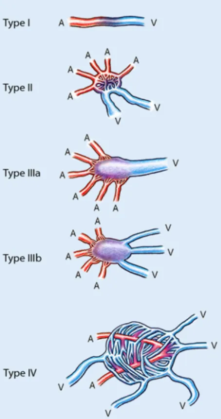
The arteriographic classification of AVMs was proposed to better define po-tential treatment strategies. Several classi-fication systems have been published. A cerebrovascular congenital AVM classi-fication system published in 1993 [3] and a peripheral vascular AVM classification system [4, 5] published in 2006 are strik-ingly similar regarding the angioarchitec-ture of high-flow AVMs.
The arteriographic classification was further adjusted by angioarchitecture ad-ditions not described in these two classi-fication systems by W. Yakes, adding the Yakes type I [direct artery-to-vein con-nections, e.g., as is seen in pulmonary ar-teriovenous fistulas (AVFs)] and Yakes type IV AVMs (characterized by a net-work of innumerable AVFs without a de-fined nidus infiltrating entire tissue with capillary beds interspersed among the
in-numerable AVFs) and a further modifi-cation of the previous two classifimodifi-cation systems with Yakes types II, IIIa, and IIIb (. Tab. 2, . Fig. 1).
Management
AVMs remain the most challenging mal-formation among various congenital vas-cular malformations due to their potential impact on the cardiovascular system in
Fig. 1 8 Angioarchitecture of high-flow arterio-venous malformations. A artery, V vene
the affected tissue and the subsequent he-modynamic consequences related to arte-rial steal and venous hypertensive chang-es. Clinical manifestations associated with AVM are dependent on the anatomical lo-cation and may produce cardiac failure, local venous hypertension with abnormal tissue changes, and arterial steal. In addi-tion, local effects of AVMs may include ulcerations that may be nonhealing and chronic, bleeding and infections in those ulcerated tissues, necrosis of the affected tissues, and gangrene if the arterial vascu-lar steal is significant.
Hormonal factors known to trigger progression of clinical symptoms in vas-cular malformations that have been pre-viously quiescent include the onset of pu-berty/menarche, pregnancy, birth con-trol pills, direct trauma, surgery with par-tial resection of the AVM lesion, proxi-mal ligation of prominent feeding
arter-ies, “skeletonization” surgical exclusion procedures, etc. All these produce neo-vascular stimulation enlarging the AVM and worsening its symptoms in the Schob-inger classification.
An ill-planned and improper treat-ment strategy (incomplete resection, sur-gical ligation/skeletonization and/or prox-imal coil embolization of the feeding ar-teries) can stimulate an AVM lesion to transform from a dormant state to a pro-liferative state, resulting in AVM growth with advancement of the Schobinger clas-sification to higher stages of symptomo-tology.
The main goal of treatment should be to elimination the nidus by vascular oc-clusion procedures or complete surgical extirpation of the AVM. Partial resection will result in lesion recurrence and en-largement with increased symptoms by neovascular stimulation.
Overview of endovascular
occlusive procedures
Endovascular therapy with various em-bolization and sclerotherapy modalities is reported in the literature by many vas-cular malformation centers as the pre-ferred therapeutic option for the majority of AVM lesions, rather than surgical extir-pation [7–10]. Precise delivery of the em-bolic agent to ablate the AVM nidus is re-quired for successful endovascular ther-apy, to minimize complications, and to achieve a long-term cure.
A combination of approaches utilizing any of the three routes of delivery of em-bolic agents (transarterial, transvenous, direct puncture) may be required to ablate the AVM. Multisession endovascular ther-apy is preferred and every effort should be exercised to minimize the risks of embo-lo-sclerotherapy during each session. The most appropriate embolic agents for pri-mary control of AVM include transcathe-ter embolization, direct puncture emboli-zation, and retrograde vein embolization approaches primarily using ethanol in Yakes types II, IIIa, and IIIb AVMs. Coils and other mechanically occlusive devic-es can be curative in Yakdevic-es type I AVFs. Coils can also lessen complications when densely packed in the aneurysmal veins of Yakes types IIIa and IIIb AVMs. In Yakes type IV AVMs, a 50–50% mixture of etha-nol and contrast as an embolic agent is cu-rative in these infiltcu-rative lesions and can spare the capillary beds from occlusion. The use of nBCA (n-butyl-2-cyanoacry-late) and/or Onyx are completely inade-quate to cure or provide long-term control for AVMs as a primary form of therapy. In the literature, it is reported that these po-lymerizing embolic agents are palliative at best and recurrences are common [11–15].
Embolic agents
Many embolic agents have been used in the treatment of AVMs. Particulate agents, which include polyvinyl alcohol (PVA) particles, microspheres, gelfoam, and col-lagen powders, have been used to emboli-ze AVMs. Their use, alone or in combina-tion with other agents, is well documented in the literature and is palliative with high rates of recurrences of the AVM [16–21].
Tab. 1 Schobinger clinical classification of AVMs symptomatology Stage I Quiescence: may or may not have a vascular skin stain, warmth of the affected tissues, and AV shunts can be detected by Doppler US scanning. The AVM is present but causes no clinical symptoms Stage II Expansion of the AVM lesion: stage I plus enlargement, pulsations, palpable thrill, au-dible bruit and enlarged arterialized tortuous/tense veins Stage III Destructive tissue changes: stage II plus dystrophic skin changes, skin ulcerations that can be nonhealing, bleeding from the ulcerated areas in the skin or mucosal surfaces, overt tissue necrosis, and lytic lesions of bone may occur Stage IV Decompensation: stage III plus congestive cardiac failure with increased cardiac output, abnormally lowered PVR, and venous hypertension secondary tissue and skin changes
AV arteriovenous, AVM arteriovenous malformation, PVR peripheral vascular resistance, US ultrasound.
Tab. 2 Yakes classification of high-flow AVM angioarchitecture [6] Type I A direct artery/arteriole to vein/venule connection
Direct AV connections can be treated with fibered coils, wires, Amplatzer plugs. Mechanical device occlusion can be curative
Type II Multiple arteries/arterioles connecting through an intervening “nidus” without any in-tervening capillary beds draining into multiple out-flow veins.
Ethanol embolization superselectively delivered by transcatheter and direct puncture tech-niques is curative
Type IIIa Multiple inflow arterioles shunting into an aneurysmal vein that has a single vein out-flow. (AV fistulae which infiltrate the wall of a draining aneurysmal vein with single outflow vein.)
Ethanol and/or coils can be curative
Type IIIb Multiple inflow arterioles shunting into an aneurysmal vein with multiple outflow veins. (AV fistulae which infiltrate the wall of the aneurysmal vein with multiple outflow veins).
More challenging to be treated with coils as multiple veins must be occluded
Type IV Multiple arteries/arterioles which form innumerable microfistulae that diffusely infil-trate the affected tissue. Interspersed within these innumerable AVFs are capillary beds that maintain the viability of the affected tissue. The innumerable AVF drain into mul-tiple veins. Venous hypertensive changes can occur as the capillary bed out-flow veins have greatly restricted venous access competing with the arterialized AVM draining veins that have arterialized venous pressures (Schobinger II and III changes).
A 50–50% mixture of ethanol and non-ionic contrast can be curative in this lesion type
326 |
Gefässchirurgie 4 · 2014Unfortunately, these agents do not possess properties that are well suited for treating AVMs. The particles are often ei-ther too large and occlude the vessels prox-imal to the nidus, or too small traveling through the AV shunt causing nontarget embolization. Since they are not well suit-ed to treat AVMs, their primary purpose is to alter the hemodynamics of the lesion to improve the possibility for surgical re-section, which is difficult to achieve.
Fibered and non-fibered coils
Coils are designed to mechanically oc-clude larger vessels and are not able to penetrate into the AVM nidus, as they are an endovascular occluding device sized to the diameter of the artery/vein that is be-ing embolized. When used as a transarte-rial embolic occlusive device, the result is proximal arterial ligation that is not cu-rative and will cause neovascular stimula-tion of the AVM, thus, stimulating growth of new arteries/arterioles to the AVM and worsening of the situation [22].
A major disadvantage of coil emboliza-tion therapy is that its mechanism of ac-tion is limited to the occlusion and subse-quent thrombosis of the artery or vein in which it is placed. Permanent damage to the blood vessel endothelium does not oc-cur, thus, allowing for subsequent regen-eration or recovery of the endothelium. This can result in recanalization with re-currence of the lesion.
However, coils can be extremely effec-tive and curaeffec-tive in AVM treatment when placed in the aneurysmal outflow veins and is a definitive treatment. This is particular-ly effective where there is a single AV con-nection (Yakes type I) and multiple arter-ies connect to a single draining vein (Yakes types IIIa/IIIb AVM lesions). In these types of AVM, occlusion of the venous outflow with coils can be very effective and dramat-ically reduces the risk of tissue injury and complications associated with transarteri-al ethanol embolization.
In AVMs that have an aneurysmal dilatation of the draining vein, with sin-gle outflow vein (Yakes IIIa) and multi-ple outflow veins arising from it (Yakes type IIIb), coil embolization can be used in combination with ethanol to achieve a definitive treatment. Once flow has been
slowed in the outflow veins by the place-ment of the coils, the injection of abso-lute ethanol can then reflux into the many vein fistulae in the wall of this aneurys-mal vein to allow permanent occlusion of the AVM. This “retrograde vein oc-clusion” technique in curative treatment of high-flow vascular malformations was first described by Yakes et al. in 1990 [23] and later confirmed by Jackson et al. [24] and Cho et al. [25].
Absolute ethanol
The curative potential of absolute ethanol as an AVM embolic/sclerosant agent lies in the fact that it destroys the endotheli-al cells on the vascular wendotheli-all by precipitat-ing its protoplasm (endothelial cells lin-ing arteries, veins, capillaries, lymphatics) and causes fractures of the vascular wall to the level of the internal elastic lamina. Be-cause the endothelial cells are destroyed, the phenomena of neovascular stimula-tion with new vascular inflow due to se-Gefässchirurgie 2014 · 19:325–330 DOI 10.1007/s00772-013-1303-9
© Springer-Verlag Berlin Heidelberg 2014
W. Yakes · I. Baumgartner
Interventional treatment of arterio-venous malformations
Abstract Background. Due to their hemodynamic ef- fects and tendency to progress, the major- ity of congenital arterio-venous malforma- tions (AVM) require treatment. AVM are classi-fied according to clinical severity (Schobinger classification, stages I–V) and angiographic appearance (types I–IV). Treatment. Endovascular embolization is the treatment of choice. However, because complications such as necrosis and neuro- pathy occur in up to 15% of cases, treat-ment is challenging and is dependent on the angio graphic appearance. Critical for treat-ment success is elimination of the so-called nidus, which is the location of the short cir-cuit connection between the artery and the often extended aneurysm-like drainage vein. Embolization of only the feeding artery with- out the actual nidus or the incomplete elim-ination of drainage veins should be avoided. Absolute 96% alcohol in addition to coiling of the nidus has been established as the most effective technique. Conclusion. Unlike other embolic agents, al-cohol leads to a definitive destruction of the vessel wall. Recanalization and recurrence are excluded due to adequate elimination of the nidus. Keywords AVM classification · Angioarchitecture · Coils · Congenital vascular malformation · Ethanol embolizationInterventionelle Behandlung von arteriovenösen Fehlbildungen
Zusammenfassung Hintergrund. Angeborene arteriovenöse Malformationen (AVM) stellen aufgrund ihrer hämodynamischen Auswirkungen und Pro- gressionstendenz mehrheitlich eine Thera-pieindikation dar. AVM werden nach ihrem klinischen Schweregrad (Schobinger-Klas- sifikation, Stadium I–V) und ihrem angio-graphischen Erscheinungsbild (Typ I–IV) eingeteilt. Behandlung. Therapie der Wahl ist die kathe tertechnische Embolisation wobei die Behandlung aufgrund der hohen Kom- plikationsraten mit Nekrosen und Neuro- pathien in bis zu 15% der Fälle anspruchs- voll ist, und sich die Technik nach dem angio- graphischen Erscheinungsbild richtet. Ent- scheidend für den Therapieerfolg ist die Aus-schaltung des sog. „Nidus“, der den Ort der Kurzschlussverbindung zwischen der Arte-rie und der oft aneurysmartisch erweiterten Drainagevene darstellt. Zu vermeiden ist die alleinige Embolisation der zuführenden Arte-rie ohne den eigentlichen Nidus zu erreichen oder die inkomplette Ausschaltung von Drainagevenen. Absoluter 96%-iger Alkohol in Verbindung mit einem Coiling des Nidus ist als die effektivste Technik etabliert. Schlussfolgerung. Im Gegensatz zu anderen Embolisaten führt Alkohol zu einer defini-tiven Zerstörung der Gefäßwand, wodurch eine Rekanalisation und ein Auftreten eines Rezidivs bei adäquater Ausschaltung des Ni-dus ausgeschlossen ist. Schlüsselwörter AVM-Klassifikation · Angioarchitektur · Coils · Kongenitale vaskuläre Fehlbildungen · Alkoholcretion of angiogenesis factors, and the recanalization phenomenon due to secre-tion of chemotactic cellular factor to carry out intravascular debris, is noticeably ab-sent and the potential for a permanent oc-clusion is now possible [26–34].
Because the endothelial cells are de-nuded from the vascular wall, its proto-plasm precipitated, and the vascular wall is fractured to the level of the internal elas-tic lamina, platelet aggregation occurs on the denuded vascular wall surface. This thrombotic process progressively occludes the vascular lumen from the vascular wall surface to the central lumen. There will be no more chemotactic cellular factors and angiogenesis factors secreted since the endothelial cells are completely de-stroyed [35].
However, the risk of cardiopulmonary complications during ethanol sclerother-apy administration is significant; there-fore, appropriate measures should be tak-en which include administration of gtak-ener- gener-al anesthesia and close cardiopulmonary monitoring. Use of a pulmonary artery catheter during ethanol sclerotherapy will allow continuous pulmonary artery pres-sure monitoring. Shin et al. [36] report-ed that if no more than 0.14 ml ethanol/kg body weight is embolized every 10 min, no cardiopulmonary complications will oc-cur [37, 38].
Pulmonary hypertension is a poten-tially fatal complication associated with
ethanol sclerotherapy and occurs when a significant dose of ethanol is allowed to reach to the lungs. The etiology of pul-monary hypertension is felt to be related to either pulmonary arterial spasm or ex-tensive microthromboembolization. The development of pulmonary hypertension can lead to subsequent cardiopulmonary arrest if not controlled effectively.
A total dose of ethanol used during an embolization procedure should be less than 1 ml/kg, since volumes greater than this can be toxic. Limiting ethanol in-jections to 0.14 ml ethanol/kg ideal body weight every 10 min will be able to obvi-ate the need of a pulmonary artery cath-eter when anticipating large injections of ethanol in large lesions [39]. By adher-ing to these principles, the risk of etha-nol flowing to the pulmonary circulation, thus, causing pulmonary vascular spasm with subsequent acute right heart failure, is obviated.
Because absolute ethanol is associated with various complications and morbidity, safe use of ethanol in AVM embolization requires accurate delivery into the nidus by precise placement solely in the AVM nidus vasculature that is nonnutritive and without capillaries. Proximal injection of ethanol into a feeding artery would cause severe tissue necrosis by destroying nutri-tive capillary beds.
In order to enhance the denaturat-ing or sclerosdenaturat-ing effect on the
endotheli-al cells, lowering the flow in the AVM it-self is a highly effective technique to al-low the injected ethanol to remain in lon-ger contact with the cells. Decreasing the flow through the lesion can be achieved in different ways (. Fig. 2):
F arterial approach: this approach can make use of occlusion balloons,
F direct puncture injection of the scle-rosing material, or
F direct puncture or retrograde trans-catheter venous approach. Occlusion of the aneurysmal vein with coils can be curative.
To decrease postembolization swelling in the endovascularly treated area, intra-venous dexamethasone is routinely used prior to the procedure as well as nonroidal anti-inflammatory agents and ste-roid therapy for 5 days postprocedure, thus, greatly reducing the risk of devel-oping compartment syndrome or subse-quent possible nerve injuries due to ede-ma. Accurate and meticulous emboliza-tion techniques, treating only the AVM, total avoidance of ethanol embolization of capillary beds, use of fibered coils in the vein approach in Yakes types I, IIIa, IIIb AVMs, will greatly diminish compli-cations from vascular malformation en-dovascular treatment procedures in these complex vascular lesions [40, 41].
Conclusion
F Due to their hemodynamic effects and tendency to progress, the major- ity of congenital arterio-venous mal-formations (AVM) require treatment. F AVM are classified according to clini-cal severity (Schobinger classification, stages I–V) and angiographic appear-ance (types I–IV). F Depending on the type of AVM, com-bined intraarterial, percutaneous or transvenous embolization is the treat- ment of choice, but requires special-ized interventional expertise. F Important for treatment success is the elimination of the nidus, whereby the interventional treatment of congen- ital AVM differ from other emboliza-tion treatments. Fig. 2 8 Type IIIb arterio-venous malformation a before and b after combined percutaneous ethanol embolization (21 ml, 96% ethanol) and coiling (12 Nester® coils) of the aneurysmal vein that has a sin-gle vein out-flow. Total cure was achieved in a single procedure328 |
Gefässchirurgie 4 · 2014Leitthema
F The use of 96% alcohol is the most ef-fective, but also associated with the most complications. F Transvenous or percutaneous coiling is a treatment option in AVM with an aneurysm-like nidus.
Corresponding address
W. Yakes Swedish Vascular Malforma-tion Center 501 E. Hampden Avenue, Suite 4600, 80113 Englewood, CO USA wayne.yakes@ vascularmalformationcenter.com Prof. I. Baumgartner Division of Angiology, Swiss Cardiovascular Center, INSELSPITAL, University Hospital 3000 Bern Switzerland Iris.Baumgartner@insel.chCompliance with ethical
guidelines
Conflict of interest. W. Yakes and I. Baumgartner
state that there are no conflicts of interest. The accompanying manuscript does not include stud-ies on humans or animals.