HAL Id: hal-00171465
https://hal.archives-ouvertes.fr/hal-00171465
Submitted on 12 Sep 2007
HAL is a multi-disciplinary open access
archive for the deposit and dissemination of
sci-entific research documents, whether they are
pub-lished or not. The documents may come from
teaching and research institutions in France or
abroad, or from public or private research centers.
L’archive ouverte pluridisciplinaire HAL, est
destinée au dépôt et à la diffusion de documents
scientifiques de niveau recherche, publiés ou non,
émanant des établissements d’enseignement et de
recherche français ou étrangers, des laboratoires
publics ou privés.
Using your head: Cognition and sensorimotor functions
in microgravity
Gilles Clément
To cite this version:
Gilles Clément. Using your head: Cognition and sensorimotor functions in microgravity. Gravitational
and Space Biology, 2007, 20 (2), pp.65-78. �hal-00171465�
Gilles Clément, Ph.D.
CNRS, Toulouse, France
ABSTRACT
To be aware of the environment, one must sense or perceive that environment. The body senses the environment by the interaction of specialized sensory organs with some aspect or another of the environment. The central nervous system utilizes these sensations in order to coordinate and organize muscular movements, shift from uncomfortable positions, and adjust properly. One relevant question is “What is the relative contribution of gravity to these sensory and motor functions?” This manuscript reviews the effects of microgravity on the functioning of the sensory organs primarily used for balance and spatial orientation. Disorientation and malaise so frequently encountered during early exposure to microgravity and on return to Earth are described. Theories and actual data regarding the role of the central nervous system in the adaptation of sensorimotor functions (including the control of posture, eye movements, spatial orientation, and cognition) to changes in the gravito-inertial force level during space flight are explored.
INTRODUCTION
Spaceflight creates a challenge for sensorimotor functions that depend on gravity, which include postural balance, locomotion, eye-hand coordination, and spatial orientation (Clément, 2005). Sensorimotor functions rely on organs which sense the acceleration environment, nerves which transmit this information to the spinal cord and brain, the central nervous system (CNS) which integrates this information so that we can determine our position and orientation relative to the environment, and on muscles for generating the appropriate reaction. The vestibular organs in the inner ear detect and measure linear and angular accelerations. These responses, already complex, are further integrated with visual and somesthetic inputs. These sensorimotor functions must adapt to weightlessness on entering orbit, and again to normal gravity upon return to Earth. During this period of adaptation, some of the sensory signals are modified, leading to misinterpretation and non-adequate responses by the brain.
One example of non-adequate responses is space motion
sickness (SMS). SMS is a special form of motion sickness that is experienced by some individuals during the first several days of exposure to microgravity. The syndrome may include such symptoms as depressed appetite, malaise, lethargy, gastrointestinal discomfort, nausea, and vomiting. Gastrointestinal symptoms have their onset
from minutes to hours after orbital insertion. Symptom resolution usually occurs between 30 and 48 hours, with a reported range of 12 to 72 hours, and recovery is rapid (for review, see Crampton, 1990). However, there is often a recurrence of symptoms at landing, similar to the “mal de débarquement” in seafarers, especially after long-duration space missions (Gorgiladze and Bryanov, 1989). Microgravity by itself does not induce space sickness. There were no reports of motion sickness during the Mercury and Gemini spaceflights. However, as the volume of spacecrafts has increased (allowing more mobility) the incidence of SMS has increased as well. Movements that produce changes in head orientation seem necessary to induce SMS symptoms. In particular, many crewmembers report that vertical head movements (rotation in the pitch or roll planes) are more provocative than horizontal (yaw) head movements (Oman et al., 1990).
About two-thirds of the Space Shuttle astronauts experienced some symptoms of SMS. There are no statistically significant differences in symptom occurrence between pilots versus non-pilots, males versus females, different age groups, or novices (first time flyers) versus veterans (repeat flyers.) An astronaut’s susceptibility to SMS on his/her first flight correctly predicted susceptibility on the second flight in 77% of the cases (Davis et al., 1988; Jennings, 1997). Although anti-motion sickness drugs (i.e., scopolamine, promethazine) offer some protection against SMS, some drugs may interfere with the adaptation process, and symptoms controlled by these drugs are experienced again once treatment ceases.
Several theories have been proposed to for the occurrence of SMS. The sensory conflict theory of motion sickness assumes that human orientation in 3-D space, under normal gravitational conditions, is based on at least four sensory inputs to the central nervous system. The otolith organs provide information about linear accelerations and tilt relative to the gravity vector; angular acceleration information is provided by the semicircular canals; the visual system provides information concerning body orientation with respect to the visual scene or surround; and somatosensory systems supply information about limb and body position. In normal environments, information from these systems is compatible and complementary, and matches that expected on the basis of previous experience. When the environment is altered in such a way that information from the sensory systems is not compatible and does not match previously stored neural patterns, motion sickness may result (Reason and Brand, 1975). It has been proposed that motion sickness results from the activation of a vestibular mechanism
____________________
* Correspondence to: Dr. Gilles Clément Centre de Recherche Cerveau et Cognition
UMR 5549 CNRS-Université Paul Sabatier Toulouse 3 Faculté de Médecine de Rangueil
31062 Toulouse Cedex9, France Email: [email protected] Phone: 33-562-173779; Fax: 33-562-172809
whose physiological function is the removal of poisons from the stomach. Nausea and vomiting would tend to keep a disoriented or dizzy individual from moving about the environment in search of food when he would be at risk doing so (Money, 1990).
The sensory conflict theory postulates that motion sickness occurs when patterns of sensory inputs to the brain are markedly rearranged, at variance with each other, or differ substantially from expectations of the stimulus relationships in a given environment. In microgravity, sensory conflict can occur in several ways. First, there can be conflicting information (i.e., regarding tilt) transmitted by the otolith organs and the semicircular canals. Sensory conflict may also exist between the visual and vestibular systems during motion in space; the eyes transmit information to the brain indicating body movement, but no corroborating impulses are received from the otolith organs (such as during car sickness). A third type of conflict may exist in space because of differences in perceptual habits and expectations. On Earth, we develop a neural store of information regarding the appearance of the environment and certain expectations about functional relationships (e.g., the concepts of “up” and “down”). In space, these perceptual expectations are at variance, especially during the inversion illusions described above.
Some investigators have proposed a mechanism complementary to the sensory conflict theory to explain individual differences in SMS susceptibility. They suggest that some individuals possess slight functional
imbalances (for example, weight differences) between the right and left otoconia that are compensated for by the central nervous system in 1 g. A weight imbalance between the left and right otolith crystals is reasonable since there is a continual turnover of otoconia, and it is unlikely that the otolith crystals on both sides would ever weight exactly the same. This compensation is inappropriate in 0 g, however, since the weight differential is nullified and the compensatory response (either central or peripheral) is no longer correct for the new inertial environment. The result would be a temporary asymmetry producing rotatory vertigo, inappropriate eye movements, and postural changes until the imbalance is compensated or adjusted to the new situation. A similar imbalance would be produced upon return to 1 g, resulting in postflight vestibular disturbances. Individuals with a greater degree of asymmetry in otoconia morphology would thus be more susceptible to SMS.
A sensory compensation hypothesis has also been proposed. Sensory compensation occurs when the input from one sensory system is attenuated and signals from others are augmented. In the absence of an appropriate graviceptive signal (or perhaps the presence of atypical signals) in microgravity, information from other spatial orientation receptors such as the eyes, the semicircular canals, and the neck position receptors would be used to maintain spatial orientation and movement control. The
increase in reliance on visual cues for spatial orientation could be explained by this mechanism (Parker et al., 1985).
Although past research has yielded a great deal of information applicable to SMS, a definitive solution to this vexing problem is needed. Among the objectives of current SMS research is the development of: (a) more precise predictive indices; (b) more effective drug treatments; (c) more efficient preflight adaptation procedures; (d) methods to evaluate performance impairment induced by SMS and anti-motion sickness drugs; and (e) the early detection of incipient symptoms.
VESTIBULAR RECEPTORS Physiology
The inner ear contains two balance-sensing organs: one is sensitive to linear acceleration, the other to angular acceleration. The linear acceleration sensitive organ is comprised of the saccule and utricle, which send messages to the brain as to how the head is translated or positioned relative to the force of gravity. The angular acceleration sensitive organ is comprised of three semicircular canals, which detect angular acceleration through the inertial movement of the endolymph within each canal and provide the brain with information about rotation about the three axes: yaw, pitch, and roll (for review, see Highstein et al., 2004). The semicircular canals do not react to the body’s position with respect to gravity. They react to a change in the body’s position. So, unlike the otolith organs, the semicircular canals are not affected by spaceflight.
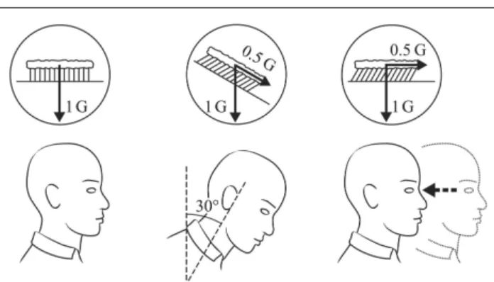
Figure 1. The otolith crystals bend the hair cells of the utricles
the same way when the head is maintained at a constant tilt angle of 30 deg relative to gravity and when the whole body is translating backwards at 0.5 g. Drawing Philippe Tauzin, SCOM, Toulouse.
When our head is horizontal the hair cells in the utricles are not bent and this stimulation is interpreted as signifying “normal posture”. If our head is tilted forward, the otolith crystals shift downward under the action of gravity, bending the hair cells. If we translate backward, again there is a shift of the otolith crystals forward due to the inertial forces. Thus, an equivalent displacement of the otolithic membrane (and consequently the same
information is conveyed to the CNS) can be generated when the head is tilted 30 deg forward, or when the body is translating at 0.5 g backward, for example (Figure 1). This example simply illustrates Einstein’s principle stating that, on Earth, all linear accelerometers cannot distinguish between an actual linear acceleration and a head tilt relative to gravity.
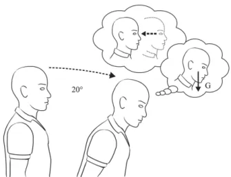
On Earth, the otolith signals can be interpreted as either linear motion (translation) or as tilt with respect to gravity. Because stimulation from gravity is absent in weightlessness, interpretation of otolith signals as tilt is inappropriate. Therefore, it is possible that during adaptation to weightlessness, the CNS reinterprets all otolith signals to indicate translation. This hypothesis is known as the Otolith Tilt-Translation Reinterpretation (OTTR). This central reinterpretation would persist following return to Earth, and be at the origin of spatial disorientation, until re-adaptation to the normal gravity environment occurs (Parker et al., 1985; Young et al., 1986).
Figure 2. In microgravity, the otolith organs are stimulated by
head translation, but not by head tilt. Consequently, it is hypothesized that, after a period of adaptation, the brain reinterprets all otolith signals as signaling head translation. According to this hypothesis, on return to Earth, a static tilt of the head relative to gravity could be perceived as an actual tilt or a translation in the opposite direction. Drawing Philippe Tauzin, SCOM, Toulouse.
Evidence for the OTTR hypothesis comes from subjective reports by astronauts returning from spaceflight who have a sense of body translation when they voluntarily pitch or roll their head (Clément and Reschke, 1996). For example, many experience a backward translation when they pitch their head forward (Figure 2), or a rightward translation when they roll their head to the left. Such a misleading interpretation of the otolith signals might be responsible for the staggering posture of the astronauts as soon as they land. The astronauts tend to lean to the outside of the turn when walking and turning corners immediately after landing, also suggesting a misevaluation of the apparent vertical from the otolith signals.
The OTTR hypothesis has been the theoretical basis of much space research on the neuro-vestibular system for the past 15 years. I was fortunate enough to be able to perform a space experiment that tested this hypothesis in 1998 (Clément et al., 2003). This experiment, which flew on board the Neurolab mission (STS-90), used a human-rated centrifuge constructed by the European Space Agency. On Earth, when an individual is rotated in a centrifuge in darkness, he senses the direction of the resultant gravito-inertial force and regards this as the vertical. If a centrifugal force equivalent to 1 g is directed sideways, the gravito-inertial force is displaced 45 deg relative to the upright body, and the subject has a sense of being tilted by 45 deg to the outside. In microgravity, however, the gravitational component is negligible and the gravito-inertial force is equivalent to the centrifugal force. This force could be interpreted either as a 90 deg tilt of the body, or a whole body translation in the opposite direction. During the Neurolab mission, four astronauts were asked to report their perceived angle of tilt during steady-state centrifugation in darkness throughout the flight and during the postflight re-adaptation period. Centrifugation was always perceived as tilt, not translation. Therefore the findings do not support the OTTR hypothesis. Despite the fact that the otoliths do not respond to head tilt in orbit, the brain continues to sense a steady-state linear acceleration applied to the otolith organs as the upright in all circumstances (Clément et al., 2001).
The debate regarding the OTTR hypothesis is still raging. Some have proposed that the OTTR only occurs during voluntary head movements, or only during rotational head movements, or that OTTR has to be frequency dependent. Centrifugation, by applying very low frequency passive linear acceleration to the entire body, would thus not elicit OTTR. I am currently conducting a follow-up study on astronauts returning from spaceflight, by spinning them about a tilted axis (off-vertical axis rotation) at various frequencies to further address this hypothesis.
The Neurolab centrifuge experiment, however, brought another interesting result. At the beginning of the flight, during 1-g centrifugation in darkness, the astronauts perceived a 45-deg tilt to the side, very much like on Earth. However, as the mission progressed, they felt more and more tilted, until a 90-deg tilt to the side on flight day 16. This simple result indicates that the brain does not continuously calculate the direction of gravity, but uses an internal estimate of gravity whose weighting changes during spaceflight. The internal estimate normally used on Earth (1 g) carries over to the early period of exposure to weightlessness, and therefore the astronauts continue to perceive a 45-deg tilt, despite the absence of sensed gravity. After a period of adaptation, the internal estimate declines to zero and the astronauts perceive a full body tilt to the side (Clément et al., 2001; Clément et al., 2003).
Changes in the Vestibular Receptors during Spaceflight
Although it is difficult to measure changes in the vestibular end organs directly, several attempts have been made to examine the question “Is there anatomical and physiological changes in the vestibular end organs and their primary afferents after exposure to microgravity?” Experiments on frogs have revealed no alteration of the sensory epithelium of the vestibular organ of adults returned from an 8-day stay aboard the Russian Mir space station, or following larval development in microgravity. However, changes in the structure of the otoconia in rats had been observed during earlier missions (Cosmos-782). This degeneration of the otolith crystals could occur because of changes in body calcium, protein metabolism, and calcium exchange. In addition, it is unclear how much of these changes were due to the high accelerations experienced by the animals during take-off and landing. More recent Spacelab experiments indicated no deleterious effects in the otoconia of the otolith organs from rodents who flew as compared with the ground controls. However, an unexpected change found during the Spacelab SLS-1 mission, and later confirmed during the Neurolab mission, was an increase (by a factor of 12) in the number of synapses in hair cells from the in-flight maculae as compared with the control data. These findings suggest that mature utricular hair cells retain synaptic plasticity, permitting adaptation to an altered environment (Ross et al., 1993; 1994). Consistent with these results is data that show a decrease in synapse activity in centrifuged rats. These data suggest that the maculae adapt to g-forces changes in either direction by up- or down-regulation of synaptic contacts in an attempt to modulate neural inputs to the CNS (Ross and Tomko, 1998).
Primary afferent fibers of the vestibular nerve are relaying the information originating at the hair cells to the brainstem (within each nerve are also efferent fibers from the CNS which provide neural feedback to modulate the activity of the peripheral organs). The resting activity of single otolith afferents and their response to centrifugal forces were found to be different in microgravity compared to the ground condition in frogs (Braachi et al., 1975). Recently, a study recording the vestibular nerve impulse data from the oyster toadfish during the Neurolab mission confirmed these results (Boyle et al., 2001). On the other hand, the spontaneous firing rate of single horizontal semicircular canals afferents did not change postflight relative to preflight in two flight monkeys (Correia et al., 1992). However, these monkeys were restrained in a laboratory chair, thus preventing any movements of the head during the flight. It is known that movement and interaction with the environment are necessary factors to drive adaptive changes. For example, vestibular patients show a faster recovery when moving around after vertigo crisis or unilateral surgery.
Few experiments on the early development of the vestibular system have been carried out in space (cf. Clément and Slenzka, 2006). This is an interesting research topic since in all species the vestibular system begins to respond to stimulation (linear or angular acceleration) prior to hatching or birth, in contrast to hearing or vision, which can be postnatal in some species. Mammalian offspring emerge from the birth canal in a species-typical orientation, which, for rats and humans, is head first. Fetuses typically achieve the appropriate orientation via active, in-utero behavior. Perhaps the vestibular system is employed for this early task. Indeed, many infants born in the breach position are born with vestibular disorders. Also, the so-called righting response, by which the newborn mammals actively adjust from a supine to a prone position, is disrupted by induced vestibular disorders during development (Romand, 1992). In the development of the visual system, activity in the retinal pathway influences the specification of those connections that determine how visual information is processed in the cerebral cortex. In every other sensory systems known, especially those that make up the neural space maps in the brainstem, sensory stimulation has been implicated in the initial specifications of the connections and physiological properties of the constituent neurons. Only in the utricle and saccule gravitational pathway has it been impossible to study the role of sensory deprivation, because there is no way to deprive the system of gravitational stimulation on Earth. For this reason, experiments in microgravity should be planned to test the hypothesis that gravity itself plays a role in the development and maintenance of the components of the vestibular system. These components include both the vestibular receptors of gravity (i.e., the sensory hair cells in the utricle and saccule, vestibular ganglion cells that form synapses with vestibular hair cells, and vestibular nuclei neurons) and the motor neurons. The latter receive input from axons of the vestibular nucleus neurons composing the vestibular reflex pathways. The vestibular system also receives inputs from the proprioceptive system, involved in the control of muscle length and tension, and from the visual system, involved in the control of eye movements. Little is known about the exact nature of these interactions and virtually nothing concerning the development of these connections.
POSTURE AND MOVEMENT
Postural activity is the complex result of integrated orientation and motion information from visual, vestibular, and somatosensory inputs. These inputs collectively contribute to a sense of body orientation and, additionally, coordinate body muscle activities that are largely automatic and independent of conscious perception and voluntary control. The absence of gravity modifies the stimuli associated with proprioception and impact spatial orientation, including knowledge of position in the passive limb, difficulty of pointing accurately at targets during voluntary limb movement, modification of tactile sensitivity, and changes in the
perception of mass (for a review, see Lackner and DiZio, 2000). However, the nature of proprioceptive changes in microgravity has been poorly studied. There is almost no space study of neck and joint angle sensors, and on the role of localized tactile cues in the perception of body verticality.
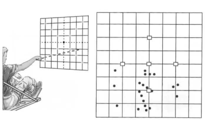
When crewmembers point at remembered target positions with they eyes closed, they make considerable errors and tend to point low (Figure 3). When they are asked to reproduce from memory the different positions of a handle, the accuracy of setting the handle to a given position is significantly lower with an error towards a decrease of handle deflection angle (Bock et al., 2001). Also, when trying to touch various body parts, they usually note that their arms are not exactly where expected when vision is restored. The problem is that these examples are suggestive of either degradation in proprioceptive function, or an inaccurate external spatial map, or both (Watt, 1997; Young et al., 1993).
Figure 3. This experiment measured the pointing accuracy
during spaceflight. The subjects looked at one of the 5 cardinal targets, then closed their eyes and pointed a laser beam at the remembered target position. The inflight results for one subject are shown on the right. In microgravity, this subject exhibited a pronounced downward pointing bias. The divisions are in inches. Courtesy of NASA.
Human factor studies, after investigating photographs taken during Skylab missions, have led to the NASA
Neutral Body Posture model. This model is characterized by a forward tilt of the head (with the line of sight 25 deg lower than the body-centered horizontal reference), shoulders up (like a shrug), and arms afloat, up and forward with hands chest high. Recent investigations, taking into account body size, gender, and mission duration suggest, however, that the neutral body posture model is too generalized, and should be modified with additional data to provide more representative spaceflight crew postures. However, it is unclear how the direction of the line of sight has been evaluated from the Skylab photographs. Also, the downward deviation of gaze in microgravity in this model is in contradiction with the results of several space experiments that actually measured the eye deviation during spaceflight.
Vestibulo-Spinal Reflexes
Two of the more dramatic responses to orbital flight have been postural disturbances and modified reflex activity in the major weight-bearing muscles. For example, monitoring the Hoffman reflex (or H-reflex), which takes advantage of the anatomical pathways that link the otolith organs and spinal motoneurons, has been selected as a method of monosynaptic spinal reflex testing to assess otolith-induced changes in postural muscles. By contrast to doctor tapping a patient’s knee to produce the proverbial “knee jerk” reflex, during H-reflex the stimulus is an electrical shock to sensory fibers coming from stretch receptors in the calf (soleus) muscle, and the response is the electrical activity recorded from the muscle. Each time a subject is tested, the number of motoneurons that have been excited by a standard volley of sensory impulses is counted. That number is an indicator of spinal cord excitability. Interestingly enough, this number fell in ISS crewmembers, quite quickly at first and then more gradually over many days. A return to normal was observed within days after landing (Watt, 2006).
When performed in conjunction with linear acceleration (such as “falls” simulated by bungee cords) the H-reflex amplitude is low in-flight, but very large postflight (Reschke et al., 1986). Interestingly, sudden drops are perceived as falls or drops on Earth, and felt in-flight much as they did preflight. Later in-flight as well as postflight, drops were perceived as more sudden, fast, and hard. During those drops, the subjects did not have a falling sensation, but rather a feeling that “the floor came up to meet them”.
Second, extensive dynamic postural testing with a moving platform was performed before and after space missions. Balance control performance has been systematically tested before and after the flight using a computerized dynamic posturography system widely employed for evaluation of balance disorders (Paloski et al., 1993). Postflight measurements revealed significant deviations from the results obtained before flight. The strategy used by the individuals for balance is modified, and their behavior indicates a decrease in awareness of the direction and magnitude of the motion. On landing day, every subject exhibited a substantial decrease in postural stability. Some had clinically abnormal scores, being below the normative population 5th percentile. After flights ranging from 5-13 days, postflight re-adaptation took place in about 8 days and could be modeled as a double-exponential process, with an initial rapid phase lasting about 2.7 hours, and a secondary slower phase lasting about 4 days (Paloski et al., 1993). The effects of demographic factors like age, gender, and longer mission duration on these responses are currently evaluated. Information obtained from these investigations is promising for ground-based clinical research. A relatively large number of individuals on Earth suffer from prolonged, frequently life-long, clinical balance disorders.
Disorders like Ménière’s disease and traumatic injuries to the inner ear can severely influence quality of life. Falls are the leading cause of injury-related deaths in the elderly and these numbers continue to grow. Inner ear disorders are thought to account for 10–50% of falls among senior citizens. Currently, human spaceflight is the only means available for studying the response to sustained loss and recovery of inner ear information. Comparison between data from astronaut-subjects and similar data from patients and elderly subjects demonstrates similarities between these balance disorders. One sensible difference is that the posture problems recover in a few days for the astronauts, whereas it can take weeks (or never recover) in the patients. It is hoped that a better understanding of the strategies used during the recovery process in the astronauts, and of the plasticity of this system in general, will help to improve rehabilitation treatments for patients with balance disorders on Earth.
Locomotion
The cautious gait of astronauts descending the stairs of the “white room” docked with the Space Shuttle and walking on the runway is an obvious example of changes in sensorimotor coordination. Typically, locomotion in microgravity poses no problem and is quickly learned. However, adaptation continues for about a month. The astronauts who just visit the ISS note that the long-duration crewmembers move more gracefully, with no unnecessary motion. They can hover freely in front of a display when the new comers would be constantly touching something to hold their position.
When locomoting in space, the astronauts stop using the legs. Instead they use the arms or fingers to push or pull themselves. For clean one-directional movements, push must be applied through the center of gravity, i.e., just above the hips for a stretched-out body. When translating though, the natural place for the arms is overhead to grab onto and push off from things as they come whizzing by. This is the worst possible place from the physics of pushing and pulling for clean movements, for by exerting forces with arms overhead, some unwanted rotations will invariably occur, which have to be compensated with ever more pushes and pulls, giving an awkward look to the whole movement. “To cleanly translate, the best to keep the hands by the hips when exerting forces and boldly go headfirst. This way the pushing and pulling is directed through the body’s center of gravity and gives nice controlled motions without unwanted rotations” (Pettit, 2003).
Movement in a weightless environment obeys to the Newton’s laws of motion. Friction forces are negligible and the angular momentum is always conserved unless acted on by an outside torque (Jones, 1997). Since the legs are less used for locomotion, new sensorimotor strategies emerge in microgravity. Some of this newly developed sensorimotor program “carries over” to the
postflight period, which leads to postural and gait instabilities upon return to Earth. Both U.S. astronauts and Russian cosmonauts have reported these instabilities even after short-duration (5-10 days) spaceflights. Subjects experienced a turning sensation while attempting to walk a straight path, encountered sudden loss of postural stability especially when rounding corners, perceived exaggerated pitch and rolling head movements while walking, and experienced sudden loss of orientation in unstructured visual environments. In addition, oscillopsia and disorienting illusions of self-motion and surround-motion occurred during head movement induced by locomotion (Clément, 2001).
After spaceflight, changes have been documented in both head-trunk and lower limb patterns of coordination. Bloomberg et al. (1997) reported changes in head pitch variability, a reduction of coherence between the trunk and compensatory pitch head movements, and self reports from crewmembers indicating an increased incidence of oscillopsia (the illusion of a visual surround motion) during postflight treadmill walking. A number of characteristics of walking also appear to be changed after spaceflight. For example, during the contact phase of walking, the foot “thrusts” onto the support surface with a greater force than that observed before flight.
The alterations in locomotion seen after spaceflight raise some concern about the crew capability for unaided egress from the Space Shuttle or the Soyuz in a case of emergency. As discussed earlier, many crewmembers experience marked vertigo when making head movements during entry, landing, and afterwards. This vertigo could be a major obstacle to successful egress if vision were impaired, as with a smoke-filled cabin. An interesting investigation was performed by Bloomberg et al. (1999), in which the ability for crewmembers to repeat a previously seen trajectory without vision was examined. When attempting to walk a triangular path after flight, blindfolded subjects showed both under- and over-estimations of the distances walked, but a correct estimation of the angle turned. These results suggest a difficulty for reconstructing motion cues from the otolith organs, but not from the semicircular canals. However, the changes found could also be related to the lower walking velocity during postflight testing.
These results imply that mechanisms like computing self-displacement and updating spatial information (both of which being also called navigation) are disturbed by spaceflight and have to be reacquired after return to Earth.
Body Movement
On the Earth’s surface, gravity significantly affects most of our motor behavior. It has been estimated that about 60% of our musculature is devoted to opposing gravity. For example, when making limb movements during static balance, anticipatory innervations of leg muscles compensate for the impending reaction torques and the changes in location and projection of the center of mass
associated with these movements (Clément et al., 1985). Similar patterns of anticipatory compensations are seen in-flight, although they are functionally unnecessary. Also, rapidly bending the trunk forward and backward at the waist is accompanied on Earth by backward and forward displacements of hips and knees to maintain balance. The same compensatory movements of hips and knees are made in weightlessness. Since the effective gravity torques are absent during spaceflight, the innervations necessary to achieve these synergies in weightlessness are different from those needed on Earth. Consequently, these in-flight movements must reflect reorganized patterns of muscle activation. Dorsi-flexor muscles (e.g., the Tibialis anterior leg muscle) assume a larger role in space than on Earth in regulating the orientation of the individual relative to his/her support. This is in contrast with the general use of muscle extensors on Earth, which are used to counteract gravity. This transfer of motor strategies from one muscle group to another explains the forward tilted posture of crewmembers placed in darkness when instructed to maintain a posture perpendicular to the foot support (Clément and Lestienne, 1988).
Using a simple ball catching experiment in weightlessness, it has been elegantly shown that the central nervous system uses an internal estimate of gravity in the planning and execution of movements. During the act of catching a ball on Earth, the brain estimates the trajectory of the ball, accurately taking into account its downward acceleration due to gravity. In space, a seated astronaut had to catch a ball traveling at a constant velocity, in contrast to the constant acceleration that would occur on Earth. The ability to anticipate and predict is one of the nervous system’s basic functions. When we catch a ball, the brain does not wait for it to touch the hand before stimulating arm flexor muscle contraction to compensate for the impact. About one third of a second before impact, the brain elicits just the right amount of contraction to counteract the force exerted, which itself depends on the weight of the object combined with the acceleration of its fall. The experiment led to the conclusion that the brain works by anticipating the effects of gravity on the ball rather than by making direct measurements of its acceleration. This anticipation ability remains even in conditions of weightlessness. Thanks to childhood experience, the brain possesses internal models of the gravity laws governing the behavior of a falling object, and perhaps more generally, Newton’s law of mechanics. We see here the beginnings of an adaptation to new laws. A longer period in weightless flight would now be needed to assess how such an adaptation might develop (McIntyre et al., 2001).
Eye Movement
Eye movement is probably the response of the vestibular system that has been the most studied during spaceflight. For several decades, the study of eye movements has been a source of valuable information to both basic scientists and clinicians. The singular value of studying eye
movements stems from the fact that they are restricted to rotations in three planes and the eyeball offers very little inertia to the eye. This facilitates accurate measurement (for example using video eye recording in near infrared light), a prerequisite for quantitative analysis.
Eye movements must continuously compensate for head movements so that the image of the world is held fairly steady on the retina, and thus appears clear and stationary. During head movements, the vestibular apparatus measures head velocity and relays this information to those centers controlling eye position to generate compensatory eye movements; this reflex behavior ensures that vision is not blurred. When performed in darkness, this leads to a pattern of rhythmic eye movements known as nystagmus, consisting of slow phases in the direction opposite to the head and fast phases which bring the eye back when it reaches the extreme of its travel. The nystagmus response to a rapid head movement outlasts the changes in signals in the semicircular canals, through the activation of a velocity storage mechanism located in the brainstem.
This so-called vestibulo-ocular reflex has been studied systematically in orbital flight, both during active (voluntary) and passive movements of the head (for review, see Clément, 1998). With my co-investigators, we were the first to report that the amplitude of vertical eye movements was decreased during the first three days of weightlessness compared to normal value on Earth. In this experiment, the eye movements of an astronaut were recorded when he voluntary moved his head while either fixating a visual target or imagining that target in darkness. After four days in orbit, the vestibulo-ocular reflex gain returned to the pre-flight level, perhaps as a consequence of substituting neck receptor cues for vestibular receptor cues. Several investigations have later reported that after short-duration spaceflight, the pattern of eye and head movements was significantly altered when subjects moved their heads and eyes to fixate a laterally displaced target (Clarke et al., 1993).
Problems in hand-eye coordination and blurriness of the visual scene when reentering in normal gravity have also been reported after long-duration missions. Tracking of moving visual targets seems also to be altered, especially in the vertical direction. These deficits might pose a problem for piloting tasks during landing. The vestibular nuclei located in the brain stem are part of a system that allows one to fix the gaze on a stationary target during voluntary head motions as well as to track moving targets. This system appears to be disturbed during spaceflight, presumably as a consequence of altered vestibular receptor function due to the absence of gravity (Clément, 1998).
One problem in studying eye movements by asking subjects to perform voluntary head movements is that the central nervous system is “aware” of the movement to be performed. A copy of the motor command (the so-called
coordination control system, and this helps to achieve the adequate, compensatory eye movements. For this reason, scientists also use passive rotation generated by servo-controlled rotating chair or sled in order to generate unpredictable inertial stimulation of the vestibular system, and to study the resulting responses. Several of these devices have flown on board the Spacelab.
In 1985, a 4-m linear sled generated sinusoidal oscillations in subjects sitting either facing the track, or perpendicular to it, or lying on their back (Merfeld, 1996). The peak linear acceleration was 0.2 g. Absolute thresholds for the perception of linear acceleration in-flight and postin-flight were found to be elevated in some astronauts and lowered in others for some axes, relative to ground-based controls. Another measure of linear acceleration sensitivity, the time elapsed from acceleration onset to reports of self-motion (which varies inversely with magnitude of acceleration) have been more consistent. Results indicate an elevation of the sensitivity when linear accelerations are exerted along the body longitudinal axis, and a decrease in sensitivity for the other axes (Merfeld et al., 1994). It is, however, difficult to rule out a contribution of the somatosensory sensation in these results.
In 1992, a rotating chair flew on board the Spacelab IML-1 mission, allowing the evaluation of the vestibulo-ocular reflex evoked by passive rotation of 4 crewmembers about the yaw, or pitch or roll axis, during the course of a 7-day spaceflight. Results showed that the responses generated by rotation in pitch and roll were the most affected in space (Clément and Reschke, 1996).
More recently, in 1998, a human-rated centrifuge flew on the Neurolab mission, in which crewmembers were both exposed to angular and linear acceleration). One objective of this experiment was to study the adaptation of the CNS by measuring the eye movements in response to the angular and linear acceleration in space (Moore et al., 2001). Eye rotations can compensate for both the rotational and the translational components of head motion. As mentioned above, in microgravity, the otoliths are not stimulated by head tilt, and therefore the eye movements in response to head pitch or roll are likely to be altered during and after spaceflight. The results of the centrifuge experiment have not confirmed this hypothesis, though: the torsional (along the line of sight) eye movement elicited by the linear acceleration (known as
ocular counter-rotation), and the optokinetic nystagmus were unchanged in-flight and postflight relative to preflight (Moore et al., 2001, 2005). More investigations are therefore necessary to fully understand the adaptation of the compensatory eye movements during spaceflight. New tests of the otolith function are currently introduced in order to evaluate the re-adaptation of eye movements in response to body tilt after spaceflight. The eye movements and the perception of crewmembers exposed to body rotation about an axis tilted from Earth vertical offer interesting capabilities. This off-vertical axis
rotation (OVAR) causes, when rotation is in darkness at a constant low velocity, the perception of being successively tilted in all directions. Consequently, both a counter-rotation of the eyes and a perception of moving along the edge of an inverted cone, appear. At higher rotation velocity the illusion is that of being upright, but moving along the edges of a cylinder (hence more translational motion), and eye movements are predominantly horizontal (Clément et al., 1995). Another otolith test is achieved using a centrifuge where sitting subjects are displaced minimally from the rotation axis, so that one labyrinth becomes aligned on-axis, while the second labyrinth alone is exposed to the centripetal acceleration. This technique allows investigating subjective vertical and otolith-ocular responses during stimulation of the otolith on one side at a time (Wetzig et al., 1990). These tests should allow to more accurately document a change in the reinterpretation of the otolith signals from tilt to perception in returning crewmembers, and validate or not the OTTR hypothesis.
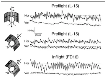
Very recently, scientists have discovered that, on Earth, the eye movements also reflect an orientation to the resultant linear accelerations during turning. During either passive rotation, as in a centrifuge, or while walking or running around a curved path, the axis of eye rotation tends to align with the resultant axis of the summed linear accelerations. The same phenomenon occurs when viewing a visual scene that moves in the horizontal plane, but with the head tilted to the side. The optokinetic
nystagmus is then oblique relative to the visual scene, as if the eye movements tried to align with the resultant of visual motion and gravity (Figure 4). Space experiments have showed that this gravity-oriented response was absent in microgravity, and that a return to the normal preflight response was observed only two days after return to Earth (Clément, 1998).
Figure 4. Horizontal (Hor) and vertical (Ver) components of eye
movement during horizontal optokinetic stimulation in a subject with the head upright or roll tilted over the trunk on Earth (L-15) and in space (FD16). When the head is tilted, on Earth the optokinetic nystagmus is oblique, whereas during the flight the optokinetic nystagmus is purely horizontal. This result indicates a loss of eye movement orientation relative to the spatial vertical.
SPATIAL ORIENTATION
Vision may compensate in large measure for modified otolith sensitivity. It helps in spatial orientation, and is essential to motor coordination (Howard and Hu, 2001). Astronauts working in microgravity must rely much more on vision to maintain their spatial orientation, since otolith signals no longer signal the direction of “down”. It has long been known that moving visual scenes can produce compelling illusions of self-motion (“seeing is believing”) (Howard and Hu, 2001). These visually-induced illusions become even stronger in space, since visual cues are unhindered by constraints from the otoliths, which in microgravity do not confirm or deny body tilt. This has been confirmed with experiments where crewmembers observing a rotating visual field felt a larger sense of body rotation in space than on Earth (Young and Shelhamer, 1990). It is interesting to note that frogs born in microgravity also showed stronger behavioral response to moving visual scenes when tested after their return on Earth than control animals born on Earth.
Crewmembers who remained seated in the relatively small Soyuz, Mercury, Gemini, and Apollo capsules rarely encountered orientation problems. However crews of the larger Skylab and Shuttle reported occasional disorientation, particularly when they left their seats, and worked in unpracticed, visually unfamiliar orientations. The problem occurred both inside the spacecraft, and also outside, as when performing EVA. For example, Bernard Harris, an astronaut of the STS-63 Shuttle mission reported: “As I was getting ready to step out of the spaceship, it felt like gravity was going to grab hold of me and pull me down toward Earth. Your natural response is to hesitate and grab on harder. I felt myself hanging on to the handrail and saying: “No, you’re not going to fall toward the Earth, this is the same thing you’ve been seeing for the last five days.”
Although episodes of visual disorientation are observed by many crewmembers, some seem more affected than other. In some individuals static visual cues become increasingly dominant in establishing spatial orientation in microgravity. Other subjects are more “body oriented” and align their exocentric vertical to be along their longitudinal body axis, and perceive the body axis relative to placement. Such individuals exhibit no problems in spatial orientation aloft even in the absence of visual cues for vertical orientation. Further, these individuals appear able to strengthen their perception of subjective verticality by using localized tactile cues, especially by pressure exerted on the soles of their feet (Lackner and DiZio, 2000).
Part of the difficulty of the people who predominantly rely on visual cues for spatial orientation is due to the natural tendency to assume that the surface seen beneath our feet is the floor. When working “upside down” in the spacecraft, the walls, ceiling, and floors then frequently exchange subjective identities. Also, when viewing
another crewmember floating upside down in the spacecraft, they often suddenly feel upside down themselves, because of the subconscious assumption carried over from life on Earth that people are normally upright. Fluid shift and the absence of otolith cues also contribute, and make some crewmembers feel continuously inverted, regardless of their actual orientation in the spacecraft (Young, 2000). The inversion illusion may be understood using a model that includes an internal (idiotropic) orientation vector. This vector may also explain the sensation of the “downs” (Mittelstaedt and Glasauer, 1993; Glasauer and Mittelstaedt, 1998). There is also a natural tendency to perceive the Earth as “down”. Consequently, when looking at the Earth out of a window “above” their head, some crewmembers may feel that they are just standing on their head. Astronauts often report that “if you lose something in weightlessness, you instinctively look down, which of course is not the solution” (Pettit, 2003).
It was once thought that these inversion illusions could trigger attacks of SMS during the first several days in weightlessness. Many crewmembers have reported to get sick when looking out the Shuttle middeck window and find the Earth at the top of the window frame instead of the bottom. However, though space sickness susceptibility eventually subsides, crewmembers on long-duration flights say that visual illusion episodes continue to occur. The observation that inversion illusions do not provoke SMS as the flight progresses indicates a resolution of the factors that triggered the motion sickness early on. As a countermeasure for these visual illusions, it is thought that visual experience of working in unfamiliar orientations during preflight neutral buoyancy training (in a water tank) and virtual reality might help maintain spatial orientation while on orbit.
COGNITION
The word cognition is often used in computer science-related fields to denote the level of activities that require “understanding” of what is going on, rather than merely signal-level reaction. We will review here the few cognitive functions that have been investigated during and after spaceflight.
Navigation
Vertebrate brains form and maintain multiple neural maps of the spatial environment that provide distinctive, topographical representations of different sensory and motor systems. For example, visual space is mapped onto the retina in a two-dimensional coordinate plan. This plan is then remapped to several locations in the central nervous system. Likewise, there is a map relating the localization of sounds in space and one that corresponds to oculomotor activity. An analogous multi-sensory space map has been demonstrated in the mammalian hippocampus, which has the important function of providing short-term memory for an animal’s location in a
specific spatial venue. This neural map is particularly focused on body position and makes use of proprioceptive as well as visual cues. It is used to resume the location at a previous site; a process called navigation.
This system of maps must have appropriate information regarding the location of the head in the gravitational field. So it follows that the vestibular system must play a key role in the organization of these maps. Only recently has this been demonstrated by experiments carried out in space. During an experiment performed on board Neurolab, rats ran a track called the Escher staircase, which guided the rats along a path such that they returned to their starting location after having made only three 90 deg right turns. On Earth, rats could not run this track. But in space, it provided a unique way to study the “place cells” in the hippocampus that encode a cognitive map of the environment. The rats had multi-electrode recording arrays chronically implanted next to their hippocampal place cells. Recordings in space indicated that the rats did not recognize that they were back where they started, after only three 90-deg right turns (Knierim et al., 2000). Such studies could help to explain the visual inversion illusions and the navigation difficulties experienced by some astronauts when they arrive in space. A weightless environment presents a true three-dimensional setting where Newton's laws of motion prevail over Earth-based intuition. We normally think in terms of two dimensions when we move from place to place. However, in orbit, one might decide the best way is to go across the ceiling and then sit on the wall. In addition, each module of the ISS provides a local visual frame of reference for those working inside. Once the ISS construction will be complete, the modules will eventually be connected at 90-deg angles, so not all the local frames of reference will be co-aligned. It might sometimes be difficult to remain oriented, particularly when changing modules Even after living aboard for several months, it could be difficult to visualize the three-dimensional spatial relationships among the modules, and move though the modules instinctively without using memorized landmarks (Mast and Oman, 2004). Crewmembers will not only need to learn routes, but also develop three-dimensional “survey” knowledge of the station. Disorientation and navigation difficulties could be an operational concern in case an emergency evacuation is required in the event of a sudden depressurization or fire.
Mental Rotation
On Earth, gravity provides a convenient “down” cue. Large body rotations normally occur only in a horizontal plane. In space, the gravitational down cue is absent. When astronauts roll or pitch upside down, they must recognize where things are around them by a process of mental rotation that involves three dimensions, rather than just one.
It is well known that on Earth, a familiar visual environment, a face or a printed text cannot be recognized
or analyzed when it is tilted by more than roughly 60 deg. In a very simple experiment, I once asked one crewmember to report the tilt angle of his body with respect to the inside of the spacecraft from which he had more difficulty in mentally rotating the visual features. The reported angle was about 60 deg on the first day flight, 90 deg on the second day, but after three days in-flight his perception was independent of the respective orientations (Clément et al., 1987). One interpretation is that weightlessness, by providing a release of the gravity-dependent constraint on mental rotation, would facilitate the processing of visual images in any orientation with respect to the body axis.
In a series of subsequent missions, a mental rotation paradigm with pictures of three-dimensional objects was tested on several cosmonauts. Responses showed that the average rotation time per degree was shorter in-flight than on the ground. This difference seems to be particularly marked for stimuli calling for mental rotation about a roll or a pitch axis (an actual body rotation around both of these axes would induce different responses from the otolith organs in weightlessness compared to Earth). However, a later study in which the repertoire of objects was different between all experimental sessions to avoid a learning effect, showed no significant differences in rotation time in space versus ground data (Léone, 1998). So, the results are inconclusive at this point and further studies are needed to investigate whether mental rotation is facilitated or not in microgravity. One concern is that a poorer ability to mentally rotate the visual environment could be a determinant factor for the apparition of space motion sickness. Another concern is the ability for the astronauts to recognize their fellow crewmembers when upside-down. However, preliminary tests suggest that after a few days in space it is less hard to identify an upside-down face (the so-called “inversion effect”) in space than on Earth. There was one instance on a Shuttle mission where a crewmember was “lost”. Several of his crewmates looked for this individual but couldn’t find him…yet all the while he was right in front of them. The lost crewmember was actually inverted relative to those looking for him (Millard Reschke, personal communication).
Other experiments have investigated whether it was easier to detect the presence of a symmetry axis in absence of gravity. For example, it is well known that on Earth, a vertical axis of symmetry is faster to identify than a horizontal and an oblique axis of symmetry. A change in the position of the head relative to the trunk on Earth influences symmetry detection. One experiment performed in space on five astronauts indicated that both vertical and horizontal axes of symmetry were equally faster to identify (Léone, 1998).
Interestingly enough, mental tasks that demand logical reasoning, decision-making, as well as memory retrieval functions, seem unimpaired during spaceflight. This result is in conflict with the frequent report by crewmembers of a difficulty in evaluating time periods while in space.
Mental Representation of Space
An accurate representation of the visual environment is crucial for successful interaction with the objects in that environment. It is clear that humans have mental representations of their spatial environment and that these representations are useful, if not essential, in a wide variety of cognitive tasks such as identification of objects and landmarks, guiding actions and navigation, and in directing spatial awareness and attention.
In physics, a coordinate system, which can be used to define position, orientation, and motion is called a
reference frame. It has been argued that the Earth’s gravitational field is one of the most fundamental constraints for the choice of reference frames for the development and the use of cognitive representations of space. For example, a subject looking at a diamond-shaped figure (in retinal coordinates) perceives a square-shaped figure when he/she and the figure are both tilted relative to gravity (for review see Howard, 1982). This result indicates that an object’s form perception generally depends more on the orientation of this object in world (spatial) coordinates than on its orientation in retinal coordinates. In other words, gravity is critical for the extraction of an object’s reference frame.
One problem with the ground-based studies is that tilting the observer’s relative to gravity on Earth creates a conflict between perceived gravitational (extrinsic) vertical and retinal- or body-defined (intrinsic) vertical, but does not suppress the gravitational information. On the other hand, the loss of the gravitational reference in spaceflight provides a unique opportunity to differentiate the contribution of intrinsic and extrinsic factors to the spatial orientation system in astronauts.
Measuring the changes in the mental representation of an object throughout a space mission is a simple way to assess how the gravitational reference frame is taken into account for spatial orientation. Results of space studies by our group suggest that the absence of the gravitational reference system, which determines on Earth the vertical direction, influences the mental representation of the vertical dimension of objects and volumes. For example, I once asked one French astronaut to write with the eyes closed his name vertically and then horizontally on a notebook attached by Velcro to his knee. These tests are variants of tests traditionally used in oriental medicine (the Fukuda Writing test, the Square Drawing test) to diagnose patients with an impairment in motor function (when the size of all characters is irregular) from those with vestibular disorders (the writing or drawing is deviated to one side. Interestingly enough, the astronauts responses are close to those of patients with otolithic disorders on Earth. The length of these words was compared between in-flight and preflight tests. Results showed that the length of the written words decreased in-flight for both vertical and horizontal directions, but the vertical direction was the most affected. In another astronaut, the reduction in the vertical length of words
was observed during several days after returning from a 28-day space mission. It is interesting to note that in both experiments, the size of the letters did not change in-flight or postflight, but the vertical distance between them was decreased. These results suggest that adaptive changes in the mental representation of a vertical layout of letters take place when the gravitational frame of reference is removed (Clément et al., 1987).
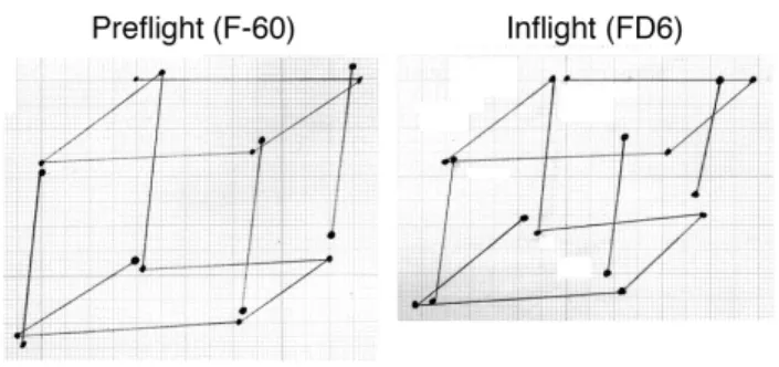
During another test, two crewmembers had to draw the well-known Necker’s cube. This figure is the simplest representation of a three-dimensional object in a two-axis coordinate system. Comparison between the length of line between the cubes drawn on the ground and the cubes drawn in space revealed a 9% decrease in size in the vertical dimension (i.e., the height) of the cubes drawn in weightlessness (Lathan et al., 2000) (Figure 5). Similar results have been found in another study involving two astronauts. The trajectory of hand-drawn ellipses in the frontal plane in the air with the eyes closed revealed a 10-13% decrease in the vertical length of the ellipses, whereas the horizontal length of the ellipses were basically unchanged (Bock et al., 2001; Gurfinkel et al., 1993). This result supports the hypothesis that the mental representation of the vertical dimension of objects or volumes is altered during exposure to weightlessness.
Figure 5. These graphs show the average of each end point of
lines for ten Necker’s cubes drawn with the eyes closed by an astronaut on the ground and during a spaceflight. The length of the vertical lines is significantly smaller in microgravity compared to normal gravity.
The results of these studies may have important consequences for human performance during spaceflight. For example, if an astronaut cannot accurately visualize the station, navigation of the station may cause delays and frustration. There may also be consequences for space habitat design if squared volumes do not look square to astronauts. Virtual reality training may be a way to train the astronauts to compensate for such altered spatial representation.
Further investigations carried out in space will perhaps reveal that other higher cortical functions are impaired in weightless conditions. The combination of virtual reality with multi-EEG recordings (for the measurement of evoked-related potentials and brain mapping), both equipments being available on ISS, should soon provide
exciting results on the adaptive mechanisms of cerebral functions in absence of gravity.
How the cognitive processes of spatial orientation will differ from the terrestrial norm after a long absence of a gravitational reference? It can be speculated that the way of processing three dimensions will be more developed. Creativity will certainly be more three-dimensional and definitely thinking will be out of the gravitational box. Like the way culture and language influences our ability to creatively think, being free from gravity will entice thoughts never before possible for the human mind, and thus give opportunities for new art and scientific discoveries (Pettit, 2003).
ACKNOWLEDGEMENTS
Funding for the author's research is provided by Centre National de la Recherche Scientifique (CNRS), Centre National d'Etudes Spatiales (CNES), and European Space Agency (ESA). Special thanks to Angie Bukley for editing the manuscript.
REFERENCES
Bloomberg, J.J., Peters, B.T., Smith, S.L., Huebner, W.P., and Reschke, M.F. 1997. Locomotor head-trunk coordination strategies following spaceflight. Journal of
Vestibular Research 7: 161-177
Bloomberg, J.J., Layne, C.S., McDonald, P.V., Peters, B.T., Huebner, W.P., Reschke, M.F., Berthoz, A., Glasauer, S., Newman, D., Jackson, D.K. 1999. Effects of spaceflight on locomotor control. In: Extended Duration
Orbiter Medical Project, Final Report 1989-1995. (Sawin, C.F., and Taylor, G.R., Eds.) Houston, TX: NASA SP-1999-534, Chapter 5.5 pp. 1-57
Bock, O., Fowler, B., and Comfort, D. 2001. Human sensorimotor coordination during spaceflight: An analysis of pointing and tracking responses during the Neurolab Space Shuttle mission. Aviation, Space, and Environmental Medicine 72:877-883
Boyle, R., Mensinger, A.F., Yoshida, K., Usui, S., Intravaia, A., Tricas, T., and Highstein, S.M. 2001. Neural adaptation to Earth's gravity following return from space,
Journal of Neurophysiology 86:2118-2122
Bracchi, F., Gualtierotti, T., Morabito, A., and Rocca, E. 1975. Multi-day recordings from the primary neurons of the statoreceptors of the labyrinth of the bullfrog. Acta
Oto-Laryngologica, Supplementum 334:1-26
Clarke, A.H., Teiwes, W., and Scherer, H. 1993. Evaluation of the three-dimensional VOR in weightlessness. Journal of Vestibular Research 3:207-218
Clément, G. 1998. Alteration of eye movements and motion perception in microgravity. Brain Research
Reviews 28:161-172
Clément, G. 2001. The human sensory and balance system. In: A World Without Gravity. (Fitton, B. and Battrick, B., Eds.) Noordwijk: European Space Agency Publication Division ESA SP-1251, pp. 93-111
Clément, G. 2005. Fundamentals of Space Medicine. El Segundo: Microcosm Press and New York: Springer Clément, G., Berthoz, A., and Lestienne, F. 1987. Adaptive changes in perception of body orientation and mental image rotation in microgravity, Aviation, Space,
and Environmental Medicine 58:A159-A163
Clément, G., Darlot, C., Petropoulos, A., and Berthoz, A. 1995. Eye movements and motion perception induced by off-vertical axis rotation (OVAR) at small angles of tilt after spaceflight. Acta Oto-Laryngologica 115:603-609 Clément, G., Gurfinkel, V.S., Lestienne, F., Lipshits, M.I., and Popov, K.E. 1985. Changes of posture during transient perturbations in microgravity, Aviation, Space,
and Environmental Medicine 56:666-671
Clément, G., and Lestienne, F. 1988. Adaptive modifications of postural attitude in conditions of weightlessness. Experimental Brain Research 72:81-389 Clément, G., Moore, S., Raphan, T., and Cohen, B. 2001. Perception of tilt (somatogravic illusion) in response to sustained linear acceleration during spaceflight.
Experimental Brain Research 138:410-418
Clément, G., Moore, S., Raphan, T., and Cohen, B. 2003. Perception of the spatial vertical during centrifugation and static tilt. In: The Neurolab Mission: Neuroscience
Research in Space. (Buckey, jr., J.C. and Homick, J.L., Eds.) Houston: Government Printing Office, NASA SP-2003-535, pp. 5-10
Clément, G., and Reschke, M.F. 1996. Neurosensory and sensorimotor functions. In: Biological and Medical
Research in Space: An Overview of Life Sciences Research in Microgravity. (Moore, D., Bie, P., and Oser, H., Eds.) Heidelberg: Springer-Verlag, pp. 178-258 Clément, G., and Slenzka, K., eds. 2006. Fundamentals of
Space Biology. Research on Cells, Animals, and Plants in
Space. El Segundo: Microcosm Press and New York: Springer
Correia, M.J., Perachio, A.A., Dickman, J.D., Kozlovskaya, I.B., Sirota, M.G., Yakushin, S.B., and Beloozerova, I.N. 1992. Changes in monkey horizontal semicircular canal afferent responses after spaceflight.