HAL Id: hal-02325710
https://hal.archives-ouvertes.fr/hal-02325710
Submitted on 10 Nov 2020HAL is a multi-disciplinary open access archive for the deposit and dissemination of sci-entific research documents, whether they are pub-lished or not. The documents may come from teaching and research institutions in France or abroad, or from public or private research centers.
L’archive ouverte pluridisciplinaire HAL, est destinée au dépôt et à la diffusion de documents scientifiques de niveau recherche, publiés ou non, émanant des établissements d’enseignement et de recherche français ou étrangers, des laboratoires publics ou privés.
Investigation of titanium dioxide nanoparticles toxicity
and uptake by plants
Camille Larue, H. Khodja, N. Herlin-Boime, F. Brisset, A. Flank, B. Fayard,
S Chaillou, Marie Carrière
To cite this version:
Camille Larue, H. Khodja, N. Herlin-Boime, F. Brisset, A. Flank, et al.. Investigation of titanium dioxide nanoparticles toxicity and uptake by plants. Journal of Physics: Conference Series, IOP Publishing, 2011, 304, pp.012057. �10.1088/1742-6596/304/1/012057�. �hal-02325710�
Investigation of titanium dioxide nanoparticles toxicity and uptake by plants
This article has been downloaded from IOPscience. Please scroll down to see the full text article. 2011 J. Phys.: Conf. Ser. 304 012057
(http://iopscience.iop.org/1742-6596/304/1/012057)
Download details:
IP Address: 89.90.186.122
The article was downloaded on 03/04/2013 at 17:50
Please note that terms and conditions apply.
View the table of contents for this issue, or go to the journal homepage for more
Investigation of titanium dioxide nanoparticles toxicity and
uptake by plants
C Larue1, H Khodja2, N Herlin-Boime3, F Brisset4, AM Flank5, B Fayard6, S Chaillou7 and M Carrière1
1
Laboratoire de Structure et Dynamique par Résonance Magnétique UMR 9956 CEA-CNRS-IRAMIS, Gif-sur-Yvette, France
2
Laboratoire d’Etude des Eléments Legers, UMR 9956 CEA-CNRS-IRAMIS, Gif-sur-Yvette, France
3
Laboratoire Francis Perrin URA 2453 CEA-CNRS-IRAMIS, 91191 Gif-sur-Yvette, France
4Institut de Chimie Moléculaire et des Matériaux d’Orsay, UMR8182 CNRS-Univ. Paris sud, Orsay, France
5
LUCIA beamline, SOLEIL synchrotron, Saint-Aubin, France
6
Laboratoire de Physique du solide, Orsay, France and ID21 beamline, ESRF, Grenoble, France
7
Unité de Nutrition Azotée des Plantes, INRA, Versailles, France E-mail: [email protected]
Abstract. Nanoparticles (NP) are introduced in a growing number of commercial products and
their production may lead to their release in the environment. Plants may be a potential entry point for NP in the food chain. Up to now, results describing NP phytotoxical effects and plant accumulation are scarce and contradictory.
To increase knowledge on titanium dioxide NP (TiO2-NPs) accumulation and impact on plants,
we designed a study on three plant species, namely wheat (Triticum aestivum), oilseed rape (Brassica napus) and Arabidopsis thaliana. These plants were exposed in hydroponics to a panel of well-characterized TiO2-NPs, with diameters ranging from 12 to 140 nm, either
anatase or rutile. Their accumulation in plant tissues is currently being assessed by complementary imaging techniques: scanning electron microscopy (SEM), transmission electron microscopy (TEM), X-ray fluorescence (SR-µ-XRF) imaging and micro-particle induced X-ray emission (µ-PIXE) imaging. Moreover, the impact of TiO2-NP exposure
on germination rate, root elongation, dry biomass and evapotranspiration is evaluated. Preliminary results are presented here, with data collected on wheat plants exposed to 12 nm and 25 nm anatase TiO2-NPs. These results show that TiO2-NPs are taken up by plants, and do
not significantly alter their germination and root elongation. These results underline the necessity of deeper evaluation of nanoparticle ecotoxicity, and particularly on their interaction with plants.
Nanosafe2010: International Conference on Safe Production and Use of Nanomaterials IOP Publishing Journal of Physics: Conference Series 304 (2011) 012057 doi:10.1088/1742-6596/304/1/012057
1. Introduction
Nanoparticles (NP) are introduced in a growing number of commercial products. Among them, TiO2
anatase nanoparticles (TiO2-NPs) are one of the most produced NPs in the world (see Nanowerk
database, www.nanowerk.com). They are used as a pigment in paint, paper, ink and plastic. They are also introduced in cosmetics such as sunscreens for their UV protective properties (see Nanowerk database, www.nanowerk.com). Finally, they are presently used in pilot water-purification reactors, because of their ultraviolet irradiation-induced bactericidal effects (photocatalysis). For these reasons, these NPs will surely be released in the environment [1]. The knowledge of their potential effects on human health is rapidly increasing, however little is known on their potential toxicological effects on environment, i.e. destabilization of the ecosystems and trophic transfer, but also on their potential transfer to the food chain via plant ingestion. The literature relating NP toxicity and accumulation in plants is scarce; authors mostly relate NP inhibitory effects on seed germination and root elongation [2-4]. A few studies relate NP uptake by plant roots [3, 5], and root to shoot transfer [5]. Concerning TiO2-NPs, TiO2-based nanocomposites have recently been shown to be accumulated and translocated
to the shoots of Arabidopsis thaliana [6]. These translocation studies are based on elemental analysis of the ionic form of metal in plants, but neither address the presence of metal as NPs, nor the spatial distribution of NPs throughout the plant. Actually, TiO2-NP dissolution has been demonstrated to be
low in aqueous solution [7], however its occurrence has never been tested in plant exposure solution and/or during NP transfer to the roots or from the roots to the shoots. Synchrotron radiation micro X-ray fluorescence (SR-µ-XRF), coupled to micro X-X-ray absorption spectroscopic (µ-XAS) analysis is the ideal method to address metal distribution after plant uptake, together with speciation analysis which would permit the identification of this metal as NPs. It has already been used to assess ionic metal internalization and distribution in plants [8].
We used a combination of complementary imaging techniques: scanning electron microscopy (SEM), transmission electron microscopy (TEM), micro-X-ray fluorescence (SR-µ-XRF) imaging and micro-particle induced X-ray emission (µ-PIXE) imaging, to assess TiO2-NP accumulation in plants,
and correlated their uptake with their impact on plant development. This study is currently under process in our laboratory with several plant models and several TiO2-NPs varying in their diameter,
crystalline phase and shape; we present here some preliminary results obtained on wheat (Triticum
aestivum), exposed in hydroponics to 12 nm and 25 nm anatase TiO2-NPs. These results show that
TiO2-NPs are taken up by plants, and do not significantly alter their germination and root elongation.
These results underline the necessity of deeper evaluation of nanoparticle ecotoxicity, and particularly on their interaction with plants.
2. Methods
2.1. Nanoparticles, characterization and suspension
For TiO2-NP dispersion studies and impact studies on plant development, we used commercial Evonik
Aeroxide® P25 TiO2-NPs. The NPs used in plant uptake experiments were synthesized by laser
pyrolysis in Laboratoire Francis Perrin (LFP) [9], then annealed under air at 400°C during 3 h. Physico-chemical characterization of these NPs has been reported in two previous studies [10, 11]. Briefly, LFP NPs are 95% anatase and 5% rutile. Their mean diameter is 12 ± 3 nm, they are spherical-shaped and their specific surface area, measured according to Brunauer, Emmett and Teller (BET) [12], is 82 ± 4 m².g-1. Their point of zero charge, measured by assessment of zeta potential at increasing pH, is 6.4. Evonik P25 nanoparticles are 75% anatase and 25% rutile. Their mean diameter is 25 ± 7 nm, they are spherical-shaped and their specific surface area, measured according to Brunauer, Emmett and Teller (BET) [12], is 46 ± 1 m².g-1. Their point of zero charge is 7.
These TiO2-NPs were suspended in ultrapure water and sonicated using a high energy probe,
during 30 mn, at 4°C, in pulsed mode (1 s on, 1 s off). Agglomeration status was evaluated using photon correlation spectroscopy (PCS) using a Zetasizer 3000HS (Malvern). Z-average values are given in the result section.
Nanosafe2010: International Conference on Safe Production and Use of Nanomaterials IOP Publishing Journal of Physics: Conference Series 304 (2011) 012057 doi:10.1088/1742-6596/304/1/012057
2.2. Plant exposure and sample preparation for imaging
For imaging, seeds were germinated on sand, and their roots were directly immersed in the NP suspension (100 mg.L-1), and placed in a growth chamber under controlled conditions: day/night photoperiod: 16/8 h; temperature (day/night), 24/20 ± 1°C and relative humidity (day/night), 70/75%. After 7 days, plant roots were thoroughly rinsed with deionised water. For µ-XRF, SEM and µ-PIXE analysis, plant roots and leaves were cryofixed, immediately embedded in Tissue Tek OCT™ resin (Agar Scientific, Essex, UK) and cross sections were cut and freeze-dried.
The distribution of Ti, P, Cl and Ca in plant thin sections were mapped using synchrotron radiation µ-X-ray fluorescence (SR-µ-XRF), images presented in this study were recorded on LUCIA beamline (SOLEIL synchrotron, France). The spot size was 3 × 3 µm². XAS spectra were recorded between 4.95 and 5.05 keV, i.e. around Ti X-ray absorption edge (4.966 Kev). On each sample, at least 10 spectra were collected, in situ on map locations containing high concentrations of Ti. These spectra were then averaged and compared to XAS spectra collected on Ti standards: 12 nm, 25 nm and 140 nm TiO2 anatase NPs, 20 and 50 nm TiO2 rutile NPs. Spectrum was then normalized to the
second-order polynomial to be equal to one, with Athena software [13]. SEM image were acquired on a Zeiss-Supra 55VP Field Emission Gun Scanning Electron Microscope (FEG-SEM) equipped with a X-ray Energy Dispersive Spectrometer (EDS), operated at 15 kV, at ICMMO, Orsay, France.
2.3. Nanoparticle impact on plant development
For NP impact on plant development assessment, 10 seeds were germinated in Petri dishes in deionised water containing 10 to 100 mg/L NPs. According to US-EPA guidelines (US-EPA, 1996), seeds with roots longer than 5 mm were considered germinated. For root elongation experiment, after 7 days, roots were measured on a picture thanks to ImageJ software.
3. Preliminary results and discussion
3.1. Characterization of TiO2-NPs and dispersion in aqueous solution
In order to obtain properly dispersed and stable TiO2-NP suspensions, sonication was applied to TiO2
-NP powders dispersed in water at 10 g.L-1. For this study, TiO2-NPs from Evonik (Aeroxide® P25)
were used. Optimal TiO2-NP concentration for proper measure was evaluated, and concentrations of
50-1000 mg.L-1 were found to be appropriate, whereas at lower concentrations, the Zetasizer was not able to properly measure the mean hydrodynamic diameter (Table 1). At concentrations higher than 1000 mg.L-1, the suspension is too opaque to obtain a proper measure.
Table 1. Mean hydrodynamic diameter of nanoparticle agglomeratesa
Concentration (mg.l-1) 10 50 100 500 1000 2000 Diameter (nm) 140 0.6 24.2 38.7 43.1 66.6 12.5 Standard deviation 333. 4 11.1 15.4 22.6 26.1 7.3 a
Mean hydrodynamic diameter was determined by PCS after a 30 mn sonication, in water, using high energy probe. TiO2-NP concentration in these suspensions ranged from 10 to 2000 mg.l
-1
.
When diluted in Hoagland medium, these suspensions of TiO2-NPs immediately agglomerated. To
prevent agglomeration, various dispersing agents were tested. The obtained suspensions are presented in Figure 1, which shows that NP agglomeration occurs in most conditions. Mean hydrodynamic diameters of TiO2-NP agglomerates are presented in Table 2.
Nanosafe2010: International Conference on Safe Production and Use of Nanomaterials IOP Publishing Journal of Physics: Conference Series 304 (2011) 012057 doi:10.1088/1742-6596/304/1/012057
Figure 1. TiO2-NP suspensions, use of a dispersing agent. From left to right: TiO2-NPs sonicated in
water, sorbitol, 1 g.l-1 PVP, Evian water, oxalic acid, 0.9% NaCl, or sonicated in water and then diluted in Hoagland medium.
Table 2. Mean hydrodynamic diameter of NP agglomerates when using a dispersing agenta Dispersing agent 20 mM oxalic acid 1 g.l-1 PVP 10 g.l-1 PVP 1 mM sorbitol 0.9% NaCl Evian water After sonication 1536 46 245 41 1000 1668 Hoagland 1234 896 41 853 1126 746 a
Mean hydrodynamic diameter was determined by PCS at 50 mg/L after a 30 mn sonication, in water containing a dispersing agent (oxalic acid or polyvinylpyrrolidone (PVP) or sorbitol or in physiologic serum (0.9% NaCl) or Evian water). Diameters are determined directly after sonication, or after sonication and dilution in Hoagland medium.
As regards these dispersion results, and considering that we aimed at exposing plants in a well-dispersed TiO2-NP suspension, we chose to expose plants in TiO2-NPs prepared in water without
addition of any dispersing agent, which would not increase suspension quality. 3.2. Impact of TiO2-NPs on plant development
The impact of TiO2-NP exposure on plant development was evaluated on wheat seeds and plantlets,
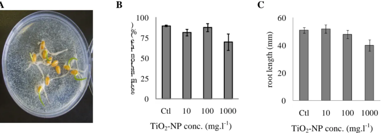
exposed to Evonik (Aeroxide® P25) TiO2-NPs. Exposure did not induced any statistically significant
modification in wheat seed germination (Figure 2B), nor in root elongation (Figure 2C). However, during root development, TiO2-NPs strongly stuck onto roots and agglomerated in exposure medium
(water, Figure 2A).
A B 0 25 50 75 100 Ctl 10 100 1000 g er m in at io n r at e (% ) TiO2-NP conc. (mg.l -1 ) C 0 20 40 60 Ctl 10 100 1000 ro o t le n g th ( m m ) TiO2-NP conc. (mg.l -1 )
Figure 2. Seed germination test. Wheat seeds were exposed to Aeroxide® P25 TiO2-NPs (A), and
number of germinated seeds was counted (B), then the mean length of roots was measured (C).
H 2O m Q S O R B IT O L P V P H 2O E V IA N O X A L IC A C ID 0. 9% N aC l H O A G L A N D
Nanosafe2010: International Conference on Safe Production and Use of Nanomaterials IOP Publishing Journal of Physics: Conference Series 304 (2011) 012057 doi:10.1088/1742-6596/304/1/012057
3.3. TiO2-NP accumulation in plants
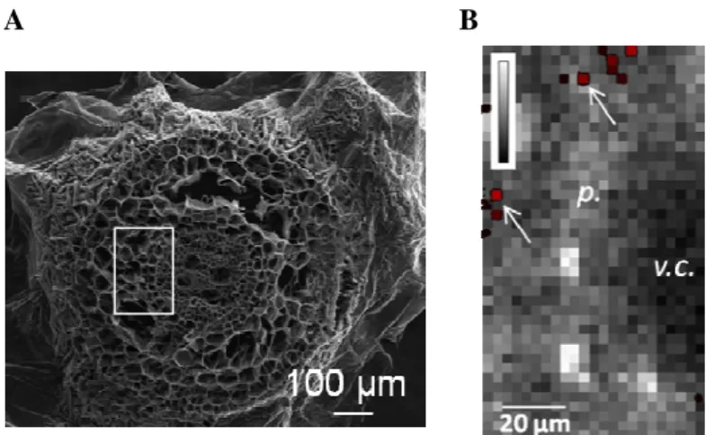
Elemental analysis, in scanning mode, enabled to draw images of P content, representative of plant tissues, and of Ti content, representative of TiO2-NPs, in plant roots and leaves after exposure. An
example is presented in Figure 3, mapped on wheat plant roots after exposure to anatase, 12 nm TiO2
-NPs. We used these nanoparticles rather than Evonik nanoparticles because they were smaller, and thus would more probably be taken up by plants. Their agglomeration status and impact on plant development was close to that of Evonik nanoparticles (not shown). After 7 days of exposure, Ti was observed in the parenchyma of wheat roots (Figure 3, arrows), but also in the vascular cylinder, demonstrating that TiO2-NPs were transferred from the exposure suspension to vegetal tissues.
A B
Figure 3. Ti distribution in wheat roots. Transversal sections of a root analyzed by SEM (A) and by
µ-XRF image of a region related to the white square in A (B) after a 7-day exposure to 12 nm anatase TiO2-NPs. In B: maps of P distribution (gray) and Ti distribution (red, clusters are also indicated by
arrows) are superimposed. SEM image was recorded in ICMMO and µ-SR-XRF image on LUCIA beamline, SOLEIL synchrotron.
As described in several publications, Ti pre-edge features are characteristic of the crystalline phase and size of TiO2-NPs. Anatase displays a typical triplet feature (A1, A3 and B peaks), with a weak
shoulder on the low-energy side of the central A3 peak (A2) [14]. The intensity of A2 peak is related to the distortion of the octahedral TiO6 unit, particularly the distortion observed on the surface of NP.
The intensity of this peak is thus related to the size of the particle: the smaller the NP, the higher this A2 peak [15]. Meanwhile, the intensity of the A1 peak also decreases as the particle size decreases [16]. Analysis of this pre-edge region may then inform on changes in the crystalline phase of NPs after cellular internalization, and on their dissolution. In the present experiment, no modification of these pre-edge features was observed after plant internalization of anatase TiO2-NPs (Figure 4), meaning
that Ti is still in the nanoparticulate TiO2 chemical form inside plants, and that their crystalline phase
did not change. Moreover the size of NPs remained constant after plant internalization meaning that no partial dissolution of NPs occurred.
Nanosafe2010: International Conference on Safe Production and Use of Nanomaterials IOP Publishing Journal of Physics: Conference Series 304 (2011) 012057 doi:10.1088/1742-6596/304/1/012057
Figure 4. XANES spectra of Ti in plant roots exposed to TiO2-NPs. Plants were exposed for 7 days to
anatase TiO2-NPs. Spectra were recorded at the Ti K-edge (4.966keV) at room temperature, locally on
two Ti-rich regions determined on µ-SR-XRF maps (black spectra), and compared to the spectrum of a reference compound (red spectrum). A: pre-edge feature; B: XANES spectra. Image recorded on
LUCIA beamline, SOLEIL synchrotron.
In the post-edge region, the oscillations show some differences in the samples as compared to reference compounds. However these spectra are noisy and their interpretation is difficult. Still, these changes may be due to surface modifications of TiO2-NPs exposed to root exudates or plant fluids. A
future experiment will aim at studying these changes which may occur in these conditions.
4. Conclusion
We determined sonication conditions enabling to properly disperse anatase NPs, the conclusion is that dispersion has to be performed in water, by ultra-sonication during 30 min. In these conditions, dilution in Hoagland medium leads to agglomeration, and none of the dispersing agent studied permits to maintain a stable dispersion.
Plant exposure to anatase, 25 nm NPs neither affect their seed germination rate, nor their root elongation. However 12 nm anatase NPs are efficiently taken up by plant roots and localize in the parenchymal region and vascular cylinder. Their nutrient solution-to-root transfer do not induce their dissolution, nor localized change in their crytalline structure.
µPIXE experiment are under process and will permit to have a global view of the perturbations in major elements homeostasis which may be caused by TiO2-NP uptake. Moreover, on all these plant
samples, transmission electron microscopic observations will now enable to evaluate if isolated NPs are taken up by plants, or if these NPs are grouped into clusters. The present results prove that TiO2
-NPs can be transferred to plants by their roots in case of an accidental release in the environment, and consequenltly may enter the food chain.
Acknowledgements
This work was funded by ADEME, CEA and the region Ile de France through the C’Nano framework. The authors would like to thank the European Synchrotron Radiation Facility and SOLEIL for provision of synchrotron radiation facilities at ID21 and LUCIA. This work was performed in the framework of the iCEINT research consortium.
References
[1] Gottschalk F, T Sonderer, R W Scholz, and B Nowack 2009 Environ Sci Technol 43. [2] Lin D and B Xing 2007 Environ Pollut 150.
[3] Lin D and B Xing 2008 Environ Sci Technol 42.
[4] Yang F, F Hong, W You, C Liu, F Gao, C Wu, and P Yang 2006 Biol Trace Elem Res 110. [5] Zhu H, J Han, J Q Xiao, and Y Jin 2008 J Environ Monit 10.
Nanosafe2010: International Conference on Safe Production and Use of Nanomaterials IOP Publishing Journal of Physics: Conference Series 304 (2011) 012057 doi:10.1088/1742-6596/304/1/012057
[6] Kurepa J, T Paunesku, S Vogt, H Arora, B M Rabatic, J Lu, M B Wanzer, G E Woloschak, and J A Smalle 2010 Nano Lett
[7] Schmidt J and W Vogelsberger 2009 J Sol Chem 38.
[8] Isaure M P, B Fayard, G Sarret, S Pairis, and J Bourguignon 2006 Spectrochim Acta, Part B [9] Pignon B, H Maskrot, Y Leconte, S Coste, C Reynaud, N Herlin-Boime, M Gervais, V Guyot
Ferreol, T Pouget, and J F Tranchant 2008 Eur J Inog Chem 6.
[10] Simon-Deckers A, B Gouget, M Mayne-L'hermite, N Herlin-Boime, C Reynaud, and M Carriere 2008 Toxicology 253.
[11] Simon-Deckers A, S Loo, M Mayne-L'hermite, N Herlin-Boime, N Menguy, C Reynaud, B Gouget, and M Carriere 2009 Environ Sci Technol 43.
[12] Brunauer S, P H Emmett, and E Teller 1938 J Am Chem Soc 60. [13] Ravel B and M Newville 2005 Phys Scr T115.
[14] Wu Z Y, J Zhang, D Ibrahim, D C Xian, G Li, Y Tao, and T D Hu 2002 App. Phys. Lett. 80. [15] Luca V, S Djajanti, and R F Howe 1998 J Phys Chem B 102.
[16] Chen L X, T Rajh, Z Y Wang, and M C Thurnauer 1997 J Phys Chem B 101.
Nanosafe2010: International Conference on Safe Production and Use of Nanomaterials IOP Publishing Journal of Physics: Conference Series 304 (2011) 012057 doi:10.1088/1742-6596/304/1/012057