Collective Migration of Epithelial Sheets
by
Michael Peter Murrell
Submitted to the Department of Biological Engineering
in partial fulfillment of the requirements for the degree of
Doctor of Philosophy in Biological Engineering
at the
MASSACHUSETTS INSTITUTE OF TECHNOLOGY
April 2009
@Michael P. Murrell, 2009.
/u
Author ... ''I
... ... ... ...Department of Biological Engineering
May 21, 2009
Certified by.
Certified by...
Roger D. Kamm
Professor, MIT
Thesis Supervisor
Paul Matsudaira
Professor, NUS
Thesis Supervisor
1/
AcceDted by ...
Alan Grodzinsky
Chairman, Department Committee on Graduate Students
ARCHIVES
MASSACHUSETTS INSTITTE
OF TECHNOLOGY
6 L
Io
Collective Migration of Epithelial Sheets
by
Michael Peter Murrell
Submitted to the Department of Biological Engineering on May 21, 2009, in partial fulfillment of the
requirements for the degree of
Doctor of Philosophy in Biological Engineering
Abstract
The varied movements of the epithelium play vital roles in the development and renewal of complex tissues, from the separation of tissues in the early embryo, to homeostasis in the adult. Their movement is intricately connected to their proper functioning as selective barriers of the intestinal mucosae, as well re-epithelialization in the healing of wounds. Yet, considering their ubiquity and relevance, the basic origin of the collective motion of sheets has eluded a clear and quantitative interpre-tation in physical terms, prohibited by the lack of understanding of the relationship between motility, cell-cell contact, and their mediation by the mechanical properties of the substratum to which they adhere. Therefore, within this context, this the-sis defines the prerequisites for both equilibrium and non-equilibrium coordinated cell motion. The timescales and lengthscales of the in vitro migration of an epithe-lial monolayer were calculated and compared under imposed constraints designed to mimic various states of in vivo epithelia. These constraints include assays that recre-ate the wound response of the epithelium such as what is seen in the cornea and epidermis, by unequivocally separating the influence of free space from cell damage in the induction of coordinated motion. The motion of the epithelium was further explored by the generation of gradients that reproduce asymmetry in the capacity for cells to migrate, divide, or undergo apoptosis, such as what is found along the crypt-villus axis of the intestine. Finally, as the epithelium adheres and migrates against the basal lamina, a substrate of uncertain in vivo mechanical properties, we explored the contribution of substrate viscoelasticity to the dynamics of coordinated migration. Parameterized this way, multiple modes of motility emerge, each distinct dynamically, phenotypically, and in their dependence on cell-cell contact.
Thesis Supervisor: Roger D. Kamm Title: Professor, MIT
Thesis Supervisor: Paul Matsudaira Title: Professor, NUS
Acknowledgments
Being able to perform research at MIT, and in the Department of Biological Engi-neering has been an extraordindary blessing. Therefore, I have to give thanks to He from whom all blessings flow, my Lord and Savior, Jesus Christ. His love is great, and his mercy is never-ending.
I devote this thesis, to my grandparents, Dr. and Mrs. Peter C. Murrell. I would
have never been able to get this far, without their love and support. They raised me, provided for me, and sacrificed for me to bring me to this point, so this PhD belongs to them more than it belongs to me.
In addition, I have also benefited greatly from the body of Christ here in Cam-bridge, the Black Graduate Ministries (BGM) Group. They have been a huge source of support, and without them, this would not have been possible. They have become family to me over the last couple of years. This includes my close friends Renee Smith, Pierre Fuller, Christopher Rhodes, and Heather McLetchie-Leader. In addition BGM, my good friends Alex and Karen Sheh have not only been a source of support, but a source of inspiration of the years. Without them, this work also would not have been possible. Their constant, unwavering faith, serves as an example that I intend to emulate.
And, of course I have to give thanks to the members of my Thesis Committee. I have had unparalleled freedom to pursue my interests. I have always had open access to the resources necessary for accomplishing the work.
Thesis Committee
A ccepted by...
---.---Roger D. Kamm
Professor of Biological Engineering and Mechanical Engineering (MIT) Thesis Supervisor
Accepted by... ... Paul Matsudaira
Professor of Biology (National University of Singapore) Thesis Supervisor
Accepted by... - - . - - - -
-Douglas Lauffenburger
Professor of Biological Engineering and Chemical Engineering (MIT) Committee Chairman
Contents
1 Introduction
31
1.1 In vitro models of Sheet Motion . . . . 31
1.1.1 Wound Healing . . . . 33
1.1.2 Model Wounds . . . . 34
1.1.3 Cell Sorting . . . . 36
1.2 Summary of Open Questions . . . . 38
1.3 Summary of Testable Hypotheses . . . . 42
1.4 A ppendix . . . . 46
1.4.1 A note on adhesion in the intestinal epithelium . . . . 46
2 Initial and Boundary Conditions in the Collective Migration of
Ep-ithelial Sheets
57
2.1 Introduction . . . . 572.2 Materials and Methods . . . . 59
2.3 Mathematical Analysis . . . . 61
2.4 Free Space is Sufficient for Generating Collective Motion . . . . 62
2.4.1 Dynamics of the border . . . . 63
2.4.2 Dynamics of the bulk . . . . 64
2.5 Free Space is Necessary for Generating Collective Motion . . . . 65
2.5.1 Closed Boundaries . . . . 65
2.6 Conclusions . . . . 66
2.7 Supplemental Information . . . . 67
2.7.1 Density Dependence for Equilibrium Movement . . . . 67
2.8 A ppendix . . . . 2.8.1 Shear Stress in the Microfluidic Channel . . . .
2.8.2 Alternate Gradients in Motility . . . .
2.8.3 Viability in the Microfluidic Chamber . . . .
2.9 P rotocols . . . . 2.9.1 Soft Lithography of 100 pm high channels . . . .
2.9.2 Immunostaining Adhesions inside the microfluidic channel . .
3 Substrate Mechanics and Cell Contact in Epithelial Cell Motion
3.1 Introduction . . . .
3.2 Materials and Methods . . . .
3.3 Coupling of monolayer and bulk . . . . 3.4 Correlated Dynamics follow a second-order transition with substrate
viscoelasticity . . . .
3.5 Substrate Viscoelasticity inversely modulates positional and dynamic correlation . . . .
4 Motion of Epithelial Sheets as a function of Population Size
4.1 Introduction . . . . 4.2 Materials and Methods . . . .
4.3 Dynamics vary only with density, not population . . . .
4.4 Confinement on Substrates of Varying Rigidity . . . . 4.5 Appendix . . . . . . . ..
4.5.1 Free Space sufficient for generating "Purse-String" . . .
4.6 P rotocols . . . .
4.6.1 Preparing Aldehyde Terminated PDMS Surfaces . . . .
4.6.2 Stamping onto PDMS . . . . 4.6.3 Ninhydrin Test for Primary Amines . . . .
5 Supplemental Studies To Sheet Motion
5.1 Computational Methods . . . . 80 80 82 84 85 85 88
95
95 96 100 101 102 119 . . . . 119 . . . . 120 . . . . 123 . . . . 124 . . . . 124 . . . . 124 . . . . 126 . . . . 126 . . . . 128 . . . . 130139
1395.1.1 Dynamic Monte Carlo Simulations . . . .
5.1.2 Velocity Correlations and the Elastic Wave Equation . .
5.1.3 Dynamic Structure Factor and Collective Modes . . . . . 5.1.4 Dynamic Susceptibility . . . .
5.1.5 Modeling the Sheet as a 2D XY Ferromagnetic Material
5.2 Cell M echanics . . . .
5.2.1 Cell Shear Modulus Calculation . . . .
5.2.2 C ontrols . . . . . . . 139 . . . 141 . . . 142 . . . 146 . . . 151 . . . 157 . . . 157 . . . 167
6 Conclusions and Review of the Initial Hypotheses
6.1 The influence of free space in coordinated migration . . . .
6.2 Density and Contact Dependence in Compliant and Non-Compliant M odels . . . .
7 Future Work
8 Derivations and Tutorials
8.1 Why Statistical Mechanics?
Definitions . . . . How do we approach the constructio 8.4 Important Principles 8.5 8.6
8.7
8.8
8.98.10
8.11 8.12Microcanonical Ensemble (N,V,E) The Canonical Ensemble (N,V,T) Freely Jointed Chain . . . . Worm-Like Chain . . . . Derivation of the Master Equation.
The Fokker-Planck Equation . . . .
Fluctuation-Dissipation . . . . Basic Thermodynamics . . . . 8.13 Useful Mathematics .
179
. . . . 179 . . . . 18 1 i of a S.M . problem? . . . . 183 . . . . 185 . . . . 18 7 . . . . 189 . . . . 192 . . . . 19 6 . . . . 200 . . . . 20 1 . . . . 202 . . . . 20 7 . . . . 2098.14 Thermodynamic Variables and the Partition Function . . . .2
173 174 175 177 8.2 8.3
210
8.15 Example: Proving the Ideal Gas Law . . . . 211
8.16 Example: Proving Hooke's Law in an Ideal Polymer . . . . 214
8.17 Example: Force-Dependent Equilibrium Constants(1) . . . . 218
8.18 Example: Force-Dependent Equilibrium Constants (2) . . . . 220
List of Figures
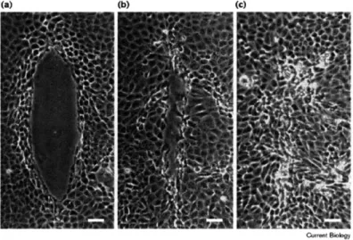
1-1 WOUND HEALING IN AN MDCK MONOLAYER. Wound closure in
MadinDarby canine kidney (MDCK) epithelial cell monolayers. (ac) Phase-contrast micrographs of MDCK cells after wounding and mi-croinjection with 2.5 mg/ml solution of OG dextran into the first three rows of cells around the wound margin (concentration in cells after mi-croinjection is 1/10th that of the microinjected solution or 250 g/ml). Cells are shown (a) immediately after wounding and microinjection, (b) after 6 h, and (c) after 18 h. The scale bar represents 50 m. All figures for each treatment in this paper are representative of experiments per-formed in triplicate on at least three separate occasions (n=9). Figure and caption taken from [1] . . . . 48
1-2 ACTIN PURSE STRING IN WOUND CLOSURE. Repair of in vitro wounds made in monolayers of the gut epithelial cell line Caco-2BBE is achieved by lamellipodial crawling or purse-stringing, or a combination of both. In this wound, one group of leading-edge cells is being drawn forwards by contraction of an actin cable (arrows), as occurs during embryonic repair, while other cells are clearly extending lamellae (ar-rowheads) and crawling forwards, as occurs during repair of an adult skin wound (image courtesy of J. Brock). Green staining is fluorescein isothiocyanate/ phalloidin-tagged filamentous actin; red staining is the nuclear dye 7AAD. Image and caption taken from [2] . . . . 49
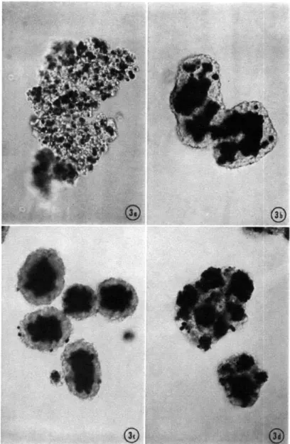
1-3 CELL SORTING. Sorting in mixtures of 7-day chick embryo neural
retina (clear) and pigmented retina (black). (a) control culture after 2 hours (initial aggregate), (b) control culture at an intermediate stage of mixing, (c) complete sorting occurs in control culture after one day,
(d) partial sorting occurs membrane ruffling and pseudopod formation
are disrupted. Image taken from
[3]
. . . .
50
1-4 DIAGRAM OF "MODEL" WOUND. Thin PDMS films with holes in the shapes of long strips are placed on a cell culture surface, and cultured with MDCK cells (top panel). After the cells have bound, the film is removed, leaving behind cells exposured to a virgin surface. The cells them move migrate out into free space (bottom panel). Cells are shown for (a) 90 minutes, (b) 13 hours, (c) 25 hours, or (d) 37 hours after removal. This is the predominant, and most widely accepted model thus far. Image taken from [4] . . . . 51
1-5 MOVEMENT OF THE INTESTINAL EPITHELIUM.
Histoautora-diographs of small intestine at length 33% after 1 month of exposure
(270). A, dl; B, d2; C, d5 after Pu ingestion. Image and Caption taken
from : [5]. . . . . . . . . 52
1-6 HISTOLOGY OF THE SMALL INTESTINE. Histological Image of
the villi of the small intestine. Taken from Kansas University Medical
Center (http://www.kumc.edu/instruction/medicine/anatomy/histoweb/) 53
1-7 EPITHELIAL CELLS OF THE SMALL INTESTINE. Epithelial cells
of the small intestine are columnar, and have no visible actin-based ex-tensions, such as lamellipodia, filopodia, or pseudopodia. Taken from
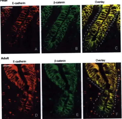
1-8 CADHERIN IN THE INTESTINE. Distribution of E-cadherin and
#-catenin in fetal (AC) and adult (DF) human small intestine. Frozen sections of fetal intestine between 18 and 20 weeks of gestation were costained with a rabbit antibody to E-cadherin and a mouse
anti-#-catenin.
The secondary antibodies, anti-rabbit conjugated to rho-damine, and anti-mouse conjugated to FITC, allowed the detection of E-cadherin (A and D) and 0-catenin (B and E) independently. Over-lays of both stainings (C and F) show the differential localization of the two proteins. Numbers in overlay pictures point to individual cell position in the crypts, position 1 indicating the bottom. Scale bars =15 pm. Image and Caption taken directly from [6] . . . . 54
1-9 CADHERIN IN THE INTESTINE. Quantitation of E-cadherin (A),
#-catenin
(B), and the difference E-cadherin minus -catenin (C) flu-orescence stainings shown in Fig. 1. Quantifications were performed as described in Materials and methods on native images. Graphics present results obtained with four crypt/villus axes for 10 fetal and 7adult tissues. Cell positions in the crypt are indicated by the number below the histograms. Bars 1620 represent means of cells at positions
1620 from each crypt side; these values were used as references for
statistics (*p < 0.05, * * p < 0.02). Image and Caption taken directly from [6] . . . . 55
1-10 LAMININS AND INTEGRINS IN THE INTESTINE. Distribution of
laminins and their corresponding integrins along the crypt-villus axis of the adult human small intestine. Laminin-1 (al 3 '1l) and laminin-2 (a2o1-1) show complementary locations while laminin-5 (a3#3Y2) is restricted to the villus. Among integrins, a604, which can bind to these three laminins, is distributed uniformly, while the basal distribution of a301 in intestinal cells coincides with the location of its specific ligand, laminin-5. In contrast, the laminin-1 binding integrin a7#1 is primarily expressed in the differentiating compartment. Image and Caption taken directly from [7]. . . . . 56
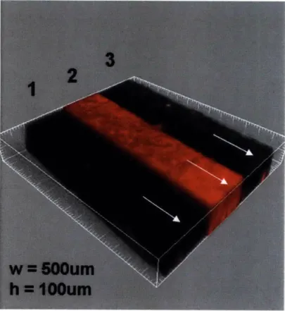
2-1 DIAGRAM OF MICROFLUIDIC SYSTEM. (A). Three inlets lead into a main channel, each 500 pm wide, and 100 pm high. When the volumetric flow rate that retracts fluid into the channel from the inlets is greater than 10 pL/min, the flow is laminar, and the fluid from the three inlets flow side by side, separated over short distances into the channel, (B). The trypsin channel can either be at the side, or in the middle. (C) All migration experiments utilize a flow of 30 pL/min. At this rate to enzymatically cleave cells with trypsin, carrying the away with the flow. After they have been removed, media is switched to the trypsin inlet, and the flow is reduced to 0.5 pl/min, and we image the movement of the viable cells. . . . . 68
2-2 DIMENSIONS AND FLOW IN THE MICROFLUIDIC CHANNEL.
3D Confocal Image of the microfluidic channel, with three separate
fluid streams (all cell media, at a volumetric flow rate of 30 pL/min), with two separate fluorescent dyes, 568 Alexa Fluor (inner stream) and 647 Alexa Fluor (outer streams). The shear, T, across the cell monolayer varies with the inverse square of the channel height (ap-proximately, as we cannot say w >> h), and the inverse of the width. The initial flow used to separate the laminar flows, yields an initial shear stress of 4.4dyn/cm2, which is held for 5-10 minutes. After the
cells have been cleaved from the channel, the flow is reduced, and the shear drops to 0.073dyn/cm2. This flow is chosen to be arbitrily small,
such that the shear stress is negligible, but yet fresh media is constantly being delivered to the cells. . . . . 69
2-3 MEAN CELL VELOCITIES FOR WOUND AND MODEL WOUND.
Cells are stained with 1:1000 CMFDA, and imaged over the course of
12-36 hours, on either a Mattek Dish (Wound Healing Assay, denoted
WH), or inside the microfluidic channel (having isolated free space alone, with no cell damage, denoted as FS). The mean velocities for cell motion over this time are shown for experiments with no free space (control), or with denudation at low and high densities. Also at high density, 200 pg of anti E-cadherin antibody was added. The mean
velocities are significantly increased when cell contact is blocked. . . . 70
2-4 HIGH DENSITY MOVEMENT AFTER ENZYMATIC
DENUDA-TION. (A). Brightfield image of 0.05% Trypsin cleaving cells from the
left hand side of a 500 pm wide channel. At high densities, fingering instabilies ofter occur, and result in little net movement after (B) 0,
(C) iday, (D) 1.5 days, all stained with Dil, a calcium stain. The
in-stability itself is highly motile in the direction perpendicular to, but not parallel to the axis of initial penetration. . . . . 71
2-5 LOW DENSITY MOVEMENT AFTER ENZYMATIC DENUDATION. At low density, there is much faster net movement, and the reoccupa-tion of free space, although principally, cells will tend to scatter, rather than more uniformly, or form secondary structures, like the fingering
instability. Images are taken after (A) 0, (B) 1 day (C) 1.5 days. . . . 72
2-6 E-CADHERIN AT DIFFERENT DENSITIES. E-cadherin staining from
enzymatically denuded microfluidic experiments, taken at different den-sities. (A) 25x image of a cleaved (T=0) sheet inside of a channel, with an intermediate density. Within this fully confluent sheet, there are regies of both high density and low density, as can be seen by the het-erogeneous expression of E-cadherin. Similarly shown at 100x, at both low (B) or high (C) densities. . . . . 73
2-7 CADHERIN TIME COURSE. E-cadherin staining on samples taken (A) 0 hrs, (B) 6 hrs, or (C) 12 hours after being denuded by phase
separated flow. Fingering instabilities arise slowly, due to the clear persistence of cadherin at high densities at the leading edge (arrows in (C )). . . . . 74
2-8 LATENCY IN MIGRATION FOLLOWING PHYSICAL WOUND.
Ensemble averaged real time velocity correlation function for wound (WH) (A) and model wound (FS) (B) experiments. The red plot is the leading edge cells, followed by cells 50 pum (gray) and 100pum (black) behind the leading edge. Both models show a separation of timescales for the onset of correlated behavior, although there is a significantly greater latency for the the submaginal cells in the model wound system than the wound healing assay. This is indicated by the sudden jump in correlation for the gray and black curves, after some time delay. . . 75
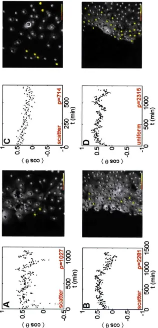
2-9 MEAN COSINE FOR LOW DENSITY SHEETS. Movement for
sub-confluent sheets (A,C) is generally random, either progressing directly to zero post denudation, or constantly oscillating back and forth. The yellow dots are cells at the beginning of the time course that will even-tually persist for (cosO) > 0.3. At low density, they are dispersed
throughout the sheet, while at intermediate to high densities, they are relegated to the periphery. For sheets of increased density (B,D), there is a mean direction, with most cells moving out into free space. . . . . 76
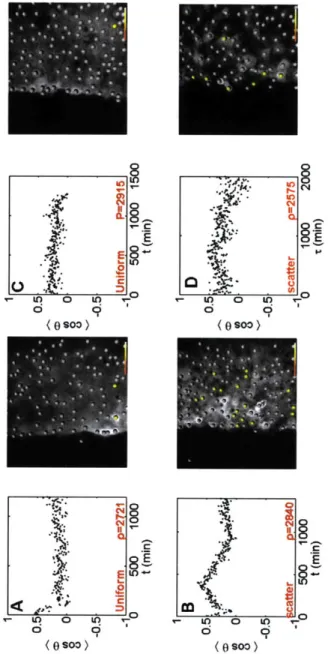
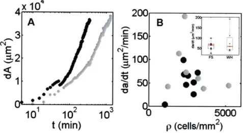
2-10 MEAN COSINE FOR HIGH DENSITY SHEETS. As the density of the cell monolayers reaches a maximum (A,C), nearly all directed move-ment is ablated. Cell movemove-ment is relegated purely to the periphery, with virtually no net directed movement in the bulk. When epithelial E-cadherin is blocked with monoclonal antibody (B,D), this effect is reversed, and even at high density, highly coordinated, directed move-ment is observed. In fact, the transition to coordinated movemove-ment is highly linear in time, as is its subsequent randomization when the density evens out (when both sides of the denudation meet each other). 77 2-11 DYNAMICS OF THE BORDER. The rate of re-epithelialization of the
free space depends highly on density, being slow at both very low den-sity, where cells scatter, and very high densities, which are retarded
by cell contact. This is evident in the slopes of the recovery of this
area (A). This can be seen in the slope plotted for wounds (WH) and "model" wounds (FS) at different densities (B). Both methods of de-nudation, the wound (gray), and the model wound (black) show this same dependence, although the speed of recovery is faster in the wound. 78 2-12 GRADIENT IN CONTRACTILITY. 200pM blebbistatin was used to
make the left side of the monolayer non-contractile, by decreasing the affinity of Myosin II for filamentous actin (A). The right side is un-treated. The results show that there is no directed movement by the untreated cells in preference for the non-contractile side (B). . . . . . 78
2-13 GRADIENT IN APOPTOSIS. 0.0875%H
20
2was delivered to the left
side, to induce apoptosis (A). This was imaged by a live/dead stain over 12 hours. (The tracked data in (B) however, was over roughly 24 hours, at half that concentration (0.042%)). . . . . 792-14 (A) Live Dead Stain with the middle channel filled with paraformalde-hyde, and ran for just 2 minutes. The right side remains alive, but only for a short period of time. Fixing one side of the cells, even with no diffusion to the right, still killed the cells on the right hand side. There is likely a cell-cell communication signal that makes cell death proceed quickly across the entire length of the sheet. Thus, there was no chance to observe any migration in the viable population. (B)
Brightfield image of both fixed and unfixed cells. . . . . 83
2-15 FUNCTIONAL BOUNDARIES. (A) Viable cells interface with
apop-totic cells (red: apopapop-totic, green: viable), and (B) Motile cells interface with Non-Motile cells (Gray: Motile, Red: Non-motile). In neither case to the fully functional, viable cells push. No extension of border. . . . 83
2-16 CYTOCHALASIN D. The left channel was filled with 5pM
cytocha-lasin D, creating a gradient across the lateral axis. The cytochacytocha-lasin has a dye that emits at 468nm, and the right hand side was filled with normal medium, with 1:10000 DiI. Thus the red side has an altered cytoskeleton, with its actin unable to polymerize. The right hand side is normal. There was also no gradient in migration observed here. . . 90
2-17 LIVE/DEAD IN MICROFLUIDICS. After the trypsin cut, all of the
2-18 MEAN COSINE. Calculation of the mean cosine. For equilibrium (no
free space) movement (A), the cosine of the angle between vectors sepa-rated by an elapsed time, T are averaged for the ensemble of trajectories over all t (B). That allows us to characterize the PDF for the angle of movement (C). If movement is non-equilibrium, as in wound healing and microfluidic denudation studies, it is calculated in real time, as the cosine of the angle between successive vectors (D,E). . . . . 91 2-19 DENSITY DEPENDENCE. The density dependence of the mean
co-sine calculation for equilibrium movement. As the density is increased, the correlation lengthscale of the angle between successive vectors dur-ing the migration of cells within a sheet, decreases. With the smaller the magnitude of the scaling exponent, the longer the paths will persist as straight . . . . 92 2-20 LEADING EDGE CELL MORPHOLOGY. Morphology of a leading
edge CMFDA-stained cell in front of a fully confluent sheet, imaged at 100x. Grid = 2pm . . . . 93
2-21 SUBMARGINAL CELL MORPHOLOGY. Morphology of a submarginal GFP-cell in a fully confluent sheet, imaged at 100x. Grid = 2pm. Z slices are at 0.8um, 2.8um, and 10.8 um from the surface. . . . . 93 2-22 SURFACE PROTEIN REMOVAL. The denudation of cells, whether
by physical scratching (A) with a 1ml pipette tip, or by enzymatic
cleavage (B), removes suface protein (FN+FITC Collagen). In both cases, the new protein is added by the medium (that contains 10% FBS). 94
2-23 RETENTION OF ADHERENS JUNCTIONS DURING SHEET MO-TION. During Wound Healing, as the sheet moves forward, the leading
edge displays both focal adhesions (A, characteristic to single cell move-ment), as well as maintaining adherens junctions (B, characteristic to sheets). This is a fixed sample, stained with rhodamine-phalloidin. . . 94
3-1 BASAL LAMINA ANALOG. Rheology of Matrigel (red), and 1/80
PDMS (black). Storage (.), Loss (o) Moduli. For this composition of PDMS, the rheology nearly matches that for Matrigel, for low fre-quencies. We stop our comparsion at 10-1 radians/s, as below this
frequency, we are uncertain concerning the rise in G' and G". . . . . . 104
3-2 SURFACE PROTEIN AS A FUNCTION OF STIFFNESS.
FITC-stained gelatin bound to fibronectin, attached nonspecifically (A-C) or covalently (D) to PDMS in a ratio of (A) 1/10, or (B,C,D) 1/80, which is the critical combination of stiffness and wettability that char-acterizes the most correlated state, and is thereby marked with a red star. . . . . 105
3-3 CELL TRAJECTORIES. Trajectories of dense cell populations on
non-compliant (1/10) low density (A) and high density (B) or compli-ant (1/80) high density (C) surfaces. At low densities on non-complicompli-ant substrates, the trajectories are very coherent and coordinated. High density generally ablates this coherence on non-compliant substrates, but small domains of highly coordinated movement persists when the substrate is compliant. . . . . 106
3-4 COUPLING OF CELL MOTION AND BULK MOTION. Velocity au-tocorrelations for cell movement (black) and bead displacement (gray) in substrates of varying viscoelasticity. As the stiffness of the surface dips below 67.7 Pa, the timescales converge, with the substrate move-ment mirroring the cell movemove-ment. . . . . 107
3-5 DECOUPLING OF CELL MOTION AND BULK MOTION. (A) Cell
autocorrelation on 1/80 PDMS, with fibronectin covalently linked. (B) Cell autocorrelation on 1/80 PDMS, with covalently linked fibronectin, and 200pg anti-E-cadherin antibody to block cell-cell contact. The per-sistence of cell motion is not altered on this non-compliant substrate with the removal of cell-cell contact, but the correlation is no longer conferred to the substrate (same bead scaling as 1/10 PDMS compo-sition ). . . . . 108
3-6 COUPLING BETWEEN CELL MOTION AND BULK AS A FUNC-TION OF CELL CONTACT. Velocity autocorrelations for cell
move-ment (black) and bead displacemove-ment (gray) on both hard (and elastic) and soft (and viscous) substrates. For hard, elastic substrates, the ab-sence of cell-cell contact, does not alter the persistence of single cell motion. However, on soft substrates, blocking E-cadherin separates the timescales between the cell motion and the persistence, as well as decreasing the magnitude and scaling of the autocorrelation function. 109
3-7 PERSISTENCE AS A FUNCTION OF DENSITY. Both deformable
(gray) and non-deformable (black) substrates have roughly the same scaling behavior for all highly confluent densities. Cell motion persists for longer (and hence have a lower power law exponent) when the density is low, which is not possible on deformable substrates, as cells w ill com pact. . . . .. 110
3-8 DISTINCT VELOCITY CORRELATIONS. Mutual Velocity
Correla-tion FuncCorrela-tions (1hr), for dense cell populaCorrela-tions on 14.4 Pa (light gray),
3-9 SECOND ORDER TRANSITION IN MUTUAL CORRELATION.
Taking the scaling exponent, a', of the mutual velocity correlation function for each value of |G|, maps out a continous transition from
the non-compliant and less correlated (- 1/r'0) to the compliant and
more correlated (~ 1/ro 3). The inset is the mean velocity of the cells,
with the velocity of the beads subtracted, as the instability causes a net drift. This relative velocity is constant for all measured moduli. . 111
3-10 VAN HOVE CORRELATIONS. (A) radial distribution function for
two systems, compliant (gray) and noncompliant (black). (B) Timescale of the evolution of the spring and damping constants, characterized by time evolution of their ratio, ( = K/'r - t"/*. (C) Time-scaling of
beads in PDMS. (D) Relationship between timescale of cell organiza-tion and timescale of bead displacement. . . . . 112
3-11 PDMS RHEOLOGY. Rheology of PDMS of different curing agent
con-centrations. G(w) dark, G(w) light, and follow: 1/10, 1/20, 1/40,1/80 (red) and 1/160 . . . . 113
3-12 STRUCTURAL INSTABILITY. The onset of the most correlated state
on compliant (1/80) PDMS is also accompanied by holes that open up in the sheet, which can grow in time. (A) 10x, (B) fluorescence image of E-cadherin on a 1/80 PDMS sheet. On the same sample, local tension induces these instabilities, which retain functional E-cadherin around the circumference (C) fluorescence, (D) brightfield. . . . . 114
3-13 SHEET REMAINS FLAT. (A) 25x, 3D image of stained nuclei on
nearly pure prepolymer (1/200). For long times, the cells retain their sheet-like character, although scattering is possible by moving under-neath other cells (3D reconstruction and contour plot, B). . . . . 115
3-14 3D MORPHOLOGY FOR DIFFERENCE STIFFNESSES. Cells on non-compliant surfaces, |G| > |G* , (A) are spread, while cells on com-pliant surfaces, |G| < |G*|, (B) create long projections and have less interfacial area with the surface (grid = 2pm). . . . . 115
3-15 CONTROLS FOR THE VAN HOVE CORRELATION FUNCTION.
Dynamic Monte Carlo simulation (A) of the same size image as most experiments, (D), at high density yields the RDF in (B) and spectrum in (C ). . . . . 116
3-16 FIBRONECTIN ADSORBED TO PDMS. (1) 0.4 mg/ml, (2) 0.2 mg/ml, (3) 0.1 mg/ml stained fibronectin on 1/10, 1/60, and 1/160 PDMS,
spun onto Mattek dishes. On the 1/10, and 1/60 PDMS, the fi-bronectin is rigidly adhered. On the 1/160 plates, where the PDMS is nearly purely viscous, the fibronectin does not stick, and instead is diffuse across the plate. . . . . 117 3-17 DYNAMIC INSTABILITY. On compliant substrates, (i1/80), the sheet
will slowly begin to aggregate. This results in the deformation of the substrate over millimeters (A). Structural inhomogeneities appear dur-ing the collapse, such as the appearance of holes in the sheet, and invaginations (B,C). . . . . 118
4-1 STAMPED FIBRONECTIN. 1mg/mL Fibronectin transferred
non-specifically to 1/10 PDMS spun onto a Matek dish, and stained with lug/ml FITC-pig skin collagen. . . . . 122 4-2 CELLS BOUND TO STAMP. Brightfield image of Mouse Mammary
Epithelial Cells bound nonspecifically to 1/10 PDMS. . . . . 122 4-3 LOW DENSITY CELL POPULATION. 1/10 PDMS trated with 5
min H202
/HCl,
10% PEG-amine, to generate the reactive/passivatedsurface. 1mg/ml fibronectin added to stamp and transferred. Mouse Mammary Epithelial Cells bound, and stained with 1/1000 CMFDA. 131
4-4 LOW DENSITY CELL TRAJECTORIES. Trajectories of cell move-ment on stamped pattern for the low density system. Low density epithelial cell populations under confinement are marked for their ad-herence to the boundaries, and high speed. . . . . 132
4-5 HIGH DENSITY CELL POPULATION. High Density of Cells, iden-tically treated as 4-3 ... ... 132
4-6 HIGH DENSITY CELL TRAJECTORIES. Trajectories of cell move-ment on stamped pattern for the high density system. More highly dense systems also show preferences for the boundaries, although the effect is significantly less pronounced than the lower density. . . . . . 133
4-7 PROBABILITY DENSITY FUNCTION FOR CELL VELOCITIES (LOW). Probability Density Function for the velocity of cell trajecto-ries in low density populations with diameters 50 ptm (red), 150 pim (gray), and 250 pm(black). Each overlaps, and contains the same non-gaussian character, and slight fast subpopulation. . . . . 133
4-8 PROBABILITY DENSITY FUNCTION FOR CELL VELOCITIES
(HIGH). Probability Density Function for the velocity of cell
trajec-tories in highly dense populations with diameters 50 pm (red), 150 pm (gray), and 250 pm(black). Each overlaps, and contains the same non-gaussian character, and slight fast subpopulation. . . . . 134
4-9 MUTUAL CORRELATION FUNCTION FOR SEPARATE
POPU-LATIONS. Mutual Velocity Correlation Functions for the separate
populations as defined by the spatial patterning. This function isn't really valid above the half-width of the patterns, which is 50pm, but nevertheless, illustates an important conclusion. There is no real differ-ence in the level of correlated activity. (red: 50 pm inner radius, yellow:
150 pim inner radius, green: 250 pm inner radius, blue: 350 pum inner
radius. The modulus of the PDMS is 492.2 Pa, with G"=154.5Pa, and G '= 470.2 . . . . 135
4-10 STIFFNESS ALTERATION BY OXIDATION?. Twelve minute plasma oxidation of a 1/10 PDMS surface, and subsequent non-specific stamp-ing of fibronectin (A) 10x Fluorescence image of the fibronectin, (B) brightfield 5x image of cracks throughout the dish. Stamping on sur-faces of different stiffness was not pursued, largely because we were unsure whether or not by oxidation of the PDMS surface, we were al-tering the stiffness as well as the chemical composition. Then we have both the effect of the covalent attachment as well as the stiffness to contend with when analyzing the movement of the cells. . . . . 136
4-11 ACTIN CONTRACTILE BUNDLES IN PSEUDO PURSE-STRING. 1:200 rhodamine-phalloidin was used to stain the filamentous actin bundles in a pattern of mouse mammary epithelial cells. It shows that there is a large concentration of actin bundles arranged radially, and preferentially towards the center and the inner diameter of each ring. Thus, the formation of these bundles is not dependent upon the wound, as in the "purse-string" closure of a small wound, but is sufficiently explained by free space. . . . . 136
4-12 10x image of fibronectin stamped onto two separate samples, PDMS surfaces of a crosslinker/prepolmer concentration 7/400. The surfaces were treated as described above, with FITC-Pig Skin Gelatin as con-trast. Collagen is good as a contrast agent, but also adheres cells more quickly than fibronectin. This can be a valuable parameter, as even given the cell exclusion chemsitry, the longer that cells sit on the sur-face, the more likely it is they can still bind the surface where you do not want them . . . . 137
5-1 DYNAMIC SUSCEPTIBILITY. Using a technique quite common to
granular physics, we seek to identify timescales involved with the ex-change of neighbors. As cell monolayers that move uniformly versus those that scatter may have the same magnitude of their velocities, we wanted an order parameter that would be able to distinguish between the two. Here, we show the results of three simulations. In the top row, there is a crystalline system with low displacement, and nearly no neighbor exchange. Therefore the order parameter barely dips, and there is no identifiable timescale by the susceptibility parameter, x. As the temperature of the system is increased (middle row), there is now a finite timescale, as thermal energy is sufficient to drive the scatter-ing of each of the particles. Takscatter-ing the same density, but biasscatter-ing the movement (bottom row), we get the the magnitude of x decreasing, reflecting the increased uniformity, as each cell has a preferred motion. As this system could not be easily compared across densities, we opted for just the mean cosine as a superior method. . . . . 150
5-2 DYNAMIC SUSCEPTIBILITY PREDICTS A SECOND ORDER
TRAN-SITION. The location of the peak in the x parameter, T*, for the movement of monolayers on substrates of varying viscoelasticity pre-dicts the same transition as does the divergent power law lengthscale calculated in Chapter 2, from the 2pt. velocity correlation function. The parameter ( is identical to a' from Chapter 2, but only for very high densities. . . . . 156
5-3 DIAGRAM OF MUTUAL 2PT CORRELATION FUNCTION. The correlation function whose power law exponent is used to outline the transition from uncorrelated to correlated behavior, is taken as the product of the velocities in the directions that conenct them, in the radial, perpendicular direction. . . . . 156
5-4 DIAGRAM OF THE RADIAL DISTRIBUTION FUNCTION. Each cell is at the origin of its own radial coordinate system. In successively larger intervals, at a distance, r away from the origin, the density of cells is counted. This function is solidly periodic for crystalline materials, oscillatory for weakly interacting or dense systems, and flat for non-interacting and very low density systems. . . . . 158 5-5 HARD DISK TRAJECTORIES. Trajectories of 300 hard disks. The
potential is either 0 or infinity, and are walked around a lattice, and captured at 80 time points . . . . 158
5-6 VAN HOVE CORRELATION OF HARD DISK SYSTEM
MIMICK-ING CELL MOVEMENT. We use dynamic monte carlo simulations to reproduce cells in the same quantity and with the same dimensions as the cells we image at 25x. After collecting the trajectories of each
in silico cell, we can calculate a control for the van Hove correlation
function. At T = 0, there is significant order, out many cell diameters. This correlation is lost quickly over time, however. . . . . 159 5-7 VAN HOVE CORRELATION OF LENNARD-JONES SYSTEMS
MIM-ICKING CELL MOVEMENT. Lennard-Jones systems make use
simi-lar short-range exclusion as hard disk systems, but also have a very long ranged attraction. Using the canonical ensemble described in Chapter
8, we can lower the temperature to see the emergence of long ranged
correlations in the van Hove correlation. . . . . 160
5-8 THE DYNAMIC STRUCTURE FACTOR FOR THE HARD DISK SYSTEM. The two-dimensional fourier transform in time and space
peaked at 27r/0.35 = 18 pm, the diameter of a single hard disk. . . . 161
5-9 THE STATIC STRUCTURE FACTOR FOR A SHEET ON A VIS-COELASTIC SUBSTRATE. The fourier transform of the radial
ditri-bution function, showing a peak at the principal cell-cell spacing. This is the only relevant lengthscale. . . . . 162
5-10 THE DYNAMIC STRUCTURE FACTOR FOR A SHEET ON A VIS-COELASTIC SUBSTRATE. The same two-dimensional fourier
trans-form was calculated on the van Hove correlation function calculated for the movement of cells within a plane, for every separation distance r, between cells, and every elapsed time, r, during the timecourse of the imaging. There are no significant collective modes, what would be seen as diagonals in the Dynamic Structure Factor. The only peaks remain for most time, and represent the characteristic lengthscales of the separation distances between cells. . . . . 163 5-11 2PT VELOCITY CONTROL. The C,, has a 1/r decay, and the
mag-nitude is much greater along the mutual direction that connects two beads, than in the parallel direction, or crossing parallel and perpen-dicular . . . . 164
5-12 MEAN-SQUARED DISPLACEMENT FOR SINGLE ENDOCYTOSED
PARTICLES. 0.5 pm rhodamine coated, carboxylated, internalized mi-crospheres were imaged over 60s in mouse mammary epithelial cells cultured on coverslips. Each line is a separate microsphere, located in a distinct local mechanical, microenvironment. . . . . 168 5-13 ENSEMBLE AVERAGED MSD FOR CELL ENDOCYTOSED
PAR-TICLES. 0.5 pum rhodamine coated, carboxylated, internalized
micro-spheres were imaged over 60s in mouse mammary epithelial cells cul-tured on coverslips. The plot is the ensemble average of all of the different microspheres, so that we might fit it to a power law, and calculate the mean diffusivity of the particles. . . . . 168
5-14 POWER LAW SCALING FACTOR FOR MSD. At short times, the embedded microspheres are often confined by the cellular cytoskeleton, and thus are "sub-diffusive" (a < 1), unable to sample space at the
rate that is expected by diffusion. As more time elapses between frames however, the bead is able to break free, and diffuse freely (a ~ 1) . . 169
5-15 FREQUENCY-DEPENDENT SHEAR MODULUS FOR MOUSE
MAM-MARY EPITHELIAL CELLS. From the MSD, the particle size, and the temperature, we can calculate the frequency-dependent shear mod-ulus. G' is the loss modulus, an indication of how the cell stores elastic energy that is applied. G" is the loss modulus, what indicates how the cell dissipates energy applied to it. Clearly from this figure, we can see that for nearly all timescales, the cell is more viscous that it is elastic. 169
5-16 MEAN ELASTIC MODULUS FOR DIFFERENT EPITHELIAL CELLS.
Mean Elastic Modulus, for both mouse mammary epithelial cells, as well as Caco-2 colon epithelial cells. The mean Elastic modulus for the mammary epithelial cells, what we use throughout the experiments in this thesis, is 0.5631 + / - 0.0903 Pa, or 5.631dyn/cm2 (n=3). The
mean Elastic Modulus for the colon epithelial cells, which are more likely more similar to the intestinal epithelium, to which we draw many analogies, is 0.1915+/-0.0464 Pa, or 1.915dyn/cm2 (n=3). Both mea-surements are taken at 1 Hz. . . . . 170 5-17 BEADS IN GLYCEROL. 1 pm beads in a 50:50 mixture (by volume),
imaged over 100 seconds. The trajectories are color-coded, with dark being old, and light colors being more recent . . . . 170 5-18 MEAN-SQUARED DISPLACEMENT FOR A 50:50 GLYCEROL/WATER
MIXTURE. Perfectly linear scaling for the MSD, over nearly all timescales. The only limitation is the ability to track the beads for every frame, as the beads can diffuse out of the plane. . . . . 171 5-19 POWER LAW SCALING FACTOR FOR GLYCEROL MSD. The
mean-squared displacement scales as T1 0 for nearly all timescales,
con-firming that all motion is strictly diffusive. . . . . 171 5-20 VISCOSITY OF A 50:50 GLYCEROL/WATER MIXTURE. The
vis-cosity of the 50:50 Glycerol/Water mixture is - 3mPa s, as measured in
Properties of Ordinary Water-Substance N.E. Dorsey, p. 184., Hafner
Publishing Co., New York (1940). . . . .
172
8-1 Particles occupy right side, membrane ruptures, and the volume avail-able to the system has increased . . . . 212 8-2 Length versus time traces of the RNA hairpin at various constant forces
Chapter 1
Introduction
1.1
In vitro models of Sheet Motion
The canonical description of cellular motility is defined at the level of a single cell, in the protrusion, contraction, and detachment of membrane lamellipodia or fillopo-dia [8, 9]. The intracellular machinery that drives the cell forward is largely actin, coupled with the stabilization of dynamic, multiprotein plaques at the surface, called focal adhesions. There is extensive knowlege of the mechanisms involved - the key intracellular molecular components, both in terms of the proteins that link together to comprise the machinery, as well as the molecules responsible for the signaling cas-cades that drive the system forward. The morphologies of migrating cells have been detailed in a wide variety of cell types, for varying conditions and dimensionality, from the flattened to the amoeboid [9]. In addition, there is large and growing knowledge of the external, physico-chemical factors that modulate adhesion. The adhesivity of the surface, as defined by the receptor-ligand binding affinity [10], or the quantity and availability of surface ligand, is a principal variable. The mechanical properties of the substrate, the time-dependent deformation characteristics have also emerged as influential in the determination of movement [11, 12, 13] and geometry [14, 15]. Cells also respond to gradients in these parameters. Spatial asymmetry in the surface localization of adhesivity [16, 17], as well as in the compliance of the surface itself
the less compliant (following a bimodal relationship).
Thus, despite the abundance of detail in the mechanisms that determine single cell motion, little is understood of how coherent patterns of movement can emerge when they migrate as groups. It is still unclear how the introduction of cell-cell contact contributes explicitly to coordinated movement en masse in any of the aforemen-tioned scenarios, despite the ubiquity and importance of this phenomenon in a wide variety of physiological processes, from development in the embryo, to regulation and homeostasis in the adult. Equally as varied, are the manners in which the sheets have been observed to move. In embryonic development, epithelial sheet fusion oc-curs in the ventral enclosure of Caenorhabditis elegans [20, 211, and dorsal closure of the Drosophila embryo [22, 23, 24, 2]. In certain wounded scenarios, such as in the stratified epithelium of the cornea [25, 26, 27], cells have been shown to slide as a contiguous sheet [27]. In normal adult tissue, the unobserved (but experimen-tally verified) unixaxial movement of the intestinal epithelium constitutes the fastest turnover of tissue in the body [28, 29, 30]. Thus, the coordination and self-similarity in the movement of cells in each of these cases is extraordinary, despite (or perhaps because of) the retention of their cell-cell contact.
The cell-cell contact in epithelial tissue, for example, is moderated by E-cadherins, calcium-dependent transmembrane proteins that are anchored intracellularly by actin-based adherens junctions [31]. These junctions are belts of filamentous actin, around the periphery of each cell, which is known to provide the structural integrity of the cell (cadherins are also bound to hemidesmosomes, which are anchored to interme-diate filaments, with less well studied physical properties). Given that each cell is adhered to each other by these strong interactions, yet they each have their own fo-cal adhesions that drive locomotion, how does one influence the other? It has been shown that increased expression of E-cadherin slows the migration of in vivo epithelia
[32]. Although, it has also been associated with a reduction in proliferation and an
still not been isolated (nor imaged directly, as these are based on histological studies of sacrificed mice). Thus far, efforts to understand collective migration of epithelial cells at all, limit themselves to (1) characterizing the extent that the movement is collective (tracking cell motion in time, then using mathematical analyses to quantify its correlation), and (2) correlating movement to distinguishing morphological char-acteristics. And, as the predominant method for inducing coordinated sheet motion is through the denudation of confluent sheets in culture, in vitro efforts center on the role of free space and cell damage in inducing it[33, 4].
1.1.1
Wound Healing
As previously stated, the predominant method for generating coordinated group motility in culture, is by denudation of space, such as what is found in the classi-cal wound healing assay. The most common method for wounding, is to physiclassi-cally scratch the surface denuding the sheet (Figure 1-1), and cells on either side of the scratch will move to close the gap. The cells in the front of the denuded sheet, called the leading edge, produce large, clear actin-based extensions that actively search their environment, and are extended in the direction of the edge. The cells behind the leading edge, termed sub-marginal cells, are still confined, and have only been shown recently to extend mild lamellopodium along their basal surface in between their neighbors, in the direction of the wound[34]. Thus, there is massive, coordi-nated motion along the leading edge, but it does not penetrate deep into the rear of the sheet. In an alternative, mechanistically distinct method, when the wound in an epithelial sheet is small and circular, closure proceeds by what has been called an actin 'purse string' (Figure 1-2), [2]. During this event, the cells at the periphery of this small wound, organize large actin filaments that extend around the circum-ference of the wound. These filaments are anchored by the adherens junctions, and are linked between cells by cadherins. Thus, short range coordinated movemement in this sense is mediated by lateral junctions, and contractions that extend further than the lengthscale of a single cell. As both processes involve the physical denudation
of the sheet, from these techniques it would be impossible to know with certainty whether the movement resulted from the opening of free space, or from the induced cell damage, and release of factors into the medium, or through gap junctions, the in-tercellular communication channels. Thus, while there is coordinated movement, and cells all move perpendicular to the direction of the wound to initiate closure, causality is obscure, and there is an inconsistency with what should exist in the natural ep-ithelium, that cells in the rear (which are in the vast majority of the population) are not very motile. Nevertheless, this is the standard model, as this is the only method (or its variants, described below) in culture that is known to force cells to move in a directed, uniform direction (after previously moving randomly) or "break symmetry".
1.1.2
Model Wounds
As an attempt to address this 'damage vs space' dichotomy, other studies have fo-cused on creating removable barriers to denude the sheet, thereby creating a 'model wound'. Either by the placement of non-adhesive PDMS blocks, or by the removal of stencils (Figure 1-4), various studies claim to be able to discern that the onset of collective migration is sufficiently explained by the availability of free space. By these methods, the implication is that if scratching can be avoided, then there will be no induced damage, and no signal to instruct the sheet to migrate forward. This assumption is inherently flawed however, as will be shown in the chapters that follow, that these methods still do induce damage - only less. The application of physical force to remove cells, will always result in the induction of some kind of signal, either
by the death that results, or by the transmission of physical force into a variety of
known mechanotransduction signals among the remaining viable cells. Therefore, the issue of free space as it pertains to the onset of collective migration remains unsolved, given that we assume it is a parameter relevant to in vivo studies. Further, these studies still suffer from the latter of the two aforementioned issues, which is that submarginal motility is still not explained.
As there is an apparent lack of submarginal motility, some studies have tried to conclude that this is due to a lack of traction, by experimentally measuring the migration-induced deflection of high density arrays of micro-fabricated pillars[35]. This is a similar approach to the 'denudation models', explicitly using the default assumption that sheet motion must be entirely based on traction against a non-compliant substrate. They compare the traction exerted by isolated cells and cells within a sub-confluent monolayer. As expected, cells within the monolayer exert less traction than isolated cells - completely confluent cells exert none (compare to fully confluent, polarized epithelia). Moreover, the tractional stress is greatest at the cell edge, both of an isolated cell and cohered cell at the boundary. This is consistent with other experiments that measure the speed of cells in the sheet and at the edge
[33]. These results are not surprising, as they mirror what is observed by imaging
the denudation models. The leading edge cells have actin-based searching machinery, which is clearly visible by light microscopy. The cells further towards the rear do not appear to have similar machinery. However, this experimental system is extraordi-narily relevant, in that it sums up the assumptions and inadequacies of the models before it. It makes most clear the central assumption of each of the models that (1) the denudation of space on non-compliant surfaces (continuous or not) is required to induce motion, and (2) that traction of all or part of the population is the principal physical mechanism for that motion.
The explanation for this lack of movement, which is put forth explicitly in the mi-cropillar study, but inferred in many others, is that the cells are in a dynamic balance of pushing and pulling, and therefore there is no net movement on these epithelial islands suspended by pillars. Inverting this inference, the denudation models would therefore have a net movement, because now there is an imbalance. The implicit arguments of each of the above studies, is that only this imbalance breaks symmetry (i.e. post denudation). However, as we understand that whether we are talking about the contiguous sliding of a wounded cornea, or the renewal of the intestine, motion is required from more than just the first couple of rows of cells. Thus net movement is
achieved over long lengthscales, presumably much longer than what can be accounted for by an imbalance in physical cell contention. This being the case, moving forward, we can accept reduced submarginal motility and either continue to force an analogy of this model back onto in vivo scenarios, or abandon the model system that causes this migratory phenotype in the first place.
To expand on the second assumption in the aforementioned studies, what en-courages the idea that cells are in a dynamic state of pushing and pulling, is the assumption that cells in a sheet will migrate using the same mechanism as single cells
- only together. They are either in contention, or they cooperate. Free space makes them cooperate, because as the imbalance at the leading edge encourages those cells to walk forward, the cells in the rear can move in step. And, while this may be true for the situations in which they are currently observed, where traction against a non-compliant substrate is the only option for coordinated migration, there is sufficient reason to doubt this as an in vivo mechanism. First, we expect longer lengthscale migration than what denudation yields. Further, there is a discrepancy between mor-phologies that we see in vivo (often columnar in vivo, spread in culture; Figure 1-9), with those that we see in these experiments. From a separate perspective however, there is doubt, because there are a variety of additional variables that these methods view as obstactles to movement, foremost among them being cell-cell contact (i.e. constraining cells that desire autonomy, causing no net motion). Thus, to explain the movement of tissue, we should depart from these models, and focus on what makes it tissue, which is the multicellularity from the cell-cell contact.
1.1.3
Cell Sorting
Along this direction, for studying movement of the epithelium as tissue, we draw our nearest analogies to embryonic development, where tissue movement is in part explained by the competition between cohesion and adhesion, the tensions generated between cells versus those generated at the cell-matrix interface. This phenomenon
is detailed by the differential adhesion hypothesis (DAH)[36, 37, 38], which generally states that mobile cell aggregates organize themselves and move in order to mini-mize their interfacial free energy (Figure 1-3). This has appropriately described the equilibrium spatial distribution of initially heterogeneous cellular aggregates by the differential expression of cell-cell adhesion molecules - a perspective which accu-rately captures the role of cell-cell contact in the spatial integration of varied adhe-sive stresses that drive non-equilibrium movement (e.g. sorting) on the way to the equilibrium structure. In experiments on mixed populations of neural retinal and pigmented retinal cells, over time, cells in the mixed population will aggregate with their own cell type, in the end sorting and recovering the two original tissues [3]. Each cell type has different cell-cell adhesion receptors which prefers homophilic in-teraction, and thus by the DAH, cells search space in preference of self, minimizing their own free energy. What is particularly notable, and further underscores the role of cell contact in largescale movement, is that this movement can still occur in the absence of a functional actin cytoskeleton. When cytochalasin B was added to the initially mixed aggregate, all filopodia and pseudopodia, the actin 'search' machinery
described earlier, is ablated, and the mixtures are left with only their mutual cell adhesions, and random fluctuations in shape. Still, these two populations sorted, although the sorting was incomplete. The cell populations never fully reach the con-figuration observed before their ability to search actively was eliminated. In polarized epithelia, there are also no distinguishable pseudopods or membrane ruffling, and so either active search does not exist, or at the very least, it does not exist in the same form. Thus, there is a cell-cell mediated phenomenon, not dependent on (although faciliated by) actin-based motility, that can account for large scale coordinated mo-tion. Noticeably, this type of coordinated motility is within a cellular aggregate, and not on a non-compliant substrate. While the conceptual framework for differential adhesion has been presented as a description of cell sorting in heterogeneous aggre-gates, we merely use this as a springboard to motivate a simpler extension of this theory, that cell-cell contact may plays an elevated role in coordinated cell motion of the epithelia (cornea, kidney, colon, etc) rather than being a connection for pushing
and pulling, that it contributes (in the previous example energetically) to the move-ment of cells over long timescales and long lengthscales. I therefore use this general relationship, the mechanical balance between cell-cell and cell-matrix adhesions (pref-erence towards the former), as the primary target for measurement in our studies of the mechanics of the epithelium, using the intestinal epithelium as a reference system.
The intestinal epithelium is a cellular monolayer that is folded into crypts and villi that form invaginations and projections into the intestinal lumen for the purpose of absorbing nearly all of the body's nutrients (Figure 1-8). Roughly half of the cells in the intestinal tract rapidly proliferate at the bottom of the crypt, and migrate against the basement membrane (Figure 1-5) to the top of the villus, while they mature into either absorptive or secretory cells. At the tip of the villus, mature cells undergo apoptosis [39], completing a lifecycle on the order of - 3.3 days for mouse
([40]). In this time, cells move at roughly 1-1.5 cell diameters per hour, ranking them among the most motile cell types, renewing the lining of the intestinal epithelium faster than any other tissue. Further, the importance of this movement as a physical process is underscored by the fact that they are all mutually adherent. They migrate collectively, forming and breaking adhesions with the basal lamina and each other in a way that characterizes a motile sheet. Astoundingly, this non-equilibrium process is stable, as the rates of proliferation, predominantly at the bottom of the crypt, balance the cell death that occurs at the tip of the villus. The motility of cells is precisely balanced with their lifecycle, to generate a dynamic equilibrium state. Thus, this presents an interesting model for migration (and reference for our experiments), as it has coordinated, directed movement of a monolayer, with no wounds, and a unique combination of boundary conditions intricately tied to the movement itself.
1.2
Summary of Open Questions
While there continues to be debate in the epithelial cell motion community regard-ing the mechanisms of coordinated migration surroundregard-ing in vitro models of wound