by Dong Wang B. S. Chemistry
Peking University, China, 1998
SUBMITTED TO THE DEPARTMENT OF CHEMISTRY IN PARTIAL FULFILLMENT OF THE REQUIREMENTS FOR THE DEGREE OF
DOCTOR OF PHILOSOPHY IN BIOLOGICAL CHEMISTRY AT THE
MASSACHUSETTS INSTITUTE OF TECHNOLOGY September 2004
C Massachusetts Institute of Technology, 2004 All rights reserved
7 Signature of Author: ( -. -Departmfnt of hemistry
-- Augus',4004
Certified By: Accepted by: MASSACHUSETTS INSTITUTE OF TECHNOLOGY SEP 1BRARIE5 2004LIBRARIES
Stephen J.Lippa& Arthur Amos Noyes Professor of ChemistryThesis Supervisor
Roberi W. Field Chairman, Departmental Committee on Graduate Studies
'ARCHIVES A- .. _ __ __ __
j
-,, -~ ~~~~~~~~, - _'This doctoral thesis has been examined by a committee of the Department of
Chemistry as follows:
Alexander M. Klibanov Committee Chairman Professor of Chemistry and Bioengineering
V Sepher. Lippard
Arthur Amos Noyes Professor of Chemistry Thesis Supervisor
Catherine L. Drennan Cecil and Ida Green Career Development Assistant Professor of Chemistry
Cisplatin-Induced Nucleosome and RNA Polymerase II Modifications Mediate Cellular Response to the Drug
by Dong Wang
Submitted to the Department of Chemistry on August 18, 2004, in partial Fulfillment of the requirements for the Degree of Doctor of Philosophy
Abstract
Chapter 1. Cellular Processing of Platinum Anti-Cancer Drugs - Identifying Pathways for Chemogenotherapeutic Drug Design
Cisplatin, carboplatin, and oxaliplatin are widely used in cancer chemotherapy. Platinum-DNA adducts, formed following uptake of the drug into the nucleus of cells, activate several cellular processes that mediate the cytotoxicity of these platinum drugs. This review focuses on recently discovered cellular pathways that are activated in response to cisplatin, including those involved in regulating drug uptake, the signaling of DNA damage, cell cycle checkpoints and arrest, DNA repair, and cell death. Such knowledge of the cellular processing of cisplatin adducts with DNA provides valuable clues for the rational design of more efficient platinum-based drugs as well as the development of new therapeutic strategies.
Chapter 2. Nucleotide Excision Repair from a Site-Specifically
Platinum-Modified Nucleosome
Nucleotide excision repair is a major cellular defense mechanism against the toxic effects of the anticancer drug cisplatin and other platinum based chemotherapeutic agents. In this study, mononucleosomes were prepared containing either a d(GpG) or a d(GpTpG) intrastrand cross-link. Comparison of
the extent of repair by mammalian cell extracts of free and nucleosomal DNA containing the same platinum-DNA adduct reveals that the nucleosome significantly inhibits nucleotide excision repair. The effects of post-translational modification of histones on excision of platinum damage from nucleosomes were investigated by comparing native and recombinant nucleosomes containing the same intrastrand d(GpTpG) cross-link. Excision from native nucleosomal DNA is
- 2-fold higher than the level observed with recombinant material. The in vitro system established in this study will facilitate the investigation of platinum-DNA damage by DNA repair processes and help elucidate the role of specific
post-translational modification in NER of platinum-DNA adducts at the physiologically relevant nucleosome level.
Chapter 3. Nucleotide Excision Repair of Site-Specifically Platinum-Modified Tetrasomes
The nucleosome, the basic structural unit of chromatin, is composed of a histone (H3/H4)2 tetramer flanked by two H2A/H2B dimers, around which is wrapped 146 base pairs (bp) of DNA. The (H3/H4)2 tetramer plays a central role
in the structural integrity and positioning of the nucleosome core particle. Site-specifically platinated tetrasomes were prepared to investigate the modulating effects of histone tetramers on excision repair of cisplatin-intrastrand cross-links. In addition, {Pt(DACH))2+-modified tetrasome was prepared to investigate the
effect of spectator ligands on excision repair of platinated tetrasomes. The NER results reveal that the (H3/H4)2 tetramer is sufficient to block excision of both cisplatin-DNA and Pt(DACH))2+-DNA adduct in vitro. The efficiency of excision
of cisplatin-modified tetrasomal DNA is about half (53%) that of the
{Pt(DACH)}2+-modified tetrasome.
Chapter 4. Cisplatin Adducts Change the Rotational Positioning of DNA on
the Nucleosome
Understanding the interaction of cisplatin-DNA adducts with nucleosomes and the effect of cisplatin-DNA adduct formation on nucleosome structure is important for improving the cytotoxicity of this drug. Here we use hydroxyl radical and DNAse I footprinting analysis to characterize site-specifically cisplatin-modified nucleosomes. Preexisting cisplatin-DNA adducts change the rotational setting of DNA with respect to the histone core particle
surface during nucleosome formation.
Chapter 5. Cisplatin-Induced Post-Translational Modification of Histones H3 and H4
The anticancer drug cisplatin kills cells by damaging DNA and inducing apoptosis. Understanding the detailed mechanisms by which cancer cells respond to cisplatin has the potential to improve substantially platinum-based therapy. Post-translational modification of histones alters chromatin structure, facilitating the binding of nuclear factors that mediate DNA repair, transcription and other processes. In the present study, we have investigated the effects of cisplatin treatment on histone post-translational modification in cancer cells. We discovered that specific phosphorylation of histone H3 at Ser 10, mediated by the p38 MAPK pathway, is induced in response to cisplatin treatment. In addition,
alterations, providing mechanistic information about how cells respond to platinum-induced stress.
Chapter 6. The Modulating Effect of DNA Methylation on the Cellular
Processing of Cisplatin-Damaged DNA
Epigenomic regulation of genes is essential for proper cellular function. One major way of conveying epigenetic information in mammalian cells is DNA methylation, with 5-methylcytosine within the CpG dinucleotide being the major component. DNA methylation has profound effects on the mammalian genome. Cancer cells frequently exhibit altered genomic methylation patterns, mostly exhibiting global hypomethylation with region-specific hypermethylated
sequences that occur within the promoter region of certain genes in these cells. Here we investigate the role of DNA methylation on the processing of cisplatin-modified DNA, specifically, the role of DNA methylation in the formation of DNA-platinum adducts as well as on protein-DNA interactions and on the repair of platinum damage. The rates and extent of platination of DNA probes containing 5-methylcytosine residues adjacent to the platinum binding site were compared to similar properties of non-methylated DNA. The effects of DNA methylation on the interaction between a protein and platinated DNA were investigated by studying HMGB1 domA binding with methylated and non-methylated probes. Finally, the effect of DNA-methylation on the NER of cisplatin-damaged DNA was examined with 195mer substrates containing site-specific cisplatin-DNA cross-links with flanking or base-paired 5-methylcytosine residues.
Chapter 7. Transcription-Coupled and DNA Damage-Dependent
Ubiquitination of RNA Polymerase II In Vitro
Transcription-coupled repair (TCR) is essential for the rapid, preferential removal of DNA damage in active genes. The large subunit of RNA polymerase (Pol) II is ubiquitinated in cells following UV-irradiation or cisplatin treatment, which induces DNA damage preferentially repaired by TCR. Several human mutations, such as Cockayne syndrome CSA and CSB, are defective in TCR and incapable of Pol II ubiquitination upon DNA damage. Here we demonstrate, for the first time, a correlation between ubiquitination of RNA Pol II and arrest of transcription in vitro. Ubiquitination of Pol II is significantly induced by alpha-amanitin, an amatoxin which blocks Pol II elongation and causes its degradation in cells. Pol II undergoes similar ubiquitination on DNA containing cisplatin
adducts that arrest transcription. Stimulation of ubiquitination requires the addition of template DNA, and it does not occur in the presence of an antibody to the general transcription factor TFIIB, indicating the transcription dependence of the reaction. We propose that components of the reaction recognize elongating polymerase-DNA complexes arrested by alpha-amanitin or cisplatin lesions, triggering ubiquitination.
In the second part, site-specifically platinated-DNA probes for in vitro transcription studies were synthesized by both primer-extension and enzymatic ligation methods. In vitro study revealed that the presence of cisplatin-DNA adducts on the template strand significantly block the RNA polymerase.
Chapter 8. Synthesis and Characterization of Platinated Dumbbell DNAs for In Vitro Transcription and Repair Studies
Site-specifically platinated double-stranded DNA probes were prepared as valuable templates for in vitro studies of cellular processing of cisplatin,
indicating the formation protein-DNA complexes, transcription, and repair. However, the blunt-ends of double-stranded probes permit non-specific transcription, protein-binding, and exonuclease degradation. Here we report the synthesis of site-specifically platinated dumbbell DNA probes from a pair of hairpins with 3' overhangs and three or four central fragments using enzymatic
ligation. Formation of the modified dumbbell DNA probes were confirmed by exonuclease and restriction enzyme digestion assays. The dumbbell DNA probes were then used as templates for in vitro nucleotide excision repair and transcription studies.
Appendix Al. The Interaction of Human SWI/SNF with Platinated
Nucleosomes and Its Effect on NER
In eukaryotic cells, DNA is packaged into chromatin. Two major classes of factors have been identified to alter DNA accessibility in chromatin: histone modified complexes and ATP-dependent remodeling complexes. SWI/SNF, one of best characterized members of the latter class, has been linked to nucleotide excision repair stimulation. The stimulatory effect of SWI/SNF on NER depends upon the nature of the DNA lesion. SWI/SNF stimulates the excision of AAF-adducts and the (6-4) photoproduct from nucleosomal DNA, whereas it has a negligible effect on the repair of TT dimers. These differential effects of SWI/SNF on the repair of UV photoproducts and AAF adducts promoted our interest in investigating the effect of SWI/SNF complexes on repair of cisplatin damage within the nucleosome. In this section, the effect of SWI/SNF on the repair of cisplatin-DNA adducts in a mononucleosome will be addressed. A moderate
stimulatory effect of SWI/SNF on the repair from cisplatin-DNA adducts was observed.
Thesis Supervisor: Stephen J. Lippard
For my mother, my farther, and my sister, who taught me to love, to care,
and many other things.
And for Francis Harry Compton Crick (1916-2004), who revealed such a perfect structure,
Acknowledgments
First and foremost I must acknowledge my thesis advisor, Stephen J. Lippard. His great insights and guidance throughout the course of this work will be always appreciated. His constant enthusiasm and dedication for science always inspire me. I really appreciate his unconditional support and scientific freedom he gave to me. His excellent style for doing science has transplanted into my mind as a great "template."
I thank Prof. Philip A. Sharp and Dr. Keng-Boon Lee for their close collaboration on the Pol II ubiquitination project. I really appreciate those insightful discussions with Phil regarding the present and future of science, which I am certainly touched by his amazing love for science and his friendly personality. I also thank K.B. who taught me a lot of hardcore biological techniques. She is a wonderful and quiet person. I wish her and her family well.
Prof. Aziz Sancar, Dr. Ryujiro Hara, and Dr. J. T. Reardon from Univ of North Carolina gave me tremendous help on the repair project. I went to Aziz's lab for about one month to learn the techniques of nucleosome assembly and purification. I really enjoyed my stay there. Aziz's sharp insight and stimulating discussion have benefited me a lot. I really missed those wonderful afternoons at his office and of course, the world cup of soccer.
I also thank Prof. Thomas D. Tullius and Mr. Qun Wang (Boston Univ.) for the collaboration of footprinting project; Dr. Hua-Ying Fan, Prof. Robert E. Kingston (Harvard Medical School), Prof. Danny Reinberg (Univ. of Medicine and Dentistry of New Jersey), and Prof. Karolin Luger (Colorado State Univ) for providing me materials for experiment or technical help. I have also benefited a lot from several members of MIT faculty. I thank my thesis chair Prof. Alexander M. Klibanov, Prof. Catherine L. Drennan, Prof. Alice Y. Ting, Prof. John Essigmann, Prof. JoAnne Stubbe, Prof. Jianzhu Chen, Prof. Alexander Rich, and Dr. Shuguang Zhang for thoughtful discussion of my project and general scientific matters.
I have been extremely lucky to involve in such an excellent research group, which I really enjoyed. I appreciated their help and contribution. In particular, I thank Dr. Christa Kneip, Dr. Qing He, Dr. Sudhakar Marla, Dr. Yuji
Mikata, and Ms. Amanda Yarnell who taught me experimental skills in my early days in the lab. I also thank Dr. Chuan He, Dr. Min Wei, Dr. Christiana Xin Zhang, Dr. Juan Carlos Mareque-Rivas, Dr. Edna Ambundo, Dr. Edit Y. Tshuva, Dr. Christopher J. Chang, Dr. Seth Cohen, Dr. Elizabeth R. Jamieson, Olga Burenkova, Alyssia Dangel, Dr. Jane Kuzelka, Dr. Douglas A. Whittington, Dr. Daniel Kopp for their support and friendship. As for current members, I especially thank Ms. Katie Barnes for her help in tissue culture and her patience
to read most of my manuscripts. I also thank Andrew Danford, Dr. Christian Goldsmith and Andrew Tennyson for proofreading some chapters of my thesis. In addition, I thank Yongwon Jung, Matthew H. Sazinsky, Sungho Yoon, Carolyn C. Woodroofe, Lisa L. Chatwood, Elizabeth Nolan, Dr. Elisabeth R. Cadieux, Dr. Jessica Blazyk, Dr. Dong Xu, Evan Guggenheim, Mi-Hee Lim, Dr. Sumitra Mukhopadhyay, Richard C. Girardi, Emily C. Carson, Dr. Laurance Beauvais, Amy Kelly, Leslie Murray, Michael McCormick, Dr. Jeremy Kodanko, Dr. Rayane Moreira, Rachel Niehuus, Sonya Tang, Annie Won, Cindy Yuan, Caroline Saouma, Pamela Chang, and Cindy Liang for their friendship. Finally, but not least, I want thank my three great UROPs, Andrew Danford, Gita Singh, and Tim Davenport, for their excellent work, especially for Andrew. We have been worked together for over three years. We shared a lot of ups and downs during the progress of project and we are happy to get through and overcome those difficulties. We also have a lot of interesting talks beyond chemistry. He is a really great and fun person to know. I hope for his great success on whatever he pursues.
On a more personal level, I thank my Mom, Dad, my dear sister for their support, encouragement, and love throughout the years. Also special thanks go to Q.C. for her support, love and understanding.
Table of Contents
A bstract ... ...3 D edication ...8Acknowledgments ...
9
Table of Contents ... 11 List of Tables ... 21 List of Figures ... 22Chapter 1. Cellular Processing of Platinum Anti-Cancer Drugs -Identifying Pathways for Chemogenotherapeutic Drug Design ...27
Introduction
...
28
Mechanisms of Cisplatin Transport into Cells ... 30
Cisplatin-DNA Damage Recognition Proteins - A Postulated Key Role for HMGB1 ...32
Cell Cycle Checkpoints and Arrest ... 36
p53 and p73 Pathways ... 37
MAPK and Other Related Pathways ... 42
Akt Pathway ... 42
c-Abl and Bcr-Abl
.
...
43
MAPK/JNK/ERK Pathways ... 46
DNA Repair Pathways ... 50
Cell Death Pathways: Apoptosis and Necrosis ... 55
Cisplatin and Nucleosomes ...
57
A cknow ledgm ents ... 61
References ... 61
Chapter 2. Nucleotide Excision Repair from a Site-Specifically Platinum-Modified Nucleosome ... 101
A bbreviations ... 102
Introduction
...
103
Materials and Methods ... 105
Materials
...
105
Synthesis of Platinated Oligonucleotides ... ...105
Characterization of Oligonucleotides ... 106
Preparation of Site-Specifically Modified Platinum-DNA Probes ...106
Cell-Free Extract Preparation ... 107
Native Histone Octamer Preparation ...107
Expression and Purification of Recombinant Histone Proteins ...107
Nucleosome Reconstitution and Purification ... 107
Excision Repair Assay ... 108
Results ... 109
Synthesis and Purification of DNA 199GG -Pt and 199GTG-Pt Probes ...109
Reconstitution and Purification of Nucleosomes ... 109
Excision Assay on Nucleosomal DNA ... 110
Discussion ... 111
Comparison of NER from Nucleosomes with Native vs Recombinant Histones ...114
C onclusion ... 117
Acknowledgment...117
References ... 118
Chapter 3. Nucleotide Excision Repair of Site-Specifically Platinum-Modified Tetrasomes ... 129
Introduction ...130
Method and Materials ... 132
M aterials ... 132
Synthesis of Platinated Oligonucleotides ...132
Preparation of Site-Specifically Modified Platinum-DNA Probes ...132
Cell-Free Extract Preparation ... 133
Expression and Purification of Recombinant Histone Proteins ...133
Reconstitution and Purification of Tetrasome and Nucleosome ...134
Excision Repair Assay ... ...134
Results and Discussion ... 135
Acknowledgment
...
139
References ... 139
Chapter 4. Cisplatin Adducts Change the Rotational Positioning of DNA on the Nucleosome ... 150
Introduction ...151
M aterials ... 152
Synthesis of Platinated Oligonucleotides ... 153
Preparation of 5'-End Radiolabeled Site-Specifically Platinum-Modified D N A Probes ... 153
Nucleosome Reconstitution ... 153
Restriction Enzyme Digestion ... 154
DNase I Footprinting Assay ... 154
Hydroxyl Radical Footprinting Assay ... 154
Results and Discussion ... 155
Synthesis of 5'-End Radiolabeled Site-Specifically Platinum-Modified DNA Probes ... 155
Restriction Enzyme Assay ... 155
DNAse I Footprinting Characterization of Nucleosome ...156
Characterization of Nucleosome by Hydroxyl Radical Footprinting ...157
A cknow ledgm ents ... 159
References ... 160
Chapter 5. Cisplatin-Induced Post-Translational Modification of Histones H3 and H4 ... 171
Introduction
...
172
Methods and Materials ... 173
Reagents
...
173
Preparation of Nuclear Extracts ... 174
Immunoblotting
...
175
Results
...
175
Cisplatin Strongly Induces Histone H3 Ser 10 Phosphorylation in HeLa Cells ...175
Cisplatin Induces Histone H3 Phosphorylation in Other Cell Lines ...176
SKF86002 Inhibits Phosphorylation of H3 at Ser 10 Activated by Cisplatin ...176
Cisplatin Induces Other Histone Modifications ... ... 176
Discussion ... 177
Acknowledgment
...
180
R eferences ... 181
Chapter 6. The Modulating Effect of DNA Methylation on the Cellular Processing of Cisplatin-Damaged DNA ...190
Introduction ...
191
Materials and Methods ... 192
M aterials ... 192
Synthesis of Oligonucleotide Fragments ... 193
Preparative-Scale Platination of 16mer Fragments ... ...193
Time Course of Platination of Double-Stranded 14mer and Single-Stranded 16mer Substrates ... 194
Maxam-Gilbert Sequencing of 16mer and 36mer DNAs ... 194
Synthesis of Site-Specifically Platinated 195mer Substrates ...196
Gel Electrophoresis Mobility Shift Assay of HMGB1 DomA with ds-16mers ...197
Results
...
197
Synthesis and Purification of Platinated Oligonucleotides for 195mer Repair Substrates. ... 197
Sequencing of 16mer and 36mer Substrates ... 198
Time Course of Platination of 14mer and 16mer Substrates ...199
EMSA Assay of dsDNA with HMGB1 Dom A ... 199
Effect of Methylation on NER of 195mer Substrates ...200
D iscussion ... 201
DNA Methylation Modulates the Formation of Platinum-DNA Adducts ...201
DNA Methylation Modulates the Interaction between HMGB1 Dom A
and Cisplatin-DNA Adducts .
...
203
DNA Methylation Modulates NER of Cisplatin-DNA Adducts ...205
Implications in Cancer and Processing of Cisplatin. ... 207
Acknowledgment
...
209
References ... 209
Appendix 6.1 Oligonucleotides sequences . ... 233
Chapter 7. Transcription-Coupled and DNA Damage-Dependent Ubiquitination of RNA Polymerase II In Vitro ... 234
Introduction
...
235
Materials and Methods ... 236
Preparation of Nuclear Extracts . ... 237
Platination of Plasmid DNA . ... 237
In Vitro Transcription Assay ... 238
In Vitro Ubiquitination Assay ... 239
R esults ... 240
Alpha-amanitin Stimulates Ubiquitination of RNA Pol II ... 240
Dependence of Ubiquitination on DNA ... 243
Ubiquitination is Induced by Arrest of Transcription on Cisplatin-Damaged DNA ... 244
Dependence of Ubiquitination on Transcription . ... 245
D iscussion ... 246
Implications for Transcription-Coupled DNA Repair . ... 246
The Cellular Signals Triggering Ubiquitination ... 248
Implications for the Cisplatin Mechanism of Action ...250
Acknowledgments
...
251
References ... 252
Part II. Synthesis of Site-Specifically Cisplatin-Modified DNA Templates and In Vitro Transcription Study ... 262
Introduction ...263
Materials and Methods ... 265
Synthesis and Purification of DNA Fragments . ... 266
Synthesis of DNA 296-Pt by the Enzymatic Ligation Method ...267
Site-Directed-M utagenesis (SDM ) ...268
Synthesis of DNA 296-Pt by the Primer Extension Method ...269
In Vitro Transcription with a Reconstituted Transcription System ...271
In Vitro Transcription Assay with Nuclear Extracts ... 272
Results and Discussion . ... 273
Platination Purification and Characterization of DNA Primer 20GG ... ...273
Synthesis of DNA 296-Pt by the Enzymatic Ligation Method ...273
Site-Directed-Mutagenesis
...
273
Synthesis of DNA 296-Pt by the Primer Extension Method ... 274
In Vitro Transcription Assay ... ...276
Acknowledgments
...
279
References ... 279
Appendix 7.1. Sequence of 296 mer ... 290
Chapter 8. Synthesis and Characterization of Platinated Dumbbell DNA for In Vitro Transcription and Repair Studies ... 292
Introduction
...
293
Experimental Section ... 294
Synthesis and Characterization of the Dumbbell DNA probe ...294
Excision Assay of Dumbbell DNA ... ...295
Reconstitution of Site-Specifically Cisplatin-Modified Nucleosome ...295
Assessment of Transcription from Dumbbell DNA using RNase
Protection Assay ... 296
Results and Discussion ... 297
Synthesis and Characterization of Dumbbell DNA Probes ...297
Nucleotide Excision Repair from Dumbbell Probes ...299
Nucleosome Assembly of Dumbbell DNA ...300
Cisplatin Blocks the Transcription from Dumbbell Probes ...300
TFIIB Dependent Transcription from Dumbbell Probes ...300
Acknowledgment
...
301
References ... 301
Appendix 8.1. Dumbbell GG and GTG sequences ...314
Appendix Al. The Interaction of Human SWI/SNF with Platinated Nucleosomes and Its Effect on NER ... 315
Introduction ... 316
Materials and Methods ... 319
M aterials ... 319
Synthesis and Purification of Site-Specifically Platinated DNA Probes ...319
Proteins Preparation ... 319
Nucleosome Assembly of Site-Specifically Cisplatin-Modified Nucleosomes ...320
Glycerol Gradient Purification ... 320
DNase I Footprinting Assay ... ...320
Restriction Enzyme Characterization ... 321
Results and Discussion .
...
322
Characterization of Nucleosomes by Footprinting ... ... 322
Restriction Enzyme Characterization ... 322
The Stimulatory Effect of hSWI/SNF on NER ... 323
SWI/SNF and Histone Modifications ... 325
The Role of SWI/SNF in Cancer ...325
Acknowledgment
. ... 326
R eferences ... 326
Biographical N ote ... 333
Curriculum Vitae ...334
List of Tables
Table 5.1 Stimuli that Activate a MAPK Pathway to Induce Histone
Phosphorylation ...185
Table 6.1 The Ion Exchange HPLC Gradient for Isolation of Platinated 16mer
Products
.
... 215
Table 6.2 Buffer Conditions for the Time Course of Platination of Single-Stranded 16mer Substrates by Ion Exchange HPLC ...215
Table 6.3 Buffer Conditions for the Time Course of Platination of Double-Stranded 14mer Substrates by Reverse Phase Ion Exchange HPLC ...216
Table 6.4 Electrospray Mass Spectrometry Results for 16mer Substrates ...216
Table 6.5 UV/Vis and AA Spectroscopy Results for 16mer Substrates ...216
Table 6.6 Extent of Platination for the Single-Stranded 16mer Substrates ...217
List of Figures
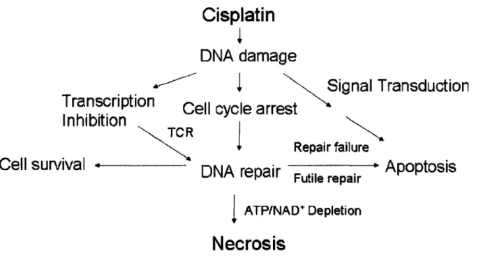
Figure 1.1 Cisplatin-DNA adducts cause a wide variety of cellular responses ...95 Figure 1.2 Mechanisms of cisplatin uptake and efflux ...96 Figure 1.3 HMGB1 protein has multiple roles ... 97 Figure 1.4 The p53 pathway can partially mediate cisplatin cytotoxicity ...98 Figure 1.5 Cisplatin activates MAPK pathways ... 99 Figure 1.6 Cisplatin induces necrosis and apoptosis, two different modes
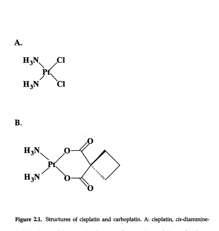
of cell death ... 100 Figure 2.1 Structures of cisplatin and carboplatin ... ... 122 Figure 2.2 Strategy for synthesizing site-specifically platinated DNA probes ...123
Figure 2.3 Denaturing and non-denaturing PAGE gel of ligation products of
site-specifically platinated DNA probes ...124 Figure 2.4 SDS-PAGE gel of recombinant histones and histone octamer ...125 Figure 2.5 Non-denaturing PAGE gel of nucleosome and free DNA ...126 Figure 2.6 Excision assay with nucleosomal and free DNA ... 127 Figure 2.7 Analysis of repair of native and recombinant nucleosomal
platinated DNA by the excision assay . ... 128 Figure 3.1 Strategy for synthesizing the blunt-ended site-specifically
platinated DNA probe ...144 Figure 3.2 4-20% SDS-PAGE gel of recombinant histones, histone (H3/H4)2
tetramer and histone octamer ... 145 Figure 3.3 Non-denaturing PAGE gel of purified double-stranded DNA
Figure 3.4 Figure 3.5 Figure 3.6 Figure 4.1 Figure 4.2 Figure 4.3 Figure 4.4 Figure 4. Figure 4.( Figure 4.5 Figure 4.E Figure 5.1 Figure 5.2 Figure 5.' 146-mer GTG-Pt ... 146
Analysis of site-specifically cisplatin-modified tetrasome,
{Pt(DACH))2+-modified tetrasome, and cisplatin-modified nucleosome ...147 Nucleosome and tetrasome inhibition of nucleotide excision
repair of cisplatin-DNA adduct ...148 Tetrasome inhibition of the excision repair of
cisplatin-and {Pt(DACH))}2-DNA adduct ...149 Strategy for synthesizing site-specifically platinated-DNA probe ...163 Non-denaturing PAGE gels of nucleosomal and free DNA ...164
Non-denaturing gel of 5'-32P-labeled nucleosomal DNA of
199GTG (unmodified) and 199GTG-Pt (cisplatin-modified) ...165
Restriction enzyme digestion of nucleosome 199GTG and
199GTG-Pt
...
166
5 DNase I footprinting of cisplatin-modified and unmodified
nucleosom es ...167 6 Hydroxyl footprinting of the 146GTG and 146GTG-Pt nucleosomes ...168 7 Model of the n146GTG nucleosome ... 169 38 Hydroxyl footprinting of the n146GTG and n146GTG-Pt nucleosomes ...170
1 Cisplatin-induced phosphorylation of histone H3 at Ser 10 ...186 2 Inhibition of cisplatin-induced phosphorylation of H3
Ser 10 by SKF86002 ... 187
MCF-7 (top) and Ntera II (bottom) cell lines ...188
Figure 5.4 Cisplatin induction of phosphorylation of H3 Ser28 and
acetylation of H4 ... 189 Figure 6.1 Strategy for synthesizing site-specifically platinated 195mer
DNA probes ... 218 Figure 6.2 HPLC trace of platination products of 16mer DNAs
after 18 h incubation ... 219 Figure 6.3 Denaturing PAGE analysis of Maxam-Gilbert sequencing of
16GG and 16GGMe substrates ...220
Figure 6.4 Maxam-Gilbert sequencing results for the 16GG and 16GGMe ...221 Figure 6.5 Maxam-Gilbert sequencing of 16GTG and 16GTGMe substrates ...222 Figure 6.6 Maxam-Gilbert sequencing results for the 16GTG and 16GTGMe ...223 Figure 6.7 PAGE analysis of Maxam-Gilbert sequencing of 36mer substrates ...224 Figure 6.8 Maxam-Gilbert sequencing results for the 36CC and 36 MM ...225 Figure 6.9 The Maxam-Gilbert sequencing results for the 36mer CAC
and 36mer MAM ...226 Figure 6.10 Comparison between platinated and non-platinated substrate
of Maxam-Gilbert sequencing . ... 227
Figure 6.11 Time course of platination of 16GG and 16 GGMe ...228 Figure 6.12 Time course of platination kinetics of 16GTG and 16GTGMe ...229
Figure 6.13 Time course of platination kinetics of ds14 CC and ds14MM ...230 Figure 6.14 EMSA of dsl6mers with HMGB1-dom A . ... 231
Figure 6.15.A Non-denaturing PAGE gel of dsl95mers . ... 232 Figure 6.15.B Excision assay with methylated and non-methylated DNA probes ...232 Figure 6.15.C The relative excision efficiencies of ds 195mers ...232 Figure 7.1 Ubiquitination of Pol II is induced by a-amanitin in an in vitro assay ...257 Figure 7.2 Comparison of ubiquitination and transcription activities
of various drug-induced cell cycle stage nuclear extracts ...258 Figure 7.3 Alpha-amanitin induction of Pol II ubiquitination is DNA-dependent ...259 Figure 7.4 Ubiquitination of Pol II is induced to a similar extent
by cisplatin-modified DNA and alpha-amanitin ... 260 Figure 7.5 Stimulation of polymerase ubiquitination by alpha-amanitin
or cisplatin-modified DNA is suppressed by inhibiting transcription ...261 Figure 7.6 Two-phase enzymatic ligation strategy for synthesizing
a platinated 296mer ... 282 Figure 7.7 Procedure for synthesizing a platinated DNA 296mer
by primer extension ... 283 Figure 7.8 Maxam-Gilbert sequencing results for 20GG-Pt ... 284 Figure 7.9 Denaturing PAGE of the two-phase ligation product
and purified 296-Pt ...
285
Figure 7.10 Concentration of 296mer is estimated by using
the low DNA mass ladder ... 286 Figure 7.11 Agarose gel electrophoresis of purified single-stranded DNA ...286 Figure 7.12 Agarose gel electrophoresis of DNA primer extension products ...287
Figure 7.13 In vitro transcription assay with cisplatin-modified template ... 288 Figure 7.14 In vitro transcription assay with cisplatin-modified template ...289 Figure 8.1 Strategies for the synthesis site-specifically platinated dumbbell
DNA Probes ... 305 Figure 8.2 Denaturing PAGE gel analysis of dumbbell ligation products ...306 Figure 8.3 Denaturing PAGE gel analysis of dumbbell ligation products
(six-fragment strategy) ... 307 Figure 8.4 Digestion sites of restriction enzymes on the dumbbell DNA ...308 Figure 8.5 Non-denaturing gel of restriction enzyme digestion of
dumbbell DNA ... 309 Figure 8.6 Denaturing gel of T7 exonuclease digestion of dumbbell DNA ...310 Figure 8.7 Denaturing PAGE gel of repair assays for dumbbell DNA ...311 Figure 8.8 Reconstitution of dumbbell into nucleosome ...312 Figure 8.9 Effect of anti-TFIIB on transcription from dumbbell DNA ...313 Figure A1.1 Analysis of site-specifically cisplatin-modified nucleosome
on a 5% non-denaturing PAGE gel electrophoresis ...330 Figure A1.2 The effect of hSWI/SNF on nucleosome structure
characterized by Nla III digestion ... 331
Chapter 1 Cellular Processing of Platinum Anti-Cancer
Drugs - Identifying Pathways for Chemogenotherapeutic
Drug Design*
Introduction
Cisplatin and carboplatin have been widely for many years to treat several kinds of cancer, including as testicular, ovarian, head and neck, and non-small cell lung.' Over this period, thousands of platinum analogues have been synthesized and screened for anti-cancer activity. Among them, the only one that has been approved by the FDA is oxaliplatin, for treatment of colorectal cancer.2 3 Several interesting platinum compounds, including those with a trans geometry at the platinum center, platinum(IV) molecules with tethered substituents, and multinuclear species, have been reported to be as active as cisplatin in one or another assay, as has been reviewed elsewhere.4-7
Although many cellular components interact with cisplatin, DNA is the primary biological target of the drug.' The platinum atom of cisplatin forms covalent bonds to the N7 positions of purine bases to form primarily 1, 2- or 1, 3-intrastrand cross-links and a fewer number of interstrand cross-links. These linkages to DNA occur at the positions where the chloride, CBDCA, or oxalate ligands reside in the original platinum drug. Structural studies of cisplatin and
oxaliplatin DNA adducts have been reviewed elsewhere.'
I The abbreviations used are: cisplatin, cis-diamminedichloroplatinum(II); DDP, trans-diamminedichloroplatinum(II); carboplatin, cis-diammine(cyclobutane-1,1-dicarboxylato)-platinum(II); CBDCA, cyclobutane-1,1-dicarboxylate; ER, estrogen receptor; ATM, ataxia-telangiectasia mutated; tsHMG, testes-specific HMG box protein; ATR, ataxia-ataxia-telangiectasia and Rad3-related; MAPK, mitogen-activated protein kinase; JNK, c-Jun NH2-terminal kinase; ERK,
extracellular signal-regulated kinase; NER, nucleotide excision repair; TCR, transcription-coupled repair; GGR, global genomic repair; MMR. mismatch repair; XIAP, X-linked inhibitor of
apoptosis; MDM2,murine double minute-2; MEF, mouse embryo fibroblasts; PARP, poly(ADP-ribose)polymerase; SCC, serum squamous cell; Cdc25C, cell division cycle 25C; PCNA,
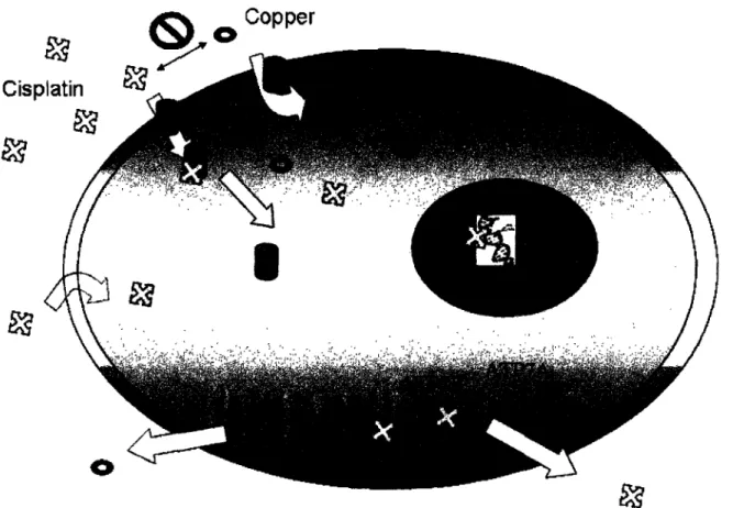
Cisplatin-DNA adducts cause a wide variety of cellular responses (Figure 1.1).8 Understanding the mechanisms of how cells process cisplatin provide important insights for designing more efficient platinum-based drugs. For example, the discovery that treatment of ER(+) cells with estradiol sensitizes them to cisplatin inspired the design and synthesis of a series of active estrogen-tethered Pt(IV) compounds.9 Information of this kind facilitates the introduction of new families of molecules in which the fundamental genotoxicity of a platinum-DNA adduct can be made selective for cancer cells by targeting the compound selectively to tumors or by interrupting a pathway used by the cancer cell to survive the chemical assault. The current trend among cancer biologists to downplay the importance of chemotherapy in favor of pathway-directed modalities is, in our opinion, failing to take advantage of a time-proven weapon against the disease. Combining the best of chemotherapeutic action with specific targets in cellular pathways offers a powerful new approach to cancer treatment that perhaps may impede the many clever ways that human cells have in becoming drug-resistant.
This purpose of the present review is to highlight recent discoveries in those pathways involved in regulating platinum drug transport, DNA damage
signaling, cell cycle checkpoints and arrest, DNA repair and mechanisms of cell death. From the understanding of these cellular pathways it has become possible to form a biological basis for the chemical synthesis of new platinum drugs for cancer treatment, a process that we term chemogenotherapy.
Mechanisms of Cisplatin Transport into Cells
The mechanisms of cisplatin cellular uptake and efflux are still not fully understood. Early studies suggested that cisplatin enters the cell mainly by passive diffusion, since its uptake proceeded linearly with time up to 60 min and could not be saturated up to a concentration of 1.0 mM. In addition, cisplatin absorption was not inhibited by structural analogues and there did not appear to be an optimum pH for its entry into the cell.1-'2
More recently, a growing body of evidence reveals a link between Ctrlp, a high-affinity copper transporter, and cisplatin uptake (Figure 1.2). Mutation or deletion of the CTR1 gene results in increased cisplatin resistance and reduction of platinum levels in both yeast and mouse cells.'3 Moreover, CTR1-deficient cells exhibit impaired accumulation of cisplatin analogues carboplatin, oxaliplatin, and ZD0473[cis-amminedichloro(2-methylpyridine)platinum(II)].14,15
Another direct link between the cellular management of copper and platinum concentrations comes from studies of cisplatin efflux. Copper-transporting P-type adenosine triphosphate (ATP7B), which plays an important role in regulating copper levels in the cell, is associated with cisplatin resistance in vitro.'6 Moreover, ATP7B levels correlate with cisplatin resistance in a variety
of cancers. For example, in the case of primary oral serum squamous cell (SCC) carcinoma, patients with ATP7B-positive tumors have reduced overall survival following cisplatin treatment than those who are ATP7B-negative.2 0 The
expression level of the ATP7B gene was significantly increased in patients with undifferentiated ovarian carcinoma and gastric carcinoma, which is less sensitive to cisplatin treatment.17-'9 Although ATP7A is expressed in a considerable fraction of many common tumor types, its role is less clear; however, enrichment of human ovarian cancer cells that express ATP7A is associated with poor response to platinum-based therapy.21 In addition, MRP2 (cMOAT), an exporter
protein, also plays a role in cisplatin resistance, presumably by promoting drug efflux. Overexpression of MRP2 is associated with increased cisplatin resistance.22- 24
The observation that both cisplatin uptake and efflux are seemingly linked to the copper metabolic pathway leads to the hypothesis that copper and cisplatin might interfere with their mutual transport.2 s In fact, both copper and
cisplatin have the ability to reduce the uptake of the other and can trigger the degradation and delocalization of Ctrlp.l3 Moreover, copper and cisplatin also exhibit bidirectional cross-resistance.2 6,27 Expression of ATP7B in human
carcinoma cells modulates sensitivity to both cisplatin and copper by causing more rapid efflux of the two agents.'6
These observations offer a potential new mechanism for targeting platinum to tumor cells by taking advantage of an emerging receptor for delivering the drug into the cancer cell. Any compound having a substituent that can direct the platinum complex to a tumor or organ tissue population in a specific manner is of potential interest for chemogenotherapy. Included in this
category would be molecules that target the tumor vasculature28-31 or deliver the molecule specifically to cells having a receptor for the appended moiety.93 2
Cisplatin-DNA Damage Recognition Proteins - A Postulated Key
Role for HMGB1
Cisplatin modifications distort the structure of the DNA duplex, bending it significantly toward the major groove and exposing a wide, shallow minor groove surface to which several classes of proteins bind. Included are high mobility group box proteins, repair proteins, transcription factors, and other proteins such as histone H1, which preferentially recognize 1,2-intrastrand cross-linked platinum-DNA adducts, as summarized elsewhere.' X35 Here we focus on recent progress in understanding the functions of the HMG box family of proteins and their putative role in facilitating the anticancer activity of cisplatin.
The abundant HMG box protein, HMGB1, also named Amphoterin, has been connected to several DNA-dependent pathways (Figure 1.3). HMGB1 stimulates RAG1/2 cleavage in V(D)J recombination and promotes binding of
sequence-specific transcription factors.-38 In addition, HMGB1 physically interacts with MutSa and plays a role in mismatch repair.3 9 Moreover, HMGB1 enhances p53 DNA binding activity and directly interacts with p53 in vitro.40 Both the C-terminal and A box domains of HMGB1 regulate p53-mediated transcription activation and its downstream effects.41 HMGB1 affinity for
cisplatin-damaged DNA is significantly enhanced by p53. Interaction between p53 and HMGB1 after platination may provide a molecular link between DNA damage and p53-mediated DNA repair.4 2 In vitro nucleotide excision repair
studies indicate that HMG box proteins such as HMGB1 and tsHMG shield the Pt-1,2-d(GpG) cross-link from repair proteins by stably binding to this DNA adduct.43-4 5 Until recently, the postulated in vivo relationship between HMG box
protein levels and cisplatin toxicity has been somewhat controversial. Several studies report that HMG box protein levels correlate with cisplatin sensitivity. For example, overexpression of HMGB1 induced by addition of the steroid hormone estradiol sensitizes breast and ovarian cancer cells to cisplatin treatment.46 Deletion of yeast Ixrl, a gene that encodes a transcriptional repressor containing two HMG boxes, results in 2-6-fold increased resistance to cisplatin, depending on the strain investigated.4 7 ,48 On the other hand, cancer cell lines that
have developed resistance to cisplatin often have elevated levels of HMGB1.49 In
addition, no significant difference in sensitivity to cisplatin was observed
between and Hmgbl knockout and wild type mouse embryonic fibroblast cell
lines.50 Moreover, S. pombe mutant deficient in the HMGB protein Cmbl is more
sensitive to cisplatin treatment.5l These discrepancies most likely reflect the importance of cell type and history in determining the ability HMGB1 and related proteins to mediate cisplatin cytotoxicity. Recent work from our laboratory using RNAi clearly demonstrate the strong correlation with HMGB1 levels and the sensitivity of cells to cisplatin.5 2
HMGB1 interacts with the nucleosome at the entry and exit points of DNA and facilitates binding of the remodeling complex ACF to nucleosomal DNA, thereby accelerating the ability of ACF to induce structural changes in the nucleosome.535 4 Of greater interest is the recent discovery that the dynamic behavior of HMGB1 is completely altered in apoptotic cells, where it binds irreversibly to chromatin, most likely due to the change of the nature of chromatin.55 In addition, HMGB1 plays an important role as a cytokine in the inflammatory response.5 6-58 In monocytes activated by inflammatory stimuli,
HMGB1 relocalizes from the nucleus to the cytoplasm, and eventually to secretory organelles.5 5 HMGB1 thus appears to act as a signal of tissue damage, inducing mesoangioblast migration and proliferation.5 9
The activity of HMGB1/2 is modulated by post-translational modifications. Histone acetyltransferase CBP acetylates HMGB1/2 proteins in vitro.6 0 The acetylated HMGB1/2 full-length protein is 3-fold more effective in inducing ligase-mediated circularization of a 111-bp DNA fragment than unmodified full length protein. In addition, acetylation of HMGB1 plays an important role in regulating relocalization of HMGB1 from nucleus to cytoplasm, and subsequent secretion when monocytes receive an appropriate second signal.6 l The export of HMGB1 from necrotic cells does not appear to require such post-translational modification.556263If viable cancer cells surrounding the necrotic core of a solid tumor took up HMGB1, it could potentiate the ability of
cisplatin to kill such cells and account for the specificity of the drug for cancer
cells.
The structure-specific recognition protein, SSRP1, contains a single HMG domain that was initially isolated from expression screening of a human B-cell cDNA library for proteins that bind to cisplatin-modified DNA.64 Human SSRP1 and Sptl6 form a complex named FACT, which facilitates RNA polymerase II transcription elongation through a nucleosome block.65,66 The isolated HMG domain of SSRP1 and FACT, but not SSRP1 alone, can bind to the major 1,2-d(GpG) intrastrand cisplatin adduct, which suggests that Spt16 primes SSRP1 for
cisplatin-damaged DNA recognition by unveiling its HMG domain.6 7 Also, SSRP1 functions as a co-activator of the transcriptional activator p63.68
Yeast Nhp6, an abundant HMG box protein, is a component of yeast FACT complex in S. cerevisiae. Nhp6 is required for proper regulated expression of a number of genes that are transcribed by RNA polymerase II or III. Either Nhp6 alone or the yFACT complex were able to bind nucleosomes and significantly reorganize their structure.6 9 In addition, Nhp6Ap binds to
cisplatin-modified intrastrand cross-links on duplex DNA with a 40-fold greater affinity than to unmodified DNA with the same sequence. Nhp6A/Bp appears to function directly or indirectly in yeast to enhance cellular resistance to cisplatin.7 0
Cell Cycle Checkpoints and Arrest
Cell cycle checkpoints monitor the proper order of events in the cell. When DNA is damaged, the cell cycle is arrested to provide time for repair. Failures to do so leads to the acquisition and accumulation of genetic alterations, ultimately causing tumorigenesis.71
The G1/S checkpoint ensures that damaged DNA is not replicated. Following DNA damage, ATM (ataxia-telangiectasia mutated) cells may control the fate of a G1/S checkpoint through direct phosphorylation of p53 or through phosphorylation of c-Abl, which in turn upregulates p21 through p73
activation.7Z73 ATM-dependent phosphorylation of p53 contributes to p53 stabilization by reducing its interaction with MDM2 (murine double minute-2).74
,75 In addition, p21, a downstream target of p53 and a universal inhibitor of
cyclin-dependent kinases, plays a major role in mediating p53-dependent cell
cycle G1 arrest. p21 inhibits G1 cyclin/cdks and blocks cell cycle progression.7 6,77
Exogenous expression of p21 exerts cell growth inhibition and enhances sensitivity to cisplatin in hepatoma cells.78
Cisplatin does not cause G1 arrest in all cases, however. In fact, the drug fails to induce a G1 arrest response in synchronized wild-type MEF cells, even though it can activate p53 and cause an S phase arrest.79
The G2/M checkpoint allows for repair of DNA that was damaged in late S or G2 phase of the cell cycle prior to mitosis to prevent damaged DNA from
being segregated into daughter cells. It has been proposed that such G2 arrest is essential to the process of engaging cell death following cisplatin treatment.80,8 l
Upon the occurrence of DNA damage, ATM and its related ATR (ataxia-telangiectasia and Rad3-related) kinase activate checkpoint kinases Chkl and 2 through phosphorylation, which in turn phosphorylate Cdc25C. The
phosphorylated Cdc25C promotes its binding to 14-3-3 adaptor proteins and
thereby is separated from Cdc2 by translocation of Cdc25c to the cytoplasm. As a result, Cdc2 phosphorylation is elevated and causes cells to arrest in G2.82-88
In addition, p21 plays a regulatory role in maintaining cell cycle arrest at G2 by blocking the interaction of Cdc25C with PCNA.8 9-9 1More recently, SHP-2 extra-cellular signal-regulated kinases (ERK) and p38 mitogen-activated protein kinases (p38 MAPK) have also been implicated in the G2/M cell cycle
checkpoint.9 2-95 Therefore, it appears that multiple pathways contribute to the
regulation of the G2/M checkpoint following genotoxic stress.
p53 and p73 Pathways
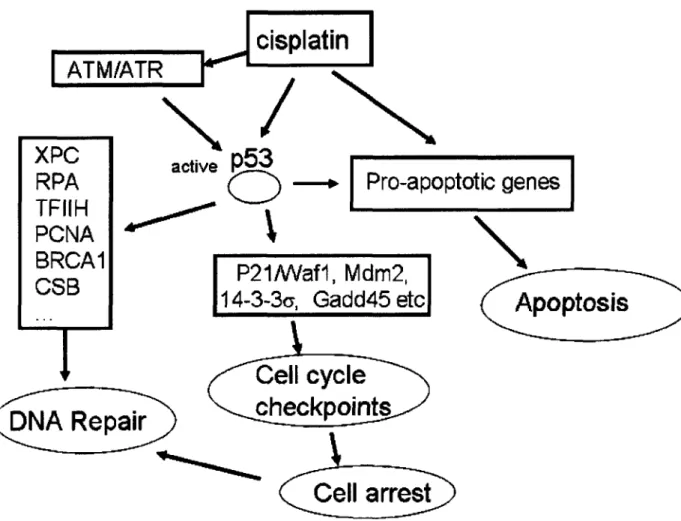
p53 is an important tumor suppressor protein which is mutated in greater than 50% of all human tumors.9 The p53 protein plays a crucial role in many cellular processes; in particular, it inhibits cell proliferation by inducing either cell cycle arrest or apoptosis in response to cellular stress. p53 functions as a transcription factor and upregulates a number of genes involved in these
processes (Figure 1.4).97- 99 In the absence of cellular stress, p53 is maintained at low steady-state levels and exerts very little effect on the cell. Upon cellular stress, p53 is overexpressed and undergoes post-translational modification.
p53 has also been connected with DNA repair, specifically nucleotide excision repair (NER) and base excision repair (BER).100-102One of the activities of
p53 involves its transcriptional activation of the Gadd45 gene. GADD45 interacts with PCNA and stimulates nucleotide excision repair activity when overexpressed.'0 3 p53 is also involved in repair through direct interactions with critical components of the NER machinery. XPC, an important damage recognition protein in NER, is induced after DNA damage in a p53-dependent manner.104 XPC also can help initiated cisplatin damage-mediated cell cycle regulation.l0 5 In addition, p53 binds to three of the subunits of the TFIIH
transcription/repair complex, XPB, XPD, and the p62 subunit, suggesting an additional role in NER.10 6 Moreover, p53 physically interacts with RPA.107 Such interaction is disrupted following UV or cisplatin damage in p53-wild type cells, but not in p53-mutant repair-deficient cells.08 These results suggest that release of RPA and p53 plays a role in facilitating NER.109 The repair defect in p53-mutant ovarian carcinoma cells IGROV-1/Pt may be attributed to a reduced removal/recycling of PCNA at repair sites.l09
Sensitivity to cisplatin correlates positively with the presence of wild-type p53 function in a National Cancer Institute (NCI) panel of 60 human tumor cell lines."1° In addition, tumor cell lines that lack functional p53 are more resistant to
cisplatin than cells that contain functional p53, but can be sensitized when reconstituted with wild-type p53.111-113Other studies, however, show no or even
negative correlation between p53 status and response to cisplatin, For example, p53 mutation promotes increased sensitivity to cisplatin, with loss of G1/S checkpoint control and decreased cisplatin-DNA adduct repair.l4 Furthermore, one study demonstrated that, in SaOS-2 osteosarcoma cells, p53 function was associated with increased cisplatin sensitivity under high serum growth conditions but with decreased sensitivity under low serum conditions.l5 Previous work from our own laboratory using murine testicular teratocarcinoma cells also revealed that, although rapid apoptosis is induced in p53-wild-type but not in p53-/- teratocarcinoma cells upon cisplatin treatment, the absence of p53 does not alter cellular sensitivity to cisplatin as measured in clonogenic assays.l6 The relationship between p53 status and cisplatin cytotoxicity depends upon several factors, including tumor cell type, activation of specific signaling
pathways, and the presence of other genetic alterations.
Besides p53, there are two other members of the p53 family, p63 and p73, which are homologous to p53 in terms of overall domain structure and conformation. Both p63 and p73 are expressed as multiple isoforms, truncated at the N- or C-terminus through alternative splicing and use of several promoters. 7,"'8 p73 can cooperate with DNA damaging agents or p53 to induce some p53 target genes and activate their promoters. Signaling pathways for p53 and p73 in inducing cell cycle arrest and apoptosis are similar but also have
important differences. p73 differentially regulates some p53 target genes, either several fold lower than p53, such as p21, or several fold higher than p53, such as 14-3-3a.119 DNA damage can stabilize p73 and enhance p73-mediated apoptosis
in a c-Abl dependent manner.73 p73 upregulates MDM2 expression, but unlike p53, it is not targeted by MDM2 for ubiquitin-dependent degradation. Instead, MDM2 downregulates p73 transactivator function by disrupting its interaction with p300/CBP, a component of the eukaryotic transcription complex.2 0
Proteins involved in p53 related pathways modulate p53 activity and cisplatin cytotoxicity. Aurora kinase A is a key regulatory component of the p53 pathway and phosphorylates p53 at Ser315, leading to its ubiquitination by Mdm2 and proteolysis. Aurora kinase A is overexpressed in many human cancers, which leads to increased degradation of p53, and down-regulation of checkpoint-response pathways. Silencing of aurora kinase A results in less phosphorylation of p53 at Ser315, greater stability of p53 and cell-cycle arrest at G2-M, and ultimately increased sensitivity to cisplatin treament.'21
MDM2 interacts with the N-terminus of p53 and inhibits p53 transcriptional activity.2 2 In addition, MDM2 acts as the E3 ligase for p53 and
thus promotes p53 ubiquitination and degradation.12 3 Post-translational modifications of p53 and MDM2, including phosphorylation, acetylation and sumoylation, modulate their interactions and thus influence p53 stability and activity.'2 4 In addition, STAT-1 interacts directly with p53 acts as a co-activator
Cyclin G is another transcriptional target gene of tumor suppressor p53. Cyclin G may play a central role in the p53-MDM2 autoregulated module. Inhibition of cyclin G by small interfering RNA (siRNA) causes the accumulation of p53 levels in response to DNA damage.'2 6 This cyclin G-mediated p53 regulation is dependent upon the status of ATM protein, which activates p53 in response to DNA damage.2 7
The human BRCA1 gene contains one RING finger domain and two BRCT domains. BRCA1 interacts directly or indirectly with numerous proteins involved in transcriptional regulation, cell cycle/checkpoint control, protein ubiquitination, chromatin remodeling, and DNA repair. BRCA1 functions as a
molecular determinant of response to a range of cytotoxic chemotherapeutic agents.l28 BRCA1 is a substrate of ATM kinase in vitro and in vivo, and is regulated by an ATM-dependent mechanism as a part of the cellular response to DNA damage.l2 9 The p53 and BRCA1 tumor suppressors are involved in repair processes and may cooperate to transactivate certain genes, including p21WAF/cm
and GADD45. BRCA1 also enhances p53 binding to the DDB2 promoter in vivo. Antisense of BRCA1 abrogates upregulation of DDB2 after cisplatin exposure. DNA repair activity is significantly restored by introduction of BRCA1 into wild-type as compared to DDB2-deficient cells, suggesting that BRCA1 plays a role in DNA damage repair-mediated cisplatin resistance.3 0,13'
These studies demonstrate that many cellular functions controlled directly or indirectly by p53 respond to cisplatin treatment. Since p53 is not essential for
cisplatin-mediated cell-killing,l1 6 however, it is unlikely that any one of them is
critical to the selectivity and curative properties of the drug for certain cancers such as testicular.
MAPK and Other Related Pathways
Akt Pathway
Akt, protein kinase B (PKB), is a member of the family of phosphatidylinositol 3-OH-kinase-regulated serine/threonine kinases.3 2 Akt promotes cell survival and down-regulates apoptotic pathways through phosphorylation and modulation of several downstream target proteins.'33 Therefore, Akt protects cells from apoptotic death induced by a variety of stimuli, including cisplatin.3 4
Akt phosphorylates X-linked inhibitor of apoptosis (XIAP) and thus stabilizes XIAP by inhibiting both its auto-ubiquitination and cisplatin-induced ubiquitination activities. Increased levels of XIAP are associated with decreased cisplatin-stimulated caspase 3 activity and apoptosis. Inhibition of XIAP and/or
Akt expression/function may be an effective means of overcoming chemoresistance in ovarian cancer cells expressing either endogenous or reconstituted wild-type p53.13-137 Akt also phosphorylates the MDM2 protein promoting its translocation to the nucleus and destabilization of p53, therefore impairing the cellular stress response and increasing the survival of tumor cells.
Akt also phosphorylates and inactivates several other apoptotic pathway proteins. For example, Akt phosphorylates BAD, promoting its association with 14-3-3 proteins in the cytosol and inactivating its proapoptotic function.lM Moreover, Akt phosphorylates and inactivates apoptosis signal-regulating kinase-1 and mixed lineage kinase 3 (MLK3), the mitogen-activated protein kinase kinase kinases (MAPKKK), leading to cell survival.'3, 39 Constitutively active Akt might be due to down-regulation of FADD-protein or the upregulation of the caspase 8 inhibitor c-FLIP to increase cisplatin resistance.l40 Expression of adenovirus Ela gene is able to block selectively Akt activation mediated by cisplatin and increases sensitivity to cisplatin.l4 0
In addition, Akt also phosphorylates IKB kinase, subsequently activating nuclear factor of KB (NFKB). NFKB plays an important role in controlling cell survival.l3 3 Increase of NFKB activity correlates with decreased apoptosis,
whereas inhibition of NFKB increases the efficacy of cisplatin both in vitro and in vivo.13 3,41
c-Abl and Bcr-Abl
c-Abl is a member of the Src family of non-receptor tyrosine kinases, which contain Src- homologue regions 2 and 3 (SH2 and SH3) at N-terminus and a large C-terminus region that contains nuclear localization motifs and nuclear export sequences. In addition, c-Abl also carries a DNA binding motif and an actin-binding domain at its extreme C-terminus. Nuclear c-Abl tyrosine kinase
activity can be stimulated by cisplatin and other DNA damaging agents and acts to transmit DNA damage signals from the nucleus to the cytoplasm.14 2 The activation of c-Abl tyrosine kinase might be a downstream event in ATM-dependent apoptosis. ATM may directly phosphorylate c-Abl at Ser465 or indirectly phosphorylate c-Abl through DNA-PK or other unidentified protein kinases.l43 c-Abl tyrosine kinase plays a role in the activation of apoptosis induced by cisplatin. Overexpression of c-Abl tyrosine kinase in Saos-2 cells and NIH3T3 cells can activate apoptosis,44 14 5 whereas c-Abl deficient mouse fibroblasts and chicken DT-40 cells are resistant to cisplatin-induced apoptosis. Re-introduction of a functional c-Abl in these Abl-deficient cells restore their sensitivity to the drug.73 In addition, knocking down c-Abl expression by RNA interference confers resistance to cisplatin-induced apoptosis.46
Retinoblastoma tumor suppressor protein (RB) plays a negative role in DNA damage-induced activation of c-Abl kinase.73 RB binds to the tyrosine kinase domain of c-Abl, thereby blocking c-Abl tyrosine kinase activity and preventing c-Abl from being activated by DNA damage signals. Phosphorylation of RB at G1/S disrupts the RB/c-Abl interaction, leading to the release of c-Abl in the nucleus of S phase cells.14 7
The transcription factors p73 and p53 are possible downstream effectors of c-Abl. Cisplatin can induce the accumulation of p73 protein in wild type and p53-deficient, but not in c-Abl-deficient, primary mouse embryo fibroblasts.7 3 Furthermore, c-Abl phosphorylates p73 both in vitro and in vivo, and the SH3
domain of c-Abl can directly bind to and phosphorylate the p73 protein.14 8s149
c-Abl is not essential to the induction and activation of p53 by DNA damage, although c-Abl can stabilize and activate p53 in transient co-transfection
experiments. 15 0
c-Abl is also an upstream effector of MEKK-1 and the stress-activated kinases, c-Jun N-terminal kinases NK) and p38 MAPK.5 - l53 The treatment of
wild-type fibroblasts, but not Abl-/- cells, with cisplatin is associated with an
increased level of tyrosine phosphorylation of MEKK-1.154 In addition, cells deficient in c-Abl fail to activate p38 MAP kinase and JNK after treatment with cisplatin, but not after exposure to UV and methyl methanesulfonate. Reconstitution of c-Abl in the Abl-/- cells restores the activation of p38 MAPK and JNK.1 55 These findings indicate that c-Abl dependent activation of p38 MAP and JNK protein kinases is specific to cisplatin. p38 MAPK and JNK are differentially regulated in response to different classes of DNA-damaging agents. Another interesting feature of c-Abl is its ability to associate with chromatin. c-Abl contains three HMG-like boxes that preferentially bind to A/T duplexes and distorted DNA structures, such as four-way junctions. Whether c-Abl could recognize and interact with cisplatin-DNA adducts like HMGB1 is a very interesting question, the answer to which might provide more insight into c-Abl activation by the drug.l56,15 7
Different subcellular localizations might contribute to the opposing effects of c-Abl and Bcr-Abl kinase on apoptosis.l5 8 Cytoplasmic Bcr-Abl can inhibit
apoptosis through the activation of Akt.147Mutagenesis assays demonstrated that cells expressing a deletion mutant of BCR/ABL that lacked the SH2 and SH3
domains of ABL (BCR/ABLAA) were sensitive to cisplatin. Moreover, Bcr-Abl tyrosine kinase can be converted into a potent inducer of apoptosis by allowing it to function in the nucleus, which is consistent with the role of the nuclear c-Abl tyrosine kinase in DNA damage-induced cell death.l4 7
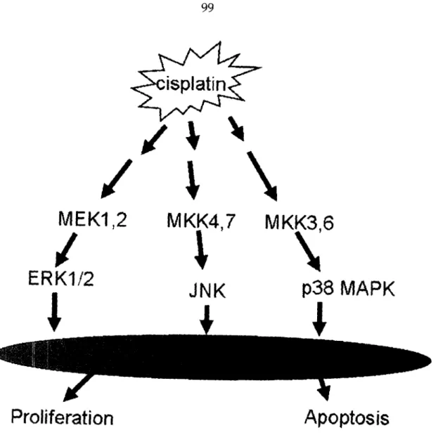
MAPK/JNK/ERK Pathways
The MAPK family of proteins play important roles in signal transduction processes through specific phosphorylation cascades in response to a variety of extracellular stimuli.l59,160 There are three major mammalian MAPK subfamilies,
ERK, JNK (also called stress-activated protein kinase), and p38 MAPK. The ERK pathway is highly induced in response to growth factors and cytokines, and it is also activated by some conditions of stress, particularly oxidative stress. In contrast, JNK and p38 are highly activated in response to a variety of stress signals including ionizing and short wavelength ultraviolet irradiation (UVC), tumor necrosis factor, and hyperosmotic stress, and they are only weakly activated by growth factors.5 9,160 Cisplatin triggers the activation of the ERK, JNK and p38 MAPK cascades in tumor cells or transformed cell lines (Figure 1.5), as discussed in following section.
p38 MAPK Path7way
A growing body of evidence suggests that activation of the p38 MAPK pathway plays an important role in the cellular response to cisplatin.l5516-1 63
Cisplatin, but not trans-diamminedichloroplatinum(II) (trans-DDP) or PtCl4, induces sustained activation of p38 MAPK.161 Cisplatin activates p38 MAPK for 8-12 hr in sensitive cells and for 1-3 hr in resistant cells.l64This difference in the
kinetics of activation could account for their differential cytotoxicity. Moreover, lack of p38 MAPK activation/function induces a resistant phenotype in human cells.l6 1"64 MAP kinases may also play a role in modifying the chromatin environment of target genes.16 5 This kinase regulates immediate-early (IE) gene
expression and other cellular responses through phosphorylation of various substrates, including transcription factors, chromatin protein constituents, and downstream Ser/Thr effector kinases.16616 7 Furthermore, p38 MAP kinase and
downstream kinase MSK-1 phosphorylate histone H3 Ser in vitro.l6 7 The p38
MAPK pathway is also involved in cisplatin-induced phosphorylation of histone
H3 at Ser 10.168
ERK Pathluay
Cisplatin treatment results in dose- and time- dependent activation of ERK1/2.169-71 However, the extent of ERK activation might reflect the nature or context of specific cell lines, since some cells show no or only weak activation of
ERK following cisplatin treatment.72 There is also conflicting evidence for the role of ERK in influencing cell survival of cisplatin-treated cells. Some studies have suggested that ERK activation is associated with enhanced survival of
cisplatin-treated cells.73,1l74 Such is not always the case, however. Although
inhibition of cisplatin-induced ERK activity by treatment with the MEK1 inhibitor PD98059 sensitizes the human melanoma cell line C8161 to cisplatin-induced apoptosis, PD98059 does not potentiate cisplatin-cisplatin-induced apoptosis in three other human melanoma cell lines. Instead, PD98059 protects against cisplatin-induced cytotoxicity in one of these cell lines, partly via an enhancement of cisplatin-induced of NFKB activation.l75-77 Moreover, inhibition of cisplatin-induced ERK activity by PD98059 results in decreased levels of p53, p21WAF1, GADD45 and Mdm2.'7 8 In addition, elevated expression of Ras, an
upstream component of the ERK signaling pathway, has been connected with enhanced sensitivity to cisplatin.7 9 HeLa cell variants selected for cisplatin resistance show reduced activation of ERK following platinum treatment.l6 9 This
discrepancy suggests that the relationship between the activity of ERKs and the cellular response to cisplatin may depend upon individual cellular context and
levels of stress.
INK Path7ay
The mechanisms of JNK1 activation in response to UV irradiation and cisplatin treatment have important differences. UV light is able to induce
translocation of JNK1 from the cytoplasm to the nuclear compartment, whereas cisplatin does not.72 Although a number of studies have established that JNK is activated in response to cisplatin treatment, a role for this signaling pathway in determining survival is far from clear. Several studies have provided evidence that the JNK pathway contributes to cisplatin-induced apoptosis,164 ,17280-184
whereas others have suggested that JNK activation in response to cisplatin signals a protective response and is important for cell survival.174,182z1s3,l5-8 7 Such
differential effects observed from one study to another could reflect the nature of different cell types and specificity or transient vs persistent activation patterns, respectively. Interestingly, one study showed that the duration of JNK induction may be the crucial factor in mediating the signaling decision resulting in either
cell proliferation or apoptosis.l88
MKP-1/CL100
Expression of phosphatase MKP-1/CL100 inhibits JNK and p38 activation after cisplatin treatment and correlates with an increase in survival after drug treatment. JNK1 induction by cisplatin is delayed and persistent, whereas trans-DDP induces JNK in a transient manner. This difference in duration of JNK induction may be due to the differential ability of inducing of MKP-1/CL-100.
trans-DDP efficiently induces MKP-1/CL100, and its expression correlates with
JNK inactivation, whereas cisplatin is only a very weak inducer of MKP-1/CL100.1 8 9 Moreover, ectopic expression of a catalytically inactive mutant of